adaptive surround modulation of mt neurons: a
TRANSCRIPT

ORIGINAL RESEARCHpublished: 26 October 2020
doi: 10.3389/fncir.2020.529345
Frontiers in Neural Circuits | www.frontiersin.org 1 October 2020 | Volume 14 | Article 529345
Edited by:
Edward S. Ruthazer,
McGill University, Canada
Reviewed by:
Yong-Jie Li,
University of Electronic Science and
Technology of China, China
Ayako Wendy Ishikawa,
Keio University, Japan
Dario L. Ringach,
University of California, Los Angeles,
United States
*Correspondence:
Parvin Zarei Eskikand
Received: 24 January 2020
Accepted: 22 September 2020
Published: 26 October 2020
Citation:
Zarei Eskikand P, Kameneva T,
Burkitt AN, Grayden DB and
Ibbotson MR (2020) Adaptive
Surround Modulation of MT Neurons:
A Computational Model.
Front. Neural Circuits 14:529345.
doi: 10.3389/fncir.2020.529345
Adaptive Surround Modulation of MTNeurons: A Computational ModelParvin Zarei Eskikand 1*, Tatiana Kameneva 1,2, Anthony N. Burkitt 1, David B. Grayden 1 and
Michael R. Ibbotson 3
1Department of Biomedical Engineering, The University of Melbourne, Parkville, VIC, Australia, 2 Faculty of Science,
Engineering and Technology, Swinburne University of Technology, Hawthorn, VIC, Australia, 3National Vision Research
Institute, Australian College of Optometry, Carlton, VIC, Australia
The classical receptive field (CRF) of a spiking visual neuron is defined as the region in the
visual field that can generate spikes when stimulated by a visual stimulus. Many visual
neurons also have an extra-classical receptive field (ECRF) that surrounds the CRF. The
presence of a stimulus in the ECRF does not generate spikes but rather modulates the
response to a stimulus in the neuron’s CRF. Neurons in the primate Middle Temporal
(MT) area, which is a motion specialist region, can have directionally antagonistic or
facilitatory surrounds. The surround’s effect switches between directionally antagonistic
or facilitatory based on the characteristics of the stimulus, with antagonistic effects
when there are directional discontinuities but facilitatory effects when there is directional
coherence. Here, we present a computational model of neurons in areaMT that replicates
this observation and uses computational building blocks that correlate with observed
cell types in the visual pathways to explain the mechanism of this modulatory effect.
The model shows that the categorization of MT neurons based on the effect of their
surround depends on the input stimulus rather than being a property of the neurons. Also,
in agreement with neurophysiological findings, the ECRFs of the modeled MT neurons
alter their center-surround interactions depending on image contrast.
Keywords: vision, neural model, motion perception, middle temporal (MT), adaptive surround modulation
INTRODUCTION
The classical receptive field (CRF) of a spiking visual neuron is defined as the region in the visualfield within which the presentation of a visual stimulus can generate spikes in that neuron. Manyvisual neurons also have an extra-classical receptive field (ECRF) that surrounds the CRF. Thepresence of the stimulus in the ECRF cannot generate a response by itself but can have an excitatoryor inhibitory effect on the neuron’s response (Barlow and Levick, 1965). For direction-selectiveneurons, the directional tuning of the surround is mainly antagonistic, which results in a reductionof the activity of the neuron when the motion in the surround is in the preferred direction ofthe center; this is called an antagonistic surround. However, in some neurons, the surround caninstead have a facilitatory effect, which reinforces the activity of the neuron when the motion inthe surround is in the preferred direction of the center; this is a facilitatory surround (Barlow andLevick, 1965).
It is commonly reported that most of the neurons in the primate middle temporal (MT) area,which is a motion specialist region, have directionally antagonistic surrounds (Albright, 1984).However, Huang et al. (2007) showed that the dominance of the antagonistic surround in theliterature is due to the characteristics of the stimuli that have been commonly used.

Zarei Eskikand et al. Adaptive Surround Modulation
Here, we present a model in which the ECRFs of MT neuronsare adaptive to the changes in the input stimulus. The surroundshave an antagonistic effect when there is a discontinuity inthe input stimulus. In our definition, any inconsistency in thecharacteristics of the stimulus are regarded as discontinuities,including changes in luminance or shape. We show in our modelthat the level of the antagonism driven by the surround increaseswith increases in stimulus contrast. The antagonistic effect of thesurround assists the MT neurons to detect discontinuities in theinput stimulus and segregates stimuli that are moving in differentdirections. The antagonistic surrounds of the MT neurons switchto having facilitatory effects when there is coherency in the inputstimulus, which facilitates the propagation of integrative signals.
Existing neurophysiological findings support the conceptof stimulus-dependent center-surround characteristics (Thiele,2007). To demonstrate alterations in surround modulation thatdepend on the input stimulus, Huang et al. (2007) evaluated theactivity of MT neurons in response to two different stimuli andobserved adaptive changes of the surround effect. According totheir observations, the surround represents integrative featuresin response to moving contours to overcome ambiguousmotion information resulting from the aperture problem. Theaperture problem refers to the generation of ambiguous motioninformation by neurons with small receptive fields that areonly able to measure the motion component in the directionorthogonal to the long edge of a bar stimulus (Born and Bradley,2005). However, the surround regions of Huang et al. (2007) MTneurons show antagonistic features in response to moving dotfields that carry unambiguous motion signals. These experimentsdemonstrate switching of antagonistic surrounds to integrativesurrounds when there is uncertainty in the CRF (Huang et al.,2007). The source of motion uncertainty could be the apertureproblem, low luminance contrast, or the presence of noise. Itis possible to make similar conclusions based on the results ofexperiments performed by Pack et al. (2005), who observed anenlargement in the spatial summation of the receptive fields ofMT neurons with decreasing stimulus contrast. The increases inthe sizes of the receptive fields are equivalent to the characteristicsof the integrative surround. Therefore, a reduction in the contrastof the stimulus, which contributes to the uncertainty in the centerof the receptive field, results in strengthening of the integrativefeatures of the surround compared to the case at high contrasts(Pack et al., 2005).
Huang et al. (2008) also showed that the modulatoryeffects of MT neurons depend on the response magnitudesof the MT neurons to different stimuli. Their experimentsshowed that a stimulus that drives low amplitude responsesproduces surround integration while a stimulus that driveshigh amplitude responses produces surround antagonism and,therefore, image segmentation.
Pack et al. (2005) investigated the effects of changes instimulus contrast on the suppressive surrounds of the neuronsin area MT. They found a reduction in the suppressive influenceof the surround in some MT neurons at low contrasts. Theresponses of neurons continuously increase with increases inthe size of the stimulus, without saturating at low contrasts. Forhigh contrasts, responses increase with increasing diameter until
an optimum is reached, and then fall as the diameter expandsfurther. Therefore, the responses of the neurons to stimuli withlarge diameters at high contrasts are significantly lower than theresponses of the same neurons to the same large-diameter stimuliat low contrasts (Pack et al., 2005).
The results of psychophysical experiments performed byTadin et al. (2003) can be also explained by the possibility of thefacilitation of MT responses at low contrasts with larger stimuli.They proposed that the transition from suppressive surround tointegrative surround occurs at around 5% contrast (Tadin et al.,2003). In their psychophysical experiments, they measured theduration threshold, which is the time that human observers needto recognize the accurate direction of motion. They observedan increase in duration threshold with increases in contrast andvice versa. They also revealed an increase in duration thresholdas the size of the moving stimulus increased when embedded inhigh contrast noise, which decreased the visibility of the stimulus(Tadin et al., 2003).
In agreement with these neurophysiological andpsychophysical findings, the MT neurons in our modelhave stimulus-dependent modulatory effects. The surroundsof the MT neurons have an inhibitory effect in the case ofdiscontinuities in the stimulus or a high level of the contrast anda facilitatory effect where there is a coherency in the stimulus.Our proposed model explains the circuitry of this modulatoryeffect of the surround in response to the properties of theinput stimulus.
METHODS
There is a clear distinction between two different categoriesof neural models, functional and mechanistic models (Kay,2018). Functional models account for the functional relationshipbetween input, namely the stimulus, and output, which arethe responses of the neurons. However, mechanistic models gofurther and not only predict the response of the neurons but alsoinclude the mechanism that associates the relationship betweenthe stimulus and the responses of the neurons (Kay, 2018). Theproposed model here is a mechanistic model that describes therelationship between different types of neurons in V1 and MTby explaining the specific role of every subtype of the modeledelements in the estimation of the correct direction of motion,which accords to the existing neurophysiological data.
The model is a two-stage process with initial visualinformation extracted by neurons that model the responses ofV1 cells and then transmit this information to the neuronsin MT for integration of local motion signals and segregationof overlapping stimuli that are moving in different directions(Figure 1). There are models of three types of neurons in V1that each have distinctive features and different roles in visualinformation processing: standard complex V1 neurons, end-stopped V1 neurons, and V1 neurons with ECRFs, which havesurrounds that are sensitive to the luminance of the stimulus andsuppress center responses (Zarei Eskikand et al., 2018).
Standard V1 neurons determine the borders of the stimulus(edges), end-stopped neurons respond only to the unambiguous
Frontiers in Neural Circuits | www.frontiersin.org 2 October 2020 | Volume 14 | Article 529345

Zarei Eskikand et al. Adaptive Surround Modulation
FIGURE 1 | A schematic diagram showing the interconnections of the neurons in MT and V1. Red arrows represent excitatory interconnections between neurons and
blue lines indicate inhibitory connections. Black solid lines indicate the effect of ECRF V1 neurons on the threshold level defined by the activity of end-stopped neurons
for the inhibitory connections between them. Black dashed lines show the adaptive center-surround interactions between MT neurons that depend on the activity of
ECRF neurons, which are expressing the changes in the contrast of the input stimulus. Depending on the input received from these V1 neurons, the surround
characteristics switch between antagonistic and integrative or, a null surround, which is something between these features. The level of the excitatory input from V1
neurons with suppressive surrounds that are sensitive to the luminance of the stimulus determines the pattern or component motion selectivities of MT neurons.
motion information of the end-points of the stimulus, and ECRFV1 neurons transmit initial form information to the next areato overcome the illusion formed at the crossing junctions ofoverlapping stimuli. The initial motion and form information areprosessed by the MT neurons in the next stage (Zarei Eskikandet al., 2018, 2019). A summary of different types of neurons inthe model is presented in Table 1. The size of the receptive fieldsin Table 1 considers only the center of the receptive fields ofthe neurons and does not include the surround of the receptivefields. In contrast to the previous computational models byZarei Eskikand et al. (2018, 2019), where there are two differenttypes of MT neurons—integration and segmentation neurons—the model presented here introduces only a single type of MTneuron that has adaptive center-surround interactions dependingon the characteristics of the input stimulus. Therefore, thecategorization of MT neurons into segmentation and integrationneurons depends on the input stimulus rather than being aproperty of the neuron. The modulatory surrounds of the MTneurons, which accord with existing neurophysiological data,improve the flexibility of the modeled neurons in response tochanges in stimuli. The observation that this change in center-surround interaction depends on contrast suggests that thestrategy of the visual system is to integrate motion signals whenthey are weak, thereby increasing sensitivity to areas of the imagewith low contrast (Pack et al., 2005).
This proposed relationship between neurons in the modelcreates the potential to easily extend the model to coverother areas of the visual cortex and explain many of the
neurophysiological findings in relation to the motion ofdifferent stimuli.
V1 NeuronsInitial motion information is extracted by standard complex V1neurons that have small receptive fields. The model of standardcomplex V1 neurons is based on the motion energy filter, whichis a modified version of the Reichardt motion detector (Adelsonand Bergen, 1985; Van Santen and Sperling, 1985). The receptivefields of these neurons are spatiotemporal filters. The spatial filterof these motion detectors is modeled by a Gabor function and thetemporal filter is a multi-stage low pass filter modeled by
gn(t) = (t
τg)n exp(−
t
τg)
[
1
n!−
( tτ g)2
(n+ 2)!
]
, (1)
where τg is the time constant of the filter and n takes values of 6and 9, simulating the delay between two different temporal filtersto compute the motion of the stimulus. The neurons are selectiveto eight different directions (Zarei Eskikand et al., 2016). Thevalue of the parameters of the model are listed in Table 2.
Standard complex V1 neurons detect only local motion signalsbecause of their small receptive fields, which results in theaperture problem. However, the neurons at the terminators(corners) of the stimulus provide unambiguous motion signalsbecause the corners are two-dimensional. To suppress theambiguous activity of standard complex V1 neurons along thelong-edges, a model of end-stopped V1 neurons is essential,
Frontiers in Neural Circuits | www.frontiersin.org 3 October 2020 | Volume 14 | Article 529345

Zarei Eskikand et al. Adaptive Surround Modulation
TABLE 1 | Different types of the neurons in the model and their functions, and receptive field (RF) sizes.
Type Definition/Role Direction
selective
Orientation
selective
Contrast
sensitive
Size of the
RF
V1 Complex V1 neurons Extract initial motion information.
Determine the borders of the stimulus.
YES YES NO 1◦
End-stopped neurons Respond only to the motion
information of the end-points of the
stimulus to overcome aperture
problem.
YES YES NO 1◦
ECRF V1 neurons Extract initial form information.
Differentiate the intrinsic terminators
from extrinsic terminators in the case
of overlapping stimuli.
NO YES YES 1◦
MT MT neurons Integration of local motion signals to
estimate the direction of motion and
segregation of overlapping stimuli that
are moving in different directions.
YES YES YES 7◦
which respond only to the terminators of the stimulus (Hubel andWiesel, 1965; Pack et al., 2003). The activity of end-stopped V1neurons is modeled using inhibitory interconnections betweenneurons. This inhibition is effective only if the neighboringneurons have activity above a threshold level (Tsui et al., 2010).The activity of these neurons is modeled by
d
dtvesx,y,θ (t) =
(
1− vesx,y,θ (t)) (
Gcx1es vcxx,y,θ (t)
)
−vesx,y.,θ (τes + Gcx2es Ŵx,y,θ (t)), (2)
where vesx,y,θ is the activity of an end-stopped neuron selective to
direction θ (eight different directions: 0◦, 45◦, 90◦, . . .) located atthe coordinate (x, y), vcxx,y,θ is the activity of the standard complex
neuron in the same location and direction, τes is a decay rate, andGcx1es and Gcx2
es are constant gains. The activities of the standardcomplex V1 neurons are the outputs of the motion energy filters,which are computed from the correlation of the input image withthe spatiotemporal filters. The parameter Γx,y,θ is the inhibitionthat the end-stopped neuron receives from standard complex V1neurons when the activity of the neighboring standard complexneurons is above the threshold, ρcxx,y,
Ŵx,y,θ =
∑8i=−8
∑8j=−8 µi,jv
cxx+i,y+j,θ vcxx+i,y+j,θ
∣
∣
∣
i=3,j=3
i=−3,j=−3> ρcxx,y
0 Otherwise,(3)
where µi,j is the inhibitory connectivity matrix that extendsacross a patch of 8x8 neighboring neurons and has a discretizedGaussian shape (Zarei Eskikand et al., 2016). These lateralinhibitory connections between standard complex V1 neurons(in our model) result in the end-stopping feature. In ourproposed scheme, there are no interactions from the neuronsthat have end-stopping features upon those that do not have thisfeature (complex V1 neurons).
The activities of end-stopped neurons at the intrinsicterminators, the actual end-points of the stimulus,
carry unambiguous motion information, while thedirection perception of the motion by end-stoppedneurons at the extrinsic terminators, formed whenobjects overlap, conflicts with the global movementof the stimulus.
Apart from the standard complex V1 neurons and end-stopped neurons, there is another set of V1 neurons in ourmodel (ECRF neurons) that play a significant role in estimatingthe accurate direction of movement of the objects when theyare overlapped. These modeled neurons have a high level ofactivity at the intrinsic terminators and their activities arestrongly inhibited at the extrinsic terminators because of thestimulation of their suppressive surrounds by the border ofthe stimulus. To consider the suppressive surround of theneurons, the receptive field is modeled as a difference ofGaussians (DoG),
Rx,y,o = AC exp
(
−
(
x2oσ 2xc
+y2oσ 2yc
))
− AS exp
(
−
(
x2oσ 2xs
+y2oσ 2ys
))
, (4)
where (xo, yo) are oriented coordinates with orientation o atspatial location (x, y). These oriented coordinates result in theorientation selectivity of the receptive fields of these neurons. Theparameters σ 2
xc and σ2yc are the standard deviations of the centers
of the receptive fields, σ 2xs and σ
2ys are standard deviations of the
surround, and AC and AS are constants.The defined model of the receptive field of the neurons is
applied on the stimulus to compute initial form information.The activity levels of standard complex V1 neurons are gated bythe activity of ECRF neurons such that the resulting standardcomplex V1 outputs have a high level of motion informationonly when ECRF neurons are active, which occurs at the intrinsicterminators (Zarei Eskikand et al., 2018).
MT NeuronsThe MT neurons in the model are adaptive to changes in thecontrast of the stimulus as the center-surround interaction ofMT neurons depends on the contrast of the input stimulus.
Frontiers in Neural Circuits | www.frontiersin.org 4 October 2020 | Volume 14 | Article 529345

Zarei Eskikand et al. Adaptive Surround Modulation
TABLE 2 | The constant parameters used in the model, their values, and their units.
Description Parameter Value
Connection strength of input to the end-stopped neurons Gcx1es 2
Connection strength of inhibitory connections on end-stopped neurons Gcx2es 3
Connection strength of complex V1 neurons to MT neurons Gcxmt 0.5
Connection strength of end-stopped V1 neurons to MT neurons Gesmt 1
Connection strength of center-surround V1 neurons to component MT neurons Gmtcs 2
Connection strength of excitatory connections to MT neurons Gmt2mt 0.1
Connection strength of inter-directional inhibitory connections Gmt1mt 1
Connection strength of long-range inhibitory connections Gmt3mt 1
Spatial extent of the inhibitory connections defined by the number of the neighboring neurons φ 6
Number of neurons at each location-selective to different directions N 8
Constant value for the threshold on the activity of complex V1 neurons ρcx 0.13
Spatial extent of the surround of MT neurons M 49
The threshold on the changes in the contrast level cr 0.2
The slope of changes in the level of suppression with contrast a −10
The slope of changes in the level of suppression with the activity level of the neurons with different direction selectivity b 2.4
The limitation constant on the level of the surround suppression k 2
Constant value for the threshold on the activity of MT neurons Tmt 0.001
Constant value for the threshold on the activity of end-stopped neurons ρes 0.3
Decay rate of the activity of MT neurons τmt 0.01
Decay rate of the activity end-stopped neurons τes 0.01
Simulation time step 1t 0.01
Time constant of the temporal filter τg 0.01
Time delay of inhibition between MT neurons T in 0.4
Spatial frequency f 1.1
Standard deviation of horizontal spatial Gaussian filter σx 0.5
Standard deviation of vertical spatial Gaussian filter σy 0.5
Standard deviation of center portion of horizontal spatial Gaussian filter σx_c 0.35
Standard deviation of center portion of vertical spatial Gaussian filter σy_c 0.4
Standard deviation of surround portion of horizontal spatial Gaussian filter σx_s 0.4
Standard deviation of surround portion of vertical spatial Gaussian filter σy_s 0.5
Strength of center portion of spatial Gaussian filter Ac 1
Strength of surround portion of spatial Gaussian filter As 0.72
TABLE 3 | Description of the different components of equation 15.
Component Description Contribution
Gcxmtv
cxx,y,θ Input from standard complex V1 neurons Provides initial motion information
Gesmtv
esx,y,θ Input from the end-stopped V1 neurons Deal with the aperture problem
Gmt1mt λx,y,θ Excitation from neighboring MT neurons Propagation of the activity of the MT neurons
Gmtcs κx,y,θ Interaction of ECRF neurons with standard complex V1 neurons Discriminates the extrinsic terminators from the intrinsic terminators
Gmt2mt γx,y,θ Inter-directional inhibition Implements of the winner-take-all operation
Gmt3mt ξx,y,θ Long range inhibition Assists in propagation of the activity of the MT neurons
τmtvmtx,y,θ Decay term for the activity of the neurons Low-pass filtering due to membrane dynamics
χx,y,θ The effect of the surround of the MT neurons Modulatory effect of the surround
The neurons have an integrative surround in response to inputmotion coherence when the motion of the center is in the samedirection as themotion of the surround. However, this integrativesurround can switch to an antagonistic surround when there is a
discontinuity in the input stimulus that results in the suppressionof its activity.
MT neurons receive excitatory input from standard V1 andend-stopped neurons. The gain of the excitatory input of the
Frontiers in Neural Circuits | www.frontiersin.org 5 October 2020 | Volume 14 | Article 529345

Zarei Eskikand et al. Adaptive Surround Modulation
end-stopped neurons is higher than the activity of the complexV1 neurons to help deal with the aperture problem. The activityof MT neurons is also enhanced by the received input as theresult of the interaction of form and motion information, whichis computed by the activity of standard complex V1 neurons,gated by ECRF neurons. This excitatory input is essential tostrengthen the influence of the intrinsic terminators comparedto the extrinsic terminators. MT neurons receive excitatoryconnections from their neighboring neurons to propagate theunambiguous activity from the terminators along the whole ofthe object. The level of the excitatory connection, λx,y,θ , is gatedby the summation of the activity of ECRF neurons, vcsx,y,o, over
different orientations, and it is computed as
λx,y,θ =
∑3i=−3
∑3j=−3 v
mtx+i,y+j,θ v
mtx+i,y+j,θ − vmt
x,y,θ > Tmtx,y
and∑
o vcsx,y,o > 0
0 otherwise,
(5)
where vmtx,y,θ is the activity level of the MT neuron selective
to direction θ at location (x, y). There is an inter-directionalinhibition between MT neurons at the same location, γx,y,θcomputed as
γx,y,θ =∑
φ 6=θ
vmtx,y,φ , (6)
where vmtx,y,φ is the activity of the MT neuron selective to
direction φ. Introducing the inhibitory connections betweenneurons selective to different directions results in winner-take-all operation in the model. Apart from this inter-directionalinhibition, there is another set of inhibitory connections betweenneighboring neurons selective to different directions. Theseinhibitory connections assist MT neurons in the propagationof activity to other regions by suppressing the activity of theneurons with different direction selectivities. These interactionsare modeled by
ξx,y,θ =∑
φ 6=θ
∑
i∈8
∑
j∈8
vmtx+i,y+j,φ , (7)
where φ determines the spatial extent of theseinhibitory connections.
The last type of interconnection between neurons is definedby the interaction with each neuron’s receptive field surround.The integrative surround of the MT neurons promotes thepropagation of activity of MT neurons, while the antagonisticsurround prevents the propagation of activity of MT neurons;i.e., where there is a discontinuity in the input stimulus resultingfrom the border of the stimulus or an overlap with anotherobject. The level of coherency is determined using the forminformation provided by the activity of ECRF, which neurons thatare sensitive to the luminance of the stimulus. The level of thiscontrast-dependent input (Figure 2) is computed as
χx,y,θ = kS(
a(
k(
3x,y − cr)
− 1+ b△x,y,θ + α.β))
− 1, (8)
where S() is a sigmoid function,
S (t) =1
1+ exp(−t), (9)
3x,y indicates the level of the changes in the contrast ofthe stimulus, and △x,y,θ is the discontinuity in the motioninformation occurring when there is another object moving in a
different direction in the neuron’s surround. The contrast level,3x,y, is computed by measuring the alterations in the levelsof intensity of the neighboring pixels of the input image andnormalizing this value by
3x,y =
∑
i
∑
j
∣
∣Ix,y − Ix+i,y+j
∣
∣
M, (10)
where Ix,y is the intensity of the input image at location(x, y) and M is the spatial extent of the surround areaof the MT neuron. △x,y,θ is obtained as the result of theweighted summation of activities of complex V1 neuronsselective to different directions and normalizing this valuebetween 0 and 1,
1x,y,θ =
∑
i
∑
j
(
∑
ψ
(
1− cos(ψ − θ))
vcxx+i,y+j,ψ
)
10M, (11)
where M is spatial extent of the surround area of the neuron.The effectiveness weight of the activity of complex V1 neuronsselective to the direction other than θ depends on the angulardifference of the selective direction with θ .
The term, α, in equation (8) indicates the level of coherencyin the input stimulus when the changes in the contrast level arebelow the threshold and is given by
α = 1− (3x,y − cr). (12)
The term, β , in equation (8) is the level of coherency of motioninformation across neurons selective to different directions,which is computed by
β = 1−1x,y,θ . (13)
Other parameters in equation (8) have constant values: cr
indicates the minimum of the contrast that triggers thesuppressive surround, k limits the value of the level ofsurround suppression to saturate at 1, and a and b determinethe slope of the changes in the level of suppression withcontrast and the activity level of neurons with differentdirection selectivities.
The term, k(
3x,y − cr)
− 1, within the sigmoid functionrepresents the increase in the level of surround suppressionwhere there is a high level of variation in the intensity of the inputimage. The term, b1x,y,θ , refers to an increase in the suppressionlevel of the surround where the neighboring neurons selective toother directions are active. The term, α.β, models the integrativeaspect of the surround where there is coherency in the inputimage and the neighboring neurons selective to other directions
Frontiers in Neural Circuits | www.frontiersin.org 6 October 2020 | Volume 14 | Article 529345

Zarei Eskikand et al. Adaptive Surround Modulation
FIGURE 2 | The level of the surround suppression depends on the contrast of the stimulus and the activities of the neurons selective to other directions. The negative
values of χx,y,θ represent the excitatory effect of the surround and the positive values of χx,y,θ denote the inhibitory effect of the surround.
have a low level of activity while there is activity in the center.The surround of the neuron has a suppressive or integrativeeffect only when the center is activated by motion information.Therefore, the level of the surround effect is gated by the activityof the MT neuron at the center, described by
χx,y,θ =
{
χx,y,θ if vmtx,y,θ>0
0, otherwise(14)
The general influence of the surround suppression when thereis motion in different directions upon the strength of the MTneuron activities, 1x,y,θ , is a shift from 0 to 1, as shownin Figure 3A. Figure 3B shows the value of the surroundsuppression when the contrast level is changed at location (x, y),3x,y, from 0 to 1 when there is no motion detected at directionsother than θ .
The general temporal dynamic behavior of component MTneurons is defined by
dvmtx,y,θ (t)
dt= h
(
Gcxmtv
cxx,y,θ (t)+ Ges
mtvesx,y,θ (t)
+Gmt1mt λx,y,θ (t)+ Gmt
cs κx,y,θ (t)− Gmt2mt γx,y,θ
(
t − Tin)
−Gmt3mt ξx,y,θ
(
t − Tin)
− τmtvmtx,y,θ (t)− χx,y,θ (t)
)
, (15)
where λx,y,θ is the excitation from neighboring MT neurons,κx,y,θ is excitatory input from standard V1 neurons gated by the
activity of ECRF neurons, γx,y,θ is inter-directional inhibition,ξx,y,θ is long-range inhibition, and χx,y,θ is the effect of thesurround on these neurons that depends on the coherencyof the stimulus. These neurons receive excitatory input fromV1 standard complex and end-stopped neurons, vcxx,y,θ and
vesx,y,θ , respectively. Finally, h() is a piece-wise linear saturation
function that keeps the level of activity within a specified range(between 0 and 1),
h (x) =
1, if x ≥ 1x, if 0 ≤ x ≤ 10, if x < 0
. (16)
Table 3 summarizes the description and the role of eachcomponent in the equation 15.
There is explicit evidence of the existence of end-stoppedV1 neurons (Hubel and Wiesel, 1965; Pack et al., 2003; Tsuiet al., 2010). According to the findings of Movshon andNewsome (1996) these neurons project to area MT, which wehave incorporated into our model. There is also evidence forthe existence of the orientation selective V1 neurons that arecontrast sensitive and have suppressive surrounds (Sceniak et al.,2001). These neurons are included as ECRF neurons in ourmodel and their role in motion detection of the moving stimuliis described. There are also neurophysiological experimentsshowing the center-surround interactions of theMT neurons andthe input that they receive from special complex V1 neurons.The modulatory surround effects of the MT neurons are shownin the neurophysiological experiments by Huang et al. (2007,
Frontiers in Neural Circuits | www.frontiersin.org 7 October 2020 | Volume 14 | Article 529345

Zarei Eskikand et al. Adaptive Surround Modulation
2008). However, the circuitry of this adaptive modulatory effectis unknown. A possible mechanism for this adaptive modulatoryeffect of the center-surround interaction inMT is proposed in ourmodel (Table 4).
RESULTS
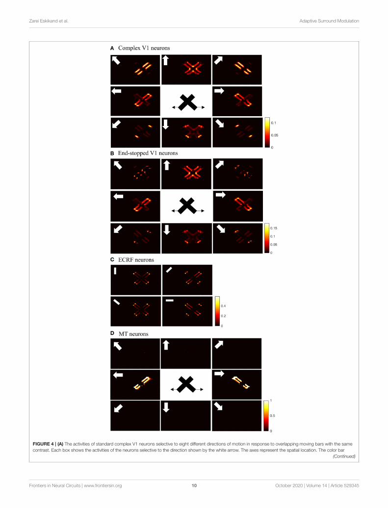
Initial motion signals are extracted by standard complex neuronsin V1. The small receptive fields of these neurons result inambiguous motion information. The activities of these neuronsin response to two overlapping moving bars with the samecontrast are shown in Figure 4A. The neurons selective todirections perpendicular to the edges of the bars have high levelsof activity at these locations because of the aperture problem. Theactivities of neurons at the intrinsic terminators express accurateestimations of the directions of movement of the bars. Theneurons selective to the upward direction also display high levelsof activity at the extrinsic terminators formed at the crossingpoints of the bars.
The existence of end-stopped neurons is essential for MTneurons to differentiate unambiguous motion information of theterminators from the ambiguous motion information that arisesfrom the aperture problem. Figure 4B shows the activities of theend-stopped neurons in response to the two overlapped movingbars. Among the neurons selective to the rightward direction,those at the intrinsic terminators of the bar that is movingto the right, have high levels of activity. Similarly, among theneurons selective to the leftward direction, the end-points ofthe bar that is moving to the left, have high levels of activity.Apart from the intrinsic terminators with unambiguous motioninformation, end-stopped neurons selective to upward directionshave higher levels of activity at the extrinsic terminators, whichrepresent upward local motion of the crossing junction of theoverlapping bars.
To discriminate the motion signals of the intrinsic fromthe extrinsic terminators, the third set of luminance sensitiveV1 neurons with suppressive surrounds play a significantrole. The activities of these neurons are shown in Figure 4C.They are strongly suppressed at the extrinsic terminatorswhere the inhibitory surrounds of the neurons are morestimulated compared to the intrinsic terminators. Therefore,excitatory connections from these neurons assist the modeledMT neurons to differentiate the unambiguous motion signals ofthe intrinsic terminators from the local motion signals of theextrinsic terminators.
The activities of the three sets of V1 neurons are transmittedto the neurons in MT. The surrounds of the MT neurons have anexcitatory effect when there is motion coherency. The excitatoryeffects of the surround assist MT neurons to propagate motionsignals. The surrounds of the neurons have an inhibitory effectat the discontinuities where the contrast level is high, whichassist MT neurons to segregate the motion signals of differentmoving objects. The disambiguation of the motion signals byMT neurons takes 30ms. Figure 4D shows the activities of MTneurons. MT neurons represent the correct direction of themotion of the overlapping bars. The MT neurons selective to the
rightward direction have high levels of activity along the edgesof the bar moving to the right and the MT neurons selectiveto the leftward direction have high levels of activity along theedges of the bar that is moving to the left. Without the contrastadaptive property of the MT neurons, the model requires twosets of MT neurons, named integration and segmentation MTneurons, for the correct estimation of motion. Introducing thecontrast adaptive feature of the MT neurons, which also accordswith existing neurophysiological findings, creates a unifiedmodelof MT neurons.
The spatial dynamics of the surround effect of theMT neuronsselective to the leftward direction is shown in Figure 5. Thesurround regions of the neurons are suppressive at the sharpedges of the bar, which is moving to the left. The surrounds of theneurons selective to the leftward direction are not activated alongthe edge of the bar, which is moving to the right. The inhibitoryeffect of the surround switches to the excitatory effect along thebar to assist MT neurons in propagating the motion informationfrom the intrinsic terminators.
To investigate the pattern motion selectivity of MT neurons,we examine the response of the model to a plaid pattern,which is obtained by occluding the intrinsic terminators of theoverlapping moving bars. The responses of the MT neurons tothe pattern or component motion of the stimulus are highlydependent on the connection strengths of the received inputscomputed by standard V1 and ECRF neurons. The activities ofthe MT neurons with strong connections associated with theinteraction of ECRF and standard V1 neurons are shown inFigure 6B.
The interaction with the ECRF neurons prevents thepropagation of motion information from the extrinsicterminators along the whole of the stimulus. Therefore, MTneurons respond to the component motion informationthat they receive from standard V1 neurons and theirresponse does not change over time, in contrast to thepattern MT neurons. The MT neurons respond to the patternmotion of the stimulus when there is a weak connectionfrom the ECRF neurons. The activities of the neuronsresponding to the pattern motion of the stimuli are shownin Figure 6A.
The motion information of the extrinsic terminators isnot suppressed when there is a weak connection fromECRF neurons to the pattern MT neurons. Therefore, theactivities of the neurons in response to the local motionof the extrinsic terminators propagate to the other regions.Pattern MT neurons represent the component motion ofthe stimulus at the beginning and they respond to thepattern motion after a delay. This is because of the timerequired for the propagation of the motion information fromextrinsic terminators.
Figure 7A shows the activities of pattern MT neuronsresponding to the pattern motion in the case of a difference inthe contrast of the bars. The MT neurons selective to the upwarddirection have a high level of activity in response to the upwardpattern motion. Figure 7B shows the activities of component MTneurons in response to the pattern motion. The neurons are ableto detect the individual component motions and, as the contrasts
Frontiers in Neural Circuits | www.frontiersin.org 8 October 2020 | Volume 14 | Article 529345

Zarei Eskikand et al. Adaptive Surround Modulation
FIGURE 3 | (A) The changes in the surround effect when the activity levels of the neurons selective to other directions change from 0 to 1 and the level of the contrast
is 0. The effect of the surround is suppressive when the level of 1x,y,θ is above 0.14 but is integrative when these neurons have a low level of activity. (B) The changes
in the level of surround suppression with contrast. The surround effect is suppressive when the contrast of the stimulus goes beyond the value of cr = 0.2. The level of
suppression increases with an increase in the contrast of the stimulus until it saturates. The surround effect is excitatory for low levels of the contrast, below cr .
TABLE 4 | A summary of the known neurophysiological findings and unknown features that are hypothesized in the model.
Existence of different subtypes of neurons/
connections/ circuitry of the mechanisms/ phenomena
Known Selected references
Direction selective standard complex V1 neurons X Hubel and Wiesel (1962), Dreher (1972), Movshon
(1975), Movshon et al. (1978a,b)
End-stopped V1 neurons X Hubel and Wiesel (1965), Sceniak et al. (2001), Pack
et al. (2003), Tsui et al. (2010)
Orientation selective V1 neurons with suppressive surround
(ECRF neurons)
X Cavanaugh et al. (2002)
Component and pattern selective MT neurons X Adelson and Movshon (1982), Albright (1984), Rodman
and Albright (1989), Livingstone et al. (2001), Born and
Bradley (2005)
Difference in the temporal dynamics of the component and
pattern MT neurons
X Smith et al. (2005, 2009)
Projection of the complex V1 neurons to MT area X Maunsell and van Essen (1983), Movshon and Newsome
(1996)
Projection of the end-stopped V1 neurons to MT area X Movshon and Newsome (1996), Sceniak et al. (2001)
Projection of the ECRF neurons to MT area Hypothesized in the model
Suppressive effect of the surround in V1 X Hubel and Wiesel (1968), Cavanaugh et al. (2002)
Center-surround interaction of MT neurons X Allman et al. (1985), Raiguel et al. (1995), Albright and
Stoner (2002), Born and Bradley (2005)
Adaptive modulatory effect of the surround X Huang et al. (2007, 2008)
Circuitry of the modulatory effect of the surround Hypothesized in the model
Contrast dependency of the pattern selectivity of the MT
neurons
X Kumbhani et al. (2008)
Contrast dependency of the suppressive effect of the
surround in MT neurons
X Pack et al. (2005)
The activity of V1 neurons is gated by the activity of the ECRF
neurons
Hypothesized in the model
The level of the excitatory connections between MT neurons
depends on ECRF V1 neurons
Hypothesized in the model
Frontiers in Neural Circuits | www.frontiersin.org 9 October 2020 | Volume 14 | Article 529345
Zarei Eskikand et al. Adaptive Surround Modulation
FIGURE 4 | (A) The activities of standard complex V1 neurons selective to eight different directions of motion in response to overlapping moving bars with the same
contrast. Each box shows the activities of the neurons selective to the direction shown by the white arrow. The axes represent the spatial location. The color bar
(Continued)
Frontiers in Neural Circuits | www.frontiersin.org 10 October 2020 | Volume 14 | Article 529345

Zarei Eskikand et al. Adaptive Surround Modulation
FIGURE 4 | shows the strength of the activity, brighter for higher values. The white arrows indicate the preferred direction of the neurons in each graph. The stimulus
is two crossing bars with the same level of contrast, as illustrated in the middle of the figure. The bar with 135◦ orientation is moving to the right and the bar with 45◦
orientation is moving to the left (black horizontal arrows). The neurons have high levels of activity at the terminators and along the edges of the bars. (B) The activities
of end-stopped V1 neurons. The neurons have high levels of activity at both the intrinsic and extrinsic terminators. (C) The activities of ECRF V1 neurons. The
preferred orientations of the neurons in each graph are shown by the white bars. The neurons have the highest levels of activity at the intrinsic terminators and their
activities are strongly suppressed at the extrinsic terminators. (D) The activities of MT neurons with adaptive surrounds. The MT neurons selective to the rightward
direction have high levels of activity in response to the motion of the bar moving to the right and the neurons selective to the leftward direction have high levels of
activity in response to the motion of the bar moving to the left. The activities of the MT neurons are suppressed at the crossing junction where there is more than one
moving object in the same depth plane.
FIGURE 5 | The surround effect of the MT neurons selective to the leftward direction with receptive fields along the vertical axis of the stimulus. The neurons have a
suppressive surround at the discontinuities along the edge of the leftward moving bar. The neurons have an excitatory effect followed by a suppression that results in
the propagation of the activity of the neurons responding to the intrinsic terminators. The surround of the neurons selective to the leftward direction are not activated
along the edge of the bar, which is moving to the right.
of the bars are different, they are also able to estimate which baris moving in front.
DISCUSSION
We developed a model of MT neurons that adapts its propertiesdepending on the input stimulus. The activities of theseneurons are influenced by their ECRFs, which have directionallyantagonistic or facilitatory effects depending on the propertiesof the input stimulus. For preferred direction motion, thesurround regions are suppressive if there are discontinuities inthe visual field or the contrast levels of the stimulus are high.The amount of suppression increases with increases in contrast.The surrounds are facilitatory when there is coherency in thedirections of motion across the visual field, which assists themodeled MT neurons in the propagation of motion signals.Our model explains the circuitry of this modulatory effect of
surrounds and provides insights into the mechanism by whichdifferent surround effects arise depending on the properties ofthe input stimulus.
The modulatory effect of the surround of MT neuronshas been reported in several neurophysiological findings.For example, the neurophysiological experiments by Huanget al. (2007) showed that integration-MT neurons switch tosegmentation-MT neurons depending on the ambiguity in themotion information of the stimulus. According to their findings,MT neurons are able to overcome the aperture problem by beingintegrative but can also code segmentation when stimulatedby random dot patterns (Huang et al., 2007, 2008). Thepsychophysical studies by Tadin et al. (2003) also provide someinsights on the possibility of the contrast dependency of thesurround. The adaptive change of the relationship between thecenter and surround regions of MT neurons with contrast hasalso been shown by other studies. The experiments by Pack et al.
Frontiers in Neural Circuits | www.frontiersin.org 11 October 2020 | Volume 14 | Article 529345

Zarei Eskikand et al. Adaptive Surround Modulation
FIGURE 6 | (A) The activities of pattern motion selective MT neurons responding to pattern motion. The neurons selective to the upward direction have the highest
levels of activity, representing the pattern motion of the stimulus. The stimulus is shown in the middle, which is two crossing bars with hidden intrinsic terminators. The
bars are actually moving to the left and right (horizontal black arrows). Each box shows the activities of the neurons selective to the direction shown by the white
arrow. The color bar shows the strength of activity (brighter for higher values) and the axes represent the spatial location. (B) The activities of the neurons selective to
the component motion in response to the pattern motion. The neurons selective to the up-right and up-left directions have the highest levels of activity, representing
the component motion of the stimulus.
(2005) showed a reduction in the suppression level as stimuluscontrast was decreased.
The surrounds of the MT neurons in our model are contrastadaptive. Therefore, they have the characteristics of integrationMT neurons when there is coherency in the input stimulus tofacilitate the propagation of motion signals from bar terminatorsto other regions. The surrounds of the neurons have a suppressiveeffect in the case of high contrast at motion discontinuities. Thepattern or component selectivity of the MT neurons dependson the input that they receive from the interaction of theECRF neurons and standard complex V1 neurons. The strongconnections from these neurons provide form information to
assist MT neurons in suppressing the effects of the extrinsicterminators and respond to the component motions of thestimuli. Weak connections from these neurons result in patternmotion selectivity of the MT neurons. The pattern selectivityof the MT neurons is highly dependent on the contrast of thestimulus. The pattern motion selectivity index drops significantlywhen the contrast of the overlapping bars differs. The differencein the contrast of the overlapping bars results in the formationof illusionary depth when it appears that one of the bars issliding over the other bar, which results in the dominance ofcomponent over pattern motion selectivity. The experiments byKumbhani et al. (2008) supports the effect of the contrast on the
Frontiers in Neural Circuits | www.frontiersin.org 12 October 2020 | Volume 14 | Article 529345

Zarei Eskikand et al. Adaptive Surround Modulation
FIGURE 7 | (A) The activities of pattern motion selective MT neurons responding to pattern motion with bars of different contrasts. The neurons selective to the
upward direction have the highest levels of activity, representing the pattern motion of the stimulus. The stimulus is shown in the middle, which is two crossing bars
with hidden intrinsic terminators. The bars are actually moving to the left and right (horizontal black arrows). Each box shows the activities of the neurons selective to
the direction shown by the white arrow. The color bar shows the strength of activity (brighter for higher values) and the axes represent the spatial location. (B) The
activities of the neurons selective to the component motion in response to the pattern motion. The neurons selective to up-right and up-left directions have the highest
levels of activity, representing the component motion of the stimulus.
pattern selectivity of the neurons. The results of their experimentsshow that the pattern selectivity index of the MT neurons dropssignificantly if the contrast of one of the gratings is reduced(Kumbhani et al., 2008).
In accordance with these neurophysiological findings, MTneurons are not categorized into integration or segmentationneurons in our proposed model. They represent the integrativeor segregated features of the MT neurons depending on thecharacteristics of the input stimulus. Therefore, the activitiesof the MT neurons are influenced by their adaptive surround,
which changes over time and space depending on the stimuli.Also, our model suggests that MT neurons are not grouped intodifferent types of pattern or component MT neurons. Their levelof pattern selectivity is only determined by the strength of theconnections from ECRF neurons. Therefore, they are selectiveto the component motions of the stimuli when they receivestrong input from ECRF neurons, and they represent the featureof the pattern selective MT neurons when the strength of theconnection from the form processing input is weak or there isno connection. This theory on the pattern motion selectivity of
Frontiers in Neural Circuits | www.frontiersin.org 13 October 2020 | Volume 14 | Article 529345

Zarei Eskikand et al. Adaptive Surround Modulation
theMT neurons contrasts with themodeling works by Simoncelliand Heeger (1998) and Rust et al. (2006). Simoncelli and Heeger(1998) suggested a linear-nonlinear model, which was furtherdeveloped by Rust et al. (2006). The non-linear normalizationin these models suppresses the unambiguous motion signalsresulting from the aperture problem, which is similar to therole of end-stopping neurons in our model. The mechanismfor pattern motion detection in the linear-nonlinear model isbased on the hierarchical relationship between component andpattern MT neurons. Our model suggests an entirely differentmechanism for pattern motion selectivity of MT neurons, whichcan explain the spatial and temporal limits on pattern motionselectivity of MT neurons observed in the experiments by Majajet al. (2007) and Kumbhani et al. (2015). However, the linear-nonlinear model is not capable of explaining these features ofpattern MT neurons. Due to the simulation of the propagationof activity, our model can replicate the temporal dynamics ofcomponent and pattern MT neurons observed by Smith et al.(2005, 2009), which shows a temporal delay in the patternmotiondetection of the MT neurons. The linear-nonlinear model lacksthese properties of the MT neurons (Simoncelli and Heeger,1998; Rust et al., 2006).
DATA AVAILABILITY STATEMENT
All datasets generated for this study are included in thearticle/supplementary material.
AUTHOR CONTRIBUTIONS
PZ, TK, MI, AB, and DG: conceptualization, methodology,validation, and writing–review and editing. TK, MI, AB, and DG:funding acquisition and supervision. DG: project administrationand resources. PZ: investigation, software, visualization, formalanalysis, and writing–original draft. All authors contributed tothe article and approved the submitted version.
FUNDING
This research was supported by the Australian Research Councilthrough Discovery Grants [DE120102210 and DP140104533],the ARC Center of Excellence for Integrative Brain Function[CE140100007] and Victorian Life Sciences ComputationInitiative (VLSCI) grant number VR0138 on its Peak ComputingFacility (Melbourne Bioinformatics) at the University ofMelbourne, an initiative of the Victorian Government.
ACKNOWLEDGMENTS
PZ acknowledges a postgraduate scholarship from the NationalInformation and Communication Technology Australia(NICTA- http://www.nicta.com.au/). NICTA was funded by theAustralian Government as represented by the Department ofBroadband, Communications and the Digital Economy (http://www.communications.gov.au/) and the Australian ResearchCouncil through the ICT Center of Excellence program.
REFERENCES
Adelson, E. H., and Bergen, J. R. (1985). Spatiotemporal energy models
for the perception of motion. J. Optical Soc. Am. A 2, 284–299.
doi: 10.1364/JOSAA.2.000284
Adelson, E. H., and Movshon, J. A. (1982). Phenomenal coherence of moving
visual patterns. Nature 300, 523–525. doi: 10.1038/300523a0
Albright, T. D. (1984). Direction and orientation selectivity of neurons
in visual area MT of the macaque. J. Neurophysiol. 52, 1106–1130.
doi: 10.1152/jn.1984.52.6.1106
Albright, T. D., and Stoner, G. R. (2002). Contextual influences
on visual processing. Annual Rev. Neurosci. 25, 339–379.
doi: 10.1146/annurev.neuro.25.112701.142900
Allman, J., Miezin, F., and McGuinness, E. (1985). Direction- and velocity-
specific responses from beyond the classical receptive field in the middle
temporal visual area (MT). Perception 14, 105–126. doi: 10.1068/p1
40105
Barlow, H. B., and Levick, W. R. (1965). The mechanism of
directionally selective units in rabbit’s retina. J. Physiol. 178, 477–504.
doi: 10.1113/jphysiol.1965.sp007638
Born, R. T., and Bradley, D. C. (2005). Structure and function
of visual area MT. Annual Rev. Neurosci. 28, 157–189.
doi: 10.1146/annurev.neuro.26.041002.131052
Cavanaugh, J. R., Bair, W., and Movshon, J. A. (2002). Selectivity and spatial
distribution of signals from the receptive field surround in macaque V1
neurons. J. Neurophysiol. 88, 2547–2556. doi: 10.1152/jn.00693.2001
Dreher, B. (1972). Hypercomplex cells in the cat’s striate cortex. Invest. Ophthalmol.
Visual Sci. 11, 355–356.
Huang, X., Albright, T. D., and Stoner, G. R. (2007). Adaptive
surround modulation in cortical area MT. Neuron 53, 761–770.
doi: 10.1016/j.neuron.2007.01.032
Huang, X., Albright, T. D., and Stoner, G. R. (2008). Stimulus dependency
and mechanisms of surround modulation in cortical area MT.
J. Neurosci. 28, 13889–13906. doi: 10.1523/JNEUROSCI.1946-0
8.2008
Hubel, D. H., and Wiesel, T. N. (1962). Receptive fields, binocular interaction
and functional architecture in the cat’s visual cortex. J. Physiol. 160, 106–154.
doi: 10.1113/jphysiol.1962.sp006837
Hubel, D. H., andWiesel, T. N. (1965). Receptive fields and functional architecture
in two non-striate visual areas (18 and 19) of the cat. J. Neurophysiol. 28,
229–289. doi: 10.1152/jn.1965.28.2.229
Hubel, D. H., and Wiesel, T. N. (1968). Receptive fields and functional
architecture of monkey striate cortex. J. Physiol. 195, 215–243.
doi: 10.1113/jphysiol.1968.sp008455
Kay, K. N. (2018). Principles for models of neural information processing.
NeuroImage 180, 101–109. doi: 10.1016/j.neuroimage.2017.08.016
Kumbhani, R., Saber, G., Majaj, N., Tailby, C., and Movshon, J. (2008). Contrast
affects pattern direction selectivity in macaque MT neurons. SFN Annual
Meeting, Washington, DC.
Kumbhani, R. D., El-Shamayleh, Y., and Movshon, J. A. (2015). Temporal
and spatial limits of pattern motion sensitivity in macaque MT neurons. J.
Neurophysiol. 7, 1977–1988. doi: 10.1152/jn.00597.2014
Livingstone, M. S., Pack, C. C., and Born, R. T. (2001). Two-
dimensional substructure of MT receptive fields. Neuron 30, 781–793.
doi: 10.1016/S0896-6273(01)00313-0
Majaj, N. J., Carandini, M., and Movshon, J. A. (2007). Motion integration
by neurons in macaque MT is local, not global. J. Neurosci. 2, 366–370.
doi: 10.1523/JNEUROSCI.3183-06.2007
Maunsell, J. H., and van Essen, D. C. (1983). The connections of
the middle temporal visual area (MT) and their relationship to a
cortical hierarchy in the macaque monkey. J. Neurosci. 3, 2563–2586.
doi: 10.1523/JNEUROSCI.03-12-02563.1983
Frontiers in Neural Circuits | www.frontiersin.org 14 October 2020 | Volume 14 | Article 529345

Zarei Eskikand et al. Adaptive Surround Modulation
Movshon, J. A. (1975). The velocity tuning of single units in cat striate cortex. J.
Physiol. 249, 445–468. doi: 10.1113/jphysiol.1975.sp011025
Movshon, J. A., and Newsome, W. T. (1996). Visual response properties of striate
cortical neurons projecting to area MT in macaque monkeys. J. Neurosci. 16,
7733–7741. doi: 10.1523/JNEUROSCI.16-23-07733.1996
Movshon, J. A., Thompson, I. D., and Tolhurst, D. J. (1978a). Receptive field
organization of complex cells in the cat’s striate cortex. J. Physiol. 283, 79–99.
doi: 10.1113/jphysiol.1978.sp012489
Movshon, J. A., Thompson, I. D., and Tolhurst, D. J. (1978b). Spatial and
temporal contrast sensitivity of neurones in areas 17 and 18 of the
cat’s visual cortex. J. Physiol. 283, 101–120. doi: 10.1113/jphysiol.1978.sp0
12490
Pack, C. C., Hunter, J. N., and Born, R. T. (2005). Contrast dependence of
suppressive influences in cortical area MT of alert macaque. J. Neurophysiol.
93, 1809–1815. doi: 10.1152/jn.00629.2004
Pack, C. C., Livingstone, M. S., Duffy, K. R., and Born, R. T. (2003). End-
stopping and the aperture problem: Two-dimensional motion signals
in macaque V1. Neuron 39, 671–680. doi: 10.1016/S0896-6273(03)0
0439-2
Raiguel, S., Hulle, M., Xiao, D., Marcar, V., and Orban, G. (1995). Shape and
spatial distribution of receptive fields and antagonistic motion surrounds in
the middle temporal area (V5) of the macaque. Eur. J. Neurosci. 7, 2064–2082.
doi: 10.1111/j.1460-9568.1995.tb00629.x
Rodman, H., and Albright, T. (1989). Single-unit analysis of pattern-motion
selective properties in the middle temporal visual area (MT). Exp. Brain Res.
75, 53–64. doi: 10.1007/BF00248530
Rust, N. C., Mante, V., Simoncelli, E. P., and Movshon, J. A. (2006). How MT
cells analyze the motion of visual patterns. Nat. Neurosci. 11, 1421–1431.
doi: 10.1038/nn1786
Sceniak, M. P., Hawken, M. J., and Shapley, R. (2001). Visual spatial
characterization of macaque V1 neurons. J. Neurophysiol. 85, 1873–1887.
doi: 10.1152/jn.2001.85.5.1873
Simoncelli, E. P., and Heeger, D. J. (1998). A model of neuronal responses in visual
area MT. Vision Res. 5, 743–761. doi: 10.1016/S0042-6989(97)00183-1
Smith, M. A., Majaj, N., and Movshon, J. A. (2009). Dynamics of pattern
motion computation. Dynamics of Visual Motion Processing. Springer. 55–72.
doi: 10.1007/978-1-4419-0781-3_3
Smith, M. A., Majaj, N. J., and Movshon, J. A. (2005). Dynamics of motion
signaling by neurons in macaque area MT. Nat. Neurosci. 8, 220–228.
doi: 10.1038/nn1382
Tadin, D., Lappin, J. S., Gilroy, L. A., and Blake, R. (2003). Perceptual consequences
of centre–surround antagonism in visual motion processing. Nature 424,
312–315. doi: 10.1038/nature01800
Thiele, A. (2007). Reconstructing the world: switching from segmentation to
integration allows neurons in area MT to make “sense” of the visual scene.
Neuron 53, 623–625. doi: 10.1016/j.neuron.2007.02.008
Tsui, J. M., Hunter, J. N., Born, R. T., and Pack, C. C. (2010). The role of
V1 surround suppression in MT motion integration. J. Neurophysiol. 103,
3123–3138. doi: 10.1152/jn.00654.2009
Van Santen, J. P., and Sperling, G. (1985). Elaborated reichardt detectors. J. Optical
Soc. Am. A 2, 300–321. doi: 10.1364/JOSAA.2.000300
Zarei Eskikand, P., Kameneva, T., Burkitt, A. N., Grayden, D. B., and Ibbotson,
M. R. (2019). Pattern motion processing by MT neurons. Front. Neural Circuits
13:43. doi: 10.3389/fncir.2019.00043
Zarei Eskikand, P., Kameneva, T., Ibbotson, M. R., Burkitt, A. N., and
Grayden, D. B. (2016). A possible role for end-stopped V1 neurons in
the perception of motion: a computational model. PLoS ONE 11:e0164813.
doi: 10.1371/journal.pone.0164813
Zarei Eskikand, P., Kameneva, T., Ibbotson, M. R., Burkitt, A. N., and
Grayden, D. B. (2018). A biologically-based computational model of visual
cortex that overcomes the X-junction illusion. Neural Netw. 102, 10–20.
doi: 10.1016/j.neunet.2018.02.008
Conflict of Interest: The authors declare that the research was conducted in the
absence of any commercial or financial relationships that could be construed as a
potential conflict of interest.
Copyright © 2020 Zarei Eskikand, Kameneva, Burkitt, Grayden and Ibbotson. This
is an open-access article distributed under the terms of the Creative Commons
Attribution License (CC BY). The use, distribution or reproduction in other forums
is permitted, provided the original author(s) and the copyright owner(s) are credited
and that the original publication in this journal is cited, in accordance with accepted
academic practice. No use, distribution or reproduction is permitted which does not
comply with these terms.
Frontiers in Neural Circuits | www.frontiersin.org 15 October 2020 | Volume 14 | Article 529345