adaptive servo-ventilation therapy using an innovative ... · original article adaptive...
TRANSCRIPT
ORIGINAL ARTICLE
Adaptive servo-ventilation therapy using an innovative ventilatorfor patients with chronic heart failure: a real-world, multicenter,retrospective, observational study (SAVIOR-R)
Shin-ichi Momomura • Yoshihiko Seino •
Yasuki Kihara • Hitoshi Adachi •
Yoshio Yasumura • Hiroyuki Yokoyama
Received: 5 March 2014 / Accepted: 11 July 2014 / Published online: 8 August 2014
� The Author(s) 2014. This article is published with open access at Springerlink.com
Abstract Adaptive servo-ventilation (ASV) therapy using
an innovative ventilator—originally developed to treat sleep-
disordered breathing (SDB)—is a novel modality of noninva-
sive positive pressure ventilation and is gaining acceptance
among Japanese cardiologists in expectation of its applicability
to treat patients with chronic heart failure (CHF) based on its
acute beneficial hemodynamic effects. We conducted a mul-
ticenter, retrospective, real-world observational study in 115
Japanese patients with CHF, who had undergone home ASV
therapy for the first time from January through December 2009,
to examine their profile and the effects on their symptoms and
hemodynamics. Medical records were used to investigate New
York Heart Association (NYHA) class, echocardiographic
parameters including left ventricular ejection fraction (LVEF),
cardiothoracic ratio (CTR), brain natriuretic peptide (BNP),
and other variables. Most of the patients were categorized to
NYHA classes II (44.4 %) and III (40.7 %). SDB severity was
not determined in 44 patients, and SDB was not detected or was
mild in 27 patients. In at least 71 patients (61.7 %), therefore,
ASV therapy was not applied for the treatment of SDB. CHF
was more severe, i.e., greater NYHA class, lower LVEF, and
higher CTR, in 87 ASV-continued patients (75.7 %) than in 28
ASV-discontinued patients (24.3 %). However, SDB severity
was not related to continuity of ASV. The combined proportion
of NYHA classes III and IV (P = 0.012) and LVEF
(P = 0.009) improved significantly after ASV therapy. CTR
and BNP did not improve significantly after ASV therapy but
showed significant beneficial changes in their time-course
analysis (P\0.05, respectively). Improvements in LVEF and
NYHA class after ASV therapy were not influenced by SDB
severity at onset. The present study suggests that ASV therapy
would improve the symptoms and hemodynamics of CHF
patients, regardless of SDB severity. A randomized clinical
study to verify these effects is warranted.
Keywords Adaptive servo-ventilation � Chronic heart
failure � Noninvasive positive pressure ventilation � Cardiac
function � Sleep-disordered breathing
Introduction
Chronic heart failure (CHF) is the end-stage pathology of
all heart diseases [1], and pharmacotherapy is the first-line
Investigators for Study on the effects of Adaptive servo-Ventilation In
patients with chrOnic heart failuRe: Real-world, multicenter,
retrospective, observational study (SAVIOR-R) are listed in
Appendix.
S. Momomura (&)
Cardiovascular Medicine, Saitama Medical Center, Jichi
Medical University, 1-847 Amanuma-Cho, Omiya-Ku,
Saitama 330-8503, Japan
e-mail: [email protected];
Y. Seino
Cardiovascular Center, Nippon Medical School Chiba Hokusoh
Hospital, Chiba, Japan
Y. Kihara
Department of Cardiovascular Medicine, Hiroshima University,
Hiroshima, Japan
H. Adachi
Division of Cardiology, Gunma Prefectural Cardiovascular
Center, Gunma, Japan
Y. Yasumura
Cardiovascular Division, Osaka National Hospital, Osaka, Japan
H. Yokoyama
Department of Cardiovascular Medicine, National Cerebral and
Cardiovascular Center, Osaka, Japan
123
Heart Vessels (2015) 30:805–817
DOI 10.1007/s00380-014-0558-8
therapy for patients with CHF. Patient prognosis was
considerably improved by the diffusion of pharmacother-
apy and by the recent striking progress in non-pharmaco-
therapy including cardiac resynchronization therapy (CRT)
[2–6]. Nevertheless, CHF is a leading cause of cardiovas-
cular death [7, 8], and patients with CHF repeat admissions
for acute exacerbation of CHF.
Noninvasive positive pressure ventilation (NPPV) has
been shown to improve pulmonary congestion of patients
with acute heart failure (AHF) and in the acute exacerba-
tion of CHF through the following hemodynamic actions:
re-opening of collapsed alveoli, prevention of small airway
obstruction, enlargement of lung volume, improvements in
oxygenation and lung compliance [9–15], amelioration of
left ventricular afterload through a reduction in transmural
pressure induced by positive intrathoracic pressure [13,
14], and relief of left ventricular preload through a reduc-
tion in venous return [10, 15]. Based on these acute ben-
eficial effects of NPPV, cardiologists had been aware of the
potential applicability of NPPV to the treatment of CHF
patients. However, such application was very difficult to
realize because conventional ventilators used for NPPV
presented poor tolerability and cumbersome operability.
Therefore, the development of an innovative ventilator
capable of solving these drawbacks was expected. The
ventilator used for adaptive servo-ventilation (ASV), a
form of NPPV, offers superior tolerability and simple
operability based on the provision of support pressure; the
device was originally developed to treat sleep-disordered
breathing (SDB) [16] and is synchronized to the respiration
patterns of individual patients through its original algo-
rithm and potentially allows for the application of home
ASV therapy to the treatment of CHF patients.
In recent years, ASV therapy diffused rapidly and
widely in Japan and is gaining acceptance among cardiol-
ogists. A number of clinical studies [17–21] on ASV
therapy have been published. However, no clinical evi-
dence of ASV therapy in real-world patients at multiple
medical institutions has been available to date. The
objectives of the present study were to investigate the
actual practice of ASV therapy for patients with CHF in
Japan and to examine the effects of ASV therapy on their
symptoms and hemodynamics.
Patients and methods
Patients
Among Japanese outpatients with CHF who had been treated
at 16 medical institutions, 116 patients were enrolled (1) who
for the first time had undergone home ASV therapy from
January through December 2009, (2) who aged 20 years or
older at the onset of ASV therapy, and (3) who did not fall
under the exclusion criterion (patients considered by their
attending physician to be ineligible for the present study).
Furthermore, one of them was excluded because of discov-
ering the non-outpatient nature of the patient after enrolment.
In consequence, 115 patients (90 males and 25 females) were
analyzed for the efficacy of ASV therapy. The present study
was conducted after the acquisition of approval by the ethics
committee at each participating institution and in accordance
with the Declaration of Helsinki.
Study design and method
The present study is a multicenter retrospective observa-
tional study in Japanese patients with CHF in real-world
settings. In principle, medical records prepared for 1 year
before and after the onset of ASV therapy were used to
investigate the following items: regarding patient back-
ground, age, gender, underlying heart disease, complica-
tions, cardiovascular events, and others; regarding efficacy,
vital signs, symptoms of CHF, New York Heart Associa-
tion (NYHA) functional class, hematology, human brain
natriuretic peptide (BNP), echocardiography determining
left ventricular ejection fraction (LVEF), left ventricular
end-systolic dimension (LVDs), left ventricular end-dia-
stolic dimension (LVDd), and left atrial dimension (LAD),
chest X-ray documenting the cardiothoracic ratio (CTR),
sleep study, estimated glomerular filtration rate (eGFR),
and others; and regarding continuity, ventilator use and
others. Examinations were performed in accordance with
the standards valid at each participating institution. The
present study did not assess the safety of ASV therapy.
We used the following two categories of definitions for
the ‘‘baseline values’’: (1) for the statistical analysis of the
pre- and post-ASV values, ‘‘the values that were obtained
closest to the onset of ASV therapy in a range from day
-363 to day 7’’; and (2) for the statistical analysis of the
time-course changes in variables, ‘‘the values that were
obtained closest to the onset of ASV in a range from day
-56 to day 14.’’ Furthermore, we used the following def-
inition for the post-ASV values: ‘‘the values that were
obtained latest since day 8 after the onset of ASV therapy.’’
In addition, we established the allowable ranges
of ± 28 days for each of the other assessment points.
Ventilator used for ASV therapy
The ventilator for ASV therapy used in the present study
was an advanced bilevel positive airway pressure unit—
AutoSetTM CS (ResMed, Sydney, Australia). The device
learns the patient’s breathing rates and patterns, provides
proper pressure support that is synchronized to them
through its state-of-the-art fuzzy logic algorithms, and
806 Heart Vessels (2015) 30:805–817
123
generates smooth pressure waveforms mimicking the
patient’s normal respiration flow patterns. ASV therapy at
home was conducted in patients whose symptoms were
stable and for whom the attending physician considered it
appropriate. The device is used confinedly in the range of
coverage by the National Health Insurance System in
Japan. The application of ASV therapy to the treatment of
SDB is currently not covered by the system.
Statistical analyses
The statistical analyses to compare the pre- and post-ASV
values were performed using paired t test, one-sample
Wilcoxon’s signed rank sum test, and McNemar’s test for
parametric, nonparametric, and binary variables, respec-
tively. Subgroup analyses were performed using the gen-
eralized estimating equation procedure to examine time-
course changes in continuous and categorical variables,
followed by the least Fisher’s significant difference method
to determine the timing for generation of a statistically
significance difference. Stratified analyses to identify the
background factors impacting on the continuity and effi-
cacy of ASV therapy were conducted using Student’s t test,
two-sample Wilcoxon’s signed rank sum test, and Fisher’s
exact probability test for parametric, nonparametric, and
binary variables, respectively. Furthermore, multivariate
logistic regression analysis using Wald v2 test was per-
formed to identify patients’ background factors associated
with LVEF improvement. A value of P\ 0.05 was con-
sidered statistically significant. All statistical analyses were
performed using a statistical software package, version 9.2
(SAS Institute Inc., Cary, NC, USA).
Results
Characteristics of CHF patients who underwent ASV
therapy
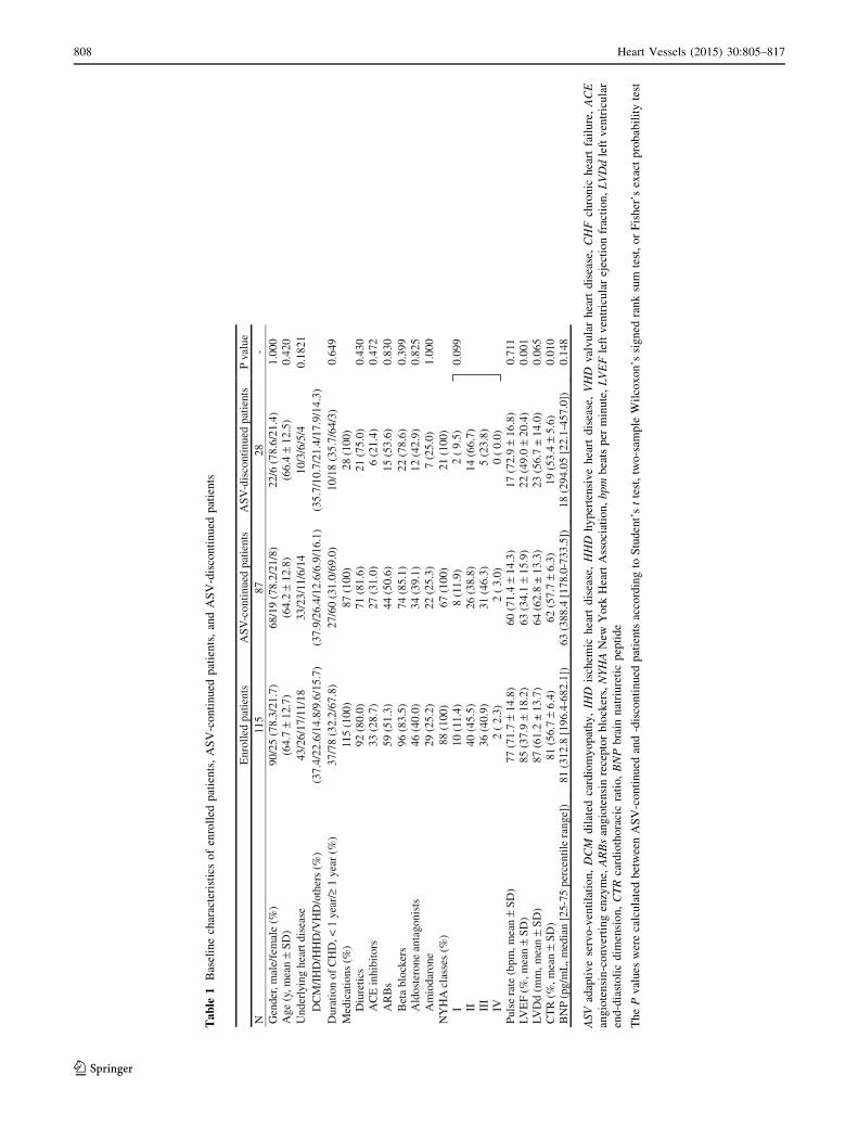
Characteristics of 115 patients at the onset of ASV therapy
are shown in Table 1. Mean age was 64.7 ± 12.7 years,
male gender was predominant—78.3 %, the proportion of
patients with dilated cardiomyopathy (DCM) was 37.4 %,
and patients having a disease duration of C1 year accounted
for 67.8 %. At onset, more than 80 % of patients received
diuretics and beta blockers, and approximately 80 % of
patients were medicated with angiotensin-converting
enzyme inhibitors and angiotensin II receptor blockers.
Despite the fact that patients had already undergone the
sufficient treatment of their heart failure (HF), the combined
proportion of patients with NYHA class III and IV HF was
as high as 43.2 %, mean LVEF was 37.9 %, mean CTR
was 56.7 %, and median plasma BNP concentration was
312.8 pg/mL. Therefore, the majority of patients on ASV
therapy were found to have severe CHF. All patients
underwent ASV therapy providing end-expiratory pressure,
minimum pressure support, and maximum pressure support.
The default values, the number of patients who used the
device under default settings, and the range for these pres-
sures were, respectively, as follows: 5, 3, and 10; 81, 112,
and 105; and 3–8, 3–4, and 8–12 cmH2O.
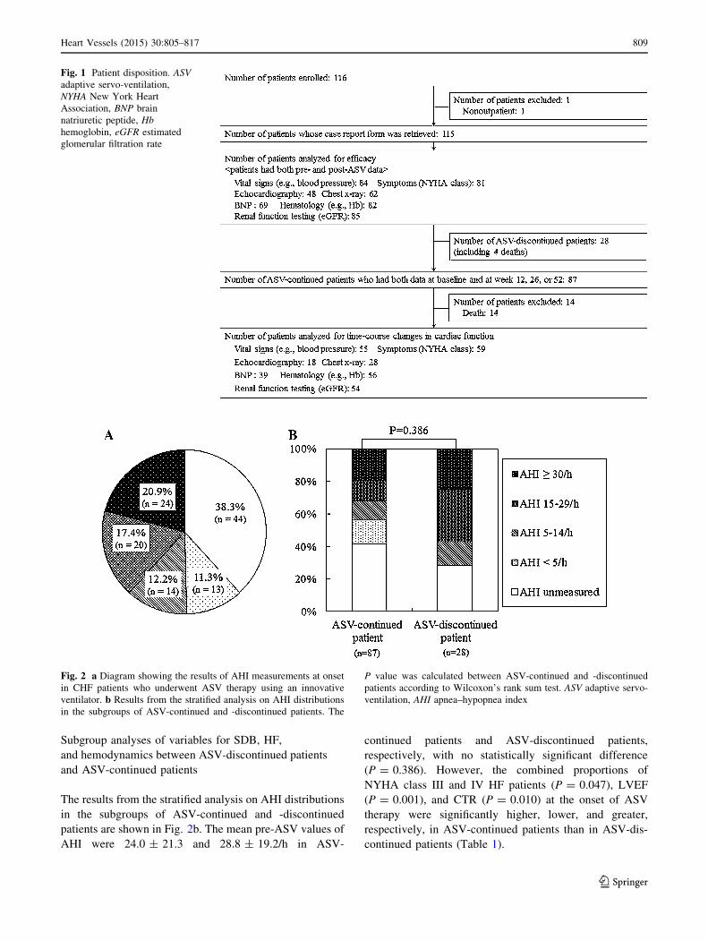
Patient disposition
Patient disposition is shown in Fig. 1. The retrieval rate of
the case report form on 115 patients who were analyzed for
efficacy was 100 %; the attending physician had discontin-
ued ASV therapy at his/her discretion in 28 patients
(24.3 %) of them (ASV-discontinued patients). The most
predominant reason for discontinuation of ASV therapy was
‘‘impatience’’ (20 patients, 71.4 %), followed by ‘‘economic
reason’’ (4 patients, 14.3 %), ‘‘improvement in HF’’ (2
patients, 7.1 %), and ‘‘deterioration of HF’’ (2 patients,
7.1 %). In contrast, the attending physician had continued
ASV therapy at his/her discretion in 87 patients (75.6 %) of
them (ASV-continued patients), 13 of whom died due to the
progression of HF. The proportions of patients were 40.9 %
(47/115) to 73.9 % (85/115), who were analyzed for seven
investigation items: vital signs [body weight, pulse rate,
systolic blood pressure (SBP), and diastolic blood pressure
(DBP)], echocardiography, BNP, renal function test, symp-
toms of CHF, chest X-ray, and hematology.
Among 115 patients who were analyzed for efficacy, 18
died within 1 year after the onset of ASV therapy: 16 died
due to the spontaneous deterioration of HF or to lethal
arrhythmias, 1 to suicide, and 1 to ileus. It was the
attending physician who had found no causality between
ASV therapy and death at his/her discretion.
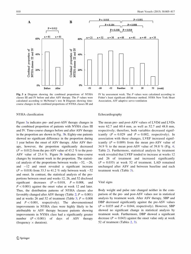
Sleep study at the onset of ASV therapy
The status of conducting the sleep study at the onset of ASV
therapy is shown in Fig. 2a. Patients, who underwent the
study, were assessed for the severity of SDB by means of the
apnea–hypopnea index (AHI). Consequently, SDB was
present in 50.4 % (58/115) of patients. The percentages of
patients with mild, moderate, and severe SDB were 12.2 %
(14/115), 17.4 % (20/115), and 20.9 % (24/115), respectively.
Patients with CHF who were complicated by moderate or
severe SDB accounted for 38.3 % (44/115) of patients. On
the other hand, the proportions of patients whose SDB
severity was not assessed because the sleep study was not
performed and of patients who were not complicated by SDB
were 38.3 % (44/115) and 11.3 % (13/115), respectively.
Namely, ASV therapy was not applied for the objective of
treating SDB in at least 61.7 % of patients.
Heart Vessels (2015) 30:805–817 807
123
Table
1B
asel
ine
char
acte
rist
ics
of
enro
lled
pat
ien
ts,
AS
V-c
on
tin
ued
pat
ien
ts,
and
AS
V-d
isco
nti
nu
edp
atie
nts
Enr
olle
d pa
tient
sA
SV-c
ontin
ued
patie
nts
ASV
-dis
cont
inue
d pa
tient
sP
valu
eN
115
8728
-G
ende
r, m
ale/
fem
ale
(%)
90/2
5 (7
8.3/
21.7
)68
/19
(78.
2/21
/8)
22/6
(78
.6/2
1.4)
1.00
0A
ge (
y, m
ean
±SD
)(6
4.7
±12
.7)
(64.
2 ±
12.8
)(6
6.4
±12
.5)
0.42
0U
nder
lyin
g he
art d
isea
se43
/26/
17/1
1/18
33/2
3/11
/6/1
410
/3/6
/5/4
0.18
21D
CM
/IH
D/H
HD
/VH
D/o
ther
s(%
)(3
7.4/
22.6
/14.
8/9.
6/15
.7)
(37.
9/26
.4/1
2.6/
6.9/
16.1
)(3
5.7/
10.7
/21.
4/17
.9/1
4.3)
Dur
atio
n of
CH
D, <
1 y
ear/
≥1
year
(%
)37
/78
(32.
2/67
.8)
27/6
0 (3
1.0/
69.0
)10
/18
(35.
7/64
/3)
0.64
9M
edic
atio
ns (
%)
115
(100
)87
(10
0)28
(10
0)D
iure
tics
92 (
80.0
)71
(81
.6)
21 (
75.0
)0.
430
AC
E in
hibi
tors
33 (
28.7
)27
(31
.0)
6 (2
1.4)
0.47
2A
RB
s59
(51
.3)
44 (
50.6
)15
(53
.6)
0.83
0B
eta
bloc
kers
96 (
83.5
)74
(85
.1)
22 (
78.6
)0.
399
Ald
oste
rone
ant
agon
ists
46 (
40.0
)34
(39.
1)12
(42.
9)0.
825
Am
ioda
rone
29 (
25.2
)22
(25
.3)
7 (2
5.0)
1.00
0N
YH
A c
lass
es (
%)
88 (
100)
67 (
100)
21 (
100)
I10
(11
.4)
8 (1
1.9)
2 (
9.5)
0.09
9II
40 (
45.5
)26
(38
.8)
14 (
66.7
)II
I36
(40
.9)
31 (
46.3
)5
(23.
8)IV
2 (
2.3)
2 (
3.0)
0 (
0.0)
Puls
e ra
te (
bpm
, mea
n ±
SD)
77 (
71.7
±14
.8)
60 (
71.4
±14
.3)
17 (
72.9
±16
.8)
0.71
1LV
EF
(%, m
ean
±SD
)85
(37
.9 ±
18.2
)63
(34
.1 ±
15.9
)22
(49
.0 ±
20.4
)0.
001
LVD
d (m
m, m
ean
±SD
)87
(61
.2 ±
13.7
)64
(62
.8 ±
13.
3)23
(56
.7±
14.0
)0.
065
CT
R (
%, m
ean
±SD
)81
(56
.7 ±
6.4)
62 (
57.7
±6.
3)19
(53
.4 ±
5.6)
0.01
0B
NP
(pg/
mL
, med
ian
[25-
75 p
erce
ntile
ran
ge])
81 (
312.
8[1
96.4
-682
.1])
63 (
388.
4 [1
78.0
-733
.5])
18(2
94.0
5[2
2.1-
457.
0])
0.14
8
ASV
adap
tiv
ese
rvo
-ven
tila
tio
n,DCM
dil
ated
card
iom
yo
pat
hy
,IH
Dis
chem
ich
eart
dis
ease
,HHD
hy
per
ten
siv
eh
eart
dis
ease
,VHD
val
vu
lar
hea
rtd
isea
se,CHF
chro
nic
hea
rtfa
ilu
re,ACE
ang
iote
nsi
n-c
on
ver
tin
gen
zym
e,ARBs
ang
iote
nsi
nre
cep
tor
blo
cker
s,NYHA
New
Yo
rkH
eart
Ass
oci
atio
n,bpm
bea
tsp
erm
inu
te,LVEF
left
ven
tric
ula
rej
ecti
on
frac
tio
n,LVDd
left
ven
tric
ula
r
end
-dia
sto
lic
dim
ensi
on
,CTR
card
ioth
ora
cic
rati
o,BNP
bra
inn
atri
ure
tic
pep
tid
e
Th
eP
val
ues
wer
eca
lcu
late
db
etw
een
AS
V-c
on
tin
ued
and
-dis
con
tin
ued
pat
ien
tsac
cord
ing
toS
tud
ent’
st
test
,tw
o-s
amp
leW
ilco
xo
n’s
sig
ned
ran
ksu
mte
st,
or
Fis
her
’sex
act
pro
bab
ilit
yte
st
808 Heart Vessels (2015) 30:805–817
123
Subgroup analyses of variables for SDB, HF,
and hemodynamics between ASV-discontinued patients
and ASV-continued patients
The results from the stratified analysis on AHI distributions
in the subgroups of ASV-continued and -discontinued
patients are shown in Fig. 2b. The mean pre-ASV values of
AHI were 24.0 ± 21.3 and 28.8 ± 19.2/h in ASV-
continued patients and ASV-discontinued patients,
respectively, with no statistically significant difference
(P = 0.386). However, the combined proportions of
NYHA class III and IV HF patients (P = 0.047), LVEF
(P = 0.001), and CTR (P = 0.010) at the onset of ASV
therapy were significantly higher, lower, and greater,
respectively, in ASV-continued patients than in ASV-dis-
continued patients (Table 1).
Fig. 1 Patient disposition. ASV
adaptive servo-ventilation,
NYHA New York Heart
Association, BNP brain
natriuretic peptide, Hb
hemoglobin, eGFR estimated
glomerular filtration rate
Fig. 2 a Diagram showing the results of AHI measurements at onset
in CHF patients who underwent ASV therapy using an innovative
ventilator. b Results from the stratified analysis on AHI distributions
in the subgroups of ASV-continued and -discontinued patients. The
P value was calculated between ASV-continued and -discontinued
patients according to Wilcoxon’s rank sum test. ASV adaptive servo-
ventilation, AHI apnea–hypopnea index
Heart Vessels (2015) 30:805–817 809
123
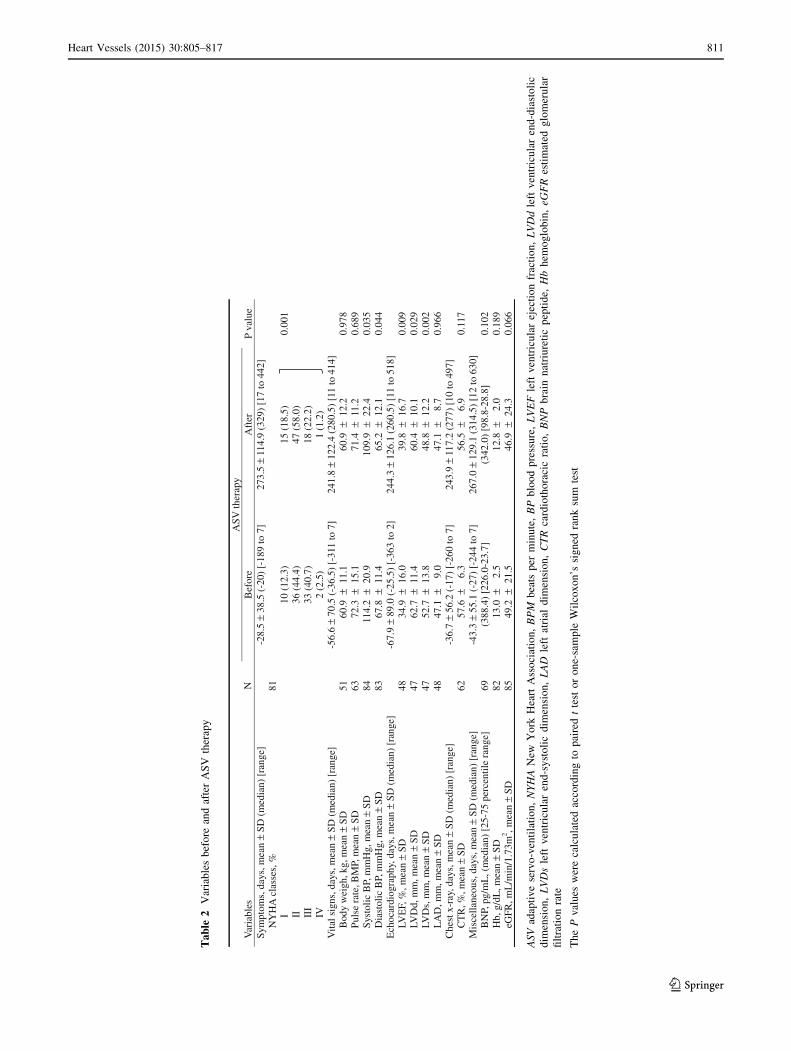
NYHA classification
Figure 3a indicates pre- and post-ASV therapy changes in
the combined proportion of patients with NYHA class III
and IV. Time-course changes before and after ASV therapy
in the proportion are shown in Fig. 3b. Eighty-one patients
showed no significant difference in the proportion during
1 year before the onset of ASV therapy. After ASV ther-
apy, however, the proportion significantly decreased
(P = 0.012) from the pre-ASV value of 43.2 % to the post-
ASV value of 23.4 %. Figure 3b indicates time-course
changes by treatment week in the proportion. The statisti-
cal analysis of the proportions between weeks -52, -26,
and -12 and onset revealed a significant increase
(P = 0.018) from 33.3 to 41.2 % only between week -52
and onset. In contrast, the statistical analysis of the pro-
portions between onset and weeks 12, 26, and 52 disclosed
significant decreases (P = 0.018, P = 0.008, and
P\ 0.001) against the onset value at week 12 and later.
Thus, the distribution patterns of NYHA classes also
favorably changed after ASV therapy (Table 2; P = 0.001)
and at weeks 26 and 52 of treatment (Table 3; P = 0.008
and P\ 0.001, respectively). The abovementioned
improvements in NYHA class were highly likely to be
attributable to ASV therapy because patients showing
improvements in NYHA class had a significantly greater
number (P\ 0.001) of days of ASV therapy
(frequency 9 duration).
Echocardiography
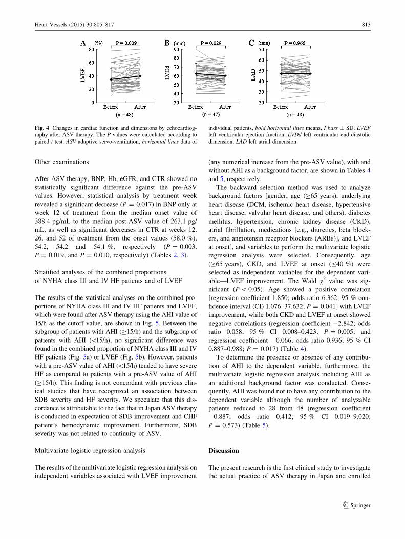
The mean pre- and post-ASV values of LVDd and LVDs
were 62.7 and 60.4 mm, as well as 52.7 and 48.8 mm,
respectively; therefore, both variables decreased signif-
icantly (P = 0.029 and P = 0.002, respectively). In
association with these changes, LVEF increased signif-
icantly (P = 0.009) from the mean pre-ASV value of
34.9 % to the mean post-ASV value of 39.8 % (Fig. 4;
Table 2). Furthermore, statistical analysis by treatment
week revealed that LVEF tended to increase at weeks 12
and 26 of treatment and increased significantly
(P = 0.035) at week 52 of treatment. LAD remained
unchanged after ASV and between baseline and each
treatment week (Table 3).
Vital signs
Body weight and pulse rate changed neither in the com-
parison of the pre- and post-ASV values nor in statistical
analysis by treatment week. After ASV therapy, SBP and
DBP decreased significantly against the pre-ASV values
(P = 0.035 and P = 0.044, respectively). However, SBP
showed no significant change in statistical analysis by
treatment week. Furthermore, DBP showed a significant
decrease (P = 0.043) against the onset value only at week
52 of treatment (Tables 2, 3).
Fig. 3 a Diagram showing the combined proportions of NYHA
classes III and IV before and after ASV therapy. The P values were
calculated according to McNemar’s test. b Diagram showing time-
course changes in the combined proportions of NYHA classes III and
IV by assessment week. The P values were calculated according to
Fisher’s least significant difference method. NYHA New York Heart
Association, ASV adaptive servo-ventilation
810 Heart Vessels (2015) 30:805–817
123
Table
2V
aria
ble
sb
efo
rean
daf
ter
AS
Vth
erap
y
NA
SV th
erap
yV
aria
bles
Bef
ore
Aft
erP
valu
eSy
mpt
oms,
day
s, m
ean
±SD
(m
edia
n) [
rang
e]-2
8.5
±38
.5 (
-20)
[-1
89 to
7]
273.
5 ±
114.
9 (3
29)
[17
to 4
42]
NY
HA
cla
sses
,%81
I10
(12
.3)
15 (
18.5
)0.
001
II36
(44
.4)
47 (
58.0
)II
I33
(40
.7)
18 (
22.2
)IV
2 (2
.5)
1 (1
.2)
Vita
l sig
ns, d
ays,
mea
n ±
SD (
med
ian)
[ra
nge]
-56.
6 ±
70.5
(-3
6.5)
[-3
11 to
7]
241.
8 ±
122.
4 (2
80.5
) [1
1 to
414
]B
ody
wei
gh, k
g, m
ean
±SD
5160
.9 ±
11.1
60.9
±12
.20.
978
Pul
se r
ate,
BM
P, m
ean
±SD
6372
.3±
15.1
71.4
±11
.20.
689
Syst
olic
BP,
mm
Hg,
mea
n ±
SD84
114.
2±
20.9
109.
9±
22.4
0.03
5D
iast
olic
BP,
mm
Hg,
mea
n ±
SD83
67.8
±11
.465
.2±
12.1
0.04
4E
choc
ardi
ogra
phy,
day
s, m
ean
±SD
(m
edia
n) [
rang
e]-6
7.9
±89
.0 (
-25.
5) [
-363
to 2
]24
4.3
±12
6.1
(260
.5)
[11
to 5
18]
LVE
F, %
, mea
n ±
SD48
34.9
±16
.039
.8±
16.7
0.00
9LV
Dd,
mm
, mea
n ±
SD47
62.7
±11
.460
.4±
10.1
0.02
9LV
Ds,
mm
, mea
n ±
SD47
52.7
±13
.848
.8±
12.2
0.00
2L
AD
, mm
, mea
n ±
SD48
47.1
±9.
047
.1±
8.7
0.96
6C
hest
x-r
ay, d
ays,
mea
n ±
SD (
med
ian)
[ra
nge]
-36.
7 ±
56.2
(-1
7) [
-260
to 7
]24
3.9
±11
7.2
(277
) [1
0 to
497
]C
TR
, %, m
ean
±SD
6257
.6±
6.3
56.5
±6.
90.
117
Mis
cella
neou
s, d
ays,
mea
n ±
SD (
med
ian)
[ra
nge]
-43.
3 ±
55.1
(-2
7) [
-244
to 7
]26
7.0
±12
9.1
(314
.5)
[12
to 6
30]
BN
P, p
g/m
L, (
med
ian)
[25
-75
perc
entil
e ra
nge]
69(3
88.4
) [2
26.0
-23.
7](3
42.0
) [9
8.8-
28.8
]0.
102
Hb,
g/d
L, m
ean
±SD
8213
.0±
2.5
12.8
±2.
00.
189
eGFR
, mL
/min
/1.7
3m2 ,m
ean
±SD
8549
.2±
21.5
46.9
±24
.30.
066
ASV
adap
tiv
ese
rvo
-ven
tila
tio
n,NYHA
New
Yo
rkH
eart
Ass
oci
atio
n,BPM
bea
tsp
erm
inu
te,BP
blo
od
pre
ssu
re,LVEF
left
ven
tric
ula
rej
ecti
on
frac
tio
n,LVDd
left
ven
tric
ula
ren
d-d
iast
oli
c
dim
ensi
on
,LVDs
left
ven
tric
ula
ren
d-s
yst
oli
cd
imen
sio
n,LAD
left
atri
ald
imen
sio
n,CTR
card
ioth
ora
cic
rati
o,BNP
bra
inn
atri
ure
tic
pep
tid
e,Hb
hem
og
lob
in,eG
FR
esti
mat
edg
lom
eru
lar
filt
rati
on
rate
Th
eP
val
ues
wer
eca
lcu
late
dac
cord
ing
top
aire
dt
test
or
on
e-sa
mp
leW
ilco
xo
n’s
sig
ned
ran
ksu
mte
st
Heart Vessels (2015) 30:805–817 811
123
Table
3V
aria
ble
sat
bas
elin
ean
dA
SV
ther
apy
wee
ks
ASV
ther
apy
Var
iabl
esN
Bas
elin
eN
12 w
eeks
N26
wee
ksN
52 w
eeks
Sym
ptom
s
NY
HA
cla
sses
(%)
59(1
00.0
)53
(100
.0)
44(1
00.0
)35
(100
.0)
I10
(16.
9)9
(17.
0)13
(29.
6)12
(34.
3)
II27
(45.
8)34
(64.
2)21
(47.
7)19
(54.
3)
III
20(3
3.9)
9(1
7.0)
10(2
2.7)
4(1
1.4)
IV2
(3.4
)1
(1.9
)0
(0.0
)0
(0.0
)
Vita
l sig
ns
Bod
y w
eigh
t, kg
2461
.4 ±
10.3
2160
.4 ±
11.3
1862
.2 ±
10.5
1564
.3 ±
10.7
Pul
se r
ate,
bpm
3971
.8 ±
16.0
3571
.5 ±
10.2
2869
.3 ±
12.1
2067
.4 ±
9.2
Syst
olic
BP,
mm
Hg
5511
9.2
±18
.853
116.
5 ±
21.1
4511
7.0
±20
.633
110.
8 ±
17.4
Dia
stol
ic B
P, m
mH
g55
71.4
±10
.653
70.9
±12
.345
69.4
±12
.133
65.2
±10
.3
Ech
ocar
diog
raph
y
LVE
F, %
1833
.1 ±
10.6
1036
.6 ±
13.6
536
.7 ±
15.0
1043
.2 ±
14.1
LA
D, m
m18
46.1
±10
.910
43.4
±9.
85
43.3
±11
.510
46.9
±8.
4
Che
st x
-ray
CT
R, %
2858
.0 ±
6.5
1954
.2 ±
5.4
1254
.2 ±
4.8
1454
.1 ±
7.5
Mis
cella
neou
s
BN
P, p
g/m
L (m
edia
n [2
5-75
pe
rcen
tile
rang
e])
39(3
88.4
[15
8.5-
691.
0])
32(2
63.1
[91
.7-4
30.5
])32
(261
.1 [
56.2
-568
.7])
21(2
78.4
[68
.0-6
63.4
])
Hb,
g/d
L56
13.3
±2.
447
12.9
±2.
046
12.9
±2.
233
13.3
±1.
9
eGFR
, mL
/min
/1.7
3 m
254
49.2
±21
.347
49.3
±23
.145
49.6
±24
.531
46.6
±23
.0
Val
ues
are
exp
ress
edas
mea
n±
SD
ASV
adap
tiv
ese
rvo
-ven
tila
tio
n,NYHA
New
Yo
rkH
eart
Ass
oci
atio
n,bpm
bea
tsp
erm
inu
te,BP
blo
od
pre
ssu
re,LVEF
left
ven
tric
ula
rej
ecti
on
frac
tio
n,LAD
left
atri
ald
imen
sio
n,CTR
card
ioth
ora
cic
rati
o,BNP
bra
inn
atri
ure
tic
pep
tid
e,Hb
hem
og
lob
in,eG
FR
esti
mat
edg
lom
eru
lar
filt
rati
on
rate
Val
ues
are
exp
ress
edas
mea
n±
SD
.S
ign
ifica
nt
dif
fere
nce
ver
sus
bas
elin
e
(Fis
her
’sle
ast
sig
nifi
can
td
iffe
ren
cem
eth
od
,P\
0.0
5o
rP\
0.0
1,
resp
ecti
vel
y)
�,��
Sig
nifi
can
td
iffe
ren
cev
ersu
sb
asel
ine
(Fis
her
’sle
ast
sig
nifi
can
td
iffe
ren
cem
eth
od
,P\
0.0
5o
rP\
0.0
1,
resp
ecti
vel
y)
812 Heart Vessels (2015) 30:805–817
123
Other examinations
After ASV therapy, BNP, Hb, eGFR, and CTR showed no
statistically significant difference against the pre-ASV
values. However, statistical analysis by treatment week
revealed a significant decrease (P = 0.017) in BNP only at
week 12 of treatment from the median onset value of
388.4 pg/mL to the median post-ASV value of 263.1 pg/
mL, as well as significant decreases in CTR at weeks 12,
26, and 52 of treatment from the onset values (58.0 %),
54.2, 54.2 and 54.1 %, respectively (P = 0.003,
P = 0.019, and P = 0.010, respectively) (Tables 2, 3).
Stratified analyses of the combined proportions
of NYHA class III and IV HF patients and of LVEF
The results of the statistical analyses on the combined pro-
portions of NYHA class III and IV HF patients and LVEF,
which were found after ASV therapy using the AHI value of
15/h as the cutoff value, are shown in Fig. 5. Between the
subgroup of patients with AHI (C15/h) and the subgroup of
patients with AHI (\15/h), no significant difference was
found in the combined proportion of NYHA class III and IV
HF patients (Fig. 5a) or LVEF (Fig. 5b). However, patients
with a pre-ASV value of AHI (\15/h) tended to have severe
HF as compared to patients with a pre-ASV value of AHI
(C15/h). This finding is not concordant with previous clin-
ical studies that have recognized an association between
SDB severity and HF severity. We speculate that this dis-
cordance is attributable to the fact that in Japan ASV therapy
is conducted in expectation of SDB improvement and CHF
patient’s hemodynamic improvement. Furthermore, SDB
severity was not related to continuity of ASV.
Multivariate logistic regression analysis
The results of the multivariate logistic regression analysis on
independent variables associated with LVEF improvement
(any numerical increase from the pre-ASV value), with and
without AHI as a background factor, are shown in Tables 4
and 5, respectively.
The backward selection method was used to analyze
background factors [gender, age (C65 years), underlying
heart disease (DCM, ischemic heart disease, hypertensive
heart disease, valvular heart disease, and others), diabetes
mellitus, hypertension, chronic kidney disease (CKD),
atrial fibrillation, medications [e.g., diuretics, beta block-
ers, and angiotensin receptor blockers (ARBs)], and LVEF
at onset], and variables to perform the multivariate logistic
regression analysis were selected. Consequently, age
(C65 years), CKD, and LVEF at onset (B40 %) were
selected as independent variables for the dependent vari-
able—LVEF improvement. The Wald v2 value was sig-
nificant (P\ 0.05). Age showed a positive correlation
[regression coefficient 1.850; odds ratio 6.362; 95 % con-
fidence interval (CI) 1.076–37.632; P = 0.041] with LVEF
improvement, while both CKD and LVEF at onset showed
negative correlations (regression coefficient -2.842; odds
ratio 0.058; 95 % CI 0.008–0.423; P = 0.005; and
regression coefficient -0.066; odds ratio 0.936; 95 % CI
0.887–0.988; P = 0.017) (Table 4).
To determine the presence or absence of any contribu-
tion of AHI to the dependent variable, furthermore, the
multivariate logistic regression analysis including AHI as
an additional background factor was conducted. Conse-
quently, AHI was found not to have any contribution to the
dependent variable although the number of analyzable
patients reduced to 28 from 48 (regression coefficient
-0.887; odds ratio 0.412; 95 % CI 0.019–9.020;
P = 0.573) (Table 5).
Discussion
The present research is the first clinical study to investigate
the actual practice of ASV therapy in Japan and enrolled
Fig. 4 Changes in cardiac function and dimensions by echocardiog-
raphy after ASV therapy. The P values were calculated according to
paired t test. ASV adaptive servo-ventilation, horizontal lines data of
individual patients, bold horizontal lines means, I bars ± SD, LVEF
left ventricular ejection fraction, LVDd left ventricular end-diastolic
dimension, LAD left atrial dimension
Heart Vessels (2015) 30:805–817 813
123
116 patients with CHF. There was only one exclusion as
mentioned previously, indicating an exclusion rate as very
low as 0.86 % (1/116). Furthermore, the retrieval rate of
the case report form on the remaining 115 patients was
100 %. Therefore, the present study may be considered
highly reliable as a field survey.
The ventilator, which had originally been developed to
treat patients with SDB, was used despite the facts that
38.3 % of patients had no diagnosis of SDB and that
23.5 % of patients did not have or had mild SDB. Namely,
we found that ASV therapy was conducted not for the
treatment of SDB but for the improvement in hemody-
namics in at least 61.7 % of patients with CHF at 16
medical institutions. This leads us to conjecture that ASV
therapy, in practice, is presumably and widely applied
to a much greater number of patients with impaired
Fig. 5 Stratified analysis in the combined proportion of NYHA
classes III and IV and LVEF before and after ASV therapy, with an
AHI cutoff value of 15/h. a Diagram showing changes in AHI in
relation to the proportion of NYHA classes III and IV after ASV
therapy. b Diagram showing changes in AHI in relation to LVEF after
ASV therapy. Values are expressed as mean ± SD. The P values
were calculated according to Fisher’s exact probability test or
Student’s t test. ASV adaptive servo-ventilation, AHI apnea–hypopnea
index, NYHA New York Heart Association, LVEF left ventricular
ejection fraction
Table 4 Logistic regression analysis of patients’ background factors associated with LVEF improvement when not including AHI
Background factors Likelihood ratios Odds ratios
B SE Wald v2 a df P valueb Exp(B) 95 % Wald CI
Age (C 65 years) 1.850 0.907 4.163 1 0.041 6.362 (1.076–37.632)
CKD -2.842 1.011 7.904 1 0.005 0.058 (0.008–0.423)
LVEF at baseline (\ 40 %) -0.066 0.027 5.741 1 0.017 0.936 (0.887–0.988)
B coefficient for the logistic regression equation to predict the dependent variable from the independent variable, SE standard error around the
coefficient, df degree of freedom for Wald v2 test, Exp(B) exponentiation of the B coefficient, an odds ratio, LVEF left ventricular ejection
fraction, AHI apnea–hypopnea index, CI confidence interval, CKD chronic kidney diseasea Wald v2 statisticb A value of P\ 0.05 was considered statistically significant
Table 5 Logistic regression analysis of patients’ background factors associated with LVEF improvement when including AHI
Background factors Likelihood ratios Odds ratios
B SE Wald v2 a df P valueb Exp(B) 95 % Wald CI
Age (C 65 years) 2.929 1.761 2.768 1 0.096 18.711 (0.594–589.686)
CKD -4.187 1.862 5.059 1 0.025 0.015 (\ 0.001–0.584)
LVEF at baseline (\ 40 %) -0.077 0.044 3.094 1 0.079 0.926 (0.850–1.009)
AHI at baseline (C 15/h) -0.887 1.575 0.317 1 0.573 0.412 (0.019–9.020)
B coefficient for the logistic regression equation to predict the dependent variable from the independent variable, SE standard error around the
coefficient, df degree of freedom for Wald v2 test, Exp(B) exponentiation of the B coefficient, an odds ratio, LVEF left ventricular ejection
fraction, AHI apnea–hypopnea index, CI confidence interval, CKD chronic kidney diseasea Wald v2 statisticb A value of P\ 0.05 was considered statistically significant
814 Heart Vessels (2015) 30:805–817
123

hemodynamics in whole Japan, regardless of the presence
or absence of SDB. Furthermore, we found that ASV
therapy represents a noninvasive therapeutic option for
patients with intractable and relatively severe CHF in real-
world settings because, at the onset of ASV therapy,
patients with IHD accounted for approximately 20 %. The
combined proportion of patients with NYHA classes III
and IV HF was 43.2 % despite the high prescription rates
of all drugs for the treatment of HF, and the median plasma
BNP concentration was as high as 388.4 pg/mL.
The proportion of ASV-continued patients in the present
study, 75.7 % (87/115), was higher than approximately
40–50 %—the values reported in previous nonrandomized
studies [17, 22, 23]. This fact probably indicates good
tolerance as the result that most of patients in the present
study who had relatively severe CHF felt better comfort or
became aware of improvements in their symptoms during
ASV therapy, and is in line with a prior clinical study
which suggested that compliance is a consequence of
subjective benefits that patients experienced in their treat-
ment [24]. Better comfort that patients obtained might have
contributed to an improvement in their adherence to ASV
therapy, and we consider that this good ASV therapy tol-
erance of CHF patients is translated into favorable NYHA
class changes.
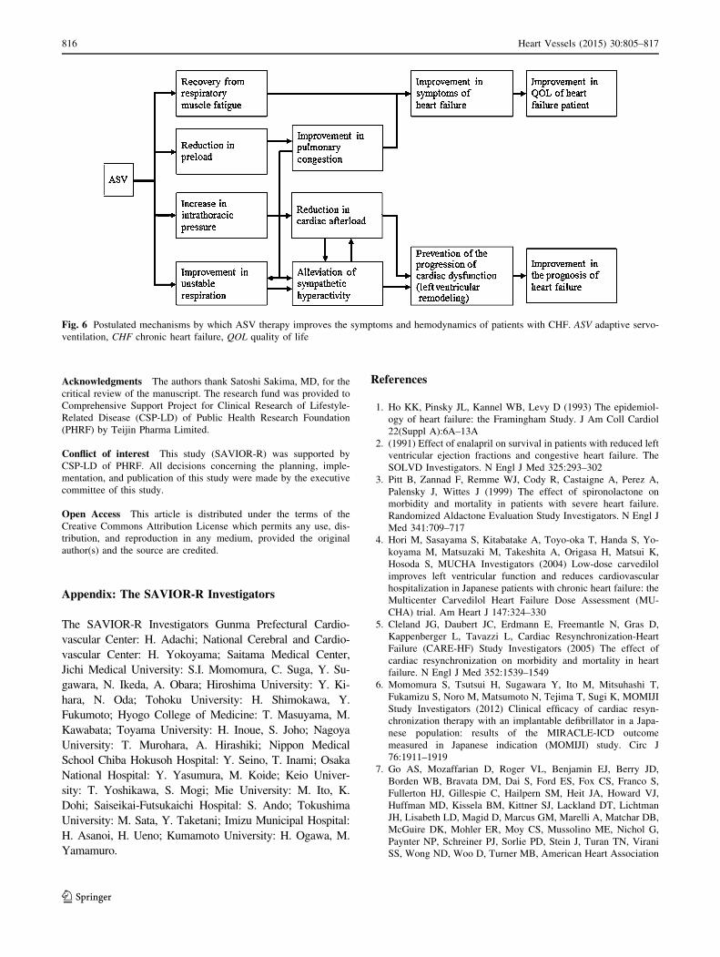
Figure 6 illustrates the postulated mechanisms by which
ASV therapy exerts its efficacy through improvements in
the symptoms and hemodynamics of patients with CHF.
ASV therapy using positive end-expiratory pressure
(PEEP) alleviates preload through a reduction in venous
return, which improves pulmonary congestion [10, 15].
PEEP ameliorates afterload by reducing transmural pres-
sure through positive intrathoracic pressure, and ASV
therapy—because of pressure support ventilation—unloads
respiration muscles [13, 14]. Furthermore, an improvement
in pulmonary congestion itself achieved by PEEP of ASV
therapy probably inhibits sympathetic nerve activity
because ASV therapy suppresses sympathetic nerve over-
activity by decreasing pulmonary capillary wedge pressure
through a reduction in venous return in CHF patients [25–
27]. Via the mechanisms described above, ASV therapy is
considered to improve symptoms of HF, to cause cardiac
reverse remodeling [17–19], and eventually to achieve the
therapeutic goals of CHF—improvements in the quality of
life (QOL) [28] and prognosis [20, 21] of CHF patients.
Despite the fact patients with advanced CHF accounted for
the majority of patients in the present study, left ventricular
dimension reduced and systolic function improved after
ASV therapy; namely, left ventricular reverse remodeling
occurred. Furthermore, comparisons between the pre- and
post-ASV values and the time-series analysis of the data
obtained revealed decreases in blood pressure and reduc-
tions in CTR and BNP [18, 23]. These results raise an
expectation that ASV may improve the prognosis of CHF
patients.
The improvement rate of LVEF concerning patients
with impaired systolic function at week 26 of treatment in
the present study was approximately 5 % and was equiv-
alent to the improvement rates of LVEF obtained in
3-month continuous positive airway pressure [29],
16-month ASV therapy [30], 6-month pharmacotherapy
[31], and meta-analysis of CRT [32] (5, 7, 7, and 5.9 %,
respectively). In consideration of the fact that all patients
had already undergone sufficient CHF therapy at the onset
of ASV therapy, this result indicates the sufficient thera-
peutic relevance of ASV therapy. The factors associated
with LVEF improvement in the present study were the low
onset value of LVEF, advanced age, and absence of CKD.
SDB severity was not related to LVEF improvement. The
findings described above suggest that the effects of ASV
therapy on patients with CHF complicated by SDB were
not exerted through the treatment of SDB, but were based
on the improvement in patients’ hemodynamics. Namely,
the direct effects on hemodynamics are probably translated
into the beneficial effects of NPPV therapy, e.g., inhibition
of sympathetic nerve activity [29, 33] and improvement in
QOL [24, 31, 34], which have been reported in previous
clinical studies in the relevant patients.
The present study has several limitations. First, sample
size is relatively small. The device was launched in
December 2007 in Japan, and most of the patients initiated
to undergo ASV therapy using the device shortly thereafter.
Therefore, the number of patients at 16 medical institutions
was as relatively small as 115. We expect findings in the
present study to be verified in a larger scale clinical study.
Second, no definitive conclusions can be drawn because the
present study is not a randomized controlled study, nor any
precise diagnosis of SDB could be made because overnight
polysomnography was not performed in real-world clinical
settings. Nevertheless, improvements in NYHA class are
highly likely to be attributable to ASV therapy. Third,
information bias cannot be ruled out because the present
study is retrospective in design. A prospective randomized
placebo-controlled study is required to solve these regards.
In conclusion, real-world practice in Japan was evi-
denced where ASV therapy is applied to patients with
relatively severe CHF, regardless of the presence or
absence of SDB. The present study suggests the following:
(1) CHF patients present long-term ASV continuity that is
affected not by SDB severity but by CHF severity; and (2)
ASV therapy improves symptoms, left ventricular con-
tractility, and remodeling in a not SDB but CHF severity-
dependent manner. Therefore, ASV therapy is expected to
become a novel and promising non-pharmacotherapy for
relevant patients. A randomized controlled study to verify
these effects is warranted.
Heart Vessels (2015) 30:805–817 815
123
Acknowledgments The authors thank Satoshi Sakima, MD, for the
critical review of the manuscript. The research fund was provided to
Comprehensive Support Project for Clinical Research of Lifestyle-
Related Disease (CSP-LD) of Public Health Research Foundation
(PHRF) by Teijin Pharma Limited.
Conflict of interest This study (SAVIOR-R) was supported by
CSP-LD of PHRF. All decisions concerning the planning, imple-
mentation, and publication of this study were made by the executive
committee of this study.
Open Access This article is distributed under the terms of the
Creative Commons Attribution License which permits any use, dis-
tribution, and reproduction in any medium, provided the original
author(s) and the source are credited.
Appendix: The SAVIOR-R Investigators
The SAVIOR-R Investigators Gunma Prefectural Cardio-
vascular Center: H. Adachi; National Cerebral and Cardio-
vascular Center: H. Yokoyama; Saitama Medical Center,
Jichi Medical University: S.I. Momomura, C. Suga, Y. Su-
gawara, N. Ikeda, A. Obara; Hiroshima University: Y. Ki-
hara, N. Oda; Tohoku University: H. Shimokawa, Y.
Fukumoto; Hyogo College of Medicine: T. Masuyama, M.
Kawabata; Toyama University: H. Inoue, S. Joho; Nagoya
University: T. Murohara, A. Hirashiki; Nippon Medical
School Chiba Hokusoh Hospital: Y. Seino, T. Inami; Osaka
National Hospital: Y. Yasumura, M. Koide; Keio Univer-
sity: T. Yoshikawa, S. Mogi; Mie University: M. Ito, K.
Dohi; Saiseikai-Futsukaichi Hospital: S. Ando; Tokushima
University: M. Sata, Y. Taketani; Imizu Municipal Hospital:
H. Asanoi, H. Ueno; Kumamoto University: H. Ogawa, M.
Yamamuro.
References
1. Ho KK, Pinsky JL, Kannel WB, Levy D (1993) The epidemiol-
ogy of heart failure: the Framingham Study. J Am Coll Cardiol
22(Suppl A):6A–13A
2. (1991) Effect of enalapril on survival in patients with reduced left
ventricular ejection fractions and congestive heart failure. The
SOLVD Investigators. N Engl J Med 325:293–302
3. Pitt B, Zannad F, Remme WJ, Cody R, Castaigne A, Perez A,
Palensky J, Wittes J (1999) The effect of spironolactone on
morbidity and mortality in patients with severe heart failure.
Randomized Aldactone Evaluation Study Investigators. N Engl J
Med 341:709–717
4. Hori M, Sasayama S, Kitabatake A, Toyo-oka T, Handa S, Yo-
koyama M, Matsuzaki M, Takeshita A, Origasa H, Matsui K,
Hosoda S, MUCHA Investigators (2004) Low-dose carvedilol
improves left ventricular function and reduces cardiovascular
hospitalization in Japanese patients with chronic heart failure: the
Multicenter Carvedilol Heart Failure Dose Assessment (MU-
CHA) trial. Am Heart J 147:324–330
5. Cleland JG, Daubert JC, Erdmann E, Freemantle N, Gras D,
Kappenberger L, Tavazzi L, Cardiac Resynchronization-Heart
Failure (CARE-HF) Study Investigators (2005) The effect of
cardiac resynchronization on morbidity and mortality in heart
failure. N Engl J Med 352:1539–1549
6. Momomura S, Tsutsui H, Sugawara Y, Ito M, Mitsuhashi T,
Fukamizu S, Noro M, Matsumoto N, Tejima T, Sugi K, MOMIJI
Study Investigators (2012) Clinical efficacy of cardiac resyn-
chronization therapy with an implantable defibrillator in a Japa-
nese population: results of the MIRACLE-ICD outcome
measured in Japanese indication (MOMIJI) study. Circ J
76:1911–1919
7. Go AS, Mozaffarian D, Roger VL, Benjamin EJ, Berry JD,
Borden WB, Bravata DM, Dai S, Ford ES, Fox CS, Franco S,
Fullerton HJ, Gillespie C, Hailpern SM, Heit JA, Howard VJ,
Huffman MD, Kissela BM, Kittner SJ, Lackland DT, Lichtman
JH, Lisabeth LD, Magid D, Marcus GM, Marelli A, Matchar DB,
McGuire DK, Mohler ER, Moy CS, Mussolino ME, Nichol G,
Paynter NP, Schreiner PJ, Sorlie PD, Stein J, Turan TN, Virani
SS, Wong ND, Woo D, Turner MB, American Heart Association
Fig. 6 Postulated mechanisms by which ASV therapy improves the symptoms and hemodynamics of patients with CHF. ASV adaptive servo-
ventilation, CHF chronic heart failure, QOL quality of life
816 Heart Vessels (2015) 30:805–817
123
Statistics Committee and Stroke Statistics Subcommittee (2013)
Heart disease and stroke statistics—2013 update: a report from
the American Heart Association. Circulation 127:143–152
8. Shiba N, Shimokawa H (2008) Chronic heart failure in Japan:
implications of the CHART studies. Vasc Health Risk Manag
4:103–113
9. JCS Joint Working Group (2013) Guidelines for treatment of
acute heart failure (JCS 2011). Circ J 77:2157–2201
10. Takano T, Endo T, Tanaka K, Seino Y, Nitta T, Matsuyama Y,
Koh M, Hayakawa H (1986) Effects of positive end-expiratory
pressure ventilation and extracorporeal ultrafiltration method in
patients with refractory heart failure. Jpn Circ J 50:359–367
11. Halter JM, Steinberg JM, Schiller HJ, DaSilva M, Gatto LA,
Landas S, Nieman GF (2003) Positive end-expiratory pressure
after a recruitment maneuver prevents both alveolar collapse and
recruitment/derecruitment. Am J Respir Crit Care Med
167:1620–1626
12. Michelet P, Roch A, Brousse D, D’Journo XB, Bregeon F,
Lambert D, Perrin G, Papazian L, Thomas P, Carpentier JP,
Auffray JP (2005) Effects of PEEP on oxygenation and respira-
tory mechanics during one-lung ventilation. Br J Anaesth
95:267–273
13. Mehta S, Hill NS (2001) Noninvasive ventilation. Am J Respir
Crit Care Med 163:540–577
14. Naughton MT, Rahman MA, Hara K, Floras JS, Bradley TD
(1995) Effect of continuous positive airway pressure on intra-
thoracic and left ventricular transmural pressures in patients with
congestive heart failure. Circulation 91:1725–1731
15. Acosta B, DiBenedetto R, Rahimi A, Acosta MF, Cuadra O, Van
Nguyen A, Morrow L (2000) Hemodynamic effects of noninva-
sive bilevel positive airway pressure on patients with chronic
congestive heart failure with systolic dysfunction. Chest
118:1004–1009
16. Teschler H, Dohring J, Wang YM, Berthon-Jones M (2001)
Adaptive pressure support servo-ventilation: a novel treatment for
Cheyne–Stokes respiration in heart failure. Am J Respir Crit Care
Med 164:614–619
17. Haruki N, Takeuchi M, Kaku K, Yoshitani H, Kuwaki H, Tamura
M, Abe H, Okazaki M, Tsutsumi A, Otsuji Y (2011) Comparison
of acute and chronic impact of adaptive servo-ventilation on left
chamber geometry and function in patients with chronic heart
failure. Eur J Heart Fail 13:1140–1146
18. Yamada S, Sakakibara M, Yokota T, Kamiya K, Asakawa N,
Iwano H, Yamada S, Oba K, Tsutsui H (2013) Acute hemody-
namic effects of adaptive servo-ventilation in patients with heart
failure. Circ J 77:1214–1220
19. Takama N, Kurabayashi M (2011) Effectiveness of adaptive
servo-ventilation for treating heart failure regardless of the
severity of sleep-disordered breathing. Circ J 75:1164–1169
20. Koyama T, Watanabe H, Igarashi G, Tamura Y, Ikeda K, Terada
S, Ito H (2012) Effect of short-duration adaptive servo-ventilation
therapy on cardiac function in patients with heart failure. Circ J
76:2606–2613
21. Koyama T, Watanabe H, Tamura Y, Oguma Y, Kosaka T, Ito H
(2013) Adaptive servo-ventilation therapy improves cardiac
sympathetic nerve activity in patients with heart failure. Eur J
Heart Fail 15:902–909
22. Oldenburg O, Bitter T, Lehmann R, Korte S, Dimitriadis Z, Faber
L, Schmidt A, Westerheide N, Horstkotte D (2011) Adaptive
servoventilation improves cardiac function and respiratory sta-
bility. Clin Res Cardiol 100:107–115
23. Koyama T, Watanabe H, Kobukai Y, Makabe S, Munehisa Y,
Iino K, Kosaka T, Ito H (2010) Beneficial effects of adaptive
servo ventilation in patients with chronic heart failure. Cir J
74:2056–2057
24. Philippe C, Stoıca-Herman M, Drouot X, Raffestin B, Escourrou
P, Hittinger L, Michel PL, Rouault S, d’Ortho MP (2006)
Compliance with and effectiveness of adaptive servoventilation
versus continuous positive airway pressure in the treatment of
Cheyne–Stokes respiration in heart failure over a six month
period. Heart 92:337–342
25. Azevedo ER, Newton GE, Floras JS, Parker JD (2000) Reducing
cardiac filling pressure lowers norepinephrine spillover in
patients with chronic heart failure. Circulation 101:2053–2059
26. Harada D, Joho S, Oda Y, Hirai T, Asanoi H, Inoue H (2011)
Short term effect of adaptive servo-ventilation on muscle sym-
pathetic nerve activity in patients with heart failure. Auton
Neurosci 161:95–102
27. Joho S, Oda Y, Ushijima R, Hirai T, Inoue H (2012) Effect of
adaptive servoventilation on muscle sympathetic nerve activity in
patients with chronic heart failure and central sleep apnea. J Card
Fail 18:769–775
28. Hastings PC, Vazir A, Meadows GE, Dayer M, Poole-Wilson PA,
McIntyre HF, Morrell MJ, Cowie MR, Simonds AK (2010)
Adaptive servo-ventilation in heart failure patients with sleep
apnea: a real world study. Int J Cardiol 139:17–24
29. Mansfield DR, Gollogly NC, Kaye DM, Richardson M, Bergin P,
Naughton MT (2004) Controlled trial of continuous positive
airway pressure in obstructive sleep apnea and heart failure. Am J
Respir Crit Care Med 169:361–366
30. Oldenburg O, Schmidt A, Lamp B, Bitter T, Muntean BG, Langer
C, Horstkotte D (2008) Adaptive servoventilation improves car-
diac function in patients with chronic heart failure and Cheyne–
Stokes respiration. Eur J Heart Fail 10:581–586
31. Maurer MS, Sackner-Bernstein JD, El-Khoury Rumbarger L,
Yushak M, King DL, Burkhoff D (2009) Mechanisms underlying
improvements in ejection fraction with carvedilol in heart failure.
Circ Heart Fail 2:189–196
32. Adabag S, Roukoz H, Anand IS, Moss AJ (2011) Cardiac re-
synchronization therapy in patients with minimal heart failure.
J Am Coll Cardiol 58:935–941
33. Yoshihisa A, Suzuki S, Miyata M, Yamaki T, Sugimoto K, Kunii
H, Nakazato K, Suzuki H, Saitoh S, Takeishi Y (2012) ‘‘A single
night’’ beneficial effects of adaptive servo-ventilation on cardiac
overload, sympathetic nervous activity, and myocardial damage
in patients with chronic heart failure and sleep-disordered
breathing. Circ J 76:2153–2158
34. Oldenburg O (2012) Cheyne–Stokes respiration in chronic heart
failure. Treatment with adaptive servoventilation therapy. Circ J
76:2305–2317
Heart Vessels (2015) 30:805–817 817
123