adaptations for nutrition by2 biology nutrition the process organisms use to get energy to...
TRANSCRIPT

Adaptations for Nutrition
BY2 Biology

Nutrition The process organisms use to get
energy to maintain life functions and matter to build and maintain their structure (from
nutrients)
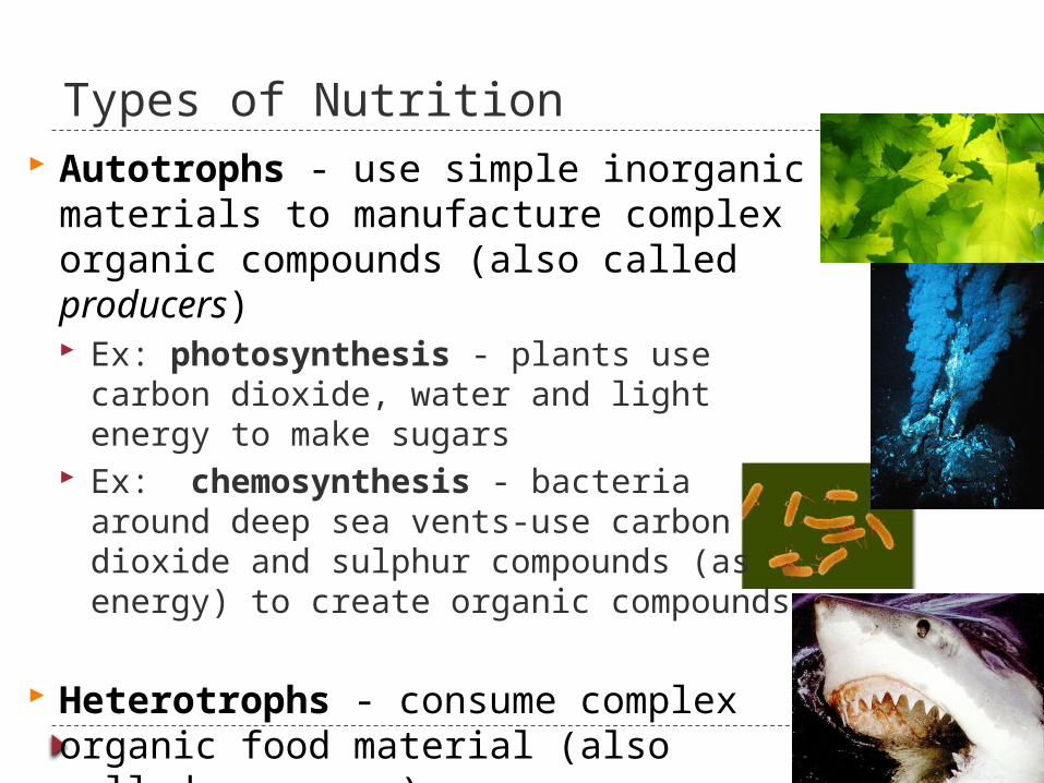
Types of Nutrition Autotrophs - use simple inorganic
materials to manufacture complex organic compounds (also called producers) Ex: photosynthesis - plants use carbon
dioxide, water and light energy to make sugars
Ex: chemosynthesis - bacteria around deep sea vents-use carbon dioxide and sulphur compounds (as energy) to create organic compounds
Heterotrophs - consume complex organic food material (also called consumers)

What do heterotrophs need to live?
O2
food
ATP
They make energy using: Food (organic compounds such as
carbohydrates, protein..) Oxygen
They build bodies using: food for raw materials
amino acids, sugars, fats, nucleotides
ATP energy for synthesis
This includes animals, fungi, some protoctists and some bacteria

Overview of food processing Ingest
taking in food Digest
mechanical digestion breaking up food into smaller pieces
chemical digestion breaking down food into soluble molecules
small enough to be absorbed into cells uses enzymes (hydrolysis)
Absorb Digested food moves across cell membrane
diffusion active transport
Assimilate use compounds in the body
Egestion undigested extracellular material passes out
of digestive system (ex: cellulose in humans)
intracellulardigestion
extracellulardigestion

Types of Heterotrophic Nutrition Holozoic feeders:
Take food into their bodies and break is down during digestion Have specialised organs in a digestive system (gut) After digestion, the nutrients are absorbed into the body
Saprophytes (saprobionts): Fungi and some bacteria Feed on dead or decaying matter No specialised digestive system Secrete enzymes onto the food source outside their body for
extra-cellular digestion and absorb the soluble products by diffusion (enzymes include protease, amylase, lipase, cellulase)
Some are decomposers and help to recycle nutrients in the ecosystem

Parasites: Highly specialised organisms Feed on other living organisms (a host) Some live inside the host (endoparasites) other live
on the outside (ectoparasites) The host is always harmed Ex: tapeworm, potato blight (fungus), plasmodium
(causes malaria) Mutualism (symbiosis):
2 different species live in a helpful relationship Ex: cows use bacteria in their stomach to help digest
cellulose and the bacteria gain nutrients from the broken plant material for growth and energy

How do animals get their food?
filter (suspension) feeding substrate feeding
fluid feeding bulk feeding

Food types/feeding mechanisms
Heterotrophs
Opportunistic Herbivore: eat autotrophs Carnivore: eat other animals Omnivore: both
Feeding Adaptations Suspension feeders - sift
food from water (baleen whale)
Substrate feeders -live in or on their food (leaf miner) (earthworm: deposit-feeder)
Fluid-feeders -suck fluids from a host (mosquito)
Bulk-feeders: eat large pieces of food (most animals)

Digestive systems

Simple More complex In simple organisms:
feeding on only one type of food the gut is undifferentiated
In more advanced organisms: Often have a varied diet, the gut is divided into various parts along its length
and each part is specialised to carry out particular functions
Organisms with a varied diet require more than one type of enzyme to carry out the digestion of the different food substrates

Human Digestive System
Overview animation

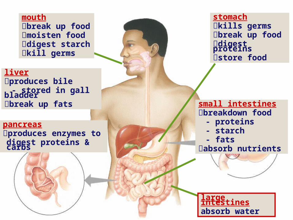
Mouth (buccal cavity)
mechanical digestion by teeth to break up food (mastication/chewing)
moistens food by mixing with saliva to lubricate it for swallowing
chemical digestion - amylase digests starchsalivaContains:• water (to soften food)•mucus (to protect the lining of the digestive system)•amylase (breaks starch to maltose)•mineral ions to keep mouth pH alkaline (act as a buffer)•Anti-bacterial chemicals to kill germs

Swallowing (& not choking)
Epiglottis problem: breathe & swallow through same orifice flap of cartilage closes trachea (windpipe) when swallowing food travels down esophagus
Esophagus move food along to stomach by peristalsis
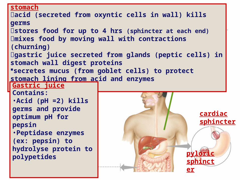
stomachacid (secreted from oxyntic cells in wall) kills germsstores food for up to 4 hrs (sphincter at each end) mixes food by moving wall with contractions (churning)gastric juice secreted from glands (peptic cells) in stomach wall digest proteinssecretes mucus (from goblet cells) to protect stomach lining from acid and enzymes
cardiacsphincter
pyloricsphincter
Gastric juiceContains:•Acid (pH =2) kills germs and provide optimum pH for pepsin•Peptidase enzymes (ex: pepsin) to hydrolyse protein to polypetides

Human Digestion
Peristalsis: rhythmic waves of contraction by smooth muscle
Sphincters: ring-like valves that regulate passage of material
Accessory glands: salivary glands; pancreas; liver; gall bladder (secrete digestive juices)

Tissue Layers The gut wall consists of four tissue layers
surrounding a central cavity (lumen)- Serosa – tough connective tissue to protect
the wall and reduce friction with other organs when it moves)
Muscle Layer – longitudinal muscle and circular muscle running in different directions Cause peristalsis when circular muscles contract and
longitudinal relax)

Submucosa – connective tissue with blood vessels and lymph vessels to carry away absorbed food Has nerves to coordinate peristalsis
Mucosa- secretes music to lubricate and protect the mucosa Secretes digestive juices in some areas Absorbs digested food in others
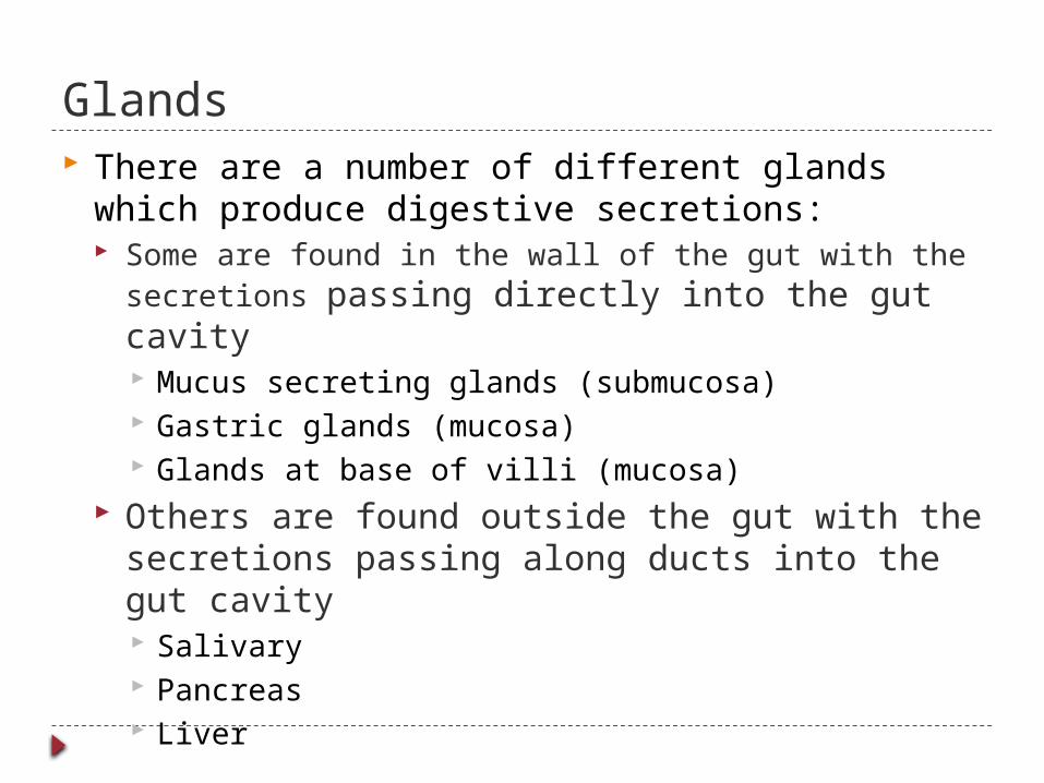
Glands There are a number of different glands which
produce digestive secretions: Some are found in the wall of the gut with the
secretions passing directly into the gut cavity Mucus secreting glands (submucosa) Gastric glands (mucosa) Glands at base of villi (mucosa)
Others are found outside the gut with the secretions passing along ducts into the gut cavity Salivary Pancreas Liver

The human alimentary canal Consists of:
buccal cavity Tongue Salivary glands Oesophagus Stomach Duodenum Ileum Colon Rectum Anus associated organs; liver and pancreas
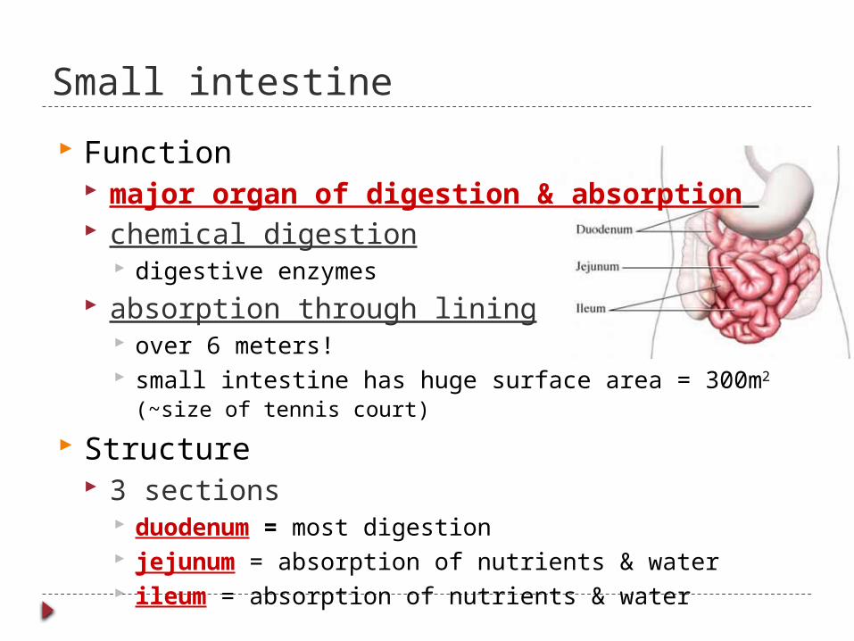
Small intestine
Function major organ of digestion & absorption chemical digestion
digestive enzymes absorption through lining
over 6 meters! small intestine has huge surface area = 300m2 (~size of
tennis court)
Structure 3 sections
duodenum = most digestion jejunum = absorption of nutrients & water ileum = absorption of nutrients & water
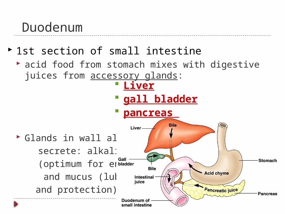
Duodenum
1st section of small intestine acid food from stomach mixes with digestive juices
from accessory glands:
Glands in wall also secrete: alkaline juice (optimum for enzymes) and mucus (lubrication
and protection)
Liver gall bladder pancreas

Duodenum Enzymes on the tip of the villi complete
digestion: Maltase Endopepsidase Exopepsidase

Liver
Digestive System Functions produces bile salts
stored in gallbladder until needed Help neutralise stomach acid breaks up fats by lowering the surface tension of lipids
act like detergents to breakup fats into tiny droplets (emulsifier)
Circulatory System ConnectionCirculatory System Connection
bile contains colors from old red blood cells collected in liver =
iron in RBC rusts & makes feces brown
bile contains colors from old red blood cells collected in liver =
iron in RBC rusts & makes feces brown

Pancreas Secretes pancreatic juice via
pancreatic duct Digestive enzymes
Endopeptidases (protein peptides)
Pancreatic amylase (starch maltose)
Lipase (lipids fatty acids + glycerol)
Buffers reduces acidity
alkaline solution rich in bicarbonate (HCO3-)
buffers acidity of material from stomach
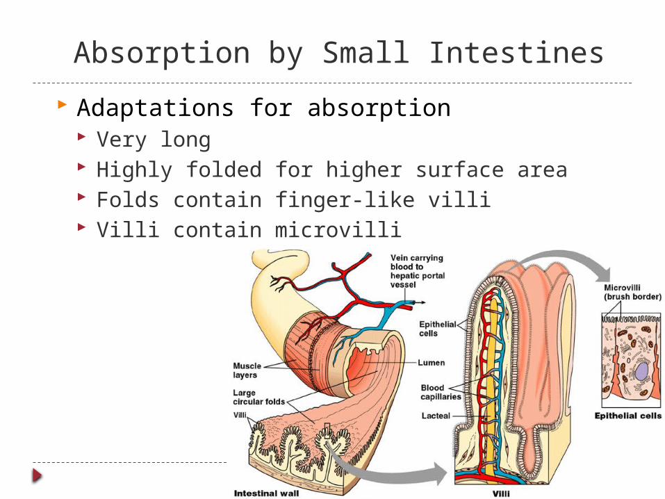
Absorption by Small Intestines
Adaptations for absorption Very long Highly folded for higher surface area Folds contain finger-like villi Villi contain microvilli
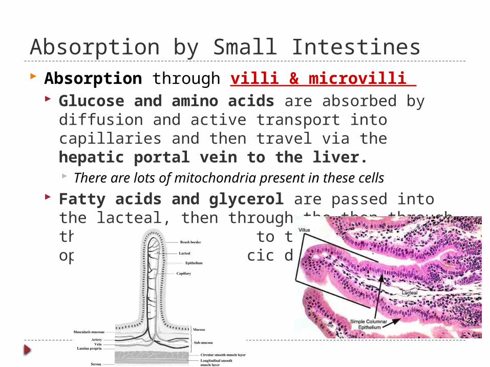
Absorption by Small Intestines Absorption through villi & microvilli
Glucose and amino acids are absorbed by diffusion and active transport into capillaries and then travel via the hepatic portal vein to the liver. There are lots of mitochondria present in these cells
Fatty acids and glycerol are passed into the lacteal, then through the then through the lymphatic system to the blood stream opening at the thoracic duct.
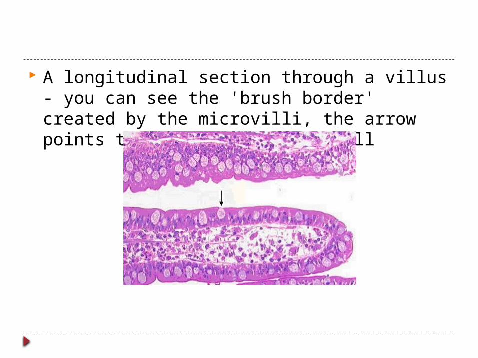
A longitudinal section through a villus - you can see the 'brush border' created by the microvilli, the arrow points to mucus in a goblet cell

small intestinesbreakdown all foods
- proteins- starch- fats- nucleic acids
absorb nutrients
stomachkills germs break up fooddigest proteinsstore food
pancreasproduces enzymes to digest proteins & starch
liverproduces bile
- stored in gall bladderbreak up fats
mouthbreak up foodmoisten food digest starchkill germs

Large intestine
4 parts: Caecum Appendix Colon Rectum
Function re-absorb water and mineral salts in colon
use ~9 liters of water every day in digestive juices > 90% of water reabsorbed
not enough water absorbed back to body diarrhea
too much water absorbed back to body constipation

Flora of the large intestine
Living in the large intestine is a richflora of harmless, helpful bacteria Escherichia coli (E. coli) bacteria produce vitamins
vitamin K, folic acid & other B vitamins
generate gasesby-product of bacterial
metabolism methane, hydrogen sulfide

Rectum Last section of large intestine
eliminate faeces undigested materials
mainly cellulose from plants
(roughage or fiber)Salts extracellular wastecells that have sloughed off masses of bacteria
Cellulose fibre is required to provide bulk and stimulate peristalsis.
stomachkills germs break up fooddigest proteinsstore food
small intestinesbreakdown food
- proteins- starch- fats
absorb nutrients
pancreasproduces enzymes to digest proteins & carbs
liverproduces bile
- stored in gall bladderbreak up fats
large intestinesabsorb water
mouthbreak up foodmoisten food digest starchkill germs

Summary of enzymes Called hydrolases as they catalyse the hydrolysis of molecules
Enzyme group
Enzyme Location Substrate
Products
Other
Carbohydrases
Amylase MouthDuodenumPancreas
Starch Maltose Maltose is further broken down
Maltase Duodenum
Maltose Glucose Glucose used as an energy source in respirationExcess glucose is converted to fat
Proteases Peptidase(ex: pepsin)
StomachDuodenumPancreas
Polypeptides then amino acids
Amino acids
Endopeptidases hydrolyse peptide bonds within the proteinExopeptidases hydrolyse peptide bonds near the end of proteinsAmino acids are absorbed for protein synthesisExcess amino acids cannot be stored sois deaminated, whereby the removed amino groups are converted to urea andthe deaminated remainder is converted to carbohydrate and stored
lipase Lipase Pancreas Fats Fatty acids & glycerol
Lipids are used for membranes and hormones, and the excess is stored asfat.

Review digestive system in humans Overview animation

Evolutionary adaptations to different diets
Adaptations of herbivore vs. carnivore specialization in teeth length of digestive system number & size of stomachs

Animals have different diets and methods of feeding. Ex: Retiles and amphibians-swallow food whole
immediately so no need for teeth for chewing
Mammals cut and chew food before swallowing and so have adapted different types of teeth
Evolutionary adaptations to different diets

Dentition Mechanical digestion (cutting and chewing
food) increases surface area for enzyme action and make swallowing easier
Mammals have evolved different types of teeth with each type being specialised for a different function

Teeth There are differences
between the teeth of carnivores and herbivores reflecting their differing diets

Carnivore teeth
Adapted for: Catching/piercing skin & killing – large, curved
pointed canines Crushing bones – premolars and molars Tearing meat – canines Scrape meat off the bone - incisors Carnassials – specialized molars (cheek teeth) that slide
past each other like scissors to cut and crush
Jaw Powerful with strong muscles Does not move side to side (to prevent dislocation) Greater vertical movement to open wide to capture prey

Herbivore Teeth Must grind plant material (due to cellulose) before swallowing
Adapted for: Cutting plants - Incisors on the lower jaw only and a
horny pad on the upper jaw to cut against or help to pull grass (no canines).
Grinding - wide molars and premolars (cheek teeth) that interlock/fit into each other. These teeth get worn down but can regrow throughout life
Jaw: Diastema – gap between front teeth and premolars at
side where the tongue can push the cut grass to the grinding surface at the back and push food to the back over again and again
Moves in a circular grinding action on the horizontal plane

Omnivore Teeth both kinds of teeth
Example: 32 Human teeth (adult) 8 incisors (chisel shape) for biting and cutting 4 canines (pointed) for tearing 8 premolars 12 molars (flat) for grinding

Length of digestive system Carnivores
No need to digest cellulose or starch so less chemical digestion in mouth
More acid in stomach (so can often eat rancid food)
short digestive system protein easier to digest than
cellulose
Herbivores & omnivores long digestive system
more time to digest cellulose symbiotic bacteria in gut

Symbiotic organisms
Ruminantsadditional mechanical digestion by chewing food multiple times after mixing it with enzymes
How can cows digest cellulose efficiently? symbiotic bacteria in stomachs help digest
cellulose-rich meals rabbit vs. cow adaptation: eat feces vs. chew cudruminantcaprophagy
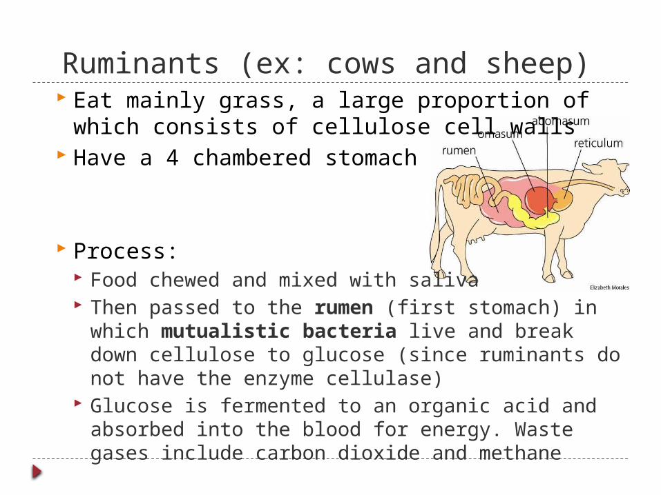
Ruminants (ex: cows and sheep) Eat mainly grass, a large proportion of which
consists of cellulose cell walls Have a 4 chambered stomach
Process: Food chewed and mixed with saliva Then passed to the rumen (first stomach) in
which mutualistic bacteria live and break down cellulose to glucose (since ruminants do not have the enzyme cellulase)
Glucose is fermented to an organic acid and absorbed into the blood for energy. Waste gases include carbon dioxide and methane

Ruminants plant material is passed up from the rumen and
reticulum back into the mouth periodically until it is completely chewed up (known as chewing the cud).
Material is then passed from the rumen and reticulum (no real function) into the omasum (next chamber) where water is absorbed
Then it is passes to the abomason (last chamber) where hydrochloric acid and protease digest protein
Then the material continues into the small intestine for absorption of products

The Bacteria Mutualistic - both benefit – mammals gets food
broken down further and bacteria gets food supply brought to it and shelter
bacteria must be kept in an isolated area with optimum pH (not killed by pH in other parts of gut)
More bacteria than in caecum, so more efficient at breaking down cellulose
When bacteria die they get passed along the digestive system as a source or protein

Rabbits Coprophagy is consumption of
faeces by animals. Rabbits do not have a complex
ruminant digestive system. They must extract excess amount of
nutrition from grass by giving their food a second pass through the gut.
Soft fecal pellets or partially digested food are excreted and consumed immediately
Consuming this matter is important for adequate nutritional intake of vitamin B 12.
http://www.bozemanscience.com/digestive-system