(adam10) regulates notch signaling during early retinal
TRANSCRIPT

RESEARCH ARTICLE
A Disintegrin and Metalloproteinase10(ADAM10) Regulates NOTCH Signalingduring Early Retinal DevelopmentJoseph A. Toonen1¤, Adam Ronchetti1, D. J. Sidjanin1,2*
1 Department of Cell Biology, Neurobiology, and Anatomy, 8701Watertown Plank Rd., Medical College ofWisconsin, Milwaukee, Wisconsin, United States of America, 2 Human and Molecular Genetics Center, 8701Watertown Plank Rd., Medical College of Wisconsin, Milwaukee, Wisconsin, United States of America
¤ Current address: Department of Neurology, Washington University School of Medicine, St. Louis, Missouri,United States of America* [email protected]
AbstractADAM10 and ADAM17 are two closely related members of the ADAM (a disintegrin and
metalloprotease) family of membrane-bound sheddases, which proteolytically cleave sur-
face membrane proteins. Both ADAM10 and ADAM17 have been implicated in the proteo-
lytic cleavage of NOTCH receptors and as such regulators of NOTCH signaling. During
retinal development, NOTCH signaling facilitates retinal neurogenesis by maintaining pro-
genitor cells in a proliferative state and by mediating retinal cell fates. However, the roles of
ADAM10 and ADAM17 in the retina are not well defined. In this study, we set out to clarify
the roles of ADAM10 and ADAM17 during early retinal development. The retinal phenotype
of conditionally abated Adam17 retinae (Adam17 CKO) did not differ from the controls
whereas conditionally ablated Adam10 retinae (Adam10 CKO) exhibited abnormal morpho-
genesis characterized by the formation of rosettes and a loss of retinal laminae phenotypi-
cally similar to morphological abnormalities identified in mice with retinal NOTCH signaling
deficiency. Additionally, Adam10 CKO retinae exhibited abnormal neurogenesis character-
ized by fewer proliferating progenitor cells and greater differentiation of early photoreceptors
and retinal ganglion cells. Moreover, constitutive activation of the NOTCH1-intracellular
domain (N1-ICD) rescued Adam10 CKO abnormal neurogenesis, as well as abnormal reti-
nal morphology by maintaining retinal cells in the progenitor state. Collectively these find-
ings provide in vivo genetic evidence that ADAM10, and not ADAM17, is indispensable for
proper retinal development as a regulator of NOTCH signaling.
IntroductionDuring retinal development, all retinal cell types are derived from a single population of plurip-otent retinal progenitor cells (RPCs). The birth order of retinal cells is unidirectional and highlyconserved although at any given developmental time point there is an overlap in the generation
PLOSONE | DOI:10.1371/journal.pone.0156184 May 25, 2016 1 / 18
a11111
OPEN ACCESS
Citation: Toonen JA, Ronchetti A, Sidjanin DJ (2016)A Disintegrin and Metalloproteinase10 (ADAM10)Regulates NOTCH Signaling during Early RetinalDevelopment. PLoS ONE 11(5): e0156184.doi:10.1371/journal.pone.0156184
Editor: Alexandre Hiroaki Kihara, UniversidadeFederal do ABC, BRAZIL
Received: November 4, 2015
Accepted: May 10, 2016
Published: May 25, 2016
Copyright: © 2016 Toonen et al. This is an openaccess article distributed under the terms of theCreative Commons Attribution License, which permitsunrestricted use, distribution, and reproduction in anymedium, provided the original author and source arecredited.
Data Availability Statement: All relevant data arewithin the paper and its Supporting Information files.
Funding: This work was funded by EY018872,EY001931, EY014537, and EY018872, National EyeInstitute, National Institutes of Health, http://www.nih.gov/. The funders had no role in study design, datacollection and analysis, decision to publish, orpreparation of the manuscript.
Competing Interests: The authors have declaredthat no competing interests exist.
of various retinal cell types [1–3]. In mice, retinal neurogenesis starts around E11 with thebirth of ganglion cells followed by the birth of cone photoreceptors, horizontal and amacrinecells, with rod photoreceptors forming around birth and finally bipolar cells and Müller glia asthe last retinal cell types born postnatally [1–3]. It has been proposed that RPCs undergo tem-porally regulated successive stages of competence to either generate a differentiated retinal celltype or to transit to the next stage of RPC competence that facilitates the birth of subsequentretinal cell types [1, 3].
NOTCH signaling is an evolutionarily conserved pathway involved in the development ofmost tissues. The role of NOTCH signaling is in the regulation of cell proliferation, cell death,cell fate determination, and differentiation [4, 5]. In mammals, there are four NOTCH recep-tors (NOTCH1-4) and five NOTCH ligands (JAG1, JAG2, DLL1, DLL3, DLL4) that exhibitboth redundant and unique functions [4]. The canonical NOTCH pathway involves binding ofa NOTCH ligand from the surface of adjacent cells to the NOTCH receptor thereby facilitatingthe subsequent NOTCH receptor cleavage at the S2 site followed by cleavage at the S3 and S4sites resulting in the release of the NOTCH intracellular domain (NICD) from the cell mem-branes; once released the NICD translocates into the nucleus and forms a complex with RBPJand MAML1 along with other cofactors to transcriptionally activate inhibitors of differentia-tion [6–8]. Therefore, one of the key roles of NOTCH signaling is maintaining progenitor cellsin their undifferentiated state. Additionally, during retinal development NOTCH signalingfacilitates neurogenesis by repressing retinal cell fates [9–15].
ADAM10 and ADAM17 are two closely related members of the ADAM family of proteinsthat proteolytically cleave or “shed” ectodomains of cell surface proteins [16, 17]. BothADAM10 and ADAM17 have been implicated as sheddases of NOTCH receptors at the S2cleavage site thereby facilitating subsequent cleavage at S3 and S4 sites by the γ-secretase com-plex [18–21]. In C. elegans, sup-17, the ortholog of ADAM10, and adm-4, the ortholog ofADAM17, are functionally redundant as NOTCH regulators [21]. In Drosophila, Kuzbanian(kuz), the fly ortholog of ADAM10, regulates NOTCH cleavage and signaling [18–20]. Pheno-typically, kuzmutants exhibit neurogenic and ommatidial defects similar to those observed inthe fly Notchmutants [18]. Mice deficient for Adam10 die at E9.5 and phenocopy Notch1 defi-cient mice [22] in contrast to Adam17-/- mice that die at birth without phenotypic similaritiesto Notch1mouse mutants [23, 24]. Although findings from knock-out mice implicateADAM10 in the proteolytic cleavage of NOTCH1, tissue culture studies have shown thatADAM17, and not ADAM10, cleaves NOTCH1 [25, 26]. Further studies determined thatADAM10 is indispensable for ligand-induced NOTCH1 signaling and ADAM17 mediatesligand-independent NOTCH1 cleavage [27, 28]. Therefore, it was proposed that differentADAMs might contribute to the NOTCH receptor cleavage in a tissue-specific manner withADAM10 as the primary regulator of NOTCH1 cleavage in vivo [22]. Recent studies supportthis hypothesis showing that in vivo ADAM10 regulates NOTCH1 during brain [29], skin [30],intestinal [31], thymocyte [32], and vascular development [33].
During retinal development, the roles of ADAM10 and ADAM17 are unclear. BothADAM10 and ADAM17 exhibit similar spatiotemporal expression in the developing chickenretina [34]. The early embryonic lethality of Adam10-/- mice prior to the optic cup formationhas precluded any evaluation of the role of ADAM10 in the retina [22]. However, pharmaco-logical inhibition of ADAM10 partially rescued upregulated NOTCH1 signaling in mouse reti-nae deficient for Srfp1 and Srfp2 implicating ADAM10 as a regulator of NOTCH1 signalingduring early retinal development [35]. By contrast, at birth Adam17-/- mice do not exhibit anyobvious retinal abnormalities suggesting that ADAM17 does not have an essential role duringthe early retinal development, although the perinatal death of Adam17-/- mice precluded evalu-ations of the postnatal Adam17 deficient mature retinae [23, 24].
ADAM10 Regulates NOTCH Signaling during Early Retinal Development
PLOS ONE | DOI:10.1371/journal.pone.0156184 May 25, 2016 2 / 18
The goal of this study was to determine the roles of ADAM10 and ADAM17 in the retinaand establish whether ADAM10, ADAM17, or both regulate retinal NOTCH1 signaling. Ourresults demonstrate that ADAM10, and not ADAM17, is indispensable for early retinal devel-opment as a key regulator of NOTCH1 signaling.
Materials and Methods
Mice and clinical eye evaluationAll animal procedures were performed in accordance with NIH guidelines and approved bythe Medical College of Wisconsin Institutional Animal Care and Use Committee (IACUC).Mice carrying a floxed Adam10 allele (Adam10tm1.1Zhu) were generously provided by Tian Xu.Mice carrying a floxed Adam17 allele (Adam17tm1.2Bbl), were generously provided by Carl Blo-bel. Six3-Cremice carrying a Cre allele driven by the promoter of the Six3 gene were generouslyprovided by Yasuhide Furuta. Gt(ROSA)26Sor tm1(NOTCH1)Dam/J. Each mouse strain was geno-typed as previously described [24, 32, 36, 37] using primers in S1 Table. Mice with Six3-Cre;Adam10tm1Zh/tm1Zhu or Six3-Cre; Adam17tm1Bbl/tm1Bbl genotypes were referred to here asAdam10 CKOmice and Adam17 CKOmice respectively. Littermates with genotypesAdam10tm1Zhu/+ and Adam10tm1Zhu/ tm1Zhu as well as Adam17tm1Bbl/+ and Adam17tm1Bbl/tm1Bbl
are referred to as controls.Mouse eyes from the eight week old control, Adam10 CKO, and Adam17 CKOmice were
examined with a Topcon SL-D8Z slit lamp biomicroscope with a Nikon SLR-based Photo SlitLamp imaging system following mydriasis with 1% atropine sulfate (Bausch & Lomb) as previ-ously described [38]. Control (n = 3) and Adam17 CKO (n = 3) animals at 8 weeks of age weredark adapted for 2 hr and ERGs were recoded after a single flash of intensity sufficient to evokethe maximum retinal response as previously described [39].
RT-PCREyes from newborn mice (n = 3) of each genotype were collected and the cornea, iris, and lenswere removed. RNA was isolated and reverse transcribed as previously described [40]. Adam10or Adam17 transcript levels were analyzed via semi-quantitative RT-PCR while in the expo-nential phase of the PCR amplification using Gapdh as an internal control as previouslydescribed [40] using primers in S1 Table. PCR band intensities were quantified using ImageJsoftware (http://rsbweb.nih.gov/ij/) and are expressed relative to Gapdh. The results representat least three independent experiments performed in triplicates and the data is expressed asmean ± SEM. Comparison between the genotype groups was analyzed by Student’s t-test andP<0.05 was considered as statistically significant.
Histology and immunohistochemistryTissues were fixed in 4% PFA, or Davidson’s solution, embedded in paraffin, sectioned to 4 μmthickness and Hematoxylin and Eosin (H&E) stained as previously described [38]. For immu-nohistochemistry, following deparaffinization and rehydration, antigen retrieval was per-formed using 1x citrate buffer pH 6.0 (Life Technologies) as previously described [38] and thensections were blocked in 10% normal serum with 1% BSA in 1xPBS for 1–2 hr and incubatedwith primary antibody in 1xPBS with 1% BSA O/N at 4°C. 2x 5 min washes in 1xPBS, 0.025%Triton X-100 were followed by secondary antibody incubation in 1xPBS with 1% BSA for 1 hrat RT. Primary antibodies used in this study include: ADAM10 (R&D Systems), BRN3B,ADAM17 OTX2, and N-cadherin (Abcam), ISL1 (Hybridoma Bank), β-catenin, NOTCH1 andHES1 (Cell Signaling). Secondary antibodies used were Alexa Fluor 546 goat-anti rabbit and
ADAM10 Regulates NOTCH Signaling during Early Retinal Development
PLOS ONE | DOI:10.1371/journal.pone.0156184 May 25, 2016 3 / 18
Alexa Fluor 488 goat-anti mouse (Life Technologies). All slides were counterstained withDAPI (Life Technologies), mounted using Prolong gold anti-fade reagent (Life Technologies)mounting media, and photographed with a Nikon DS-Fi1 camera on a Nikon eclipse 80imicroscope. The percentage of OTX2 and ISL1positive cells were determined by counting thenumbers of OTX2 and ISL1 positive cells and total DAPI positive cells from a minimum of 3separate genotypes with at least 10 sections per genotype. Significance was calculated via Stu-dents t-test (GraphPad Prism), and P<0.05 was considered significant.
EdU, BrdU, and TUNEL assaysEdU (Life Technologies) or BrdU was injected intraperitoneally at a dose of 125 mg/kg of bodyweight into pregnant dams, 13.5 days after a vaginal plug was found and sacrificed 2 hr later.Embryos were fixed in 4% PFA for 2 hours and dehydrated through a graded series of ethanolwashes, were paraffin embedded, and cut into 5 μm sections. EdU detection was performedwith Click-iT EdU Alexa Fluor 488 Imaging Kit (Life Technologies) according to the manufac-turer’s protocol and BrdU was detected using a mouse monoclonal anti-BrdU antibody conju-gated to Alexa Flour 488 (Life Technologies). TUNEL staining was performed utilizing theApopTag Plus In Situ Apoptosis Fluorescein Detection Kit (Millipore) according to the manu-facturer’s recommendations. DAPI (Life Technologies) was used to stain the DNA, slides weremounted using Prolong gold anti-fade reagent (Life Technologies) and imaged using a NikonDS-Fi1 camera on a Nikon Eclipse 80i microscope. The percentage of EdU, BrdU, and TUNELpositive cells after counting the numbers of EdU, BrdU or TUNEL positive cells and totalDAPI positive cells from a minimum of 3 separate genotypes with at least 10 sections per geno-type. Significance was calculated via a t-test (GraphPad Prism) and P<0.05 was consideredsignificant.
Results
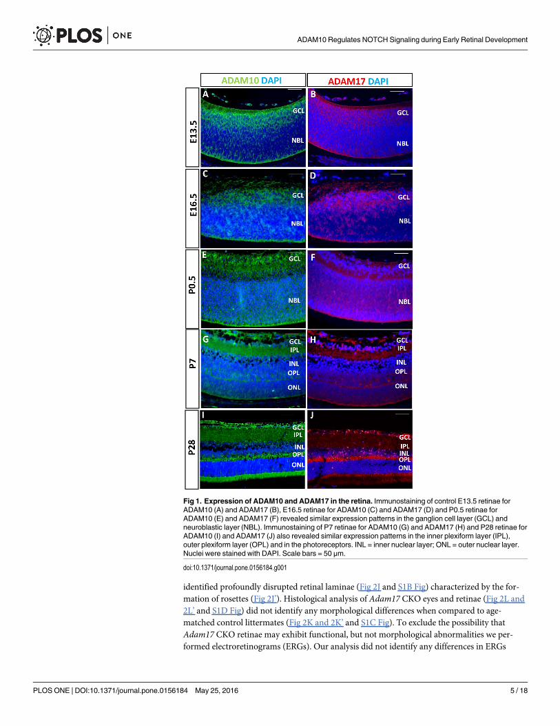
ADAM10 and ADAM17 are ubiquitously expressed retinal proteinsAs the initial step, we evaluated the expression of ADAM10 and ADAM17 during retinal devel-opment in mice. In control embryonic E13.5, E16.5 and newborn P0.5 retinae both ADAM10(Fig 1A, 1C and 1E) and ADAM17 (Fig 1B, 1D and 1F) exhibit ubiquitous expression in gan-glion cell and neuroblastic layers. By P7, as the retina starts to mature, ADAM10 andADAM17 are expressed in the ganglion cell layer, inner and outer plexiform layers, and in thephotoreceptors (Fig 1G and 1H) and this pattern of expression is maintained in the adult retinafor both ADAM10 (Fig 1I) and ADAM17 (Fig 1J).
ADAM10, but not ADAM17, is required for early retinal developmentTo determine the roles of ADAM10 and ADAM17 during early retinal development, we gener-ated mice with retina-specific ablation of Adam10 (Adam10 CKO), as well as retina-specificablation of Adam17 (Adam17 CKO) (Fig 2A and 2B). In newborn Adam10 CKO and Adam17CKO retinae, only residual levels of Adam10 transcript containing exon 3 (Fig 2C) andAdam17 transcript containing exon 2 (Fig 2D) were identified when compared to the controls.Therefore, Adam10 and Adam17 were successfully disrupted in most retinal cells in Adam10CKO and Adam17 CKOmice respectively.
The evaluation of adult (8 weeks of age) Adam10 CKO eyes by observation revealed asmaller eye size (Fig 2F) when compared to age-matched control littermates (Fig 2E). Adam17CKOmice did not exhibit any obvious eye size differences (Fig 2H) when compared to age-matched control littermates (Fig 2G). Histological analysis of adult Adam10 CKO eyes
ADAM10 Regulates NOTCH Signaling during Early Retinal Development
PLOS ONE | DOI:10.1371/journal.pone.0156184 May 25, 2016 4 / 18
identified profoundly disrupted retinal laminae (Fig 2J and S1B Fig) characterized by the for-mation of rosettes (Fig 2J’). Histological analysis of Adam17 CKO eyes and retinae (Fig 2L and2L’ and S1D Fig) did not identify any morphological differences when compared to age-matched control littermates (Fig 2K and 2K’ and S1C Fig). To exclude the possibility thatAdam17 CKO retinae may exhibit functional, but not morphological abnormalities we per-formed electroretinograms (ERGs). Our analysis did not identify any differences in ERGs
Fig 1. Expression of ADAM10 and ADAM17 in the retina. Immunostaining of control E13.5 retinae forADAM10 (A) and ADAM17 (B), E16.5 retinae for ADAM10 (C) and ADAM17 (D) and P0.5 retinae forADAM10 (E) and ADAM17 (F) revealed similar expression patterns in the ganglion cell layer (GCL) andneuroblastic layer (NBL). Immunostaining of P7 retinae for ADAM10 (G) and ADAM17 (H) and P28 retinae forADAM10 (I) and ADAM17 (J) also revealed similar expression patterns in the inner plexiform layer (IPL),outer plexiform layer (OPL) and in the photoreceptors. INL = inner nuclear layer; ONL = outer nuclear layer.Nuclei were stained with DAPI. Scale bars = 50 μm.
doi:10.1371/journal.pone.0156184.g001
ADAM10 Regulates NOTCH Signaling during Early Retinal Development
PLOS ONE | DOI:10.1371/journal.pone.0156184 May 25, 2016 5 / 18
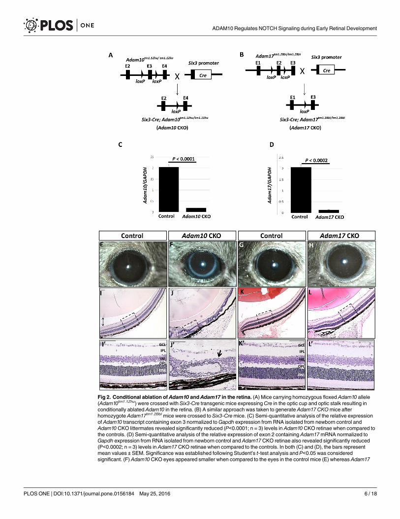
Fig 2. Conditional ablation of Adam10 and Adam17 in the retina. (A) Mice carrying homozygous floxed Adam10 allele(Adam10tm1.1Zhu) were crossed with Six3-Cre transgenic mice expressingCre in the optic cup and optic stalk resulting inconditionally ablated Adam10 in the retina. (B) A similar approach was taken to generate Adam17 CKOmice afterhomozygote Adam17tm1.2Bbl mice were crossed to Six3-Cremice. (C) Semi-quantitative analysis of the relative expressionof Adam10 transcript containing exon 3 normalized toGapdh expression from RNA isolated from newborn control andAdam10 CKO littermates revealed significantly reduced (P<0.0001; n = 3) levels in Adam10 CKO retinae when compared tothe controls. (D) Semi-quantitative analysis of the relative expression of exon 2 containing Adam17mRNA normalized toGapdh expression from RNA isolated from newborn control and Adam17 CKO retinae also revealed significantly reduced(P<0.0002; n = 3) levels in Adam17 CKO retinae when compared to the controls. In both (C) and (D), the bars representmean values ± SEM. Significance was established following Student’s t-test analysis and P<0.05 was consideredsignificant. (F) Adam10CKO eyes appeared smaller when compared to the eyes in the control mice (E) whereas Adam17
ADAM10 Regulates NOTCH Signaling during Early Retinal Development
PLOS ONE | DOI:10.1371/journal.pone.0156184 May 25, 2016 6 / 18
between the control and Adam17 CKOmice (not shown). Collectively these findings providedevidence that ADAM10, and not ADAM17, is required for early retinal development.
The onset and progression of retinal phenotypes in Adam10 CKOmiceThe first morphological abnormalities in Adam10 CKO retinae were evident at E13.5 charac-terized by disrupted cellular architecture (Fig 3B). Along the apical-basal axis nuclei fromAdam10 CKO retinae appeared rounded and disorganized resulting in misshapen apical andbasal surfaces (Fig 3B’). By contrast, the E13.5 control retinae exhibited elongated and orga-nized nuclei with distinct apical and basal surfaces (Fig 3A and 3A’). While morphologicalabnormalities were identified within the Adam10 CKO central retinae some regions of periph-eral retinae retained undisrupted retinal cell organization (Fig 3B). It was reported previouslythat Six3-Cre is expressed across the entire retina although the highest expression is within thecentral retina [36]. To explore this further, we immunostained the control and the Adam10CKO retinae for ADAM10. Within the central Adam10 CKO retinae, the majority of cells didnot express ADAM10 (Fig 3D and S2B Fig). However, at the retinal periphery, we identifiedresidual ADAM10 immunostaining (Fig 3D and S2B Fig). This finding suggested that milderretinal abnormalities identified at the retinal periphery of E13.5 Adam10 CKOmay have beencaused by the lower efficiency of CRE-mediated Adam10 ablation relative to the central retina.By E16.5 the Adam10 CKO retinae started to exhibit rosettes within the central retinae (Fig3F); the identified rosettes were of different sizes (Fig 3F’). Immunostaining of E16.5 Adam10CKO retinae for ADAM10 did not identify any residual staining (Fig 3H). We also morpholog-ically evaluated the postnatal retinal development of Adam10 CKOmice. By P0.5 in Adam10CKO retinae, the formation of rosettes expanded further towards the retinal periphery (S2Dand S2D’ Fig) and by P7 the rosettes were present throughout the entire retina (S2F and S2F’Fig).
ADAM10 inactivation disrupts retinal neurogenesisNext, we wanted to determine if Adam10 CKO retinae exhibited disrupted neurogenesis. AtE13.5 in control retinae, EdU-positive cells were highly organized and restricted to the neuro-blastic layer (Fig 4A) whereas in E13.5 Adam10 CKO retinae EdU-positive cells were highlydisorganized (Fig 4B). Quantification analysis revealed a significantly lower (P = 0.019; n = 3)percentage of EdU-positive cells in the Adam10 CKO retinae when compared to the controls(Fig 4C). Next, we hypothesized that the fewer proliferating cells identified in Adam10 CKOretinae (Fig 4A–4C) may be caused by precocious retinal cell differentiation. Immunostainingof the control retinae for orthodenticle homeobox 2 (OTX2), which is a marker for early photo-receptors [41], revealed sparse OTX2-positive staining in cells along the retinal apical surfaceconsistent with the development of early photoreceptors (Fig 4D). By contrast, Adam10 CKOretinae exhibited highly disorganized OTX2-positive cells (Fig 4E). Quantification analysisrevealed a significantly greater (P = 0.003; n = 3) percentage of OTX2-positive cells in theAdam10 CKO retinae when compared to the controls (Fig 4F). We also evaluated the ISL1expression as a marker for early ganglion cells [42]. In control retinae, ISL1 immunostaining
CKO eyes (H) that did not appear to differ from age-matched control littermates (G). H&E staining of Adam10CKO eyes at 8weeks of age (J) revealed severe retinal abnormalities characterized by the formation of rosettes (J’, arrow) in contrast to theretinal laminae in the age-matched controls (I-I’). Histological analysis of Adam17 CKO eyes (L-L’) did not identify anymorphological differences when compared to the age-matched controls (K-K’). Figures shown in (I’) through (L’) areenlarged images of the areas depicted by the dashed boxes in Figures (I) through (L). GCL = ganglion cell layer, IPL = innerplexiform layer, INL = inner nuclear layer, OPL = outer plexiform layer, ONL = outer nuclear layer. Scale bars = 50 μm.
doi:10.1371/journal.pone.0156184.g002
ADAM10 Regulates NOTCH Signaling during Early Retinal Development
PLOS ONE | DOI:10.1371/journal.pone.0156184 May 25, 2016 7 / 18
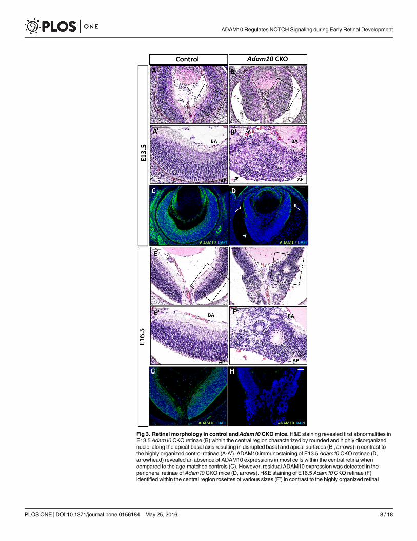
Fig 3. Retinal morphology in control and Adam10 CKOmice.H&E staining revealed first abnormalities inE13.5 Adam10 CKO retinae (B) within the central region characterized by rounded and highly disorganizednuclei along the apical-basal axis resulting in disrupted basal and apical surfaces (B’, arrows) in contrast tothe highly organized control retinae (A-A’). ADAM10 immunostaining of E13.5 Adam10 CKO retinae (D,arrowhead) revealed an absence of ADAM10 expressions in most cells within the central retina whencompared to the age-matched controls (C). However, residual ADAM10 expression was detected in theperipheral retinae of Adam10CKOmice (D, arrows). H&E staining of E16.5 Adam10 CKO retinae (F)identified within the central region rosettes of various sizes (F’) in contrast to the highly organized retinal
ADAM10 Regulates NOTCH Signaling during Early Retinal Development
PLOS ONE | DOI:10.1371/journal.pone.0156184 May 25, 2016 8 / 18
was restricted to the cells at the retinal basal layer consistent with the developing ganglion cells(Fig 4G). In the Adam10 CKO retinae, ISL1-positive cells were highly disorganized (Fig 4H).Quantification analysis revealed a significantly greater (P = 0.004; n = 3) percentage of ISL1--positive cells in E13.5 Adam10 CKO when compared to the control retinae (Fig 4I). Similarfindings were observed following immunostaining with BRN3B (S3A–S3C Fig), a marker forearly ganglion cells [42]. In both the control (Fig 4J) and Adam10 CKO retinae (Fig 4K), theOTX2-positive and ISL1-positive cells did not co-stain for EdU, indicating that OTX2-positiveand ISL1-positive cells had exited the cell cycle.
Characterization of rosettes in E16.5 Adam10 CKO retinaeMorphological evaluation of Adam10 CKO retinae revealed the formation of rosettes at E16.5(Fig 3F and 3F’). Next we set to characterize which cells formed the rosettes in E16.5 Adam10CKO retinae. In the control, the retinae BrdU-positive cells are positioned between the OTX2-positive photoreceptors on the apical side and ISL1-positive ganglion cells at the basal side (Fig5A and 5C). Immunostaining of Adam10 CKO retinae revealed that BrdU-positive cells wereforming the rosette outer cellular layers (Fig 5B) whereas OTX2 positive photoreceptors wereforming the rosette inner cellular layers (Fig 5D). However, in the Adam10 CKO retinae,OTX2-positive photoreceptors were also identified as highly disorganized cells present at theapical and basal surface as well as cells randomly scattered throughout the retina (Fig 5D). TheISL1-positive cells did not associate with rosettes and appeared to be randomly distributedthroughout the Adam10 CKO (Fig 5D). It was shown previously that both N-cadherin and β-catenin are indispensable for the formation of retinal laminae [43, 44], thus we wanted to deter-mine if N-cadherin and β-catenin expression had been altered in the Adam10 CKO retinae. Inthe control retinae both N-cadherin and β-catenin (Fig 5E–5G) were expressed throughout theretina especially in the cells forming apical and basal surfaces consistent with previous reports[44]. In the Adam10 CKO retinae both N-cadherin (Fig 5F) and β-catenin (Fig 5H) were highlyexpressed in cells forming the rosette lumens whereas cells forming apical and basal surfacesintermittently expressed both N-cadherin and β-catenin. Next, we wanted to determine if celldeath was contributing to the formation of rosettes. In E16.5 control retinae, we identified avery few TUNEL-positive cells (Fig 5I) whereas in Adam10 CKO the TUNEL-positive cellswere scattered throughout all layers of the retinae (Fig 5J). Although a significantly greater(P = 0.003; n = 3) percentage of TUNEL-positive cells were identified in Adam10 CKO retinae(Fig 5H) when compared to controls, the overall percentage of TUNEL-positive cells was verysmall (<5%) in both the WT and Adam10 CKO retinae (Fig 5K). Consistent with this, theoverall number of DAPI positive cells did not significantly differ (P = 0.19; n = 3) between thetwo genotypes (Fig 5L).
ADAM10 regulates NOTCH signalingNext, we set out to determine if disrupted NOTCH1 signaling contributed to the retinal pheno-types identified in Adam10 CKO retinae. Immunostaining of E13.5 control retinae revealedthat NOTCH1 positive immunostaining was restricted to the neuroblastic layer (Fig 6A). Bycontrast in E13.5 Adam10 CKO retinae very few cells within the central retina stained positivefor NOTCH1, although towards the retinal periphery highly disorganized NOTCH1 positivecells were evident (Fig 6B). Quantification analysis confirmed a significantly (P = 0.001; n = 3)
laminae in the age-matched control retinae (E-E’). Immunostaining of E16.5 Adam10 CKO retinae (H) did notidentify any ADAM10-positive cells in contrast to the age-matched controls (G). BA = basal surface,AP = apical surface. Scale bars = 50 μm.
doi:10.1371/journal.pone.0156184.g003
ADAM10 Regulates NOTCH Signaling during Early Retinal Development
PLOS ONE | DOI:10.1371/journal.pone.0156184 May 25, 2016 9 / 18
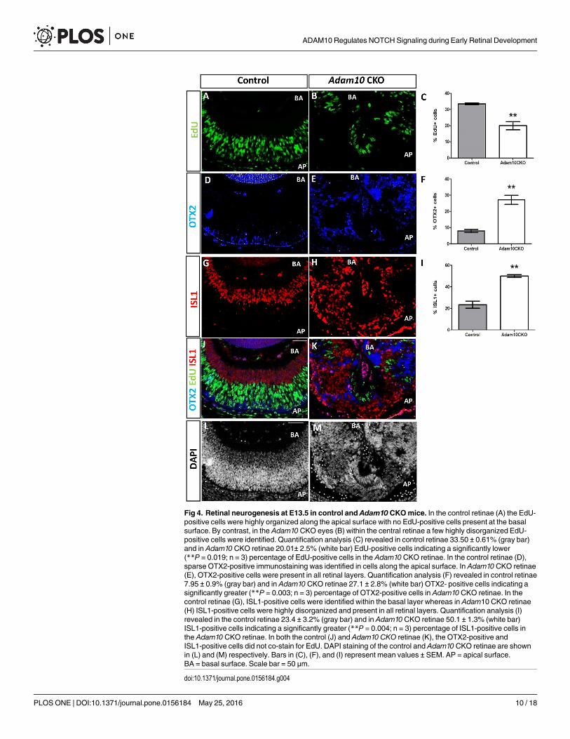
Fig 4. Retinal neurogenesis at E13.5 in control and Adam10 CKOmice. In the control retinae (A) the EdU-positive cells were highly organized along the apical surface with no EdU-positive cells present at the basalsurface. By contrast, in the Adam10CKO eyes (B) within the central retinae a few highly disorganized EdU-positive cells were identified. Quantification analysis (C) revealed in control retinae 33.50 ± 0.61% (gray bar)and in Adam10 CKO retinae 20.01± 2.5% (white bar) EdU-positive cells indicating a significantly lower(**P = 0.019; n = 3) percentage of EdU-positive cells in the Adam10CKO retinae. In the control retinae (D),sparse OTX2-positive immunostaining was identified in cells along the apical surface. In Adam10CKO retinae(E), OTX2-positive cells were present in all retinal layers. Quantification analysis (F) revealed in control retinae7.95 ± 0.9% (gray bar) and in Adam10 CKO retinae 27.1 ± 2.8% (white bar) OTX2- positive cells indicating asignificantly greater (**P = 0.003; n = 3) percentage of OTX2-positive cells in Adam10CKO retinae. In thecontrol retinae (G), ISL1-positive cells were identified within the basal layer whereas in Adam10 CKO retinae(H) ISL1-positive cells were highly disorganized and present in all retinal layers. Quantification analysis (I)revealed in the control retinae 23.4 ± 3.2% (gray bar) and in Adam10 CKO retinae 50.1 ± 1.3% (white bar)ISL1-positive cells indicating a significantly greater (**P = 0.004; n = 3) percentage of ISL1-positive cells inthe Adam10 CKO retinae. In both the control (J) and Adam10 CKO retinae (K), the OTX2-positive andISL1-positive cells did not co-stain for EdU. DAPI staining of the control and Adam10 CKO retinae are shownin (L) and (M) respectively. Bars in (C), (F), and (I) represent mean values ± SEM. AP = apical surface.BA = basal surface. Scale bar = 50 μm.
doi:10.1371/journal.pone.0156184.g004
ADAM10 Regulates NOTCH Signaling during Early Retinal Development
PLOS ONE | DOI:10.1371/journal.pone.0156184 May 25, 2016 10 / 18
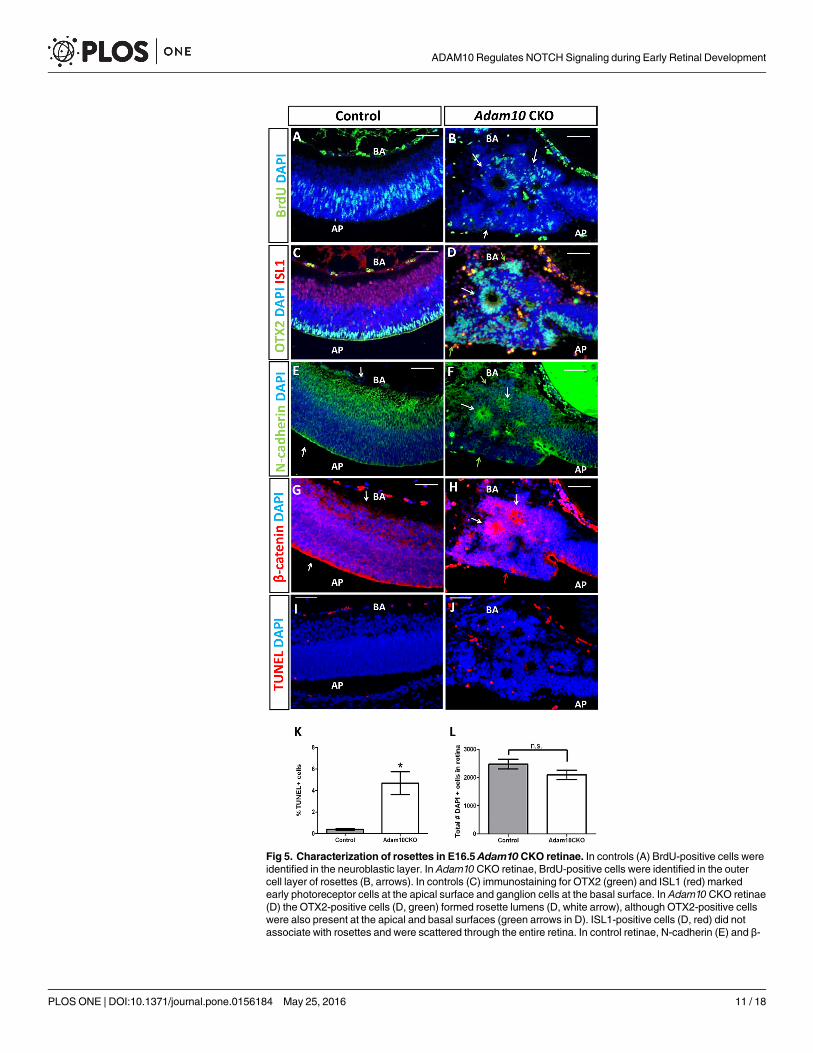
Fig 5. Characterization of rosettes in E16.5 Adam10 CKO retinae. In controls (A) BrdU-positive cells wereidentified in the neuroblastic layer. In Adam10 CKO retinae, BrdU-positive cells were identified in the outercell layer of rosettes (B, arrows). In controls (C) immunostaining for OTX2 (green) and ISL1 (red) markedearly photoreceptor cells at the apical surface and ganglion cells at the basal surface. In Adam10 CKO retinae(D) the OTX2-positive cells (D, green) formed rosette lumens (D, white arrow), although OTX2-positive cellswere also present at the apical and basal surfaces (green arrows in D). ISL1-positive cells (D, red) did notassociate with rosettes and were scattered through the entire retina. In control retinae, N-cadherin (E) and β-
ADAM10 Regulates NOTCH Signaling during Early Retinal Development
PLOS ONE | DOI:10.1371/journal.pone.0156184 May 25, 2016 11 / 18
lower percentage of NOTCH1 positive cells in E13.5 Adam10 CKO retinae when compared tothe age-matched controls (Fig 6C). Immunostaining for HES1, which is a NOTCH1 down-stream target [15], revealed a very few HES1-immunopositive cells within the central E13.5Adam10 CKO retinae that appeared highly disorganized (Fig 6D). However, at the retinalperiphery HES1 positive cells were identified restricted to the neuroblastic layer (not shown).Quantification analysis confirmed a significantly (P = 0.0004; n = 3) lower percentage ofHES1-positive in E13.5 Adam10 CKO retinae when compared to the age-matched controls.
In order to determine if disrupted NOTCH1 signaling is directly caused by a functional lossof ADAM10, we utilized a genetic approach. We focused on establishing if overexpression ofthe NOTCH1-intracellular domain (N1-ICD) can rescue aberrant morphogenesis and/or neu-rogenesis identified in Adam10 CKO retinae. We crossed Adam10 CKOmice to Gt(ROSA)26Sor tm1(NOTCH1)Dam/J mice and generated mice with genotypes Six3-Cre;Adam10 CKO;RosaN1-IC/+ (Adam10 CKO;N1-ICD) wherein the same retinal cells Six3-driven CRE recombi-nase activity deleted Adam10 exon 3 and overexpressed N1-ICD; mice with Six3-Cre;RosaN1-IC/+ (N1-ICD) genotypes, where Six3-CRE-mediated constitutive retinal activation ofN1ICD, were used as controls (Fig 6G). At E13.5 cells, both N1-ICD retinae (Fig 6H) andAdam10 CKO;N1-ICD retinae (Fig 6I), were highly organized along the apical-basal axis withwell-defined apical and basal surfaces. EdU incorporation revealed that in both N1-ICD retinae(Fig 6J) and Adam10 CKO; N1-ICD retinae (Fig 6K) proliferative cells were present in all reti-nal layers suggesting that all retinal cells in these mice were maintained in the progenitor state.The percentage of EdU positive cells did not significantly differ between the genotypes (Fig6L). To explore this further, we immunostained E13.5 N1-ICD retinae and Adam10 CKO;N1-ICD retinae for OTX2 and ISL1 as early photoreceptor and ganglion cell markers respec-tively. Within the central region of both N1-ICD retinae (S4A Fig) and Adam10 CKO; N1-ICD(S4B Fig) we did not identify any OTX2-positive or ISL1-positive cells. However, at the retinalperiphery very few OTX2-positive (<1%) and ISL1-positive cells (<3%) were identified inboth N1-ICD (S4C Fig) and Adam10 CKO; N1-ICD retinae (S4D Fig). Quantification analysisdid not identify a significant difference between N1-ICD and Adam10 CKO;N1-ICD retinae inthe percentage of OTX2-positive and ISL1-positive cells (S4E and S4F Fig).
DiscussionIn this study, we show that ADAM10 is indispensable for retinal neurogenesis as well as theformation of retinal laminae. The rescue of aberrant retinal phenotypes in ADAM10-deficientretinae by overexpression of the N1-ICD provides in vivo genetic evidence that ADAM10 is akey regulator of NOTCH1 signaling. In mice during early retinal development NOTCH1maintains retinal cells in the undifferentiated progenitor state, but it also has a role in repress-ing photoreceptor cell fate [10, 11]. As such the role of NOTCH1 signaling has been proposed
catenin (G) were expressed in all retinal cells although the highest expression was in the cells forming theapical surface (white arrows in E and G). In Adam10 CKO retinae cells forming the rosette lumens were highlyexpressing N-cadherin and β-catenin (white arrows F and H); cells at the apical and basal surface wereintermittently expressing N-cadherin (green arrows in F) and β-catenin (red arrows in H). Very few TUNEL-positive cells were identified in the control (I) and Adam10 CKO (J) retinae. Quantification analysis (K)identified in the controls 0.4 ± 0.01% (gray bar) and in Adam10 CKO 4.69 ± 0.11% (white bar) TUNEL-positivecells indicated a significantly greater (*P = 0.003; n = 3) percentage of TUNEL-positive cells in the Adam10CKO retinae. Quantification analysis (L) identified 2476 ± 173 (gray bar) DAPI-positive cells in controls and2096 ± 167 (white bar) DAPI-positive cells in Adam10 CKO retinae indicating that the overall number of retinalcells did not significantly differ (P = 0.19; n = 3) between the two genotypes. Bars in (K) and (L) representmean values ± SEM. Significance was established following analysis with Student’s t-test and P<0.05 wasconsidered significant. In all panels, nuclei were stained with DAPI. AP = apical surface. BA = basal surface.n.s. = not significant. Scale bar = 50 μm.
doi:10.1371/journal.pone.0156184.g005
ADAM10 Regulates NOTCH Signaling during Early Retinal Development
PLOS ONE | DOI:10.1371/journal.pone.0156184 May 25, 2016 12 / 18
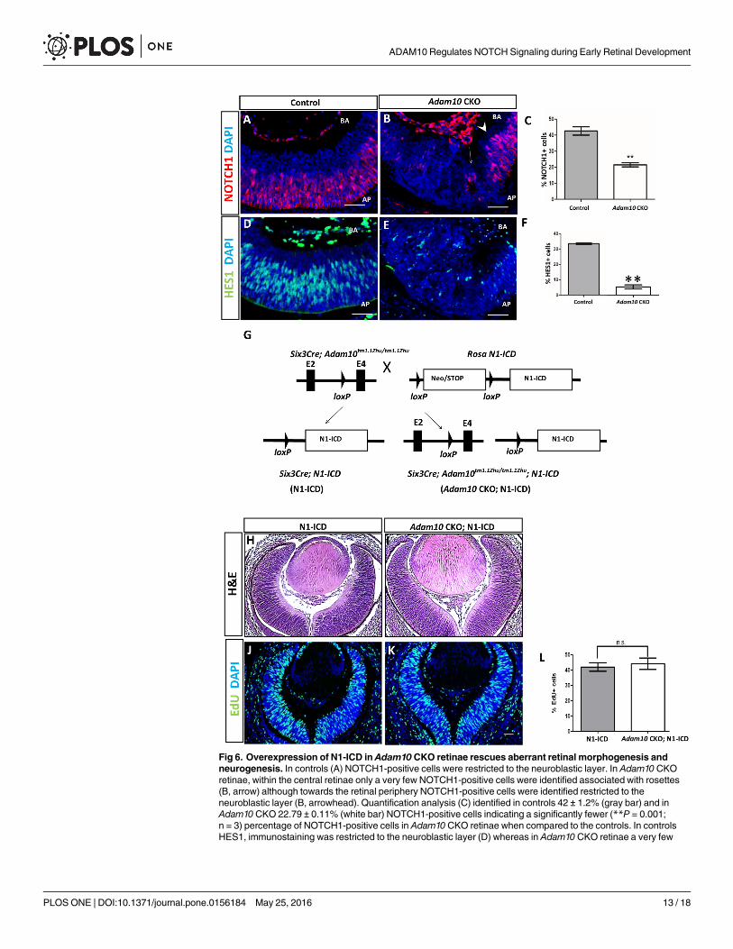
Fig 6. Overexpression of N1-ICD in Adam10 CKO retinae rescues aberrant retinal morphogenesis andneurogenesis. In controls (A) NOTCH1-positive cells were restricted to the neuroblastic layer. In Adam10 CKOretinae, within the central retinae only a very few NOTCH1-positive cells were identified associated with rosettes(B, arrow) although towards the retinal periphery NOTCH1-positive cells were identified restricted to theneuroblastic layer (B, arrowhead). Quantification analysis (C) identified in controls 42 ± 1.2% (gray bar) and inAdam10 CKO 22.79 ± 0.11% (white bar) NOTCH1-positive cells indicating a significantly fewer (**P = 0.001;n = 3) percentage of NOTCH1-positive cells in Adam10 CKO retinae when compared to the controls. In controlsHES1, immunostaining was restricted to the neuroblastic layer (D) whereas in Adam10 CKO retinae a very few
ADAM10 Regulates NOTCH Signaling during Early Retinal Development
PLOS ONE | DOI:10.1371/journal.pone.0156184 May 25, 2016 13 / 18
to have a key role in assuring the sufficient number of progenitor cells necessary for the devel-opment of all retinal cell types [10, 11]. Results from this study showed that ADAM10 defi-ciency decreased the pool of retinal progenitor cells and increased the production of earlyphotoreceptors further supporting the idea that ADAM10 is a regulator of NOTCH1 signaling.However, our results also show that ADAM10 retinal deficiency increased the production ofganglion cells, a phenotype not identified in mice with NOTCH1-deficient retinae [10, 11].ADAM10 ablation in the developing retina phenocopies aberrant retinal neurogenesis in micewith retinae deficient for both NOTCH1 and NOTCH3 [45] as well as in mice with retinaedeficient for RPBJ [12]. While it is well established that NOTCH1 is a regulator of the progeni-tor cell pool and represses photoreceptor fate, recent studies have shown both NOTCH3 andNOTCH1 contribute to the repression of ganglion cell fate [45]. Furthermore both NOTCH1and NOTCH3 signal through RBPJ as a common downstream target [12]. Therefore, it washypothesized that NOTCH1+NOTCH3>RBPJ repress retinal ganglion cell fate. We proposethat ADAM10 is a common NOTCH signaling regulator by facilitating the cleavage of bothNOTCH1 and NOTCH3 which in turn facilitates maintenance of the progenitor cell pool andrepression of both photoreceptors and ganglion cells. In support of this hypothesis are recentreports showing that in cell culture ADAM10 cleaves NOTCH3 via a similar mechanism asNOTCH1 [46]. Additionally, during retinal development expression of NOTCH1 andNOTCH3 spatially and temporally coincide [47]. However, further studies are needed tounequivocally establish if ADAM10 mediates the cleavage of both NOTCH1 and NOTCH3during early retinal neurogenesis.
In addition to aberrant neurogenesis, ADAM10 retinal ablation also results in severely dis-rupted formation of retinal laminae. Prior to differentiation, RPCs are positioned along the api-cal-basal axis and are attached to both the apical and basal surfaces [48, 49]. As RPCs start todifferentiate, they undergo detachment allowing the cells to migrate to the appropriate retinallayer thereby establishing the retinal laminae and neuronal circuitry required for vision [48, 49].Our results show that ADAM10 retinal deficiency results in the formation of rosettes similar tothe rosettes reported for mice with disrupted retinal NOTCH1 signaling [9–15]. It was shownthat RBPJ retinal deficiency causes discontinuous expression of β-catenin resulting in the forma-tion of rosettes; ectopic overexpression of β-catenin in RBPJ-deficient retinae rescued the rosetteformation without rescuing neurogenesis defects [13]. How disrupted retinal NOTCH1 signal-ing alters β-catenin expression still remains unclear. Active β-catenin is a downstream effectorof Wnt signaling [50], but active β-catenin is also bound to N-cadherin at the cell membranes[51, 52]. A functional deficiency of either N-cadherin or β-catenin disrupts the formation of ret-inal laminae without disrupted neurogenesis [43, 44, 53, 54]. However, it was shown previouslythat ADAM10mediates the cleavage of N-cadherin and as such regulates the release of active β-catenin into the cytoplasmic pool [51]. At this point, it is unclear if the rosetting phenotypes
HES1-positive cells were identified within the central retinae and HES1-positve cells appeared disorganized (E).Quantification analysis (F) identified in controls 34 ± 0.01% (gray bar) and in Adam10CKO retinae 4.7 ± 0.2%(white bar) HES1-positive cells indicating a significantly fewer (**P = 0.0004; n = 3) percentage of HES1-positivecells in Adam10CKO retinae when compared to controls. (G) Adam10 CKOmice were crossed to RosaN1-IC
(N1-ICD) mice harboring a Neo/STOP cassette flanked by loxP sites upstream of a sequence encoding theintracellular domain of theNOTCH1 gene thereby generating Adam10CKO; N1-ICDmice that exhibit constitutiveexpression of N1-ICD in the same retinal cells in which Six3-CRE led to ablation of Adam10. H&E stainingrevealed in N1-ICDmice (H) and Adam10CKO;N1-IC mice (I) highly organized retinae. In N1-ICD retinae (J) andAdam10 CKO;N1-IC retinae (K) EdU-positive cells were present in all retinal layers. Quantification analysis (L)revealed in N1-ICD retinae 41.99 ± 2.78% (gray bar) and in Adam10 CKO retinae 44.15 ± 3.76% (white bar) EdU-positive cells indicating that the percentage of EdU positive retinal cells did not significantly differ (P = 0.24; n = 3).Bars in (C), (F), and (L) represent mean values ± SEM. Significance was established with the Student’s t-test andP<0.05 was considered significant. AP = apical surface. BA = basal surface. Scale bars = 50 μm.
doi:10.1371/journal.pone.0156184.g006
ADAM10 Regulates NOTCH Signaling during Early Retinal Development
PLOS ONE | DOI:10.1371/journal.pone.0156184 May 25, 2016 14 / 18
observed in ADAM10-deficient retinae are solely caused by the disrupted NOTCH1 signalingor if disrupted ADAM10-mediated cleavage of N-cadherin also contributes to the aberrant reti-nal morphogenesis. The rescue of aberrant retinal morphology in ADAM10-deficeint retinae byoverexpression of N1-ICD supports the idea that the failure in the formation of laminae in theADAM10-deficeint retinae is solely caused by disrupted NOTCH1 signaling. However, we can-not exclude the possibility that ADAM10may be regulating the cleavage of N-cadherin as adownstream event following cleavage of the NOTCH1 receptor.
While this study establishes ADAM10 as indispensable for the early retinal development, therole of ADAM17 still remains unclear. Our results show that ADAM10 and ADAM17 exhibitan overlapping spatiotemporal expression pattern in the developing mouse retinae consistentwith the expression of ADAM10 and ADAM17 previously reported for the developing chickretinae [34]. However, it should be pointed out that another study using a different antibodyidentified that in mice, the retinal expression of ADAM17 starts at P7 [55]. Furthermore, thesame study revealed that in the adult (P175) mouse retinae ADAM17 expression is restricted tothe ganglion cell layer, the inner and outer edge of the inner nuclear layer [55]. This discrepancymay have been caused by the variable specificity of the antibodies used as well as different timepoints selected for the analysis. Nonetheless, our findings unequivocally indicate that ADAM17is dispensable for retinal development. In the developing chick retina, in addition to ADAM10,ADAM17 exhibits similar spatiotemporal expression pattern with ADAM9, ADAM12,ADAM13, ADAM22, and ADAM23 [34]. Except for ADAM9, that has been shown to have arole in the development of retinal vasculature and homeostasis [56, 57], the roles of otherADAM proteins during retinal development and homeostasis are unknown providing no infor-mation if ADAM17may have functional overlaps with other ADAM proteins. Studies focusingon the role of ADAM17 as a regulator of retinal homeostasis are currently in progress.
Supporting InformationS1 Fig. Eye phenotypes of Adam10 CKO and Adam17 CKOmice.(TIF)
S2 Fig. Adam10 CKO eye phenotypes.(TIF)
S3 Fig. BRN3B expression in E13.5 expression in E13.5 control and Adam10 CKO retinae.(TIF)
S4 Fig. Retinae of E13.5 N1-ICD and Adam10 CKOmice.(TIF)
S1 Table. A list of primers used in the study.(DOCX)
AcknowledgmentsWe would like to thank Mr. Peter Volberding and Dr. Joseph Besharse from the Department ofCell Biology, Neurobiology and Anatomy, Medical College of Wisconsin for their assistancewith performing, recording, and interpretation of ERGs in mice.
Author ContributionsConceived and designed the experiments: JT DJS. Performed the experiments: JT AR. Analyzedthe data: JT DJS. Contributed reagents/materials/analysis tools: DJS. Wrote the paper: JT DJS.
ADAM10 Regulates NOTCH Signaling during Early Retinal Development
PLOS ONE | DOI:10.1371/journal.pone.0156184 May 25, 2016 15 / 18
References1. Cepko CL, Austin CP, Yang X, Alexiades M, Ezzeddine D. Cell fate determination in the vertebrate ret-
ina. Proc Natl Acad Sci U S A. 1996; 93(2):589–95. Epub 1996/01/23. PMID: 8570600; PubMed CentralPMCID: PMC40096.
2. Young RW. Cell differentiation in the retina of the mouse. Anat Rec. 1985; 212(2):199–205. Epub 1985/06/01. doi: 10.1002/ar.1092120215 PMID: 3842042.
3. Cepko C. Intrinsically different retinal progenitor cells produce specific types of progeny. Nature reviewsNeuroscience. 2014; 15(9):615–27. Epub 2014/08/07. doi: 10.1038/nrn3767 PMID: 25096185.
4. Kopan R, Ilagan MX. The canonical Notch signaling pathway: unfolding the activation mechanism. Cell.2009; 137(2):216–33. Epub 2009/04/22. doi: 10.1016/j.cell.2009.03.045 PMID: 19379690; PubMedCentral PMCID: PMC2827930.
5. Guruharsha KG, Kankel MW, Artavanis-Tsakonas S. The Notch signalling system: recent insights intothe complexity of a conserved pathway. Nature reviews Genetics. 2012; 13(9):654–66. Epub 2012/08/08. doi: 10.1038/nrg3272 PMID: 22868267.
6. Ohtsuka T, Ishibashi M, Gradwohl G, Nakanishi S, Guillemot F, Kageyama R. Hes1 and Hes5 as notcheffectors in mammalian neuronal differentiation. The EMBO journal. 1999; 18(8):2196–207. Epub1999/04/16. doi: 10.1093/emboj/18.8.2196 PMID: 10205173; PubMed Central PMCID: PMC1171303.
7. Ohsawa R, Kageyama R. Regulation of retinal cell fate specification by multiple transcription factors.Brain research. 2008; 1192:90–8. Epub 2007/05/10. S0006-8993(07)00814-1 [pii] doi: 10.1016/j.brainres.2007.04.014 PMID: 17488643.
8. Gazave E, Lapebie P, Richards GS, Brunet F, Ereskovsky AV, Degnan BM, et al. Origin and evolutionof the Notch signalling pathway: an overview from eukaryotic genomes. BMC evolutionary biology.2009; 9:249. Epub 2009/10/15. doi: 10.1186/1471-2148-9-249 PMID: 19825158; PubMed CentralPMCID: PMC2770060.
9. Rocha SF, Lopes SS, Gossler A, Henrique D. Dll1 and Dll4 function sequentially in the retina and pV2domain of the spinal cord to regulate neurogenesis and create cell diversity. Developmental biology.2009; 328(1):54–65. Epub 2009/04/25. doi: 10.1016/j.ydbio.2009.01.011 PMID: 19389377.
10. Yaron O, Farhy C, Marquardt T, Applebury M, Ashery-Padan R. Notch1 functions to suppress cone-photoreceptor fate specification in the developing mouse retina. Development. 2006; 133(7):1367–78.Epub 2006/03/03. doi: 10.1242/dev.02311 PMID: 16510501.
11. Jadhav AP, Mason HA, Cepko CL. Notch 1 inhibits photoreceptor production in the developing mam-malian retina. Development. 2006; 133(5):913–23. Epub 2006/02/03. dev.02245 [pii] doi: 10.1242/dev.02245 PMID: 16452096.
12. Riesenberg AN, Liu Z, Kopan R, Brown NL. Rbpj cell autonomous regulation of retinal ganglion cell andcone photoreceptor fates in the mouse retina. J Neurosci. 2009; 29(41):12865–77. Epub 2009/10/16.29/41/12865 [pii] doi: 10.1523/JNEUROSCI.3382-09.2009 PMID: 19828801; PubMed Central PMCID:PMC2788434.
13. Zheng MH, Shi M, Pei Z, Gao F, Han H, Ding YQ. The transcription factor RBP-J is essential for retinalcell differentiation and lamination. Molecular brain. 2009; 2:38. Epub 2009/12/19. doi: 10.1186/1756-6606-2-38 PMID: 20017954; PubMed Central PMCID: PMC2804697.
14. Tomita K, Ishibashi M, Nakahara K, Ang SL, Nakanishi S, Guillemot F, et al. Mammalian hairy andEnhancer of split homolog 1 regulates differentiation of retinal neurons and is essential for eye morpho-genesis. Neuron. 1996; 16(4):723–34. Epub 1996/04/01. PMID: 8607991.
15. Takatsuka K, Hatakeyama J, Bessho Y, Kageyama R. Roles of the bHLH gene Hes1 in retinal morpho-genesis. Brain research. 2004; 1004(1–2):148–55. Epub 2004/03/23. doi: 10.1016/j.brainres.2004.01.045 PMID: 15033430.
16. Edwards DR, Handsley MM, Pennington CJ. The ADAMmetalloproteinases. Molecular aspects ofmedicine. 2008; 29(5):258–89. Epub 2008/09/03. doi: 10.1016/j.mam.2008.08.001 PMID: 18762209.
17. Reiss K, Saftig P. The "a disintegrin and metalloprotease" (ADAM) family of sheddases: physiologicaland cellular functions. Seminars in cell & developmental biology. 2009; 20(2):126–37. Epub 2008/12/04. doi: 10.1016/j.semcdb.2008.11.002 PMID: 19049889.
18. Rooke J, Pan D, Xu T, Rubin GM. KUZ, a conserved metalloprotease-disintegrin protein with two rolesin Drosophila neurogenesis. Science. 1996; 273(5279):1227–31. Epub 1996/08/30. PMID: 8703057.
19. Sotillos S, Roch F, Campuzano S. The metalloprotease-disintegrin Kuzbanian participates in Notchactivation during growth and patterning of Drosophila imaginal discs. Development. 1997; 124(23):4769–79. Epub 1998/01/15. PMID: 9428413.
20. Lieber T, Kidd S, Young MW. kuzbanian-mediated cleavage of Drosophila Notch. Genes & develop-ment. 2002; 16(2):209–21. Epub 2002/01/19. doi: 10.1101/gad.942302 PMID: 11799064; PubMedCentral PMCID: PMC155326.
ADAM10 Regulates NOTCH Signaling during Early Retinal Development
PLOS ONE | DOI:10.1371/journal.pone.0156184 May 25, 2016 16 / 18
21. Jarriault S, Greenwald I. Evidence for functional redundancy between C. elegans ADAM proteins SUP-17/Kuzbanian and ADM-4/TACE. Developmental biology. 2005; 287(1):1–10. Epub 2005/10/04. doi:10.1016/j.ydbio.2005.08.014 PMID: 16197940; PubMed Central PMCID: PMC1805470.
22. Hartmann D, de Strooper B, Serneels L, Craessaerts K, Herreman A, Annaert W, et al. The disintegrin/metalloprotease ADAM 10 is essential for Notch signalling but not for alpha-secretase activity in fibro-blasts. Human molecular genetics. 2002; 11(21):2615–24. Epub 2002/10/02. PMID: 12354787.
23. Peschon JJ, Slack JL, Reddy P, Stocking KL, Sunnarborg SW, Lee DC, et al. An essential role for ecto-domain shedding in mammalian development. Science. 1998; 282(5392):1281–4. Epub 1998/11/13.PMID: 9812885.
24. Horiuchi K, Kimura T, Miyamoto T, Takaishi H, Okada Y, Toyama Y, et al. Cutting edge: TNF-alpha-converting enzyme (TACE/ADAM17) inactivation in mouse myeloid cells prevents lethality from endo-toxin shock. Journal of immunology. 2007; 179(5):2686–9. Epub 2007/08/22. PMID: 17709479.
25. Brou C, Logeat F, Gupta N, Bessia C, LeBail O, Doedens JR, et al. A novel proteolytic cleavageinvolved in Notch signaling: the role of the disintegrin-metalloprotease TACE. Mol Cell. 2000; 5(2):207–16. Epub 2000/07/06. PMID: 10882063.
26. Mumm JS, Schroeter EH, Saxena MT, Griesemer A, Tian X, Pan DJ, et al. A ligand-induced extracellu-lar cleavage regulates gamma-secretase-like proteolytic activation of Notch1. Mol Cell. 2000; 5(2):197–206. Epub 2000/07/06. S1097-2765(00)80416-5 [pii]. PMID: 10882062.
27. Bozkulak EC, Weinmaster G. Selective use of ADAM10 and ADAM17 in activation of Notch1 signaling.Molecular and cellular biology. 2009; 29(21):5679–95. Epub 2009/08/26. MCB.00406-09 [pii] doi: 10.1128/MCB.00406-09 PMID: 19704010; PubMed Central PMCID: PMC2772745.
28. van Tetering G, van Diest P, Verlaan I, van der Wall E, Kopan R, Vooijs M. Metalloprotease ADAM10 isrequired for Notch1 site 2 cleavage. The Journal of biological chemistry. 2009; 284(45):31018–27.Epub 2009/09/04. M109.006775 [pii] doi: 10.1074/jbc.M109.006775 PMID: 19726682; PubMed CentralPMCID: PMC2781502.
29. Jorissen E, Prox J, Bernreuther C, Weber S, Schwanbeck R, Serneels L, et al. The disintegrin/metallo-proteinase ADAM10 is essential for the establishment of the brain cortex. J Neurosci. 2010; 30(14):4833–44. Epub 2010/04/08. doi: 10.1523/JNEUROSCI.5221-09.2010 PMID: 20371803; PubMedCentral PMCID: PMC2921981.
30. Weber S, Niessen MT, Prox J, Lullmann-Rauch R, Schmitz A, Schwanbeck R, et al. The disintegrin/metalloproteinase Adam10 is essential for epidermal integrity and Notch-mediated signaling. Develop-ment. 2011; 138(3):495–505. Epub 2011/01/06. doi: 10.1242/dev.055210 PMID: 21205794; PubMedCentral PMCID: PMC3014635.
31. Tsai YH, VanDussen KL, Sawey ET, Wade AW, Kasper C, Rakshit S, et al. ADAM10 regulates Notchfunction in intestinal stem cells of mice. Gastroenterology. 2014; 147(4):822–34 e13. Epub 2014/07/20.doi: 10.1053/j.gastro.2014.07.003 PMID: 25038433; PubMed Central PMCID: PMC4176890.
32. Tian L, Wu X, Chi C, Han M, Xu T, Zhuang Y. ADAM10 is essential for proteolytic activation of Notchduring thymocyte development. International immunology. 2008; 20(9):1181–7. Epub 2008/07/19.dxn076 [pii] doi: 10.1093/intimm/dxn076 PMID: 18635581.
33. Glomski K, Monette S, Manova K, De Strooper B, Saftig P, Blobel CP. Deletion of Adam10 in endothe-lial cells leads to defects in organ-specific vascular structures. Blood. 2011; 118(4):1163–74. Epub2011/06/10. blood-2011-04-348557 [pii] doi: 10.1182/blood-2011-04-348557 PMID: 21652679;PubMed Central PMCID: PMC3148163.
34. Yan X, Lin J, Rolfs A, Luo J. Differential expression of the ADAMs in developing chicken retina. DevGrowthDiffer. 2011; 53(5):726–39. Epub 2011/06/16. doi: 10.1111/j.1440-169X.2011.01282.x PMID: 21671920.
35. Esteve P, Sandonis A, CardozoM, Malapeira J, Ibanez C, Crespo I, et al. SFRPs act as negative modu-lators of ADAM10 to regulate retinal neurogenesis. Nat Neurosci. 2011; 14(5):562–9. Epub 2011/04/12. nn.2794 [pii] doi: 10.1038/nn.2794 PMID: 21478884.
36. Furuta Y, Lagutin O, Hogan BL, Oliver GC. Retina- and ventral forebrain-specific Cre recombinaseactivity in transgenic mice. Genesis. 2000; 26(2):130–2. Epub 2000/03/21. doi: 10.1002/(SICI)1526-968X(200002)26:2<130::AID-GENE9>3.0.CO;2-I [pii]. PMID: 10686607.
37. Murtaugh LC, Stanger BZ, Kwan KM, Melton DA. Notch signaling controls multiple steps of pancreaticdifferentiation. Proc Natl Acad Sci U S A. 2003; 100(25):14920–5. Epub 2003/12/06. doi: 10.1073/pnas.2436557100 PMID: 14657333; PubMed Central PMCID: PMC299853.
38. Toonen JA, Liang L, Sidjanin DJ. Waved with open eyelids 2 (woe2) is a novel spontaneous mousemutation in the Protein Phosphatase 1, Regulatory (inhibitor) Subunit 13 like (Ppp1r13l) gene. BMCgenetics. 2012; 13(1):76. Epub 2012/08/30. doi: 10.1186/1471-2156-13-76 PMID: 22928477.
39. Fogerty J, Besharse JC. 174delGmutation in mouse MFRP causes photoreceptor degeneration andRPE atrophy. Investigative ophthalmology & visual science. 2011; 52(10):7256–66. Epub 2011/08/04.doi: 10.1167/iovs.11-8112 PMID: 21810984; PubMed Central PMCID: PMC3207726.
ADAM10 Regulates NOTCH Signaling during Early Retinal Development
PLOS ONE | DOI:10.1371/journal.pone.0156184 May 25, 2016 17 / 18
40. Hassemer EL, Le Gall SM, Liegel R, McNally M, Chang B, Zeiss CJ, et al. The waved with open eyelids(woe) locus is a hypomorphic mouse mutation in Adam17. Genetics. 2010; 185(1):245–55. Epub 2010/03/03. doi: 10.1534/genetics.109.113167 PMID: 20194968; PubMed Central PMCID: PMC2870960.
41. Nishida A, Furukawa A, Koike C, Tano Y, Aizawa S, Matsuo I, et al. Otx2 homeobox gene controls reti-nal photoreceptor cell fate and pineal gland development. Nat Neurosci. 2003; 6(12):1255–63. Epub2003/11/20. PMID: 14625556.
42. Pan L, Deng M, Xie X, Gan L. ISL1 and BRN3B co-regulate the differentiation of murine retinal ganglioncells. Development. 2008; 135(11):1981–90. Epub 2008/04/25. dev.010751 [pii] doi: 10.1242/dev.010751 PMID: 18434421; PubMed Central PMCID: PMC2758274.
43. Malicki J, Jo H, Pujic Z. Zebrafish N-cadherin, encoded by the glass onion locus, plays an essential rolein retinal patterning. Developmental biology. 2003; 259(1):95–108. Epub 2003/06/19. PMID:12812791.
44. Fu X, Sun H, Klein WH, Mu X. Beta-catenin is essential for lamination but not neurogenesis in mouseretinal development. Developmental biology. 2006; 299(2):424–37. Epub 2006/09/09. doi: 10.1016/j.ydbio.2006.08.015 PMID: 16959241; PubMed Central PMCID: PMC3385515.
45. Maurer KA, Riesenberg AN, Brown NL. Notch signaling differentially regulates Atoh7 and Neurog2 inthe distal mouse retina. Development. 2014; 141(16):3243–54. Epub 2014/08/08. doi: 10.1242/dev.106245 PMID: 25100656; PubMed Central PMCID: PMC4197552.
46. Groot AJ, Habets R, Yahyanejad S, Hodin CM, Reiss K, Saftig P, et al. Regulated proteolysis ofNOTCH2 and NOTCH3 receptors by ADAM10 and presenilins. Molecular and cellular biology. 2014;34(15):2822–32. Epub 2014/05/21. doi: 10.1128/MCB.00206-14 PMID: 24842903; PubMed CentralPMCID: PMC4135574.
47. Lindsell CE, Boulter J, diSibio G, Gossler A, Weinmaster G. Expression patterns of Jagged, Delta1,Notch1, Notch2, and Notch3 genes identify ligand-receptor pairs that may function in neural develop-ment. Mol Cell Neurosci. 1996; 8(1):14–27. Epub 1996/01/01. PMID: 8923452.
48. Galli-Resta L, Leone P, Bottari D, Ensini M, Rigosi E, Novelli E. The genesis of retinal architecture: anemerging role for mechanical interactions? Prog Retin Eye Res. 2008; 27(3):260–83. Epub 2008/04/01. doi: 10.1016/j.preteyeres.2008.02.001 PMID: 18374618.
49. Reese BE. Development of the retina and optic pathway. Vision research. 2011; 51(7):613–32. Epub2010/07/22. doi: 10.1016/j.visres.2010.07.010 PMID: 20647017; PubMed Central PMCID:PMC2974959.
50. Logan CY, Nusse R. TheWnt signaling pathway in development and disease. Annu Rev Cell Dev Biol.2004; 20:781–810. Epub 2004/10/12. doi: 10.1146/annurev.cellbio.20.010403.113126 PMID: 15473860.
51. Reiss K, Maretzky T, Ludwig A, Tousseyn T, de Strooper B, Hartmann D, et al. ADAM10 cleavage of N-cadherin and regulation of cell-cell adhesion and beta-catenin nuclear signalling. The EMBO journal.2005; 24(4):742–52. Epub 2005/02/05. doi: 10.1038/sj.emboj.7600548 PMID: 15692570; PubMedCentral PMCID: PMC549617.
52. Maretzky T, Reiss K, Ludwig A, Buchholz J, Scholz F, Proksch E, et al. ADAM10mediates E-cadherinshedding and regulates epithelial cell-cell adhesion, migration, and beta-catenin translocation. ProcNatl Acad Sci U S A. 2005; 102(26):9182–7. Epub 2005/06/17. doi: 10.1073/pnas.0500918102 PMID:15958533; PubMed Central PMCID: PMC1166595.
53. Masai I, Lele Z, Yamaguchi M, Komori A, Nakata A, Nishiwaki Y, et al. N-cadherin mediates retinal lami-nation, maintenance of forebrain compartments and patterning of retinal neurites. Development. 2003;130(11):2479–94. Epub 2003/04/19. PMID: 12702661.
54. Erdmann B, Kirsch FP, Rathjen FG, More MI. N-cadherin is essential for retinal lamination in the zebra-fish. Developmental dynamics: an official publication of the American Association of Anatomists. 2003;226(3):570–7. Epub 2003/03/06. doi: 10.1002/dvdy.10266 PMID: 12619142.
55. Sel S, Kalinski T, Enssen I, Kaiser M, Nass N, Trau S, et al. Expression analysis of ADAM17 duringmouse eye development. Annals of anatomy = Anatomischer Anzeiger: official organ of the Anato-mische Gesellschaft. 2012; 194(4):334–8. Epub 2011/11/08. doi: 10.1016/j.aanat.2011.10.008 PMID:22055939.
56. Guaiquil V, Swendeman S, Yoshida T, Chavala S, Campochiaro PA, Blobel CP. ADAM9 is involved inpathological retinal neovascularization. Molecular and cellular biology. 2009; 29(10):2694–703. Epub2009/03/11. doi: 10.1128/MCB.01460-08 PMID: 19273593; PubMed Central PMCID: PMC2682031.
57. Parry DA, Toomes C, Bida L, Danciger M, Towns KV, McKibbin M, et al. Loss of the metalloproteaseADAM9 leads to cone-rod dystrophy in humans and retinal degeneration in mice. Am J HumGenet.2009; 84(5):683–91. Epub 2009/05/05. doi: 10.1016/j.ajhg.2009.04.005 PMID: 19409519; PubMedCentral PMCID: PMC2681008.
ADAM10 Regulates NOTCH Signaling during Early Retinal Development
PLOS ONE | DOI:10.1371/journal.pone.0156184 May 25, 2016 18 / 18