acuteandchroniceffectsofaflatoxinontheliverofdomesticand...
TRANSCRIPT

Table1Vehicle
Propylene glycolFractionB1B1LD50
(mg/kg)
0.4000.560Reference
Nesbitt et al. (38)Asao et al.(4)DimethylformamideB10.364Carnaghan
et al(18)DimethylformamideB21.696Carnaghanet al.(18)DimethylformamideG10.784Carnaghanet al.(18)DimethylformamideG23.440Carnaghanet al.(18)DimethylformamideBl0.350Lijinsky
& Butler(29)DimethylformamideG10.914Lijinsky& Butler (29)
[CANCER RESEARCH 29, 236—250,January 1969]
INTRODUCTION
The aflatoxins were isolated from peanut meal in 1961 (61)
during the investigation of an epizootic of “Turkey X―disease
in England. It was shown that these toxins were metabolites of
some strains of Aspergillus Jiavus and that they were the
etiologic agents of the disease in turkeys (7). Field outbreaks
resulted from feeding peanut meal contaminated by the moldmetabolites. Since these discoveries were made, the acute andchronic effects of metabolites of toxin-producing strains of
Aspergillus flavus have been studied in detail. Investigationshave shown that the metabolites consist of four major@fractions, referred to collectively as “aflatoxins― and
designated 1†B2 ,@ , and G2 . Individual fractions are so
designated because of their fluorescence and RF values on
thin-layer chromatographic plates. They are toxic to a large
number of species, and the mixture of metabolites iscarcinogenic to the rat, ferret, duck, and trout. The B1
fraction is carcinogenic to trout and rats but less is known
about the other three fractions.This paper will describe the pathologic changes observed in
the livers of different species after a single dose and after
continuous administration of aflatoxin.
Turkey
Histologic changes induced in the turkey by feedscontaminated with aflatoxin have been described by Suer and
Ostler (61) and by Wannop (68). Birds dying during the earlystages of intoxication had severe periportal hepatic
‘Thisstudy was supported in part by NIH Research Grant CA 08870and by Research Funds of the Medical Research Council, London,England.
2This manuscript is Contribution No. 1285 from the Department ofNutrition and Food Science, Massachusetts Institute of Technology.
3In addition to material supplied by the investigations of theauthors, we are indebted to the following: Dr. R. Allcroft and Staff,Central Veterinary Laboratory, Weybridge, Surrey, England, formaterial from cattle, turkeys, and chickens, and from chronic studieswith pigs; Professor J. H. Wales, Oregon State University, Corvallis, Ore
gon, U. S. A., for material from trout; and Dr. D. A. H. Pratt, Glaxo Research Limited, Fulmer, Buckinghamshire, England, for material fromthe monkey.
Received May 22, 1968; accepted September 8, 1968.
parenchymal cell necrosis and venous congestion. Nodularregeneration accompanied the diffuse necrosis of parenchymalcells and the concomitant biiary proliferation. There was littleor no inflammatory reaction and only a small increase incollagen. Since these early observations were reported, thepathologic changes induced in turkeys by aflatoxin havereceived little attention. Magwood et al. (39) described an“inducedtolerance― to aflatoxin poisoning which appeared tobe unrelated to the changes seen in the liver. After 23 weekson a toxic diet, the livers were nodular with a dissectingfibrosis, biiary proliferation, and marked variation in the size
of parenchymal cell nuclei.
Duckling
Hepatic lesions induced in the duckling by the aflatoxinsform the basis of the bioassay system originally described bySargeant et al. (58). The acute LD50 of the aflatoxins has beenestimated in a variety of solvents, and results from several
different laboratories indicate general agreement for the@fraction (see Table 1). Less is known about the other threefractions.
The lesion induced by a single dose of aflatoxin@ wasdescribed in detail by Butler (12). A periportal zone ofparenchymal cell necrosis with the formation of lakes of fatdeveloped over a 48-hour period. Extensive biiaryproliferation, reaching a maximum at three days, wasassociated with the parenchymal cell necrosis (Fig. 1). Thelivers of normal ducklings contain a large amount of lipid onthe first day of life as a carryover from the yolk, but this
disappears 4—5 days after hatching. In ducklings dosed withaflatoxin, there is a delay in the removal of the lipid, but thereis little or no increase in fat content compared to control
Acute LD50 dose of aflatoxin in ducklings.
236 CANCER RESEARCH VOL.29
Acute and Chronic Effects of Aflatoxin on the Liver of Domestic and
Laboratory Animals : A Review―2'@
PaulM. Newberneand William H. ButlerDepartment of Nutrition and Food Science, Massachusetts Institute of Technology, Cambridge, Massachusetts 02139, andMedica.l ResearchCouncil Laboratories, Toxicology Research Unit, Carshalton, Surrey, England

Acute and Chronic Effects of Aflatoxin
birds. At 14 days postexposure, there was an increase inmitotic activity of parenchymal cells of birds surviving anLD50 dose.
Levels representing @—@ the LD50 dose resulted in lesssevere lesions, and there was little correlation between doseand degree of biliary proliferation. A wide variation in
response to a given dose was observed among individual birds,and this contributed to the apparent lack of uniform biiaryproliferation within groups. A hemorrhagic peniportal necrosis
was often induced with doses greater than the LD50 . Thedevelopment of biliary proliferation was at first thought to be
characteristic of and specific for aflatoxin. However, a similar
lesion was described which was associated with the
administration of dimethylnitrosamine and cycasin, but not
with carbon tetrachloride, thioacetamide, or the alkaloidsretrorsine and indecine. The observation that histologic changes
similar to those observed with aflatoxin were induced by
dimethylnitrosamine was confirmed by Carlton et al. (16).
Asplin and Carnaghan (5) described an acute disease in
day-old ducklings fed toxic peanut meal. The birds failed togrow and there were macroscopic hemorrhages in many of theorgans and tissues. Extensive biliary proliferation and
parenchymal cell degeneration were observed in the liver; these
changes were followed by cirrhosis in many of the birds.
Newberne et a!. (50) have shown that the repeated
administration of purified aflatoxin or continuous feeding of
Aspergillus flavus extracts or peanut meal naturally contami
nated with the aflatoxins produced the same pathologic lesionsseen by earlier workers; they described in detail the development of the liver lesion up to four weeks. The severity of thelesion was related to the amount of aflatoxin administered.
Two days after exposure, there were parenchymal cell necrosisand early biliary proliferation; the latter progressed in parallelwith increased severity of parenchymal cell necrosis. Sevendays after exposure, mitotic figures were present in the parenchymal cells and biliary proliferation was still active. Wide
spread nodular regeneration was observed after four weeks.
Madhavan and Rao (37) reported hepatic infarcts in ducklingsgiven 10—40 hg of aflatoxin a day for five days, but this hasnot been confirmed by other workers and appears to be uncommon. Carnaghan (17) described the development ofhepatic parenchymal cell tumors in ducks fed 0.035 ppm of
aflatoxin, as a contaminant in peanut meal, for 14 months;Newberne (44) observed similar tumors in ducks after 16
months exposure to feed containing contaminated peanut
meal. Cirrhosis was present in all birds in the latter experi
ments whether or not liver cell tumors developed.
Chicken
A spontaneous disease of young chickens attributed to toxicpeanut meal was reported by Asplin and Carnaghan (5). Thelivers of birds were described as firm and pale in color. Histologically, degenerative and regenerative changes were seen,
and, by four weeks, regenerating nodules of parenchymal cells
were present. In birds fed aflatoxin-contaminated peanut meal,
there was a progressive lymphoid hyperplasia; at the end of
four months, there were no regenerative nodules, but large
multiple focal areas of lymphoid hyperplasia were observed.
Similar changes have been described by Loizelier (33) and byRaimo et a!. (56).
Carnaghan et al. (20) studied the experimental poisoning ofchickens with toxic peanut meal contained in a diet whichassayed about 1.5 ppm of the B1 fraction. Only one animalfed this diet died, and clinical manifestation of toxicity waslimited to slower growth. During the first few weeks of theexperiment, the livers of those fed the aflatoxin-contaminateddiet were enlargedand pale, and some of them containedpetechial hemorrhages. Later, there was a progressive nodularity of the surface. Microscopically, the first change was seenafter 3.5 days and consisted of a periportal fatty infiltration;this lesion progressed for the next 3—4weeks. Associated withthe fatty change were scattered liver cell necrosis, progressivebiliary proliferation, and an increase in connective tissue. Afterfour weeks on experiment, regenerating parenchymal cellswith large nuclei were arranged in ductular fashion ; these lesions were seen along with individual cell necrosis. Large aggregates of polymorphonuclear leukocytes and lymphocytes wereseen in the portal tracts. After six weeks, biiary proliferation,fibrosis, and lymphocytic hyperplasia of the portal tracts increased. Foci of regenerating parenchymal cells were presentwith some nuclear enlargement. After eight weeks exposure,when the experiment was terminated, the parenchymal cellswere surrounded by areas of bile ducts and fibrous tissue contaming large focal areas of lymphocytic hyperplasia.
Cattle
The first report of poisoning in cattle by Brazilian peanut(groundnut)mealwasthat of Loosmoreand Markson(36).Calves, 3—9 months of age, had eaten for at least six weeks acompounded food containing 15% Brazilian peanut meal (notassayed for aflatoxin content). The livers of the animals exhibited areas of fibrosis with biiary proliferation and venoocclusive disease similar to that described in ragwort (Seneciojacobea) poisoning (40). Clegg and Bryson (23) reported anoutbreak occurring at about the same time in cattle 1.5—2years old, with symptoms and lesions identical to those described above; the senior author has observed similar pathologic alterations in the liver of cattle from India.
Alicroft and Lewis (3) investigated experimental poisoningof calves and older cattle by compounded food containing 2.0ppm of aflatoxin. Liver biopsies were taken monthly and postmortem examination was performed after four months exposure (28). Progressive biiary proliferation, an increase in con
nective tissue, and some degeneration of centrilobular hepaticcells were described. The livers of animals killed after 1 1 weekson the diet had complete disruption of the lobular pattern andan increase of connective tissue which coursed throughout the
liver lobule; many of the central veins were partially or completely obliterated by fibrous tissue (Fig. 2). Throughout thelobule, parenchymal cells were isolated by strands of connective tissue. Structures resembling small bile ducts were scattered throughout the lobule, and there was a mild necrosis andpleomorphism of parenchymal cells (Fig. 3) located away fromthe peniportal area, but mitotic figures were not seen in eitherthe parenchymal or biliary cells of the material examined.
237JANUARY 1969

AnimalLD50 (mg/kg)Zone ofliverlesionDuckling0.335PeriportalRabbitapprox.0.3MidzonalCat0.55PeriportalPig0.62CentrilobularDogapprox.1.0CentrilobularGuinea
pig1.4CentrilobularRatNeonate0.56DiffuseWeanling5.5PeriportalWeanling7.4Periportal100
gm7.2Periportal150gm17.9PeriportalMouseapprox.9.0Hamster10.2Summary
of LD50values for afiatoxiii @.
PaulM. Newberne and William H. Butler
Table 2Pig
Weanling pigs, 6—7 kg, were used to determine acute effectsof aflatoxin B1 on this species (45). Oral administration indicated an LD50 of 0.62 mg/kg, and doses of 1.0 to 2.0 mg/kgresulted in acute death in 18—24 hours. Lower doses permitted some of the animals to survive and those alive after sevendays were sacrificed.
The principal lesions were similar to those seen in otherspecies, namely, liver damage and hemorrhage. The liver wasswollen, congested, and friable; occasional petechiae were vlsible on the liver surface, and animals surviving beyond 24 hoursoften had ascites and hydrothorax. The gall bladder was edematous, and the mucosa was petechiated and ecchymotic, similarto that observed in dogs. Microscopically, centnilobular necrosiswith a mild fatty change and some hemorrhage was seen (Fig.4). In subacute cases, parenchymal cell necrosis was less pronounced, but the normal lobular appearance of the liver wasaccentuated by biliary proliferation.
Field cases of the natural disease (35) and cases of experimental disease induced by feeding contaminated meal (24)were the same as those induced by multiple administration ofmixtures of toxins (69). Distortion of the lobular pattern ofthe liver by dissecting fibrosis and biliary proliferation wasseen microscopically, and there were scattered areas of degenerative changes, variation in nuclear size, and nodule formationin all livers examined (Fig. 5).
Sheep
Sheep appear to be the species most resistant to aflatoxin,with no field cases reported thus far. Abrams (2) reported thatsheep were not susceptible to low levels of aflatoxin but weresensitive to doses of 3—4 mg twice weekly for 4—6 weeks;however, details of pathologic changes, if any, were not described. Lewis et al. (31) reported long-term feeding trialsusing contaminated meal (1.75 ppm of aflatoxin in the diet)which resulted in one hepatic carcinoma at 3.5 years and twonasal tumors at four and five years.
Rats
Following the recognition of aflatoxin poisoning amongfarm animals, the rat has been used extensively to study theacute toxicity and carcinogenicity of the aflatoxins. Particularemphasis has been placed on acute toxicity of the B1 fractionof the aflatoxin complex. The youngest animals are most susceptible (see Table 2), with sex and route of administrationaffecting the response. In most experiments the rats usuallydied between three and seven days after exposure (10) ; maturefemales were considerably more resistant, a characteristicwhich appears to be lost during the latter part of pregnancy(15).
Lesions induced by an LD50 dose of aflatoxin@ include aperiportal zone of necrosis (1 1) which develops during a threeday period after dosing. This is accompanied by a markedbiliary proliferation (Fig. 6). The necrotic debris is removed bymacrophages, but rapid regeneration of parenchymal cellscomparable to that which follows either partial hepatectomy
(1) or toxic injury due to carbon tetrachloride is not seen. Atthree days postexposure, only occasional mitoses are seen inparenchymal cells, although there is active mitosis of the biliary cells, and slow recovery continues for many weeks. Thedelay in mitotic activity of the parenchymal cells was studiedby Rogers and Newberne (57), who demonstrated a two-dayinhibition of mitosis following a dose of 3 mg/kg which alsoresulted in a scattered individual cell necrosis.
Two weeks after a single LD50 dose of aflatoxin@ , prominent biliary proliferation persisted along with mild mitotic activity of parenchymal cells, but the striking feature was thedevelopment of enlarged hyperchromatic nuclei. One monthafter a single dose, this lesion was often as marked as that seenafter continuous administration. Biliary and oval cell proliferation of a magnitude that distorted the normal lobular patternwas seen in some animals, and many of the parenchymal cellshad large bizarre nuclei (Fig. 7), some of which were located inan occasional small regenerative nodule. The development ofthe lesion following a single dose has not been studied furtherin any detail but by 18 months the survivors showed a slightresidual irregularity of parenchymal nuclear size and a minimalresidual biliary proliferation, but hepatic tumors were notseen. Seven of 15 female rats surviving a dose of 7.0 mg/kg B1developed hepatocellular carcinoma after two years (18).
Although the nonpregnant, mature female is less susceptible to the acute effects of the toxin, the lesion when inducedis similar to that in the male. A periportal zone of necrosisdevelops along with biliary proliferation. The main differenceis the greater accumulation of fat in the female than in themale. In neither sex is there hemorrhagic necrosis in the liver.
Prior to the isolation of aflatoxin, Lancaster et al. (29)showed that a diet containing meal toxic to poultry also induced hepatic carcinomas in rats. Salmon and Newberne (58)reported a high incidence of hepatomas in rats fed a diet contaming peanut meal as a source of protein, and the development of the hepatic lesion induced by feeding contaminatedmeals assayed for aflatoxin was described by Butler and Barnes(14). At high dietary levels (4—5 ppm of aflatoxin B1) thelesion induced was similar to that produced by other carcinogens, including ethionine, dimethylnitrosamine, and 4-dimethylaminoazobenzene. The earliest change was seen at 3—4
238 CANCER RESEARCH VOL.29

Table3%
toxic AflatoxinBSex meal (ppm in diet1)Duration,wk. (av.)Normal
diet,wk. (av.)LivertumorsContaminated
meal(Rossetti)―Male505365/6Female101755/6Female50.58826/33Male50.58225/25Female10.1915/30Male10.110022/44Male
5059546/6Male50568312/19Male5053933/20Male5051970/13Purified
aflatoxinB1bMale14118/22Female1644/4Male0.3526/20Female0.37011/11Male0.0156812/12Female0.0158213/13Male
40 @.LgIday10days824/24Female40 pg/day10 days820/23
Acute and Chronic Effects of Aflatoxin
weeks; it consisted of a biliary and oval cell proliferation withan increasing irregularity in the size of the parenchymal cellnuclei. This lesion progressed and, by 9—12 weeks, there wasmarked biliary proliferation and many large hyperchromaticparenchymal cell nuclei (Fig. 8). Scattered foci of parenchymal cells with small uniform nuclei and deeply basophilic cytoplasm were also observed. Small ill-defined regenerative nodules were also seen, but at no stage was there a markedincrease in fibrous tissue. Cholangiofibrosis was only rarelyseen. The first hepatic carcinomas seen at 35 weeks were similar to those described for other liver carcinogens (14, 46), butthe diagnosis of a cholangiocarcinoma could be made in onlyone case.
The incidence of tumors was 100% when peanut meal contaming 4—5ppm of aflatoxin was fed. Aflatoxin levels as lowas 0.7—0.8 ppm resulted in an incidence of 100% also, butthere was a longer latent period (up to 82 weeks). At these lowlevels, the early lesions were much less obvious and were seenonly after many weeks on the diet. Lesions included mild ovalcell proliferation and a few parenchymal cells with enlargednuclei. At a later stage, when carcinomas were observed, therewas no evidence of cirrhosis (Fig. 9).
When purified aflatoxin became available, it was confirmedthat the carcinogenic action of the peanut meal was a result ofcontamination with aflatoxin (6). Subsequent investigationshave attempted to establish the lowest dose level and minimalexposure time required to induce hepatic carcinomas. Usingpurified aflatoxin B1 , Wogan and Newberne (71) have shownthat levels as low as 0.015 ppm in continuous feeding results in100% incidence of hepatic carcinomas. The intubation of 0.4mg of aflatoxin@ over a period of 14 days resulted in a lowerincidence of carcinomas (17%) and a longer latent period (upto 82 weeks). A summary of the feeding experiments reportedby Wogan and Newberne is given in Table 3. There is an appar
ent difference in susceptibility in the two groups of experiments referred to above. The feeding trials using contaminatedmeal at low dose levels showed a sex difference. Moreover,there was a reduced incidence in the males exposed to levels ofaflatoxin as a feed contaminant compared to those administered purified aflatoxin which was added to the diet. Theseobservations represent a number of variables inherent in usingdifferent strains of animals, in chemical assay versus gravimetric measurement of dosing, and others. The meal-feeding cxperiments used Wistar/Porton rats while the purified aflatoxinstudies used Fischer-strain animals. The aflatoxin content ofthe former was chemically assayed while the latter used crystalline aflatoxin B1 administered in doses measured graymetrically.
The sequential development of the lesions (induced by purifled aflatoxin B1 ) similar to those described for the contaminated meal (14) are described in detail by Newberne andWogan (49). The carcinomas were typical hepatocellular carcinomas, many of which had metastasized to the lungs. Liverdrrhosis was not observed in any of the animals, a point ofsome importance since many chemical carcinogens are associated with cirrhosis. The comparative carcinogenic activity ofthe various aflatoxins is not known at present.
Guinea Pig
Paterson et al. (54) reported the experimental induction of adisease in guinea pigs similar to a natural outbreak describedby Paget more than a decade earlier (52). The disease wasreferred to as “exudative hepatitis,― an erroneous term, sincethe lesions described were not those associated with an inflammatory process. Histologically, the livers showed a marked dilatation of the periportal lymphatics termed “tubular dilatation of the liver cell columns.― Little parenchymal cell necrosiswas seen, and biliary proliferation was not reported. The cxperimental induction of the disease by feeding a diet containing 15% peanut meal indicates that the syndrome was relatedto aflatoxin contained in the diet. Similar lesions have beenreported by Clegg and Bryson (22) and further strengthen thecase for aflatoxin contamination of the feed in earlier diseaseoutbreaks in guinea pig colonies.
Experimentally, the acute effects of single doses of aflatoxinB1 in guinea pigs have been studied by Butler (13). The LD50is 1.4 mg/kg body weight with 95% confidence limits of1.05—1.8 mg/kg. No signfficant sex difference was observed. The LD50 dose induced a centnilobular necrosis after24 hours which progressively increased in severity to 72 hours(Fig. 10). Associated with parenchymal cell necrosis was aperiportal fatty change. The portal tracts were normal after 24hours except for dilatation of the lymphatics similar to thatdescribed in the feeding experiments. After 48 hours a fewmitoses were seen in the small bile ducts but not in the paren
chymal cells. At 72 hours the mitotic activity of bile ductepitheium was prominent, and there was proliferation of smallducts. Centnilobular necrosis was well-developed at this point,but the periportal fatty change had decreased, and mitosis inthe parenchymal cells had subsided.
Necrotic areas were replaced after four days by macrophages, but there was very little evidence of parenchymal cell
Summary of the incidence of hepatic carcinomas in rats.aw H. Butler and J. M. Barnes, Fd. Cosmet. Toxicol., in press, 1968.bG. N. Woganand P. M. Newberne, Cancer Res., 27: 2370—2376,1967.
239JANUARY 1969

PaulM. Newberne and William H. Butler
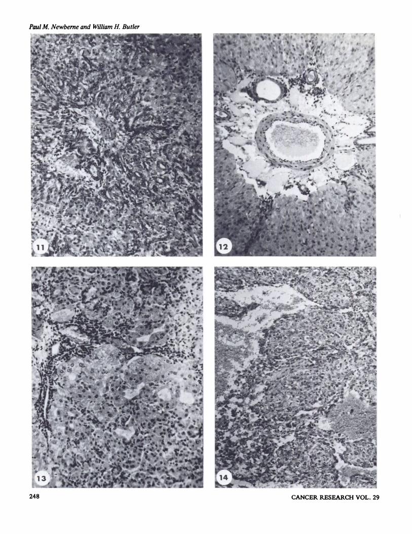
regeneration. Biliary proliferation was pronounced at this stage,with ductal cells radiating out from the portal area to separateparenchymal cells into groups or as single units. At seven dayspostexposure, a few macrophages were observed in the centrilobular zone, but most of the lobule consisted of normal parenchymal cells with an occasional mitotic figure. At the periphery of the lobule, many of the parenchymal cells wereisolated by the continued biliary proliferation (Fig. 1 1); someof the parenchymal cells contained lipid while others wereundergoing lysis. By ten days the biliary proliferation was stillmarked, but mitotic figures were not seen and there was anormal lobular pattern with no residual evidence of necrosis.Parenchymal cells were normal and only an occasional mitoticfigur e was observed. Animals studied three months after exposure had normal livers, but a few animals appeared to have aslight increase in the connective tissue component locatedabout the main portal tracts with a small residual componentof proliferated bile ducts remaining among normal parenchymal cells.
The toxic effects of groundnut meal assayed for aflatoxinwere reported by Butler and Barnes (14). Animals developedascites and edema; this had been described by previousworkers. At a level of approximately 1.5 ppm of aflatoxin inthe diet, survival was between two and four weeks. The livercontained varying degrees of biliary proliferation which cxtended into the lobules, and the periportal lymphatics weredilated (Fig. 12). In the periportal zone, the fat content of theparenchymal cells was increased and necrotic parenchymalcells were scattered throughout the lobule. At a dietary levelof 0.7—0.8 ppm of aflatoxin, the animals lived for up to eightweeks. Biliary proliferation was observed in the livers of threeanimals, but it was not as prominent as that observed at thehigher dose levels. Scattered throughout the lobules were areasof parenchymal cells undergoing lysis and a concomitant tubule formation (Fig. 13). The lowest dose level reported was0.35—0.4 ppm of aflatoxin; at this level most animals weredead by 27 weeks with only one survivor to 44 weeks. Duringthe course of the feedings, there was a progressive biliary proliferation and individual cell necrosis similar to that previouslydescribed. By the 28th week following initiation of the experiment, islands of parenchymal cells appeared as regeneratingnodules in which a few pyknotic nuclei and an occasionalmitosis were seen. The solitary survivor at 44 weeks had acoarsely nodular liver containing broad bands of fibrous tissue,collections of bile ducts, and some regenerative nodules withbizarre parenchymal cells.
In order to obtain longer survival times, the dose was furtherreduced to 0.15 ppm of aflatoxin in the diet. This dose levelresulted in a high mortality, but six animals survived an average of 106 weeks. The longest survival time was 160 weeks.One of these animals, killed at 127 weeks, had an anaplastichepatocarcinoma, the only hepatic tumor seen in guinea pigstreated with diets containing aflatoxin (Fig. 14).
Mouse
There is no detailed account of the acute lesions induced byaflatoxin in mice. The LD50 has been estimated as about 9mg/ kg. However, a problem with solvent toxicity makes this
figure unreliable. In feeding trials mice appear to be resistantto the chronic toxicity of aflatoxin. Plantonow (55) describedthree-month feeding trials which failed to produce any change.Newberne (44) fed male white Swiss mice for 16 months on apeanut meal diet contaminated with 1 .0 ppm of aflatoxin, and15% of the mice developed liver tumors; two other strains ofblack mice fed a diet containing 1.0 ppm of purified aflatoxinB1 failed to develop significant liver lesions.
The livers of mice that were fed contaminated peanut mealdeveloped liver cell tumors and widespread pleomorphism ofthe nontumorous liver cell nuclei. Large numbers of the tumorcells contained globular, eosinophilic structures in the cytoplasm which were periodic acid-Schiff positive (Fig. 15). Atthe junction of normal and neoplastic cells, mitotic figureswere occasionally seen. None of the tumors observed in thelivers of mice were large and none had metastasized; whetherthese neoplasms were malignant is open to debate.
Dog
Newberne et al. (48) have studied the effects on dogs ofsingle and repeated doses of aflatoxin. The LD50 is about0.5—1 mg/kg, with the earliest histologic change appearing asan increase in fat with congestion of the centrilobular zoneand parenchymal cell necrosis (Fig. 16). At seven days therewas a prominent biliary proliferation. Although this reviewdoes not consider organs other than the liver, the edema andhemorrhage seen in the gall bladder are such that they warrantbrief attention. The wall of the gall bladder in dogs exposed toaflatoxin was greatly thickened and microscopic examinationrevealed severe subserosal and submucosal edema and hemorrhage (Fig. 17).
Experimental studies and observations of field outbreakshave shown that the dog is very sensitive to the acute effectsof aflatoxin (48, 69). Lesions observed in spontaneous outbreaks of canine toxicosis where afiatoxin has been isolatedfrom feed samples have been remarkably similar to those seenin the experimentally induced disease using crude or purifiedaflatoxin. Lesions induced experimentally were similar tothose reported for “hepatitis X―(43), a toxic disease of kenneldogs of the southeastern United States, reported in 1955. Atnecropsy the animals fed commercial dog feed containingtoxic peanut meal were jaundiced,with swelling and yellowishdiscoloration of the liver and edema of the gall bladder identical to that seen with crude or purified afiatoxin. Histologically, fatty change with centnilobular parenchymal necrosisand biliary proliferation were seen similar to the acute singledose lesions induced experimentally.
Cat
Adult mixed-breed cats exhibited a sensitivity to purifiedaflatoxin B1 similar to the rabbit, dog, and guinea pig with asingle dose LD50 of 0.55 mg/kg. Most acute deaths occurredin 48—72 hours; grossly the liver was swollen, pale, and friablewith occasional petechial hemorrhages. Microscopically, theimmediate periportal zone was pale because of decreased staining of parenchymal cells, and there was very little lipid accumulation (Fig. 18). AnimalS sacrificed in terminal stages had
240 CANCER RESEARCH VOL.29
Acute and Chronic Effects of Aflatoxin
pale liver cell nuclei with a “washedout―appearance and littlestainable chromatin; glycogen accounted for part of the observed nuclear change (Fig. 19). There was a mixed leukocyticinfiltration in the periportal zones, and bile duct hyperplasiawas observed in the livers of cats surviving beyond 72 hours.
Rabbit
The rabbit appears to be even more sensitive to the acuteeffects of aflatoxin than other mammalian species (Table 2).The LD50 for Dutch belted males and females was about 0.3mg/kg with no significant difference between sexes or route ofadministration (i.p. or p.o.). Microscopically, there were hemorrhage and parenchymal cell necrosis in the midzone areas ofthe lobule (Fig. 20), and scattered single-cell necrosis was seenin the centnilobular area. Animals surviving the immediatelethal effects of the single dose of aflatoxin developed themild-to-moderate bile duct hyperplasia seen in other species.
Feeding experiments have not been reported in the rabbit.
Monkey
Monkeys have been shown to be susceptible to the acutetoxicity of both purified afiatoxin and contaminated peanutmeal. Doses of 500 lsg for 18 days followed by doses of 1mg/day to rhesus monkeys resulted in deaths at 32 and 34days. At higher dose levels, deaths occurred earlier. Histologically, the livers showed fatty infiltration, biliary proliferation,and portal fibrosis (38). Cuthbertson et al. (24) studied theeffects of contaminated peanut meal on cynomologusmonkeys and described liver cell damage and biliary proliferation at dietary levels of 5 ppm of aflatoxin. At lower dietarylevels (1.8 ppm of aflatoxin), animals survived three years. Oneanimal had a coarse nodular cirrhosis, while the other monkeyexhibited irregular size of parenchymal cell nuclei (Fig. 21).
Other Species
Although single dose experiments have not been reported,Alicroft and Lancaster (personal communication, manuscriptin preparation) have demonstrated that ferrets are extremelysensitive to toxic peanut meal diets. A diet containing 20% ofa toxic peanut meal resulted in the development of liver tumors in five of seven male ferrets after 24—37 months. Dccreasing the dietary level of the toxic meal to 3% resulted in100% incidence of liver tumors.
The acute LD@ of aflatoxin B1 for hamsters has been reported as 10.2 mg/kg (70), but no information is available asto the pathologic changes induced. Results of preliminaryfeeding experiments reported by Chesterman and Pomerance(22) were inconclusive. At a dietary level of 2 ppm of aflatoxin,the animalsfailed to grow and died after 8 weeks.However,both the dosed and control animals showed the same welldeveloped cirrhosis with regenerative nodules, fibrosis, and biliary proliferation. The etiology of this lesion is not understoodand implies other complicating factors.
The acute and chronic toxic effects in trout have been investigated by Halver (25) following a high incidence of hepatictumors in hatchery-raised trout in 1960. The acute LD@ of
combined aflatoxin@ and@ has been estimated at between0.5 and 1 .0 mg/kg; the principal lesion described was a hemorrhagic necrosis. Feeding experiments resulted in biliary proliferation, with some cyst formation after six months. Assoelated with this was a nodular proliferation of parenchymalcells (Fig. 22) and subsequent development of parenchymalcell carcinoma.
DISCUSSION
The first significant reports of what appears to have beenaflatoxin poisoning in domestic animals were those of Newberne et aL (43) and Burnside et a!. (10). The disease in dogswas referred to as “hepatitisX―and was traced to the dietwhich contained peanut meal (Newberne et a!.). Although thedisease was reproduced by feeding the toxic feed, the exactnature of the etiologic agent was not revealed. During the sameperiod, Burnside et a!. isolated toxin-producing strains ofAspergillus flavus and Penicillium rubrum from an outbreak oftoxicosis in swine fed moldy corn. Emphasis at the time wasplaced on the P. rubrum culture, while studies of A. flavuswere not pursued further. Le Breton et a!. (30) mention a highincidence of liver tumors in a colony of rats in Morocco fed adiet containing peanut meal. It has been pointed out (43) that“hepatitis X―has now been shown to be related, at least inpart, to aflatoxin-contaminated peanut meals in the diet. Paget(52) has described a disease of rather sporadic incidence inguinea pigs which has been shown to be similar to that produced by peanut meals known to be contaminated by aflatoxin. Schoental (61) showed that the Medical Research Counci (MRC) guinea pig diet would induce hepatic carcinoma inrats. Thus, it appears that aflatoxin has a much longer historythan current research work indicates.
Since the aflatoxins were first isolated (60), there has beenconsiderable progress in the investigation of the carcinogenicity and acute toxicity from both the structural and biochemical aspects. The structures have been elucidated (4) and racemic mixtures synthesized (9). The acute lesion in the rat hasbeen investigated by many workers; its striking features are theperiportal distribution of parenchymal cell necrosis, bile ductproliferation, slow recovery, and, in some cases, eventual development of hepatic carcinoma. It has been suggested that thecarcinogenic action of aflatoxin B1 in the rat results from acapacity to bind to DNA, a characteristic similar to that ofactinomycin D (23, 65). However, lethal doses of actinomycinD do not produce hepatic parenchymal cell necrosis. In allspecies studied, the organ most affected is the liver, althoughother organs, particularly the kidney, show signs of damage.The distribution of the hepatic lesion is not consistent fromspecies to species, i.e., rat and duckling, periportal; guinea pigand swine, centnilobular; dog, periportal and centrilobular; andrabbit, mid-zonal. In contrast, most other hepatotoxins, suchas carbon tetrachloride, regularly induce a centrilobular lesionin both rats and guinea pigs.
There is a wide range in the acute LD@ dose of aflatoxinB1, varying from 0.3 mg/kg for ducklings to 16 mg/kg formature female rats (Table 2). In species for which data areavailable, the young appear to be more susceptible than mature animals.
JANUARY 1969 241

Table4Barley
BeansCornCassavaCottonseedCowpeas
MilletPeasPeanutsRiceSesame
SorghumSoybeansSweet PotatoesWheat
PaulM. Newberne and William H. Butler
The most striking and important feature of the investigationsdescribed in this review is the carcinogenic action of the aflatoxins in fish, birds, and mammals. When one considers thatdoses of 0.015 ppm of B1 in continuous feeding or a total of0.4 mg over 14 days resulted in a high incidence of hepaticcarcinoma, and when these doses are compared with otherhepatocarcinogens, it becomes clear that aflatoxin is the mostpotent liver carcinogen so far recognized. One further interesting result has been the demonstration that choline deficiencyitself does not appear to be sufficient to induce hepatic carcinoma (47), nor is cirrhosis a prerequisite or concomitant ofaflatoxin carcinogenesis. Choline deficiency can induce a cirrhotic liver in the rat, but in recent experiments carcinoma didnot result unless aflatoxin or some other hepatocarcinogen wasincluded in the diet (46). Even at high doses, aflatoxin aloneseldom induced cirrhosis in the rat. In most of the tumorbearing animals, the carcinomas arise in liver with otherwisenormal lobular patterns.
The recognition of the possible hazard to humans consumingcontaminated foods has stimulated many programs of investigation in those areas with a high incidence of hepatic carcinoma. These have taken two main forms: (a) epidemiologicstudies to compare the pattern of disease in high and lowincidence areas, and (b) surveys of food for aflatoxin content.It has been shown (33, 61) that the toxin-producing strains ofthe fungus will grow on substrates which are being used asprotein supplements for children, but at present there is nodirect evidence that man is susceptible to the aflatoxins. Thebroad spectrum of animals that are susceptible makes it rcasonable to conclude that aflatoxin is a potential hazard toman. However, the assumption that man responds to aflatoxinexposure in a manner similar or identical to that observed inanimals is made on very tenuous grounds. We recognize thatthe association of liver disease, including carcinoma, withpotential aflatoxin exposure in certain population groups ishighly suggestive, and it provides an attractive hypothesis foruse in attempting to explain the variation in the incidence ofliver cancer. Reliable estimates of human exposure to the aflatoxins are not presently available; such information shouldderive from properly designed epidemiologic surveys, some ofwhich are now in progress in areas where the ingestion ofaflatoxin-contaminated foodstuffs may later be correlated withliver carcinoma in the indigenous population. For the present,however, we must await factual evidence and proceed withcaution in assigning the aflatoxins a role in worldwide livercarcinoma.
The epidemiologic characteristics of primary liver carcinomain Africa have been described in at least two reports (51, 66).There are wide variations within general areas of high incidence, with the highest rate of primary hepatocellular carcinoma reported in black male Africans, particularly those fromMozambique Bantu tribes (27). It is interesting that the incidence in the Mozambique group is about 500 times that reported in the U. S. population for a comparable age group(25—34 years) and 15 times that observed in natives in nearbyJohannesburg.
In India there is a highly variable incidence of liver cancer(53),witha decreasein thenorthernandwesternareasof thecountry. There are also indications for an increase in liver
cancer in the male population of Southeast Asia. Observationsin Thailand, the Philippines and Malaya (41), Indonesia (8),South China (73), Hong Kong (where about 30% of autopsiesare reported to have liver carcinoma) (W. C. Chan, personalcommunication), and Singapore (63) serve to reinforce thefeeling among pathologists and epidemiologists that there is atrend toward increased liver cancer in many areas of the world.In Singapore the highest incidence of liver carcinoma is foundin individuals who come from South China, who may havebeen exposed to environmental hazards in the early years oftheir lives.
The facts that aflatoxin-producing strains of molds are foundin so many areas of the world and, furthermore, that climaticconditions favor the growth of molds in many areas whereliver carcinoma occurs in a high incidence lend support to thehypothesis that the aflatoxins are involved in the etiology ofprimary liver cancer. We have pointed out previously (44, 46)that feed grade samples of peanut meal purchased on the openmarket have been highly contaminated with aflatoxin(1.0—5.0ppm). Evenmore significant is the extremelyimportant report by Wogan (72) that aflatoxins have been detectedat biologically significant levels in food samples collected frommany parts of the world, particularly Africa and Asia. Table 4lists food products found to contain significant amounts ofaflatoxin and confirms that most major food commodities aresubject to aflatoxin contamination. Although specific levels ofaflatoxin content were not reported, clearly the potential hazards of the aflatoxins are evident, and they must be accordedproper attention in attempts to elucidate etiologic factors related to primary liver cancer in many population groupsaround the world.
Isolated samples of food materials found to contain biologically significant amounts of aflatoxin.
REFERENCES
1. Abercrombie, M., and Harkness, R. D. The Growth ofCeIl Populations and the Properties in Tissue Culture of Segments of Liver ofthe Rat. Proc. Roy. Soc. Ser. B., 138: 544—561,1951.
2. Abrams, L. Mycotoxicoses. J. S. Afr. Vet. Med. Assoc., 36: 5—13,1965.
3. Allcroft, R., and Lewis, G. Groundnut Toxicity in Cattle. Experimental Poisoning of Calves and a Report on Clinical Effects inOlder Cattle. Vet. Rec., 75: 487—493,1963.
4. Asao, T., Btichi, G., Abdel-Kader, M.M.,Chang, S. B., Wick, E. L.,and Wogan, G. N. Aflatoxins B and G. J. Am. Chem. Soc., 87:882—886, 1965.
5. Asplin, F. D., and Carnaghan, R. B. A. The Toxicity of CertainGroundnut Meals for Poultry with Special Reference to TheirEffect on Ducklings and Chickens. Vet. Rec., 73: 1215—1219,1961.
242 CANCER RESEARCH VOL.29

Acute and Chronic Effects of Aflatoxin
6. Barnes, J. M., and Butler, W. H. Carcinogenic Activity of Aflatoxinto Rats. Nature, 202: 1016, 1964.
7. Blount, W. P. Turkey “X―Disease. Turkeys, 9: 52, 1961.8. Bonne, C. Cancer in Java and Sumatra. Am. J. Cancer, 25:
811—821, 1935.
9. BUchi, G., Foulkes, D. M., Kurono, M. and Mitchell, G. F. TheTotal Synthesis of Racemic Aflatoxin B1. J Am. Chem. Soc., 88:19, 1966.
10. Burnside, J. E., Sippel, W. L., Forgas, J., Atwood, M. B. and Doll,E. R. A Disease of Swine and Cattle Caused by Eating MouldyCorn. II. Experimental Production with Pure Cultures of Mould.Am. J. Vet. Res., 18: 817—824,1957.
11. Butler, W. H. Acute Toxicity of Aflatoxin B1 in Rats. Brit. J.Cancer, 18: 756—762, 1964.
12. Butler, W. H. Acute Liver Injury in Ducklings as a Result of Aflatoxin Poisoning. J. Pathol. Bacteriol., 88: 189—196,1964.
13. Butler, W. H. Acute Toxicity of Aflatoxin B in Guinea Pigs. J.Pathol. Bacteriol., 91: 277—280,1966.
14. Butler, W. H., and Barnes, J. M. Toxic Effects of Groundnut MealContaining Aflatoxin to Rats and Guinea Pigs. Brit. J. Cancer, 17:699—710,1963.
15. Butler, W. H., and Wigglesworth, J. S. The Effects of Aflatoxin B1on the Pregnant Rat. Brit. J. Exptl. Pathol., 47: 242—247,1966.
16. Carlton, W. W., Lord, J. E., and Friedman, L. Pathology of Dimethylnitrosamine Poisoning in Pekin Ducklings. Toxicol. App!. Pharmacol.,8:224—234,1966.
17. Carnaghan, R. B. A. Hepatic Tumors in Ducks Fed a Low Level of
Toxic Groundnut Meal. Nature, 208: 308, 1965.18. Carnaghan, R. B. A. Hepatic Tumors and Other Chronic Liver
Changes in Rats Following a Single Oral Administration of Aflatoxin. Brit. J. Cancer, 21: 811—814,1967.
19. Carnaghan, R. B. A., Hartley, R. D., and O'Kelly, J. Toxicity andFluorescence Properties of the Aflatoxins. Nature, 200: 1101,1963.
20. Carnaghan, R. B. A., Lewis, G., Patterson, D. S. P., and Allcroft, R.Biochemical and Pathological Aspects of Groundnut Poisoning inChickens. Pathol. Vet., 3: 601—615,1966.
21. Chesterman, F. C., and Pomerance, A. Cirrhosis and Liver Tumoursin a Closed Colony of Golden Hamsters. Brit. J. Cancer, 19:802—811, 1965.
22. Clegg, F. G., and Bryson, H. An Outbreak of Poisoning in StoreCattle Attributed to Brazilian Groundnut Meal. Vet. Rec., 74:992—994, 1962.
23. Clifford, J. I., and Rees, K. R. The Action of Aflatoxin B1 on RatLiver. Biochem. J., /02: 65—75,1967.
24. Cuthbertson, W. F. J., Laursen, A. C., and Pratt, D. A. H. Effect ofGroundnut Meal Containing Aflatoxin on Cynologus Monkeys.Brit. J. Nutr., 21: 893—908, 1967.
25. Halver, J. E. Aflatoxicosis and Rainbow Trout Hepatoma. In: G. N.Wogan (ed.), Mycotoxins in Foodstuffs, pp. 209—234.Cambridge,Mass.: MIT Press, 1965.
26. Harding, J. D. J., Done, J. T., Lewis, G., and Allcroft, R. Experimental Groundnut Poisoning in Pigs. Res. Vet. Sci., 4: 217—229,1963.
27. Higginson, J. The Geographical Pathology ofPrimary Liver Cancer.Cancer Res., 23: 1624—1633, 1963.
28. Hill, L. R. Comment on the Histological Appearances in SerialLiver Biopsies and Postmortem Specimens. Vet. Rec., 75:493—494,1963.
29. Lancaster, M. C., Jenkins, F. P., and Philp, J. McL. Toxicity Associated with Certain Samples of Groundnuts. Nature, 192:1095—1097,1961.
30. LeBreton, E., Frayssinet, C., and Boy, J. Sur l'Apparation d'Hepatomes “Spontanes―chex le Rat Wistar: Role de la Toxine del'Aspergillus flavus. Interet en Pathologic Humaine et Cancerologie
Experimentale. Acad. Sci. (Paris) Compt. Rend., 255: 784—786,1962.
31. Lewis, C., Markson, L. M., and Allcroft, R. The Effect of FeedingToxic Groundnut Meal to Sheep Over a Period of Five Years. Vet.Rec., 80: 312—314, 1967.
32. Lijinsky, W., and Butler, W. H. Purification and Toxicity of Aflatoxin B1. Proc. Soc. Exptl. Biol. Med., 123: 151—154,1966.
33. Loizeier, A. B. Intoxicaciones en Pollos Producidas por el Turto deCacahuete. Rev. Patron. Biol. Animal, 7: 25—33, 1963.
34. Loosmore, R. M., Allcroft, R., Tutton, E. A. and Carnaghan, R. B.A. The Presence of Aflatoxin in a Sample of Cottonseed Cake. Vet.Rec., 76: 64—65,1964.
35. Loosmore, R. M., and Harding, J. D. J. A Toxic Factor in BrazilianGroundnut Causing Liver Damage in Pigs. Vet. Rec., 73:1362—1364, 1961.
36. Loosmore, R. M., and Markson, L. M. Poisoning of Cattle by Brazilian Groundnut Meal. Vet. Rec., 73: 813—814,1961.
37. Madhavan, T. V., and Rao, K. S. Hepatic Infarction in Ducklings inAflatoxin Poisoning. Arch. Pathol., 81: 520—524,1966.
38. Madhavan, T. V., Pulpule, P. G., and Gopalan, C. Aflatoxin Induced Hepatic Fibrosis in Rhesus Monkeys. Arch. Pathol., 79:466—469, 1965.
39. Magwood, S. E., Annau, E., and Corner, A. H. Induced Tolerancein Turkeys to Aflatoxin Poisoning. Can. J. Comp. Med. Vet. Sci.,30: 17—25, 1966.
40. Markson, L. M. The Pathogenesis of the Hepatic Lesion in Calves
Poisoned Experimentally with Senecio jacobea. Proc. Roy. Soc.Med.,53: 283—284,1960.
41. Marsden, A. T. H. The Geographical Pathology of Cancer inMalaya.Brit.J.Cancer,12:161—176,1958.
42. Nesbitt, B. F., O'Kelly, J., Sargeant, K., and Sheridan, A. Toxic
Metabolites of Aspergillus flavus. Nature, 1 95: 1062—1063,1962.
43. Newberne, J. W., Baily, W. S., and Seibold, H. R. Notes on aRecent Outbreak and Experimental Reproduction of “Hepatitis X―in Dogs. J. Am. Vet. Med. Assoc., 127: 59—62, 1955.
44. Newberne, P. M. Carcinogenicity of Aflatoxin-Contaminated Peanut meal. In: G. N. Wogan (ed.), Mycotoxins in Foodstuffs, pp.187—208. Cambridge, Mass.: MIT Press, 1965.
45. Newberne, P. M. Biological Activity of the Aflatoxins in Domesticand Laboratory Animals. Trout Hepatoma Research Conference,U. S. Department Interior. Fish and Wildlife Service Report 70, pp.130—144, 1967.
46. Newberne, P. M., Carlton, W. W., and Wogan, C. N. Hepatomas inRats and Hepatorenal Injury in Ducklings Fed Peanut Meal or Aspergillusfiavus Extract. Pathol. Vet. (Basel), 1: 105—132, 1964.
47. Newberne, P. M., Harrington, D. D., and Wogan, G. N. Effects ofCirrhosis and Other Liver Insults on Induction of Liver Tumors byAflatoxin in Rats. Lab. Invest., 15: 962—969, 1966.
48. Newberne, P. M., Russo, R., and Wogan, G. N. Acute Toxicity ofAflatoxin B1 in the Dog. Pathol. Vet., 3: 331—340,1966.
49. Newberne, P. M., and Wogan, G. N. Sequential MorphologicChanges in Aflatoxin B1 Carcinogenesis in the Rat. Cancer Res.,28: 770—781, 1968.
50. Newberne, P. M., Wogan, G. N., Carlton, W. W., and Abdel-Kader,M. M. Histopathologic Lesions in Ducklings Caused by Aspergiliusflavus Cultures, Culture Extracts and Crystalline Aflatoxins. Toxicol. App!. Pharmacol., 6: 542—556, 1964.
51. Oettle', A. G. Cancer in Africa, Especially in Regions South of theSahara. J. Nat!. Cancer Inst., 33: 383—439,1964.
52. Paget, G. E. Exudative Hepatitis in Guinea Pigs. J. Pathol.Bacteriol., 47: 393, 1954.
53. Paymaster, J. C. Cancer and Its Distribution in India. Cancer, 17:1026—1034, 1964.
54. Paterson, J. S., Crook, J. C., Shand, A., Lewis, G., and Allcroft, R.
JANUARY 1969 243

PaulM. Newberne and William H. Butler
Groundnut Toxicity as the Cause of Exudative Hepatitis (OedemaDisease) ofGuinea Pigs. Vet. Rec., 74: 639—640,1962.
55. Platonow, N. Effect of Prolonged Feeding of Toxic GroundnutMeal in Mice. Vet. Rec., 76: 589—590, 1964.
56. Raimo, H. F., Correa, R., and Dc Andrade, B. M. Acao Toxica doFordo de Torta de Amendion. Produzido em Sao Paulo, Para Ayese outros Animales. Bo!. Ind. Animal (Sao Paulo) 20: 361—364,1962.
57. Rogers, A. E., and Newberne, P. M. The Effects of Aflatoxin B1and Dimethylsulphoxide on Thymidine-3H Uptake and Mitosis inRat Liver. Cancer Res., 27: 855—864,1967.
58. Salmon, W. D., and Newberne, P. M. Occurence of Hepatomas inRats Fed Diets Containing Peanut Meal as a Major Source of Protein. Cancer Res., 23: 571—575, 1963.
59. Sargeant, K., O'Kelly, J., Carnaghan, R. B. A., and Allcroft, R. TheAssay of a Toxic Principle in Certain Groundnut Meals. Vet. Rec.,73: 1219—1223, 1961.
60. Sargeant, K., Sheridan, A., O'Kelly, J., and Carnaghan, R. B. A.Toxicity Associated with Certain Samples ofGroundnuts. Nature,192: 1096—1097,1961.
61. Schoental, R. Liver Changes and Primary Liver Tumours in RatsGiven Toxic Guinea Pig Diet (M.R.C. Diet 18). Brit. J. Cancer, 15:812—815, 1961.
62. Sellachop, J. P. F., Kriek, N. P. J., and du Preez, J. C. G. Distribution and Degree of Occurrences of Aflatoxin in Groundnuts andGroundnut Products. S. Afr. Med.J., 39: 771—774, 1965.
63. Shanmugaratnam, K. Primary Carcinomas of the Liver and BiliaryTract. Brit. J. Cancer, 10; 232—246,1956.
64. Siller, W. G., and Osfier, D. C. The Histopathology of an Enterohepatic Syndrome of Turkey Poults. Vet. Rec., 73: 134—138,1961.
65. Sporn, M. B., Dingman, C. W., Phelps, H. L., and Wogan, G. N.Aflatoxin@ : Binding to DNA in vitro and Alteration of RNAMetabolism in vivo. Science, 151: 1539—1541, 1966.
66. Steiner, P. E., and Higginson, J. Definition and Classification ofCirrhosis of the Liver. Unio Intern. Contra Cancrum, 17: 58 1—592,1961.
67. Sutton, P. M., and Spurgeon, P. J. Mitoses in Bile Duct EpitheliumFollowing Acute Carbon Tetrachloride Poisoning. Brit. J. Expt!.Patho!., 47: 545—549,1966.
68. Wannop, C. C. The Histopathology ofTurkey “X―Disease in GreatBritain. Avian Diseases, 5: 371—381,1961.
69. Wilson, B. J., Teer, P. A., Barney, G. H., and Blood, F. R. Relationship of Aflatoxin to Epizootics ofToxic Hepatitis Among Animalsin Southern United States. Am. J. Vet. Res., 28: 1217—1230,1967.
70. Wogan, G. N. Chemical Nature and Biological Effects of the Aflatoxins. Bacteriol. Rec., 30: 460-470, 1966.
71. Wogan, G. N.. and Newberne, P. M. Dose-Response Characteristicsof Aflatoxin B1 Carcinogenesis in the Rat. Cancer Res., 27:2370—2376, 1967.
72. Wogan, G. N. Aflatoxin Risks and Control Measures. FederationProc., 27: 932—938, 1968.
73. Yeh, S., and Cowdry, E. V. Incidence of Malignant Tumors inChinese, Especially in Formosa. Cancer, 7: 425—436,1954.
244 CANCER RESEARCH VOL.29

Acute and Chronic Effects of Aflatoxin
Fig. 1. Liver ofduckling killed 3 days after a single LD50 does ofaflatoxin B1. Note biiary proliferation and lakes of fat. H and E, x 130.Fig. 2. Liver of cow fed toxicpeanut meal for 11 weeks. Note fibrosis of central vein and biliary proliferation. Picro-Mallory, X 160.Fig. 3. Same liver as Fig. 2 showing irregularity of parenchymal nuclear size. H and E, x 400.Fig. 4. Liver from pig killed 18 hours following a single dose ofaflatoxin B1 (2 mg/kg). Centrilobular hemorrhage and necrosis are characteristic
of the acute response. H and E, x 160.Fig. 5 . Liver from pig fed toxic peanut meal for 26 weeks. Section illustrates a small regenerative nodule, fibrosis, and pleomorphism of
parenchymal cell nuclei. H and E, x 160.Fig. 6. Liver of rat killed 2 days after a single LD50 dose of aflatoxin@ . Periportal necrosis and bile duct hyperplasia are illustrated. H and E, x
160.Fig. 7. Liver of rat killed 1 month after a single LD50 dose of aflatoxin B1. Note residual biliary proliferation and irregularity of parenchymal
cell nuclei. H and E, x 160.Fig. 8. Liver of rat fed toxic peanut meal (5 ppm) for 10 weeks, illustrating biiary proliferation, irregularity of parenchymal nuclear size, and
foci of small parenchymal cells (arrow). H and E, x 180.Fig. 9. Area of trabecular hepatocellular carcinoma from a rat fed toxic peanut meal (5 ppm) for 12 weeks followed by 54 weeks of normal diet.
H and E, x 130.Fig. 10. Liver ofguinea pig killed 2 days after a single LD50 dose ofaflatoxin B1. Centrilobular necrosis is primary change. H and E, X 135.Fig. 11. Liver of guinea pig killed 7 days after a single dose ofaflatoxin B1. Note biliary proliferation radiating from periportal zone. H and E, x
135.Fig. 12. Liver of guinea pig fed toxic peanut meal (2 ppm) for 2 weeks. Dilation of periportal lymphatics is usually severe in this species. H and
E, X 135.Fig. 13. Liver of guinea pig fed toxic peanut meal (1 ppm) for 7 weeks. Biliary proliferation and parenchymal cell necrosis with pseudotubule
formation are major morphologic alterations. H and E, x 135.Fig. 14. Area of hepatocellular carcinoma from a guinea pig fed toxic peanut meal (0.2 ppm) for 127 weeks. A moderate vascular component is
seen in this animal similar to that observed in rats. H and E, x 135.Fig. 15. Liver of mouse fed aflatoxin for 64 weeks. Note area of hepatoma compressing normal liver at left of photograph. H and E, x 140.Fig. 16. Liver of dog 5 days after a single dose of aflatoxin. Centrilobular necrosis and fatty change are characteristic for this species. H and E, X
190.Fig. 17. Gall bladder of dog killed 2 days after a single dose of aflatoxin. There is extensive hemorrhage of the mucosa and edema of muscle
layers and subserosa. H and E, x 12.Fig. 18. Liver of cat killed 2 days after a single dose of aflatoxin B (0.75 mg/kg). There is early necrosis of periportal parenchymal cells. H and
E, x 160.Fig. 19. Same liver as Fig. 18, illustrating margination of nuclear chromatin of the parenchymal cells. H and E, x 360.Fig. 20. Liver of rabbit killed 5 days after a single dose of aflatoxin (0.5 mg/kg). Note sharp delineation at midzonal area with hemorrhagic
necrosis and with lysis of cells in the centrilobular area. H and E, x 170.Fig. 21. Liver of monkey fed toxic peanut meal (1.8 ppm) for 3 years. There are a coarse nodular cirrhosis and!arge, bizarre parenchymal cells. H
and E, x 135.Fig. 22. Liver of trout at junction of proliferating liver cell carcinoma and more norma! parenchyma. H and E, x 390.
JANUARY 1969 245

PaulM. Newberne and William H. Butler
246CANCER RESEARCH VOL.29

I
.. @; -.
.‘;dI -‘@ •“@@@:@@ -..@, -
-(@@ $ -
‘@‘@ -, .‘ - (@
@ ‘@e@@
,@ @“@,.4 ‘@. -.,
.@ .,• *@‘• _I.@ @‘@.@‘5t'
-@@ 1@:@
.•.‘•@__r-@%‘@ ‘.-. @-@@@:‘‘
,@ ,-4T@@
-.@.
I;. a―
b@@ 0,.7.•%@ a• )‘, ,I. •t v@ •/
•@• @_ t@@ •@@y@ ed@@@@ @@‘-:‘;‘@:‘@ -
.-‘@ ;, - . .-. .@@ •r41@@5@‘@ @. @, , :@@ @@@J@a•4‘
:‘@.;@@@ ‘@ I 7' ‘:.ea@•@@.A@-4 ).@ .- - :@@
@ “@vi1@Z'2@I?@
,,4,__@ ft@ I@@ ,,@@@ •,4 :@‘,@
@‘,a@: .@@@
. †S ‘@@ ••@@ .@ @4 . a@ , @,, .
,‘:._a,,:@@ @.@@ . @‘@... a @-),•@@ ,@@ . , . % †a@ •@@
I -@-@@‘ø.i.@ ,s@@@ ‘“@“:;‘‘@.‘@@@a'@en• ,.iI •I@@
. .O @I.
,@
247
Acute and Chronic Effects of Aflatoxin
JANUARY 1969
2:I:@:,,[email protected] •@‘‘r@•'@―s; •; @.
:@@:@‘_@‘ @“ , ‘5 .5,
@ ‘a@ •@ • ‘I.@. —,-@
@.- a • z:-@@.@'[email protected]; @.
-%rs:,@@@@@_,Ir•_. @‘@ ,@ ; @-
- -
%
5•
.—
b' :@ : - @-‘@ a@...@
@ 3 -@‘ 44t‘ ••@_4 - @\ ‘:
,. . .. ‘ . .@
.@@
:@ @“@:
o._@ •L@•@
@ (‘
•4c'@
•@;@ :‘@ ‘@:@
@@ ::@@@@@ , :%.,,..@-@ø,
. . . %@ . a@
@@r$pV@S.. @@;U ..y@@@ 7 :- “2@@@
@p'•,..,,. @@Iaâ€. .---: •@@:;@a@@ ;4@.;@@ c r :@
.@.. .. @. @.. -._.,a,,_.@
@ ; -,,-- ..,-@- .i@ . d@-4.:.q ._.,
@.— 4 @@_-‘:@@ ... :.
--@ . -,-‘.‘ . -s:@ .-@•.@‘:‘-@@@ ‘@@
@@@ •@
. .. -.@
•a―:Z ;:@@‘a.. f@
.@tft@ .b@,
: .•?@
..p@-•@..f ,
‘@-@S@-@
. @-
@‘- t@b-@@
r.
@,@:,. .@,-
@ ‘
248 CANCERRESEARCHVOL.29
PaulM. Newberne and William H. Butler

-I-
4-'@ :- @‘
@‘ - @‘
@ -
. ,::‘@t;@ 4@' ‘
@‘‘@&.‘ r._.- .@-
a :;-@@ •
@ Pb―@
@ .-,. 5j@'@''@_ @b •f@
@ i:-,-@ . ‘‘
:.@ -s@@
@5 @J.@ @.-;:.@
@&_. 14@,@ @:@- .
@l@ -@
,-. 4.. ,. @, -.
@t@:;'s ,@‘.‘@. ‘a@@ ‘@: @:‘:-@@@
.@@ .t@
@ a@@ :1@@@ :@@@;lh@@ S@
@ ..._p@$@
.. . :
,,
.@ % -
_•_4-@
249
I,@I7
JANUARY 1969
Acute and Chronic Effects ofAflatoxin
r!@18b, -@ -

Paul M. Newberne and William H. Butler
I@ ‘
@¼@'@@ ‘@@@@
. :-@@ -@o' •@-@@ 3'.
IL'. ‘@ ‘
@1.@@@@ ‘@a@@ @Q_
@@ It..-:-@4@-ne@_'@,@@
—@ 5- __ .#.@.‘ S
:@:4 ,, :-A@
.f *@@ .@ ‘5
,• .@:
(@@‘41.
@,,. /
(@ I@,@
(5L$ t@@@
@ I―
@- :@, .
-: •@..@:-.@ .a,.t@@
@-.b' ‘@-@,@..@@
.:e@-'@.@@@ •[email protected].,- .
@ i. ;‘
I ‘@@@@
a .@d1f'@ ‘@
;@250CANCER RESEARCH VOL.29
1n4,-. @.I @@@-5S;A “