acute myeloid leukemia: importance of ancillary studies · pdf fileacute myeloid leukemia:...
TRANSCRIPT

Acute Myeloid Leukemia: Importance of Ancillary Studies in
Diagnosis and Classification
Daniel A. Arber, MDProfessor and Associate Chair
Department of PathologyStanford University

0
2
4
6
8
10
12
14
16
<5 5- 10- 15- 20- 25- 30- 35- 40- 45- 50- 55- 60- 65- 70- 75- 80- 85+
Estimated Yearly Incidence of AML
Age
Cas
es /
100,
00 0
/ Yea
r

Misconceptions Some Pathologists Have About AML
• Ruling out APL is all that is really important
• Flow cytometry is not helpful in most cases
• The clinician can correlate the pathology findings with cytogenetics, because they will not change the diagnosis

Leukemia
1976 FAB-1991
2001 WHO
1960’s Rappaport
1982 Working Formulation
1994 REAL2001 WHO
Lymphoma
What are the Significant Changes of the WHO Classification of AML?

What are the Significant Changes of the WHO Classification of AML?
• Lowered the blast cell count for acute leukemia

What are the Significant Changes of the WHO Classification of AML?
• Lowered the blast cell count for acute leukemia
• Began to introduce cytogenetics into the classification system

What are the Significant Changes of the WHO Classification of AML?
• Lowered the blast cell count for acute leukemia
• Began to introduce cytogenetics into the classification system
• Recognized the significance of multilineage dysplasia in AML

What are the Significant Changes of the WHO Classification of AML?
• Lowered the blast cell count for acute leukemia
• Began to introduce cytogenetics into the classification system
• Recognized the significance of multilineage dysplasia in AML
• Recognized the significance of prior therapy in AML and MDS

Blast Count in AML
• WHO lower the bone marrow (or blood) blast count for AML to 20% for most cases
• No blast cell minimum for cytogeneticsubtypes

RAEBT vs. AML with Multilineage Dysplasia and >30% BlastsOverall Survival
Surv
ival
Dis
trib
utio
n Fu
nctio
n
0.0
0.1
0.2
0.3
0.4
0.5
0.6
0.7
0.8
0.9
1.0
Overall Survival (mo.)
0 10 20 30 40 50 60 70 80 90 100 110 120 130
RAEBT (n=24)
AML, MLD or Therapy-related (n=113)
p = 0.5289
City of Hope Data

Cytogenetics in AML

Cytogenetics of Childhood AML
Raimondi et al. Blood 94:3707, 1999
normal 22.8%
t(8;21) 11.7%
t(15;17) 11.5%
inv(16)/t(16;16) 5.9%
11q23 18.4%
+21 alone 1.5%
+8 alone 2.1%
-7 1.9%
miscellaneous 18.6%[one abnormal 7.5%]
[two/+ abnormal 11.1%]
Rare recurrent 5.6%t(10;11)(p13;q21) 1.9%t(3:5)(q25;q34) 1.1%t(6;9)(p23;q34) 1.3%t(1;22)(p13;q13) 0.8%t(8;16)(p11;p13) 0.6%

Cytogenetics of Adult AMLnormal 40%
t(15;17) 10%
inv(16)/t(16;16) 9%
11q 7%
Other 32%
t(8;21) 8%
-5 / 5q--7 / 7q-+8inv(3) / t(3;3)Abnormality 13qi(17q)Abnormality of 17pAbnormality of 20qAbnormality of 21qt(9;22)t(6;9)del (9q)Other trisomy-X-YComplex Karyotypes SWOG Data

Surv
ival
Dis
trib
utio
n Fu
nctio
n
0.0
0.1
0.2
0.3
0.4
0.5
0.6
0.7
0.8
0.9
1.0
Overall Survival (mo.)
0 10 20 30 40 50 60 70 80 90 100 110 120 130
inv(16) AML (n=30)
t(15;17) AML (n=19)
t(8;21) AML (n=15)
11q23 AML (n=11)p = 0.0245
Arber et al Am J Clin Pathol 119:672, 2003
Recurring Cytogenetic Abnormalities in Adult AML

Cytogenetic Risk Groups
Low t(8;21)inv(16)/t(16;16)t(15;17)
Intermediate Normal karyotypeSingle abnormalities+8+11-Y12p abnormalities
High Complex (>3) abnormalities-7inv(3q)del(9q) without t(8;21)11q23, 17p, 20q or 21q
abnormalitiest(9;22)t(6;9)+13dmin/hsrs

Pure Cytogenetic Classification of AML - Overall SurvivalSu
rviv
al D
istr
ibut
ion
Func
tion
0.0
0.1
0.2
0.3
0.4
0.5
0.6
0.7
0.8
0.9
1.0
Overall Survival (mo.)
0 10 20 30 40 50 60 70 80 90 100 110 120 130
Low risk (n=69)
Intermediate risk (n=98)
High risk (n=62)p < 0.0001
Arber et al Am J Clin Pathol 119:672, 2003

Multilineage Dysplasia in AML
• WHO defines as two or more cell lines with over 50% dysplasia
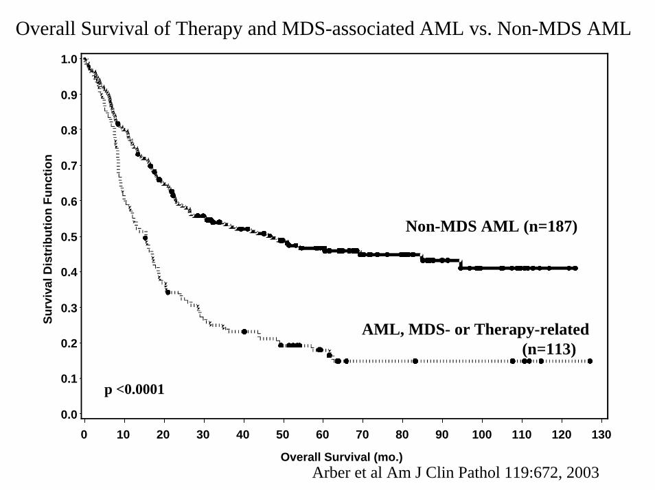
Overall Survival of Therapy and MDS-associated AML vs. Non-MDS AMLSu
rviv
al D
istr
ibut
ion
Func
tion
0.0
0.1
0.2
0.3
0.4
0.5
0.6
0.7
0.8
0.9
1.0
Overall Survival (mo.)
0 10 20 30 40 50 60 70 80 90 100 110 120 130
Non-MDS AML (n=187)
AML, MDS- or Therapy-related (n=113)
p <0.0001
Arber et al Am J Clin Pathol 119:672, 2003

WHO Classification of Acute Myeloid Leukemia
• Acute myeloid leukemia with recurrent cytogenetic abnormalities• AML with t(8;21)(q22;q22), (AML1/ETO)• AML with inv(16)(p13q22) or t(16;16)(p13;q22),
(CBFβ/MYH11)• Acute promyelocytic leukemia (AML with t(15;17)(q22;q12),
(PML/RARα) and variants)• AML with 11q23 (MLL) abnormalities
WHO Classification of Tumours, 2001

Acute Myeloid Leukemia with t(8;21) (RUNX1/RUNX1T1)
• Characteristic blast cell morphology with perinuclear hofs, Auer rods and large salmon-colored granules

Acute Myeloid Leukemia with t(8;21)
• Characteristic blast cell morphology with perinuclear hofs, Auer rods and large salmon-colored granules

Acute Myeloid Leukemia with t(8;21)
CD13 and CD33 positive blast gate

Acute Myeloid Leukemia with t(8;21)
CD19 positive CD34 positive, CD56 +/-

Acute Myeloid Leukemia with t(8;21)
• Characteristic immunophenotype of CD19+/myeloid antigen +/CD34+ blast cells in two thirds of cases. A subset are also CD56+
• These morphologic and immunophenotypic features have a high correlation with t(8;21)(q22;q22) or RUNX1/RUNX1T1 fusion
• These cases should be diagnosed as AML without regard to blast cell count (so called “oligoblastic” acute leukemia)

Does CD19+ AML = t(8;21)?

Does CD19+ AML = t(8;21)?
• Adult AML– CD19+ in 10/102 cases (9.8%)
Khalidi et al. Am J Clin Pathol 109:211, 1998

Does CD19+ AML = t(8;21)?
• Adult AML– CD19+ in 10/102 cases (9.8%)– Cytogenetics available on 7 of those 10
Khalidi et al. Am J Clin Pathol 109:211, 1998

Does CD19+ AML = t(8;21)?
• Adult AML– CD19+ in 10/102 cases (9.8%)– Cytogenetics available on 7 of those 10– One (14%) of those 7 cases had t(8;21)
Khalidi et al. Am J Clin Pathol 109:211, 1998
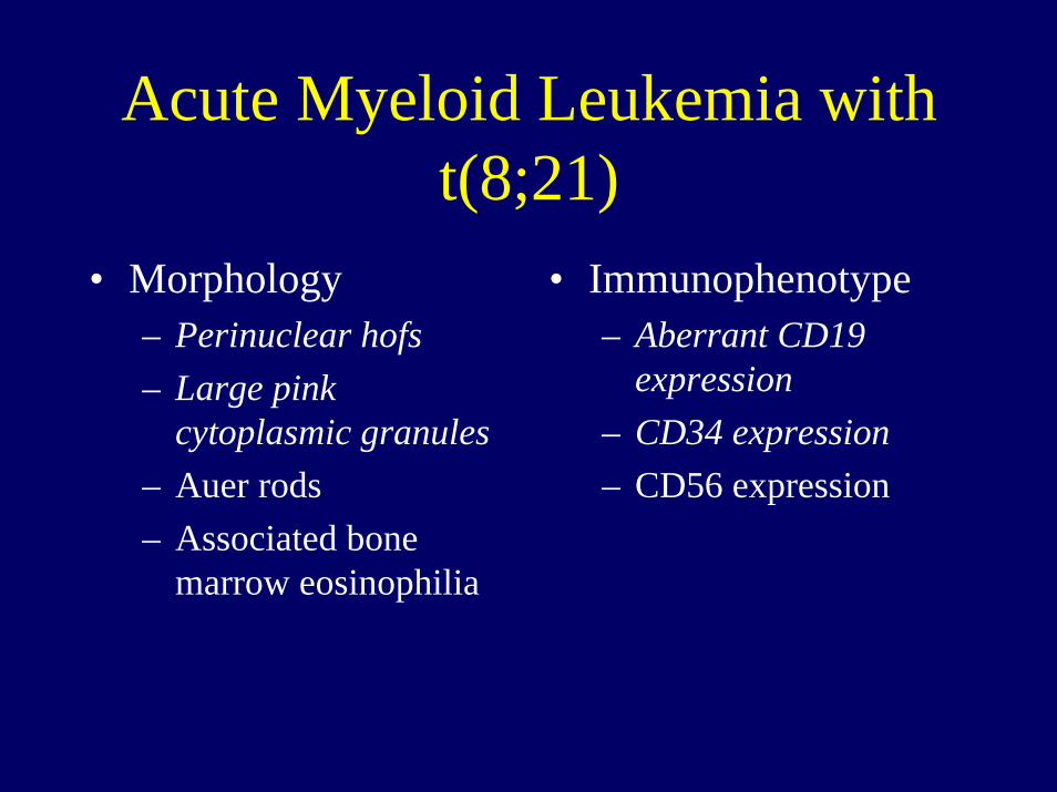
Acute Myeloid Leukemia with t(8;21)
• Morphology– Perinuclear hofs– Large pink
cytoplasmic granules– Auer rods– Associated bone
marrow eosinophilia
• Immunophenotype– Aberrant CD19
expression– CD34 expression– CD56 expression


CD13, CD33, CD34-positive with partial CD19 and CD56
DX: AML with features of t(8;21)

Acute Myeloid Leukemia with inv(16) or t(16;16) (CBFB/MYH11)
• Blast cell proliferation with or without monocytic differentiation by cytochemistry, with an associated proliferation of abnormal eosinophils

Acute Myeloid Leukemia with inv(16) or t(16;16)
• The eosinophils contain abnormal, basophilic granules

Acute Myeloid Leukemia with inv(16) or t(16;16)

Acute Myeloid Leukemia with inv(16) or t(16;16)

Acute Myeloid Leukemia with inv(16) or t(16;16)

Acute Myeloid Leukemia with inv(16) or t(16;16)
• The cases express myeloid-associated antigens and may be CD2 positive, but there is no specific immunophenotype for this disease
• These cases should be diagnosed as AML without regard to blast cell count
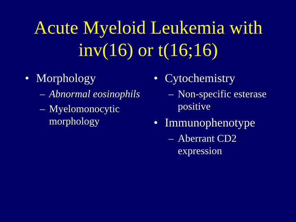
Acute Myeloid Leukemia with inv(16) or t(16;16)
• Morphology– Abnormal eosinophils– Myelomonocytic
morphology
• Cytochemistry– Non-specific esterase
positive• Immunophenotype
– Aberrant CD2 expression
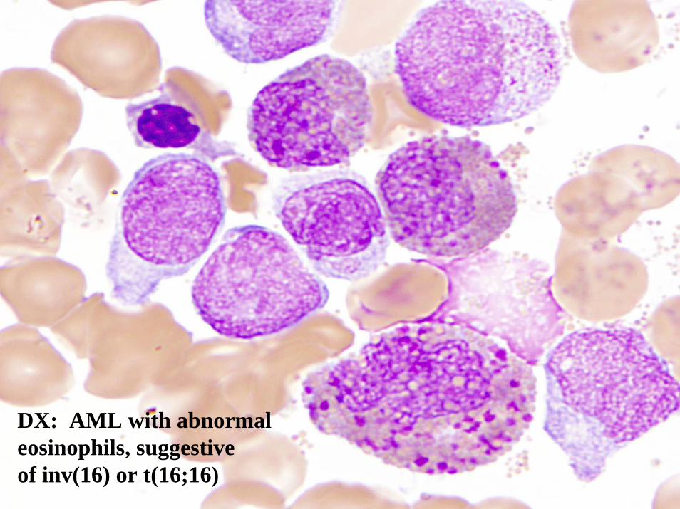
DX: AML with abnormal eosinophils, suggestive of inv(16) or t(16;16)

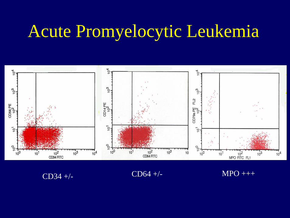
Acute Promyelocytic Leukemia
• Includes microgranular and other variants• Blasts have folded nuclei with or without
cytoplasmic granules and Auer rods

Acute Promyelocytic Leukemia
• Includes microgranular and other variants• Blasts have folded nuclei with or without
cytoplasmic granules and Auer rods

Acute Promyelocytic Leukemia
• Includes microgranular and other variants• Blasts have folded nuclei with or without
cytoplasmic granules and Auer rods

Acute Promyelocytic Leukemia
• Strong myeloperoxidase positivity

Acute Promyelocytic Leukemia
CD13 and CD33 positive

Acute Promyelocytic Leukemia
HLA-DR weak or negative
Acute Promyelocytic Leukemia
CD34 +/- CD64 +/- MPO +++

Acute Promyelocytic Leukemia
• Express myeloid-associated antigens with loss of HLA-DR in the majority of cases and often demonstrates CD2 expression
• These cases should be diagnosed as AML without regard to blast cell count

Does HLA-DR-negative AML = Acute Promyelocytic Leukemia?
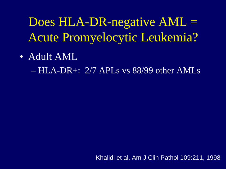
Does HLA-DR-negative AML = Acute Promyelocytic Leukemia?
• Adult AML– HLA-DR+: 2/7 APLs vs 88/99 other AMLs
Khalidi et al. Am J Clin Pathol 109:211, 1998
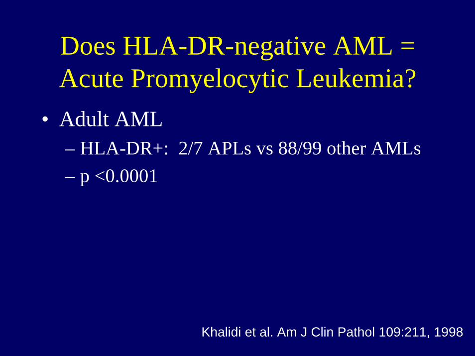
Does HLA-DR-negative AML = Acute Promyelocytic Leukemia?
• Adult AML– HLA-DR+: 2/7 APLs vs 88/99 other AMLs– p <0.0001
Khalidi et al. Am J Clin Pathol 109:211, 1998

Does HLA-DR-negative AML = Acute Promyelocytic Leukemia?
• Adult AML– HLA-DR negative in 16/106 (15.1%)
Khalidi et al. Am J Clin Pathol 109:211, 1998

Does HLA-DR-negative AML = Acute Promyelocytic Leukemia?
• Adult AML– HLA-DR negative in 16/106 (15.1%)– t(15;17) was detected in only 5 of those 16
(31%)
Khalidi et al. Am J Clin Pathol 109:211, 1998

Does HLA-DR-negative AML = Acute Promyelocytic Leukemia?
• Adult APL vs. other AMLHLA-DR negative p <0.0001
Khalidi et al. Am J Clin Pathol 109:211, 1998

Does HLA-DR-negative AML = Acute Promyelocytic Leukemia?
• Adult APL vs. other AMLHLA-DR negative p <0.0001CD4 negative p=0.0084CD11c negative p <0.0001CD36 negative p=0.0297CD117 negative p=0.0422CD2 positive p=0.0293
Khalidi et al. Am J Clin Pathol 109:211, 1998
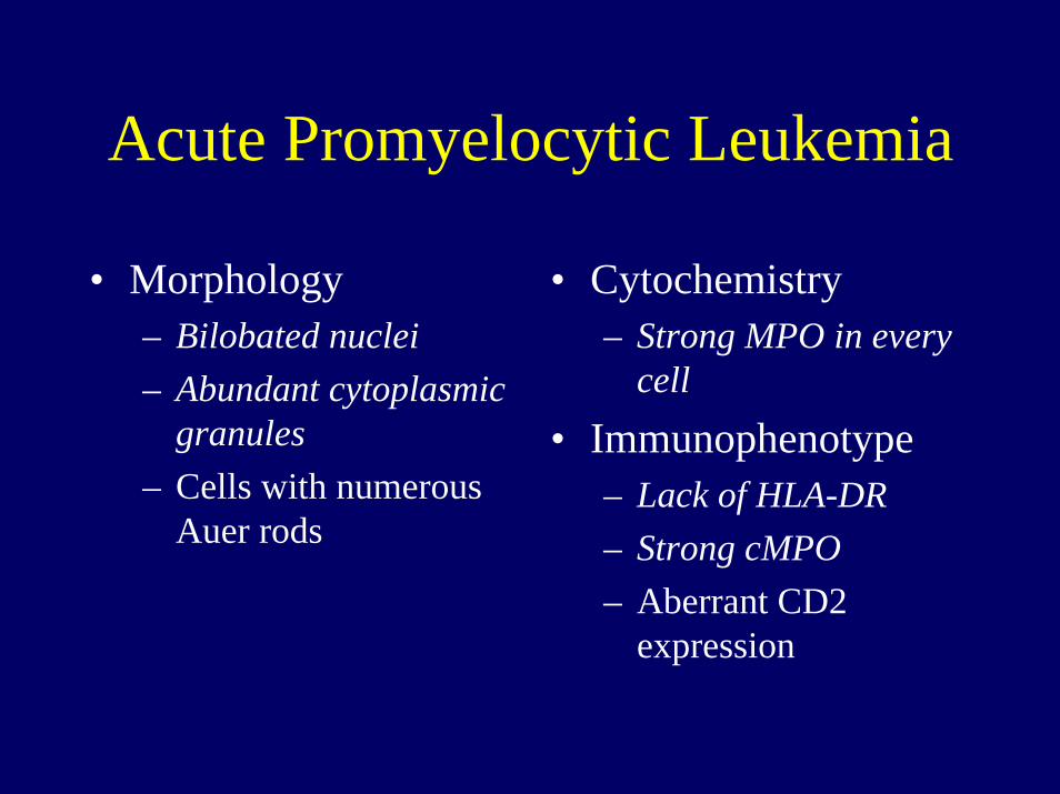
Acute Promyelocytic Leukemia
• Morphology– Bilobated nuclei– Abundant cytoplasmic
granules– Cells with numerous
Auer rods
• Cytochemistry– Strong MPO in every
cell• Immunophenotype
– Lack of HLA-DR– Strong cMPO– Aberrant CD2
expression


CD13, CD33, MPO (strong) positive, HLA-DR negative

Acute Myeloid Leukemia with 11q23 (MLL) Abnormalities
• Blast cell proliferation, usually with monocytic or myelomonocytic features

Acute Myeloid Leukemia with 11q23 (MLL) Abnormalities
• Blast cell proliferation, usually with monocytic or myelomonocytic features
• More common in children• Frequently therapy-related when occurring in
adults, although associated multilineage dysplasia is often no apparent
• No specific morphologic or immunophenotypic features

Acute Myeloid Leukemia with 11q23 (MLL) Abnormalities
CD34 negative CD56 +/- CD64 positive
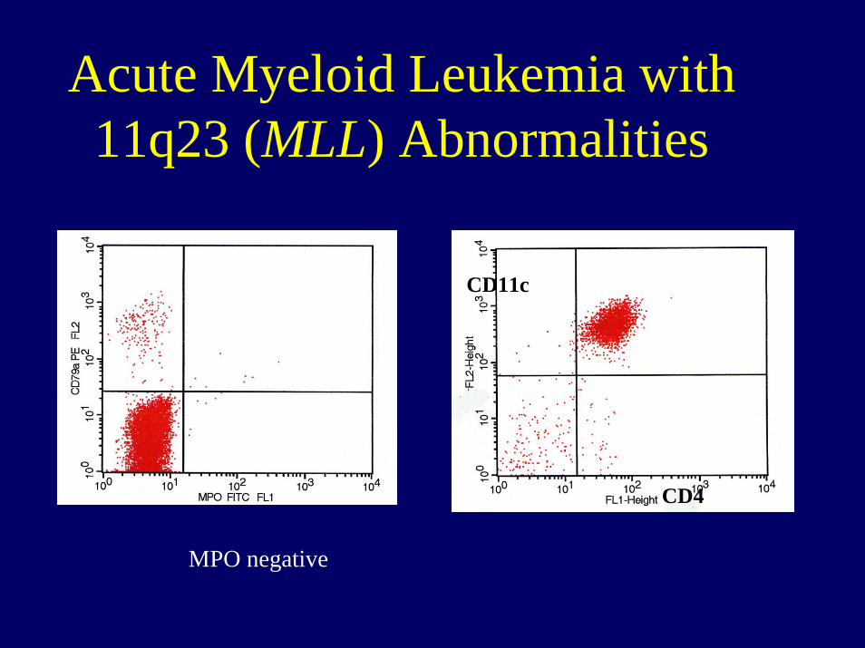
Acute Myeloid Leukemia with 11q23 (MLL) Abnormalities
MPO negative
CD11c
CD4

Acute Myeloid Leukemia with 11q23 (MLL) Abnormalities
• Diagnostic criteria

Acute Myeloid Leukemia with 11q23 (MLL) Abnormalities
• Diagnostic criteria

WHO Classification of Acute Myeloid Leukemia
• Acute myeloid leukemia with multilineagedysplasia
• Acute myeloid leukemia and myelodysplastic syndrome, therapy related• Alkylating agent related• Topoisomerase II inhibitor-related
WHO Classification of Tumours, 2001

AML with Multilineage Dysplasia

AML with Multilineage Dysplasia

Therapy-Related AML/MDS
Alkylating agent-related Topo-II inhibitor-related

WHO Classification of Acute Myeloid Leukemia
• Acute myeloid leukemia not otherwise categorized• AML, minimally differentiated• AML, without maturation• AML, with maturation• Acute myelomonocytic leukemia• Acute monoblastic and monocytic leukemia• Acute erythroid leukemia• Acute megakaryoblastic leukemia• Acute basophilic leukemia• Acute panmyelosis with myelofibrosis• Myeloid sarcoma
• Acute leukemias of ambiguous lineage

What else is there to know about AML?

What else is there to know about AML?
• What is erythroleukemia?

Acute Erythroid Leukemia• FAB (M6) requires >50% bone marrow
erythroid precursors and 30% or greater blasts (myeloblasts) in the non-erythroid cells
• WHO includes two types– M6-like except 20% or greater blasts– Pure erythroid leukemia; over 80% immature
erythroid cells• Other groups have described “M6C”
– Over 30% erythroid cells in marrow and over 30% myeloblasts


Acute Erythroid Leukemia
• Almost always associated with dysplastic changes of other cell lines
• Complex cytogenetic abnormalities are usually present, similar to the myelodysplasias and the other MDS-associated acute myeloid leukemias
• Why aren’t these just cases of MDS or AML with multilineage dysplasia?

What else is there to know about AML?
• What is acute megakaryoblastic leukemia?

What else is there to know about AML?
• What is acute megakaryoblastic leukemia?
An acute leukemia with blasts showing megakaryocytic features by immunophenotyping or platelet peroxidaseelectron microscopy

Acute Megakaryoblastic Leukemia• Cytoplasmic blebs are common, but are not
specific for this type of leukemia• Commonly associated with marrow fibrosis• Blasts are myeloperoxidase negative by
cytochemistry, but may express myeloid-associated markers and should be positive for two megakaryocyte-associated markers (CD41, CD42, CD61, vWF, Ulex)
• Demonstration of platelet peroxidase by electron microscopy may also be helpful

Acute Megakaryoblastic Leukemia
Megakaryoblast Non-Megakaryoblasts

Acute Megakaryoblastic LeukemiaAppear to be at least three types
– Adult type• Associated with multilineage dysplasia and MDS-
like cytogenetic abnormalities• May be related to acute panmyelosis with
myelofibrosis
– Childhood type• Associated with trisomy 21• Occurs at an older age (>2 yrs) than transient
myeloproliferative disorder of Down Syndrome
– Infant type• Associated with t(1;22)

Acute Megakaryoblastic Leukemia with t(1;22)(p13;q13)
• Infant leukemia• Nonspecific
cytoplasmic blebs• MPO negative;
CD41, CD61 positive• RBM15/MKL1
(OTT/MAL) fusion

What else is there to know about AML?
• Other translocations• Mutations

Recurrent Cytogenetic Abnormalities in AML with Multilineage Dysplasia
• Chromosome 5 and 7 abnormalities• Complex karyotypes• Balanced abnormalities
– inv(3)(q21q26)/t(3;3)(q21;q26), EVI1– t(6;9)(23;q34), DEK-CAN– t(3;5)(q25;q31), NPM-MLF1
Mutations in AML
• NPM1• FLT3• CEPBA• MLL• CKIT
Reviewed by Mrózek et al. Blood 109:431-448, 2007
Mutations in AML• FLT3
– Mutations occur in 10-15% of childhood AMLs and 20-28% of adult AMLs
– More frequent in APL, normal karyotypeAML or t(6;9) AML
– Mutation associated with decreased disease free survival in adults
– Clinical trials with FLT3 inhibitors are underway
Kottaridis et al. Blood 98:1752, 2001

FLT3 Mutations
Internal Tandem Duplications (ITD)
Point Mutations(D835)

Significance of FLT3 Mutations in Adult AML with Normal KaryotypesSu
rviv
al D
istr
ibut
ion
Func
tion
0.0
0.1
0.2
0.3
0.4
0.5
0.6
0.7
0.8
0.9
1.0
Disease Free Survival (mo.)
0 10 20 30 40 50 60
FLT3 Wild Type
FLT3 Mutation

Mutations in AML
• NPM1– 25-30% of all AMLs– Up to half of AMLs with normal
karyotypes– Women, high WBC and Plt counts– Mutated NPM1 associated with a good
prognosis unless associated with a FLT3 mutation
Thiede et al. Blood 107:4011, 2006

NPM1 Mutations

NPM1 Mutations
Falini, B. et al. Blood 2007;109:874-885

Mutations in AML
• CEPBA– Mutations occur in 7-11% of AMLs– Mutations associated with a favorable
prognosis, unless accompanied by a FLT3 mutation
Preudhomme et al. Blood 100:2717, 2002

Mutations in AML
• KIT– Mutations occur in 22-30% of AMLs
with t(8;21) and inv(16)– Involve exon 17 (usually D816V) or
exon 8– Mutations in these disease groups
associated with a worse prognosis
Paschka et al. J Clin Oncol 24:3904-11, 2006

Mrozek, K. et al. Blood 2007;109:431-448

Falini, B. et al. Blood 2007;109:874-885

AML Classification
• AML with Recurring Genetic Abnormalities
• AML, Myelodysplasia-Related • AML, Therapy-Related• AML, NOS

AML Classification• AML with Recurring Genetic Abnormalities
– AML with t(8;21)– AML with inv(16)– AML with t(15;17)– AML with t(9;11)– AML with t(1;22)– AML with t(9;22)– AML with NPM1 mutations– AML with CEPBA mutations
• AML, Myelodysplasia-Related • AML, Therapy-Related• AML, NOS

AML Classification
• AML with Recurring Genetic Abnormalities
• AML, Myelodysplasia-Related – AML following MDS– AML with multilineage dysplasia– AML with MDS-related cytogenetics
• AML, Therapy-Related• AML, NOS

AML Classification
• AML with Recurring Genetic Abnormalities
• AML, Myelodysplasia-Related • AML, Therapy-Related• AML, NOS

Summary
• A combined morphologic, immunophenotypic and genetic approach is necessary for the accurate classification of AML and allows for identification of prognostically significant diseases types
• The ongoing discovery of genetic subtypes of AML will impact future classification and therapy

FAB WHO