acute haemorrhagic leukoencephalitis: report of a case · acute haemorrhagic leukoencephalitis...
TRANSCRIPT

205
Acta Neurologica Taiwanica Vol 12 No 4 December 2003
INTRODUCTION
Acute haemorrhagic leukoencephalitis (AHL) wasfirst described by Hurst(1) in 1941 with five main patho-logical features including 1) perivascular cellular infil-tration, 2) multiple ball and ring haemorrhages, 3)perivascular demyelination, 4) fibrinoid necrosis ofblood vessels and 5) perivascular or confluent edema(2-6).The pathological changes usually involve subcorticalwhite matter but spare cerebral cortex and subcortical Ufibers. The involvement of subarachnoid vessels was not
well described. The clinical course of AHL is usuallycharacterized by an acute onset of headache, nausea,and vomiting, and then followed by a rapid progressivecourse of focal neurological deficits and even to deepcoma. The outcome is usually fatal and very fewsurvivors have been reported after decompressive thera-py(7-12).
Recently, we encountered a pathologically provencase of AHL who developed a slowly progressivecourse after an episode of upper respiratory tract infec-tion. Interestingly, the patient had frequent seizures and
From the Departments of 1Neurology, 2Pathology and3Neurosurgery, Chang Gung Memorial Hospital and University,Taipei, Taiwan.Received May 21, 2003. Revised June 16, 2003. Accepted August 29, 2003.
Reprint requests and correspondence to: Chin-Chang Huang,MD. Department of Neurology, Chang Gung MemorialHospital & University, No. 199, Tung Hwa North Road, Taipei,Taiwan.E-mail: [email protected]
205
Acute Haemorrhagic Leukoencephalitis: Report of a Case
Wen-Li Chuang1, Chin-Chang Huang1, Shih-Ming Jung2, Huo-Li Chuang3, and Nai-Shin Chu1
Abstract- Acute haemorrhagic leukoencephalitis (AHL) usually involves the subcortical white matter butspares the cerebral cortex and subcortical U fibers. The outcome is usually fatal and very few survivorshave been reported after decompressive therapy. An unusual presentation of a prolonged course of AHL in a39-year-old man is reported. Initial brain magnetic resonance images (MRI) showed an irregular enhancinglesion in the left frontal area and another non-enhancing lesion in the right frontal subcortical white matter.A follow-up MRI 2 months later showed a remission of the right frontal lesion but a deterioration of the leftfrontal lesion. He received a decompressive craniotomy with left frontal lobectomy and pathological studiesrevealed an inflammatory reaction with haemorrhages involving the subcortical white matter. Interestingly,the haemorrhages also extended to the cortex and even to the meninges. Unfortunately, speech disturbancedue to an epidural haematoma in the left fronto-parietal area was noted after the lobectomy. We concludethat AHL is unusual and the lesion may also involve the cortex and leptomeninges. Bleeding after decom-pression may be due to a post-operative complication or dura vessel involvement.
Key Words: Acute haemorrhagic leukoencephalitis, Epidural haematoma, MRI, Decompressive lobectomy,Pathology
Acta Neurol Taiwan 2003;12:205-210

206
Acta Neurologica Taiwanica Vol 12 No 4 December 2003
haemorrhages were also found in the cerebral cortex andthe subarachnoid spaces. In addition, the patient devel-oped an epidural haematoma after a decompressivelobectomy. The mechanisms and the lesion extension ofAHL are also discussed.
REPORT OF A CASE
A 39-year-old man was admitted to our hospital onAugust 6 because of several episodes of generalized con-vulsion. He had enjoyed good health until one weekprior to this admission. Initially he had experienced flu-like symptoms with fever, sore throat, cough and profusesputum, and then diarrhoea in the following few days.On August 6, four episodes of generalized tonic-clonicconvulsion with loss of consciousness were noted.Postictal confusion was noted for about 10 minutes. Hedenied any previous history of diabetes, hypertension,heart disease, head injury or strokes.
On evaluation, the blood pressure was 122/66
mmHg, body temperature 37.5 ˚C, pulse rate 94/min andrespiratory rate 20/min. Neurological examinationsrevealed clear consciousness. The visual field, speech,extraocular movements, sensation, and motor functionwere normal. Neither frontal releasing signs such assucking and snouting reflexes, nor palmomental signwas noted. Hemogram showed a high white count18,200/mm3 with a predominance of neutrophils (93%).Biochemical studies disclosed normal electrolytes, andliver and renal functions. Brain computed tomography(CT) scan showed an ill-defined low density in the leftfrontal lobe with normal ventricular size and no midlineshifting. Brain magnetic resonance images (MRI)revealed another smaller lesion in the right frontal sub-cortical white matter area in addition to a left frontallesion with irregular low signal intensity on T1 weightedimages (T1W) and high signal intensity on T2 weightedimages (T2W) (Fig. 1A). After gadolinium (Gd) contrastmedium injection, an enhancement was noted over theleft frontal lesion. He was treated with phenytoin 300 mg
A B
Figure 1. (A) Initial brain MRI revealed a highsignal intensity lesion on T2W overthe left frontal area and an additionallesion (arrow head) over the rightfrontal subcortical white matter. (TR:5700, TE: 91.3); (B) The follow-upbrain MRI 2 months later showed adeterioration of the left frontal lesionbut the disappearance of the rightfrontal subcor tical white matterlesion. (TR: 4000, TE: 99.0)
R

207
Acta Neurologica Taiwanica Vol 12 No 4 December 2003
daily. Under the impression of multicentric glioma, a
stereotactic brain biopsy was performed from the leftfrontal area on August 10 and revealed perivascular cel-lular infiltrations with edema and haemorrhages. Theperivascular infiltration cells included eosinophils, neu-rotrophils and mononuclear cells. There were reactiveastrocytes with positive stain for glial fibrillary acidic
protein. Therefore, a diagnosis of acute necrotizing vas-culitis was tentatively made. However the studies forrheumatoid factor, antinuclear antibody, and anti-double-stranded DNA antibody were all negative or within nor-mal limits. After the operation, the condition was sta-tionary and he was treated with phenytoin and dexam-ethasone or prednisolone 30 mg daily for about onemonth. During the follow-up period, he developed apha-sia. The follow-up brain MRI scan on Oct. 10 disclosedan ill-defined heterogeneous mass lesion over the leftfrontal area with an enhancement after Gd administra-tion. A mid-line shifting and compression of the leftfrontal horn were noted. However the right frontal sub-cortical lesion disappeared (Fig. 1B). He developed oneepisode of generalized seizures of about 5 min in dura-tion on Oct. 10 and 17 respectively. He could not speakvery well and dizziness was noted. He was placed onmethylprednisolone and admitted again.
During the second admission on Oct. 29, his con-sciousness was clear. Mild weakness with a pronatorsign was found on the right upper extremity. Deep ten-don reflexes were generally increased. However theplantar reflex was flexion. One day after admission,headache, nausea, and vomiting with a deterioration ofGlascow coma scale from 15 to 10 (E3V1M6) were
Figure 3. The pathological study of the frontal lobectomy disclosed (A) perivascular cuffings with lymphocytes and PMNs (H & E stain, 200X),(B) perivascular demyelination and edema (Luxol fast blue stain, 100X), (C) multiple ball haemorrhages in the subcortical white matter(H & E stain, 40X).
Figure 2. The lobectomy specimen revealed an involvement of thesubcortical white matter (arrow heads) and a slight sparingof subcortical white matter U fibers. In addition, the haem-orrhages were found in the cerebral cortex (arrows) andthe subarachnoid spaces (small arrow heads).
208
Acta Neurologica Taiwanica Vol 12 No 4 December 2003
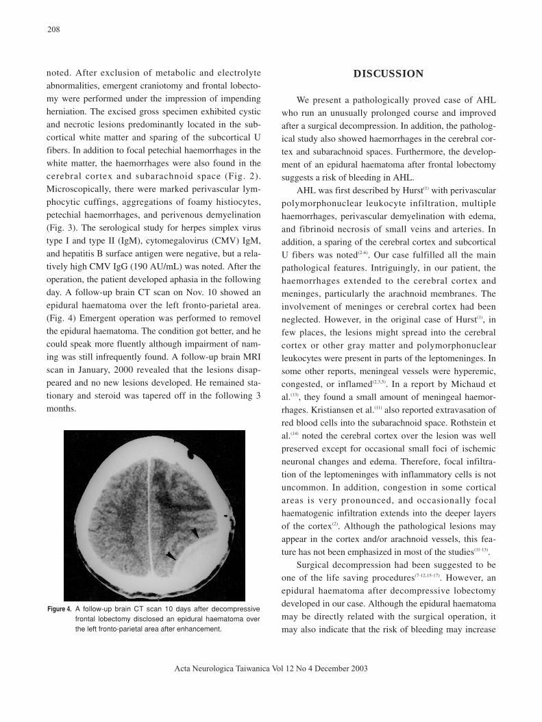
noted. After exclusion of metabolic and electrolyteabnormalities, emergent craniotomy and frontal lobecto-my were performed under the impression of impendingherniation. The excised gross specimen exhibited cysticand necrotic lesions predominantly located in the sub-cortical white matter and sparing of the subcortical Ufibers. In addition to focal petechial haemorrhages in thewhite matter, the haemorrhages were also found in thecerebral cortex and subarachnoid space (Fig. 2).Microscopically, there were marked perivascular lym-phocytic cuffings, aggregations of foamy histiocytes,petechial haemorrhages, and perivenous demyelination(Fig. 3). The serological study for herpes simplex virustype I and type II (IgM), cytomegalovirus (CMV) IgM,and hepatitis B surface antigen were negative, but a rela-tively high CMV IgG (190 AU/mL) was noted. After theoperation, the patient developed aphasia in the followingday. A follow-up brain CT scan on Nov. 10 showed anepidural haematoma over the left fronto-parietal area.(Fig. 4) Emergent operation was performed to removelthe epidural haematoma. The condition got better, and hecould speak more fluently although impairment of nam-ing was still infrequently found. A follow-up brain MRIscan in January, 2000 revealed that the lesions disap-peared and no new lesions developed. He remained sta-tionary and steroid was tapered off in the following 3months.
DISCUSSION
We present a pathologically proved case of AHLwho run an unusually prolonged course and improvedafter a surgical decompression. In addition, the patholog-ical study also showed haemorrhages in the cerebral cor-tex and subarachnoid spaces. Furthermore, the develop-ment of an epidural haematoma after frontal lobectomysuggests a risk of bleeding in AHL.
AHL was first described by Hurst(1) with perivascularpolymorphonuclear leukocyte infiltration, multiplehaemorrhages, perivascular demyelination with edema,and fibrinoid necrosis of small veins and arteries. Inaddition, a sparing of the cerebral cortex and subcorticalU fibers was noted(2-6). Our case fulfilled all the mainpathological features. Intriguingly, in our patient, thehaemorrhages extended to the cerebral cortex andmeninges, particularly the arachnoid membranes. Theinvolvement of meninges or cerebral cortex had beenneglected. However, in the original case of Hurst(1), infew places, the lesions might spread into the cerebralcortex or other gray matter and polymorphonuclearleukocytes were present in parts of the leptomeninges. Insome other reports, meningeal vessels were hyperemic,congested, or inflamed(2,3,5). In a report by Michaud etal.(13), they found a small amount of meningeal haemor-rhages. Kristiansen et al.(11) also reported extravasation ofred blood cells into the subarachnoid space. Rothstein etal.(14) noted the cerebral cortex over the lesion was wellpreserved except for occasional small foci of ischemicneuronal changes and edema. Therefore, focal infiltra-tion of the leptomeninges with inflammatory cells is notuncommon. In addition, congestion in some corticalareas is very pronounced, and occasionally focalhaematogenic infiltration extends into the deeper layersof the cortex(2). Although the pathological lesions mayappear in the cortex and/or arachnoid vessels, this fea-ture has not been emphasized in most of the studies(11-13).
Surgical decompression had been suggested to beone of the life saving procedures(7-12,15-17). However, anepidural haematoma after decompressive lobectomydeveloped in our case. Although the epidural haematomamay be directly related with the surgical operation, itmay also indicate that the risk of bleeding may increase
Figure 4. A follow-up brain CT scan 10 days after decompressivefrontal lobectomy disclosed an epidural haematoma overthe left fronto-parietal area after enhancement.
in AHL while performing a craniotomy. AHL usuallyhas a rapid progressive course leading to death. Howeverit occasionally has a fluctuant or chronic course. Ourpatient had a chronic progressive course for about 2months. In a follow-up MRI scan, the right frontal sub-cortical white matter lesion had a spontaneous remissionwhile the left frontal lesion deteriorated. The progressionon MRI appeared to correlate with the clinical deteriora-tion. In a case reported by Michaud and Helle(13), the evo-lution was biphasic with an acute onset on the secondday, followed by an improvement in the following threedays, and eventually fulminant leading to death.Kristiansen et al.(11) reported two patients who had sur-vived following decompressive craniotomy. Coxe andLuse(7) showed two biopsy-proven cases with a long sur-vival of 2.5 years and 1 year, respectively, following sur-gical decompression. The first case survived with somesequelae but the second case eventually expired. Thepatient of Lamarche et al.(10) survived 9 months after apartial frontal lobectomy. In our previous report, onepatient died 3 months from the onset and another sur-vived 1.5 months with recovery(12). The data suggest thatsome patients may have a prolonged clinical course andsurgical decompression may be helpful in prevention ofherniation.
The etiology and pathogenesis of AHL remainobscure. However an allergic reaction may play animportant role. From some clinical and histopathologicalstudies, acute brain purpura, AHL and acute disseminat-ed leukoencephalitis (ADL) may share the same mecha-nisms and can be considered as a triad of organ specificallergic conditions(2,4,18). The speed of deterioration maycorrelate with the spreading of multiple discrete smallring and ball hematomas. In hyperacute state such asbrain purpura, cellular infiltration is rarely found. InAHL, the perivascular cellular infiltrations are usuallypolymorphonuclear cells. In ADL, the infiltrations areusually mononuclear cells or lymphocytes(2). In our case,the presence of both of the lymphocytes and polymor-phonuclear cells possibly indicated an intermediate typebetween AHL and ADL.
We conclude that AHL is rare, and the pathologicallesions may also involve the cortex and leptomeninges.In addition, the procedure of lobectomy should be very
careful because that epidural haematomas may occur dueto post-operative complication or dura vessel involve-ment.
REFERENCES
1. Hurst EW. Acute haemorrhagic leucoencephalitis: a previ-
ously undefined entity. Med Aust 1941;28:1-6.
2. Gosztonyi G. Acute haemorrhagic leucoencephalitis. In:
Vinken PJ, Bruyn GW, eds. Handbook of Clinical
Neurology, Vol 3. Amsterdam: North-Holland Publishing
Co, 1978:587-604.
3. Crawford T. Acute haemorrhagic leucoencephalitis. J Clin
Pathol 1954;7:1-9.
4. Russell DS. The nosological unity of acute haemorrhagic
leucoencephalitis and acute disseminated encephal-
omyelitis. Brain 1955;78:369-76.
5. Lander H. Acute haemorrhagic leucoencephalitis. Aust Ann
Med 1958;7:55-68.
6. Vanderfield GK, Tompkins M, Jelihovsky T.
Clinicopathological features of acute haemorrhagic leu-
coencephalitis. Aust Ann Med 1960;9:29-33.
7. Coxe WS, Luse SA. Acute haemorrhagic leucoencephalitis:
a clinical and electron-microscopic report of 2 patients
treated with surgical decompression. J Neurosurg 1963;20:
584-96.
8. Litel G, Ehni G. Acute haemorrhagic leucoencephalitis:
treatment with corticosteroids and dehydrating agents. J
Neurosurg 1970;33:445-52.
9. Hart MN, Earle KM. Haemorrhagic and perivenous
encephalitis: a clinical-pathological review of 38 cases. J
Neurol Neurosurg Psychiatry 1975;38:585-91.
10. Lamarche JB, Behan PO, Segarra JM, et al. Recurrent acute
necrotizing haemorrhagic encephalopathy. Acta
Neuropathol (Berl.) 1972;22:79-87.
11. Kristiansen K, Harkmark W, Cohen MM. Acute haemor-
rhagic encephalitis. Neurology 1956;6:503-9.
12. Huang CC, Chu NS, Chen TJ, et al. Acute haemorrhagic
leucoencephalitis with a prolonged clinical course. J Neurol
Neurosurg Psychiatry 1988;51:870-4.
13. Michaud J, Helle TL. Acute haemorrhagic leucoencephali-
tis localized to the brainstem and cerebellum: a report of
two cases. J Neurol Neurosurg Psychiatry 1982;45:151-7.
14. Rothstein TL, Shaw CM. Computerized tomography as a
209
Acta Neurologica Taiwanica Vol 12 No 4 December 2003
diagnostic aid in acute haemorrhagic leukoencephalitis.
Ann Neurol 1983;13:331-3.
15. Seales D, Greer M. Acute haemorrhagic leuesencephalitis:
a successful recovery. Arch Neurol 1991;48:1086-8.
16. Markus R, Brew BJ, Turner J, et al. Successful outcome
with aggressive treatment of acute haemorrhagic leukoen-
cephalitis. J Neurol Neurosurg Psychiatry 1991;63:551.
17. Rosman PN, Gottlieb SM, Bernstein CA. Acute haemor-
rhagic leukoencephalotis: recovery and reversal of magnet-
ic resonance imaging findings in a child. J Child Neurol
1997;12:448-54.
18. Geerts Y, Dehaene I, Lammens M. Acute haemorrhagic
leucoencephalitis. Acta Neurol Belg 1991;91:201-11.
210
Acta Neurologica Taiwanica Vol 12 No 4 December 2003