acute exacerbation of ipf
TRANSCRIPT

Acute exacerbations in patients with idiopathic pulmonary fibrosis
By
Gamal Rabie Agmy , MD , FCCP Professor of Chest Diseases ,Assiut University
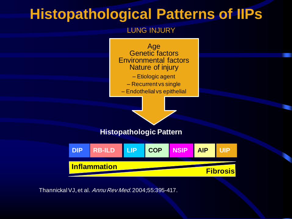
Histopathological Patterns of IIPs
Thannickal VJ, et al. Annu Rev Med. 2004;55:395-417.
Age Genetic factors
Environmental factors Nature of injury
– Etiologic agent
– Recurrent vs single
– Endothelial vs epithelial
Histopathologic Pattern
DIP RB-ILD LIP COP NSIP AIP UIP
Inflammation Fibrosis
LUNG INJURY
50%
Years
Resp
irato
ry
Fu
ncti
on
/Sym
pto
ms
1 2 3 4
FV
C
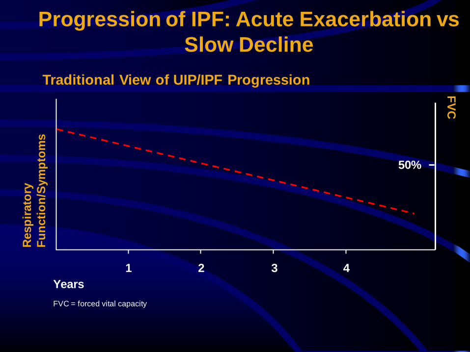
Traditional View of UIP/IPF Progression
Progression of IPF: Acute Exacerbation vs
Slow Decline
FVC = forced vital capacity
50%
Years
Resp
irato
ry
Fu
ncti
on
/Sym
pto
ms
1 2 3
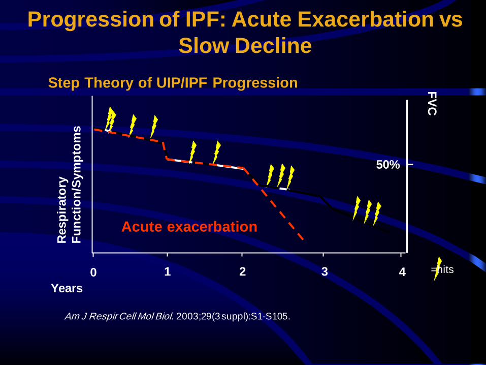
Acute exacerbation
Step Theory of UIP/IPF Progression
Progression of IPF: Acute Exacerbation vs
Slow Decline F
VC
0 4
Am J Respir Cell Mol Biol. 2003;29(3 suppl):S1-S105.
=hits
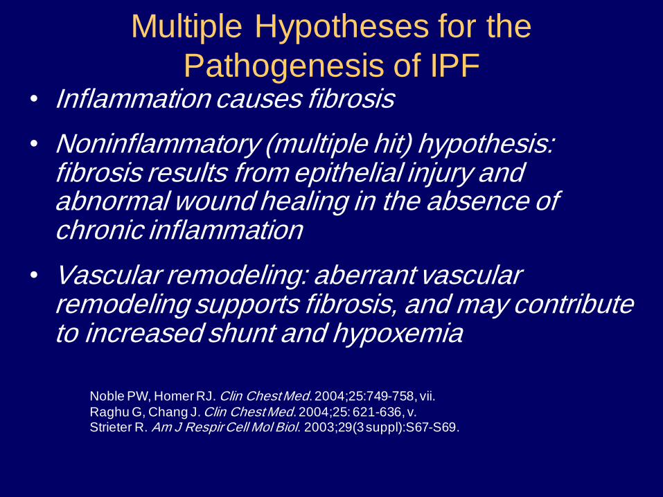
Multiple Hypotheses for the
Pathogenesis of IPF • Inflammation causes fibrosis
• Noninflammatory (multiple hit) hypothesis: fibrosis results from epithelial injury and abnormal wound healing in the absence of chronic inflammation
• Vascular remodeling: aberrant vascular remodeling supports fibrosis, and may contribute to increased shunt and hypoxemia
Noble PW, Homer RJ. Clin Chest Med. 2004;25:749-758, vii.
Raghu G, Chang J. Clin Chest Med. 2004;25: 621-636, v. Strieter R. Am J Respir Cell Mol Biol. 2003;29(3 suppl):S67-S69.
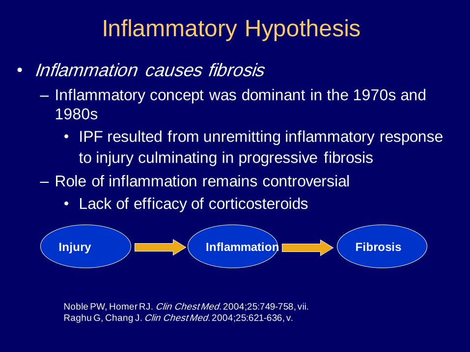
• Inflammation causes fibrosis – Inflammatory concept was dominant in the 1970s and
1980s
• IPF resulted from unremitting inflammatory response
to injury culminating in progressive fibrosis
– Role of inflammation remains controversial
• Lack of efficacy of corticosteroids
Noble PW, Homer RJ. Clin Chest Med. 2004;25:749-758, vii.
Raghu G, Chang J. Clin Chest Med. 2004;25:621-636, v.
Injury Inflammation Fibrosis
Inflammatory Hypothesis
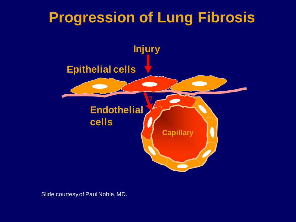
Injury
Epithelial cells
Slide courtesy of Paul Noble, MD.
Progression of Lung Fibrosis
Capillary
Endothelial
cells
?
Epithelial cells
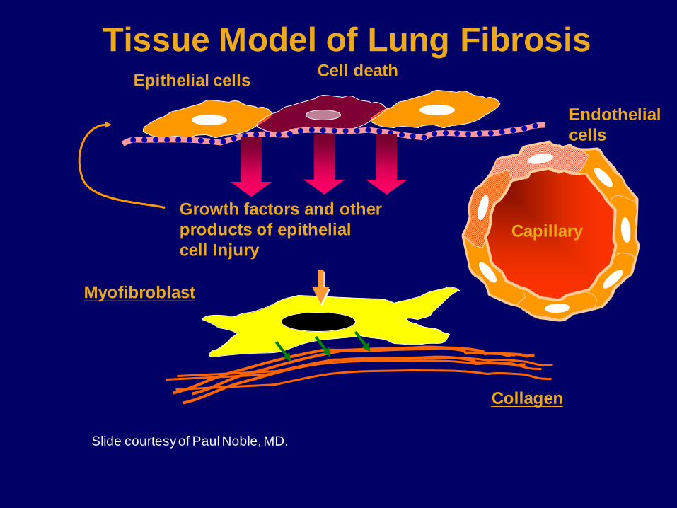
Collagen
Myofibroblast
Cell death
Growth factors and other
products of epithelial
cell Injury
Slide courtesy of Paul Noble, MD.
Tissue Model of Lung Fibrosis
Capillary
Endothelial
cells
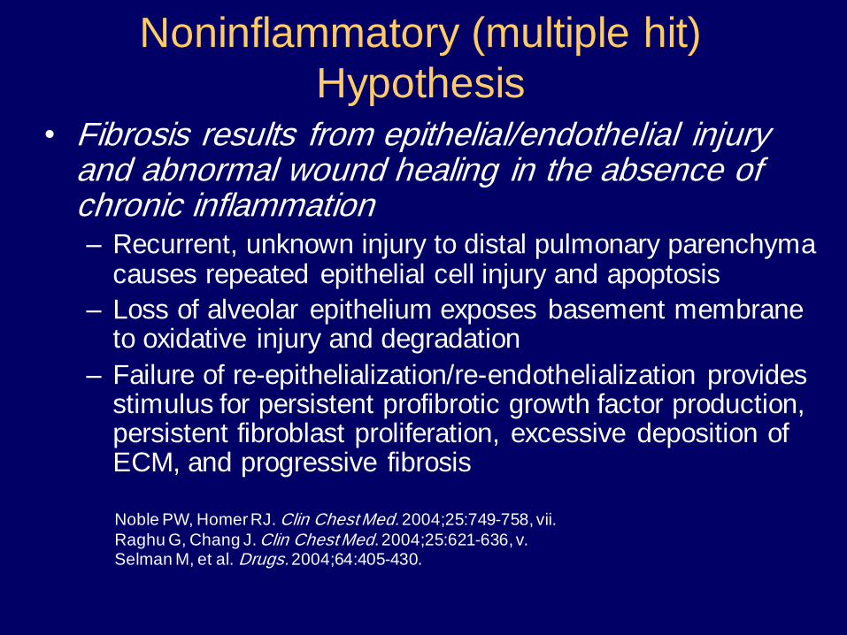
• Fibrosis results from epithelial/endothelial injury and abnormal wound healing in the absence of chronic inflammation – Recurrent, unknown injury to distal pulmonary parenchyma
causes repeated epithelial cell injury and apoptosis
– Loss of alveolar epithelium exposes basement membrane to oxidative injury and degradation
– Failure of re-epithelialization/re-endothelialization provides stimulus for persistent profibrotic growth factor production, persistent fibroblast proliferation, excessive deposition of ECM, and progressive fibrosis
Noble PW, Homer RJ. Clin Chest Med. 2004;25:749-758, vii.
Raghu G, Chang J. Clin Chest Med. 2004;25:621-636, v. Selman M, et al. Drugs. 2004;64:405-430.
Noninflammatory (multiple hit)
Hypothesis
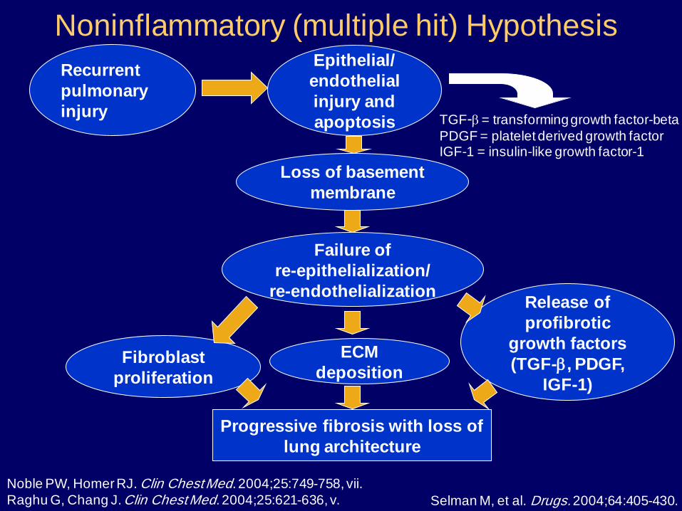
Noninflammatory (multiple hit) Hypothesis
Recurrent
pulmonary
injury
Epithelial/
endothelial
injury and
apoptosis
Loss of basement
membrane
Failure of
re-epithelialization/
re-endothelialization
ECM
deposition Fibroblast
proliferation
Release of
profibrotic
growth factors
(TGF-b, PDGF,
IGF-1)
Progressive fibrosis with loss of
lung architecture
TGF-b = transforming growth factor-beta
PDGF = platelet derived growth factor IGF-1 = insulin-like growth factor-1
Noble PW, Homer RJ. Clin Chest Med. 2004;25:749-758, vii.
Raghu G, Chang J. Clin Chest Med. 2004;25:621-636, v.
Selman M, et al. Drugs. 2004;64:405-430.
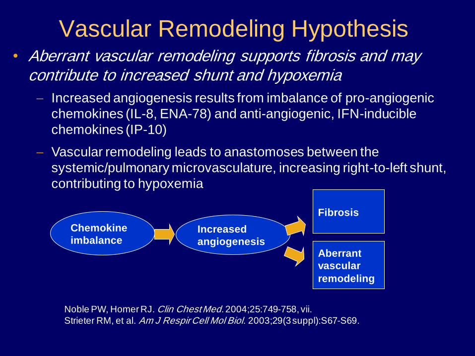
• Aberrant vascular remodeling supports fibrosis and may contribute to increased shunt and hypoxemia
Increased angiogenesis results from imbalance of pro-angiogenic
chemokines (IL-8, ENA-78) and anti-angiogenic, IFN-inducible
chemokines (IP-10)
Vascular remodeling leads to anastomoses between the
systemic/pulmonary microvasculature, increasing right-to-left shunt,
contributing to hypoxemia
Chemokine
imbalance Increased
angiogenesis
Fibrosis
Noble PW, Homer RJ. Clin Chest Med. 2004;25:749-758, vii.
Strieter RM, et al. Am J Respir Cell Mol Biol. 2003;29(3 suppl):S67-S69.
Vascular Remodeling Hypothesis
Aberrant
vascular
remodeling
Defects in Host Defense Mechanisms
May Contribute to Fibrosis
• Defects in endogenous host defense
mechanisms (eg, IFN-g, PGE2 production) that
limit fibrosis after acute lung injury may
contribute to progressive fibrosis
Noble PW, Homer RJ. Clin Chest Med. 2004;25:749-758, vii.

NHLBI in an attempt to standardize the diagnostic
criteria used across studies . This committee defined
AEx-IPF as an acute, clinically significant deterioration
of unidentifiable cause and proposed five diagnostic criteria.
Definition of AEx-IPF
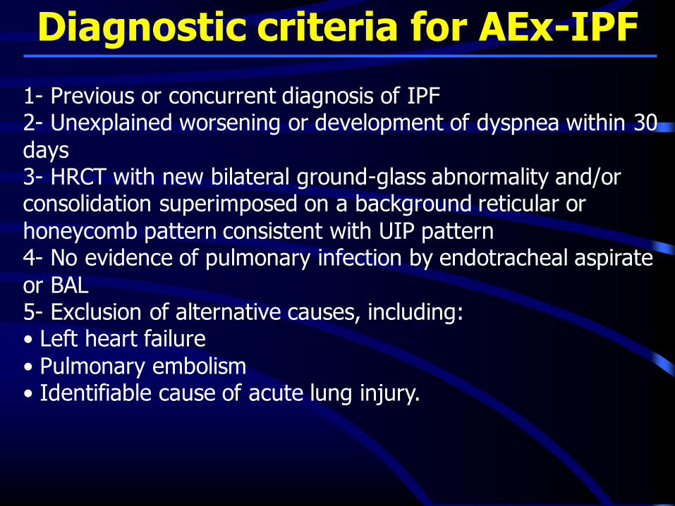
1- Previous or concurrent diagnosis of IPF 2- Unexplained worsening or development of dyspnea within 30
days 3- HRCT with new bilateral ground-glass abnormality and/or consolidation superimposed on a background reticular or
honeycomb pattern consistent with UIP pattern 4- No evidence of pulmonary infection by endotracheal aspirate
or BAL 5- Exclusion of alternative causes, including: • Left heart failure
• Pulmonary embolism • Identifiable cause of acute lung injury.
Diagnostic criteria for AEx-IPF
Incidence of AEx-IPF
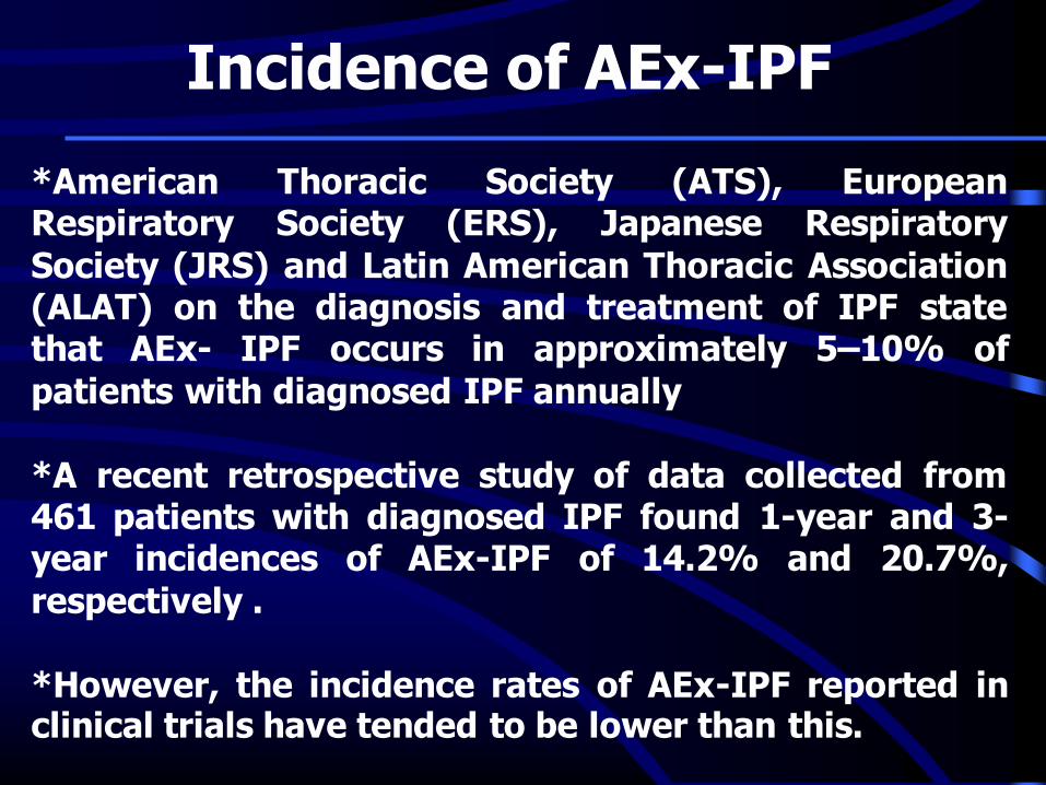
*American Thoracic Society (ATS), European Respiratory Society (ERS), Japanese Respiratory
Society (JRS) and Latin American Thoracic Association (ALAT) on the diagnosis and treatment of IPF state that AEx- IPF occurs in approximately 5–10% of
patients with diagnosed IPF annually
*A recent retrospective study of data collected from 461 patients with diagnosed IPF found 1-year and 3-year incidences of AEx-IPF of 14.2% and 20.7%,
respectively .
*However, the incidence rates of AEx-IPF reported in clinical trials have tended to be lower than this.
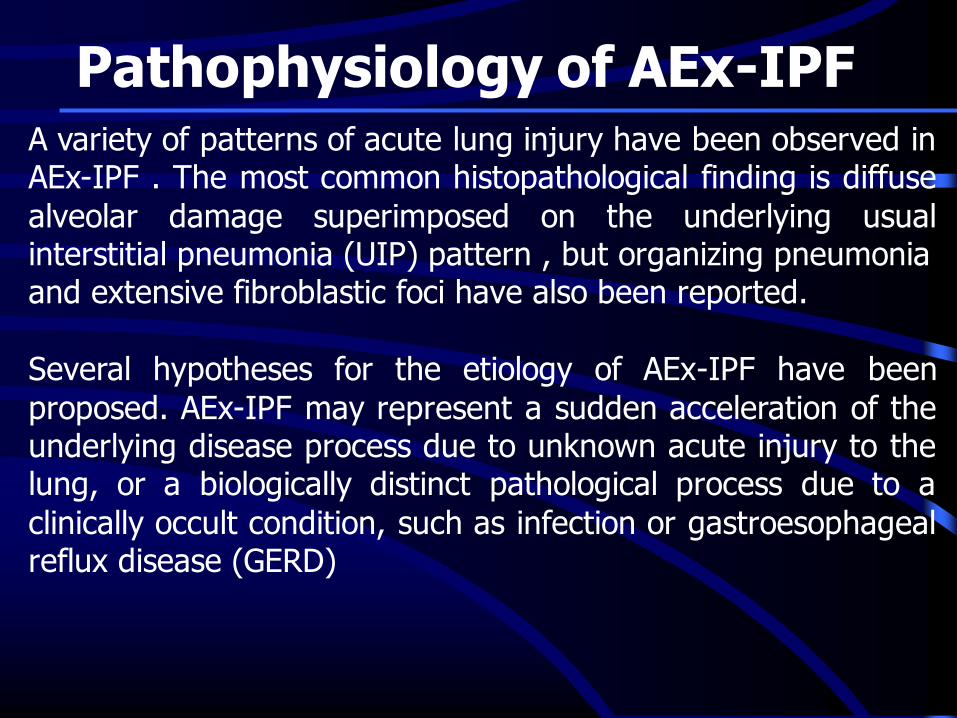
Pathophysiology of AEx-IPF A variety of patterns of acute lung injury have been observed in AEx-IPF . The most common histopathological finding is diffuse
alveolar damage superimposed on the underlying usual interstitial pneumonia (UIP) pattern , but organizing pneumonia and extensive fibroblastic foci have also been reported.
Several hypotheses for the etiology of AEx-IPF have been
proposed. AEx-IPF may represent a sudden acceleration of the underlying disease process due to unknown acute injury to the lung, or a biologically distinct pathological process due to a
clinically occult condition, such as infection or gastroesophageal reflux disease (GERD)
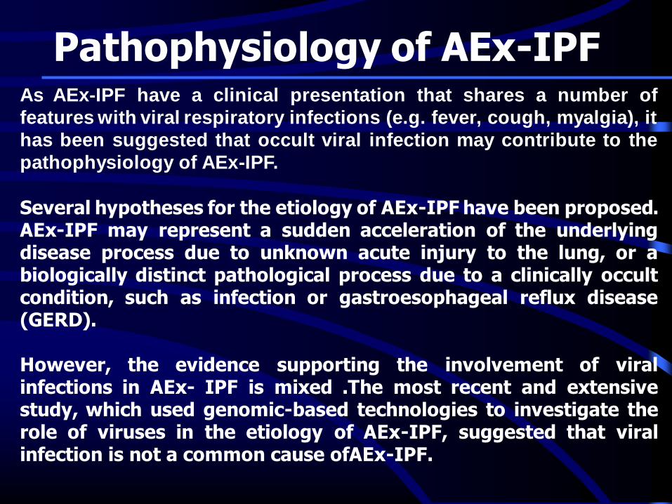
Pathophysiology of AEx-IPF As AEx-IPF have a clinical presentation that shares a number of
features with viral respiratory infections (e.g. fever, cough, myalgia), it
has been suggested that occult viral infection may contribute to the
pathophysiology of AEx-IPF.
Several hypotheses for the etiology of AEx-IPF have been proposed. AEx-IPF may represent a sudden acceleration of the underlying disease process due to unknown acute injury to the lung, or a biologically distinct pathological process due to a clinically occult condition, such as infection or gastroesophageal reflux disease (GERD). However, the evidence supporting the involvement of viral infections in AEx- IPF is mixed .The most recent and extensive study, which used genomic-based technologies to investigate the role of viruses in the etiology of AEx-IPF, suggested that viral infection is not a common cause ofAEx-IPF.
Pathophysiology of AEx-IPF Activation of the immune system, disordered coagulation/fibrinolysis, and oxidative stress may all contribute to the pathophysiology of AEx-IPF. Immune cells (e.g. neutrophils, macrophages) ,inflammatory mediators (e.g. interleukin 6, high mobility group protein B1) ,markers of coagulation/fibrinolysis (e.g. protein C, thrombomodulin, and plasma activator inhibitor-1) , and markers of oxidative stress (thioredoxin 1) are all elevated in patients with AEx-IPF. Epithelial cell damage in patients with IPF is demonstrated by over-expression of matrix metalloproteinase (MMP)-7, MMP-9 , and Krebs von den Lungen- 6 (KL-6) . Accelerated epithelial cell proliferation, with increases in the proliferation markers CCNA2 and Ki-67, in patients with AEx-IPF may be a compensatory response to injury, and is associated with epithelial cell death. Transforming growth factor (TGF)-beta, a fibrogenic cytokine, is upregulated in IPF and galectin-3, a mediator of fibrosis induced by TGF-beta, is elevated in the lungs and serum of patients with stable IPF and AEx-IPF . Circulating bone marrowderived fibrocytes may also provide a source of lung fibroblasts and myofibroblasts, as the number of circulating fibrocytes has been shown to be higher in patients with IPF and AEx-IPF, compared with healthy subjects
Risk factors and precipitating factors for AEx-IPF
*Lower total lung capacity, lower forced vital capacity (FVC) and/or lower diffusing capacity of the lung for carbon
monoxide (DLco). *A higher degree of dyspnea (score ≥2 on the modified
Medical Research Council dyspnea scale) or of fibrosis on HRCT has been shown to increase the risk of AEx-IPF, as has the
presence of concomitant conditions such as emphysema or pulmonary hypertension.
*Invasive examinations such as bronchoscopy , bronchoalveolar lavage (BAL), and pulmonary resection for
lung cancer can precipitate AEx-IPF.
Risk factors and precipitating factors for AEx-IPF
*surgical lung biopsy is a precipitating factor for AEx-IPF; however, the risk of AEx-IPF from video-assisted thoracoscopic
operation appears to be elevated only in patients with severe physiologic impairment or substantial comorbidity
*In some patients with AEx-IPF, pepsin levels were found to be
elevated in BAL fluid, suggesting a possible role for GERD in the pathogenesis of AEx-IPF .There is some evidence to suggest that the treatment of GERD in patients with IPF
reduces mortality rates .
Impact of AEx-IPF on patients
*AEx-IPF are certainly a leading cause of hospitalization and death among patients with IPF. Median survival after an AEx-
IPF has been reported to be between 22 days and 4.2 months.
*There is some evidence that patients with better lung function (FVC, PaO2, DLCO) prior to AEx-IPF are more likely to
survive an AEx-IPF , suggesting that preservation of lung function may be an important way of reducing the impact of AEx-IPF in patients with IPF.
Management of AEx-IPF
*The latest international treatment guidelines state that supportive care remains the mainstay of treatment for AEx-IPF,
but also give a weak recommendation for the treatment of the majority of patients with AEx-IPF with corticosteroids.
*In clinical practice, the treatment of AEx-IPF is variable.
Corticosteroids (e.g. prednisone, methylprednisolone) are used in the majority of patients who suffer an AEx-IPF, usually in pulse doses. Preliminary data suggest that response to high-
dose corticosteroid treatment may depend on the type of HRCT lesion, with better responses achieved in those with a
peripheral pattern
Management of AEx-IPF
*Broad-spectrum antibiotics and immunosuppressants (cyclosporin or cyclophosphamide) are sometimes used in
addition to corticosteroids .However, the efficacy of immunosuppressants in the treatment of AEx-IPF is based on a few small retrospective studies that do not provide conclusive
evidence for benefit.
*Mechanical ventilation is often used in patients with AEx-IPF, but the data on its effects on outcomes are mixed.
*Other treatments for AEx-IPF that havebeen investigated in small studies include polymyxin Bimmobilized fiber column
(PMX) hemoperfusion and tacrolimus, a cytokine transcription inhibitor ,usually administered in addition to corticosteroids.
Reducing the risk of exacerbations
*A trial of sildenafil, a phosphodiesterase-5 inhibitor, showed a numerical reduction in AEx-IPF in patients given sildenafil versus
placebo (3 [3.4%] vs. 7 [7.6%]), but the number of events was small and the difference was not statistically significant .
*Imatinib, a tyrosine kinase inhibitor , bosentan, an endothelin receptor antagonist, the anticoagulant warfarin , and inhaled
Nacetylcysteine , numerically higher rates of AEx-IPF were found in the active treatment arms compared with the placebo arms.
*triple therapy with prednisone, azathioprine,and N-acetylcysteine in patients with IPF, a significantly higher rate of
AEx-IPF was observed in patients receiving triple therapy versus placebo
Reducing the risk of exacerbations
*Pirfenidone, an anti-fibrotic molecule that has been licensed for the
treatment of IPF in Japan, India, China, Europe, and Canada, but was not approved in the United States, has shown inconsistent effects on AEx-IPF. *Nintedanib (formerly known as BIBF 1120) is a tyrosine kinase inhibitor in clinical development for the treatment of IPF. It reported a lower incidence of AEx-IPF was observed in patients treated with nintedanib 300 mg/day than placebo (2.4 vs. 15.7 AEx-IPF per 100 patient years) *It is interesting that nintedanib may have an effect on AEx-IPF whereas the tyrosine kinase inhibitor imatinib, which inhibits the platelet-derived growth factor receptor (PDGFR), did not . Nintedanib is an inhibitor of PDGFR, vascular endothelial growth factor receptor (VEGFR), and fibroblast growth factor receptor (FGFR) and this specificity of inhibition may be key to its effects on AEx-IPF
Reducing the risk of exacerbations
*Anti-acid treatment might decrease the frequency of AEx-IPF by reducing the acidity of the microaspirate .