activator of one protease transforms into inhibitor of...
TRANSCRIPT

Activator of one protease transforms into inhibitor of another in
response to nutritional signals
Jinki Yeom1 and Eduardo A. Groisman1,2
1 Department of Microbial Pathogenesis, Yale School of Medicine, 295 Congress Avenue,
New Haven, CT 06536, USA
2 Yale Microbial Sciences Institute, P.O. Box 27389, West Haven, CT, 06516, USA
Corresponding author: [email protected]
Supplementary Material
Supplemental figures S1–S8
Supplemental tables S1–S3
2
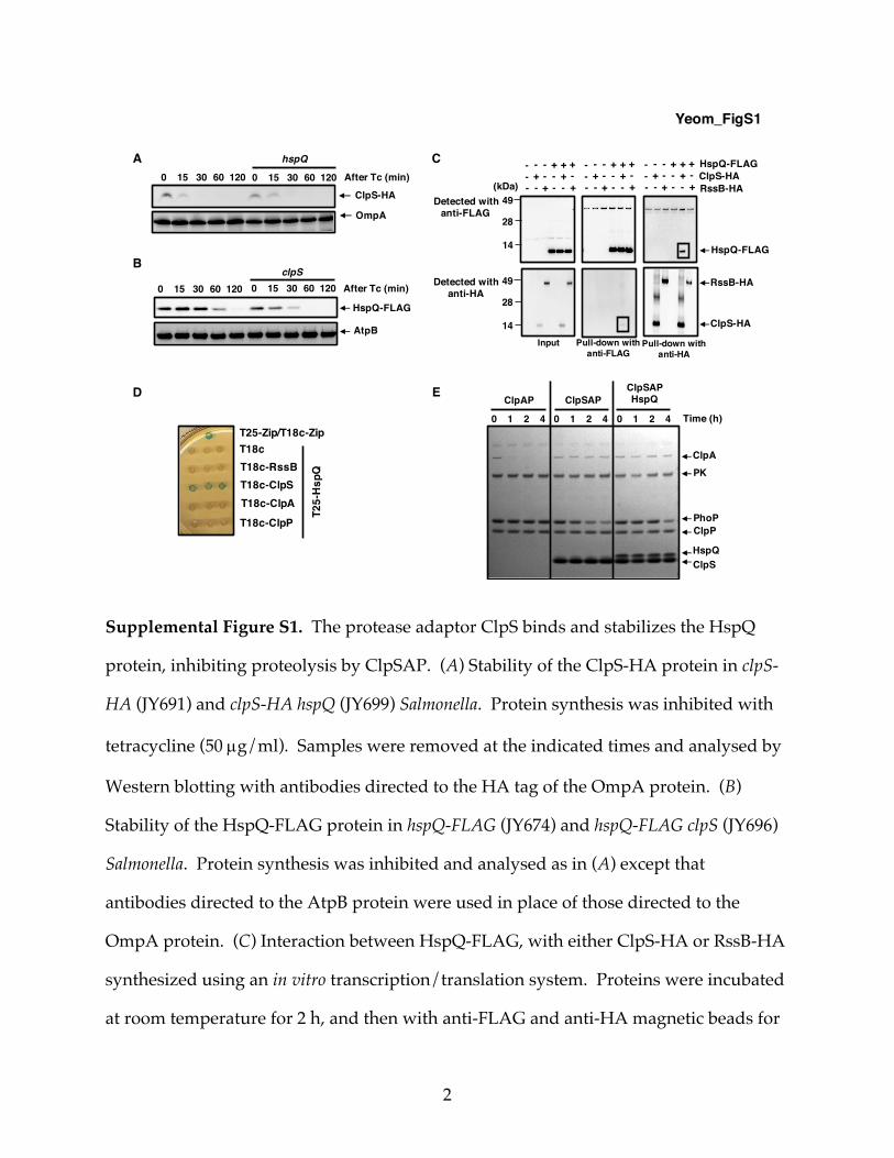
Supplemental Figure S1. The protease adaptor ClpS binds and stabilizes the HspQ
protein, inhibiting proteolysis by ClpSAP. (A) Stability of the ClpS-HA protein in clpS-
HA (JY691) and clpS-HA hspQ (JY699) Salmonella. Protein synthesis was inhibited with
tetracycline (50 µg/ml). Samples were removed at the indicated times and analysed by
Western blotting with antibodies directed to the HA tag of the OmpA protein. (B)
Stability of the HspQ-FLAG protein in hspQ-FLAG (JY674) and hspQ-FLAG clpS (JY696)
Salmonella. Protein synthesis was inhibited and analysed as in (A) except that
antibodies directed to the AtpB protein were used in place of those directed to the
OmpA protein. (C) Interaction between HspQ-FLAG, with either ClpS-HA or RssB-HA
synthesized using an in vitro transcription/translation system. Proteins were incubated
at room temperature for 2 h, and then with anti-FLAG and anti-HA magnetic beads for
A
ClpS-HA
hspQ
After Tc (min)
OmpA
0 15 30 60 120 0 15 30 60 120
HspQ-FLAG
clpS0 15 30 60 120 After Tc (min)
AtpB
0 15 30 60 120
B
ClpS-HAHspQ-FLAG
Input Pull-down withanti-HA
Pull-down withanti-FLAG
HspQ-FLAG
RssB-HA
Detected withanti-FLAG
49(kDa)
28
14
RssB-HA+ - -- + -
--
+-
-+
- - +- + ++ - -- + -
--
+-
-+
- - +- + ++ - -- + -
--
+-
-+
- - +- + +
ClpS-HA
Detected withanti-HA
49
28
14
PhoP
0 1 2 4
ClpAP
Time (h)
ClpAPK
ClpP
HspQClpS
ClpSAPClpSAPHspQ
0 1 2 4 0 1 2 4
D E
Yeom_FigS1
C
T25-
Hsp
Q
T18c
T18c-ClpST18c-RssB
T18c-ClpAT18c-ClpP
T25-Zip/T18c-Zip

3
an additional 2 h. Immunoprecipitated samples were analysed using anti-FLAG and
anti-HA antibodies. Data are representative of two independent experiments, which
gave similar results. Boxes highlight relevant bands. (D) Spots of E. coli strain BTH101
harboring plasmid pKT25-HspQ with either pUT18c-RssB, pUT18c-ClpS, pUT18c-ClpA
or pUT18c-ClpP. Negative control strains carry the plasmid vector along with pKT25-
HspQ. Positive control strains carry pKT25-Zip and pUT18c-Zip. (E) SDS-PAGE
analysis for time course in vitro degradation of the PhoP protein (0.5 µM) mixed with
ClpA (0.08 µM), ClpP (0.2 µM) in the absence or presence of ClpS (1.0 µM) and HspQ
(0.5 µM). All reactions were carried out at 30˚C for the indicated times in the presence
of an ATP regeneration system and started by the addition of substrates. After
incubation, protein amounts were determined by Coomassie-staining following
separation on a 4-12% SDS-PAGE gel. Data are representative of two independent
experiments, which gave similar results. See also Fig. 2.

4
Supplemental Figure S2. The Salmonella HspQ protein is a Lon substrate and Lon-
enhancing factor. (A) SDS-PAGE analysis for time course in vitro degradation of the
HspQ and Hha proteins. His-Hha (0.5 µM) was mixed with Lon (0.2 µM) in the absence
or presence of HspQ (0.5 µM). Reactions were carried out at 30˚C for the indicated
times in the presence of an ATP regeneration system and started by the addition of
substrates. After incubation, protein amounts were determined by Coomassie-staining
following separation on a 4-12% SDS-PAGE gel. Data are representative of three
independent experiments, which gave similar results. (B) Degradation of the His-Hha
protein in (A) was determined by quantification of bands. Relative His-Hha levels were
calculated from three independent experiments. (C) Western blot analysis of crude
extracts from hspQ-FLAG (JY674), hspQ-FLAG clpS (JY696), hspQ-FLAG clpA (JY741),
hspQ-FLAG clpX (JY701) and hspQ-FLAG lon (JY703) Salmonella. Samples were analysed
using antibodies directed to the FLAG tag of the AtpB protein. Data are representative
HspQ
0 1 2 4 Time (h)
Lon
PK
His-Hha
LonHha
0 1 2 4 0 1 2 4 0 1 2 4
LonHspQ
LonHspQ+HhaHha
A
HspQ-FLAG
AtpB
C
Yeom_FigS2
B
0 1 2 3 40
20406080
100120
Time (h)
His-
Hha
rem
aing
pro
tein
s (%
of i
nitia
l)
Hha
Hha + Lon
Hha + HspQ + Lon
1.0 0.4 0.8 1.0 4.4
PhoP
Time (h)
LonPK
HspQ
LonLon
HspQ0 1 2 4 0 1 2 4
D

5
of three independent experiments, which gave similar results. (D) SDS-PAGE analysis
for time course in vitro degradation of the PhoP protein. PhoP (0.5 µM) was mixed with
Lon (0.2 µM) in the absence or presence of HspQ (0.3 µM). Reactions were carried out
at 30˚C for the indicated times in the presence of an ATP regeneration system and
started by the addition of substrates. After incubation, protein amounts were
determined by Coomassie-staining following separation on a 4-12% SDS-PAGE gel.
Data are representative of two independent experiments, which gave similar results.
See also Fig. 2.

6
Supplemental Figure S3. Identification of the HspQ acetylation site, predicted
structures of the Salmonella HspQ and Qad proteins, and genetic organization of the
hspQ and qad genes. (A) High-resolution tandem mass spectrometry analysis of the
HspQ protein revealed acetylation at Lys96 (red arrow). (B) Structural model of the
Salmonella HspQ protein based on the structure of the Escherichia coli HspQ protein
(PDB: 5ycq). Acetylated Lys96 is shown in red. (C) Diagram of the qad (STM14_1223)-
hspQ chromosomal region. (D) The Salmonella qad gene specifies a 136 amino acid long
protein predicted to be a CoA binding domain (NCBI: COG1832). (E) Structural model
of the Salmonella Qad protein based on the structure of the Klebsiella Qad protein (PDB:
2duw). See also Fig. 3.
11/21/16, 5:11 PM
Page 1 of 1file:///Users/jinkiyeom/Desktop/__data_20161116_F498267.dat_q936_p1_mz=10.07-830.83_scoring.svg
AC
K Q L
b2
Q
b3
A
b4
P
y1
b5y(
1)
b(3)
b(2)
b*(4
)b(
4)
b(5)
200 400 600 800
m/z
0
20
40
60
80
100
% o
f bas
e pe
ak
0.0e+0
5.0e+3
1.0e+4
1.5e+4
2.0e+4
2.5e+4
3.0e+4
ion current
Lys96
D E
CoA
B
C
Qad
1 50 100 136
CoA binding domain
A
qad hspQ
Yeom_FigS3

7
Supplemental Figure S4. Acetylation of HspQ is critical for Lon-mediated
degradation. (A-C) Degradation of the HspQ (A), His-Hha (B) and PhoP (C) proteins in
Fig. 3F was determined by quantification of bands. Relative protein levels were
calculated from two independent experiments. (D) SDS-PAGE analysis for time course
in vitro degradation of PhoP (0.5 µM) and Hha (0.3 µM) in absence of acetyl-CoA.
Substrates were mixed with ClpA (0.08 µM), ClpP (0.2 µM), ClpS (0.5 µM) and/or Lon
(0.2 µM). HspQ acetylation was achieved by preincubating HspQ (0.5 µM) with Qad
(0.5 µM) and Pat (0.2 µM) at 37˚C for 3 h. Reactions were carried out at 30˚C for the
indicated times in the presence of an ATP regeneration system and started by the
addition of substrates. After incubation, protein amounts were determined by
Coomassie-staining following separation on a 4-12% SDS-PAGE gel. Data are
A
C
0 1 2 3 40
20406080
100120
Time (h)
HspQ
rem
aing
pro
tein
s (%
of i
nitia
l)HspQ + LonHha + HspQ + LonHspQAc + LonHha + HspQAc + Lon
0 1 2 3 40
20406080
100120
Time (h)
Hha
rem
aing
pro
tein
s (%
of i
nitia
l)
Hha + HspQ + Lon
Hha + HspQAc + Lon
0 1 2 3 40
20406080
100120
Time (h)
PhoP
rem
aing
pro
tein
s (%
of i
nitia
l) ClpAPSHspQ + ClpAPSLon + HspQ + ClpAPSHha + Lon + HspQ + ClpAPSHha + Lon + HspQAC + ClpAPS
B
DYeom_FigS4
0 1 2 4
HspQ
Hha
Time (h)
PK
ClpAPSHspQLon
Lon
ClpAPSHspQ
Hha
Lon
ClpS
ClpPPhoP
ClpA
0 1 2 4 0 1 2 4 0 1 2 4 0 1 2 4 0 1 2 4Pat
Qad
ClpAPSHspQLon
HspQLon
HhaHspQLon
Lon

8
representative of two independent experiments, which gave similar results. See also
Fig. 3.
9
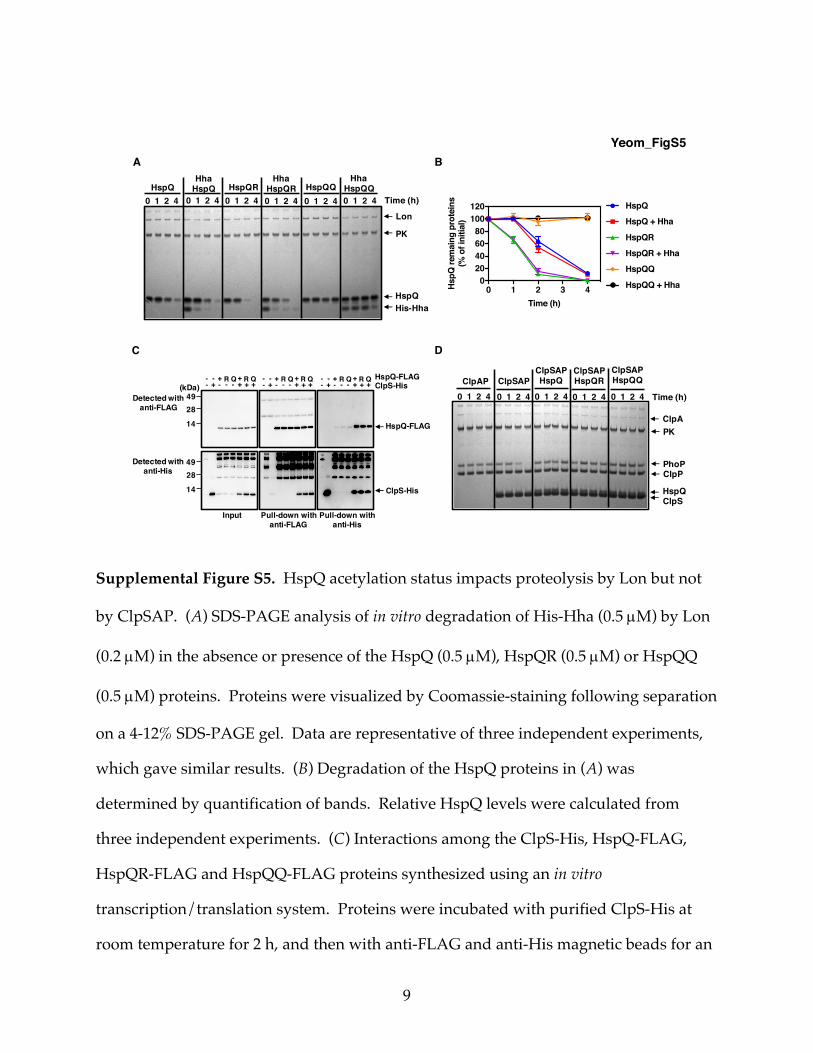
Supplemental Figure S5. HspQ acetylation status impacts proteolysis by Lon but not
by ClpSAP. (A) SDS-PAGE analysis of in vitro degradation of His-Hha (0.5 µM) by Lon
(0.2 µM) in the absence or presence of the HspQ (0.5 µM), HspQR (0.5 µM) or HspQQ
(0.5 µM) proteins. Proteins were visualized by Coomassie-staining following separation
on a 4-12% SDS-PAGE gel. Data are representative of three independent experiments,
which gave similar results. (B) Degradation of the HspQ proteins in (A) was
determined by quantification of bands. Relative HspQ levels were calculated from
three independent experiments. (C) Interactions among the ClpS-His, HspQ-FLAG,
HspQR-FLAG and HspQQ-FLAG proteins synthesized using an in vitro
transcription/translation system. Proteins were incubated with purified ClpS-His at
room temperature for 2 h, and then with anti-FLAG and anti-His magnetic beads for an
A B
PhoP
ClpAP0 1 2 4 Time (h)
ClpAPK
ClpP
HspQClpS
ClpSAPClpSAPHspQ
ClpSAPHspQR
ClpSAPHspQQ
0 1 2 4 0 1 2 4 0 1 2 4 0 1 2 4ClpS-HisHspQ-FLAG
Input Pull-down withanti-His
Pull-down withanti-FLAG
HspQ-FLAG
Detected withanti-FLAG
49(kDa)
28
14
Detected withanti-His
4928
14 ClpS-His
-- ++-
- + ++ R Q-
-+
R Q-- +
+-- + +
+ R Q-
-+
R Q-- +
+-- + +
+ R Q-
-+
R Q
HspQ
0 1 2 4 Time (h)
Lon
PK
His-Hha
HspQHha
HspQ HspQRHha
HspQR HspQQHha
HspQQ0 1 2 4 0 1 2 4 0 1 2 4 0 1 2 4 0 1 2 4
C
Yeom_FigS5
D
0 1 2 3 40
20406080
100120
Time (h)
HspQ
rem
aing
pro
tein
s (%
of i
nitia
l)
HspQ
HspQ + Hha
HspQR
HspQR + Hha
HspQQ
HspQQ + Hha

10
additional 2 h. Immunoprecipitated samples were analysed using anti-FLAG and anti-
His antibodies. Data are representative of two independent experiments, which gave
similar results. (D) SDS-PAGE analysis of time course in vitro incubation of the PhoP
protein (0.5 µM) with ClpA (0.08 µM) and ClpP (0.2 µM) in the absence of ClpS, or with
ClpS (1.0 µM) and no HspQ, HspQ (0.5 µM), HspQR (0.5 µM) or HspQQ (0.5 µM).
Substrate degradation assay was analysed as in (A). Data are representative of three
independent experiments, which gave similar results. See also Fig. 3 and 4.

11
Supplemental Figure S6. The HspQ protein does not alter the abundance of ClpS-
independent ClpAP substrates. (A) Stability of the AcnB protein in wild-type (14028s),
clpX (JY649), clpA (JY650) and clpS (JY651) Salmonella. Protein synthesis was inhibited
with tetracycline (50 µg/ml). Samples were removed at the indicated times and
analysed by Western blotting with antibodies directed to the AcnB protein. Data are
representative of three independent experiments, which gave similar results. (B)
Western blot analysis of crude extracts prepared from oat-FLAG (JY655), oat-FLAG clpS
(JY657), oat-FLAG hspQ (JY686) and oat-FLAG qad (JY774) Salmonella with a gfp-laa
expressing plasmid (pFPV25-gfp-laa). Samples were analysed using antibodies directed
to the FLAG tag, the GFP protein or the GroEL protein. Data are representative of two
independent experiments, which gave similar results. (C) Western blot analysis of
crude extracts prepared from oat-FLAG (JY655), oat-FLAG clpS (JY657) and oat-FLAG
0 1 2 4
clpX
After Tc (h)0 1 2 4 0 1 2 4 0 1 2 4
clpA clpSwild-type
Oat-FLAG
GroEL
AcnBOat-FLAG
GFP-LAA
GroEL
A
B C
AcnB
Yeom_FigS6
0 10 30 60
hspQ
After Tc (min)
clpPwild-typeD
GFP-LAA0 10 30 60 0 10 30 60

12
clpA (JY902) Salmonella. Samples were analysed using antibodies directed to the FLAG
tag, the AcnB protein or the GroEL protein. Data are representative of three
independent experiments, which gave similar results. (D) Stability of the GFP-LAA
protein in wild-type (14028s), hspQ (JY683) and clpP (JY186) Salmonella with a gfp-laa
expressing plasmid (pFPV25-gfp-laa). Protein synthesis was inhibited with tetracycline
(50 µg/ml). Samples were removed at the indicated times and analysed by Western
blotting with antibodies directed to the GFP protein. Data are representative of two
independent experiments, which gave similar results. See also Fig. 5.

13
Supplemental Figure S7. mRNA abundances of the oat and hha genes are similar in
wild-type, hspQ, and qad Salmonella when grown on glucose or glycerol. (A) mRNA
abundance of the oat gene produced by oat-FLAG (JY655), oat-FLAG hspQ (JY686), and
oat-FLAG qad (JY774) Salmonella grown on glycerol or glucose. (B) mRNA abundance of
the hha gene produced by oat-FLAG (JY655), oat-FLAG hspQ (JY686), and oat-FLAG qad
(JY774) Salmonella with a His-hha-expressing plasmid (pHis-hha) following growth on
glycerol or glucose. his-hha transcription from pHis-hha was induced with IPTG (100
µM). For all qRT-PCR analysis, mRNA abundance was normalized to those of the ompA
gene. The sequence of the primers used in qRT-PCR are presented in Table S3. Data
shown are the mean and SD from three independent experiments. See also Fig. 6.
wild-ty
pehspQ qa
d
wild-ty
pehspQ qa
d0.0
0.2
0.4
0.6
0.8
oat
mRN
A ab
unda
nce
glycerol glucose
wild-ty
pehspQ qa
d
wild-ty
pehspQ qa
d0
1
2
3
hha
mRN
A ab
unda
nce
glycerol glucose
A B
Yeom_FigS7

14
Supplemental Figure S8. The deduced amino acid sequence of the qad gene is
conserved among members of the family Enterobacteriaceae. Alignment of the deduced
amino acid sequences of the hspQ genes from the listed species of the family
Enterobacteriaceae. Red arrow indicates Lys96, which is acetylated in the Salmonella
HspQ protein. Y. pestis harbors a His at position 96.
CLUSTAL 2.1 MULTIPLE SEQUENCE ALIGNMENTFile: /Users/younsoosim/Desktop/hspQ/STM.ps Date: Fri Oct 5 13:49:27 2018 Page 1 of 1
**:**:********.* *:***::*:** *** * *::* * **: ******:*.::* .:********* * : *****::**** :**:*****:***ACY87717.1 MIASKFGIGQQVRHSLLGYLGVVVDIDPEYSLDEPSPDELAVNDELRAAPWYHVVMEDDDGQPVHTYLAEAQLRSEMRDEHPEQPSMDELARTIRKQLQAPRLRN 105CCC29975.1 MIASKFGIGQQVRHSLLGYLGVVVDIDPEYSLDEPSPDELAVNDELRAAPWYHVVMEDDDGQPVHTYLAEAQLRSEMRDEHPEQPSMDELARTIRKQLQAPRLRN 105AIZ91912.1 MIASKFGIGQQVRHSLLGYLGVVVDIDPVYSLSEPSPDELAVNDELRAAPWYHVVMEDDNGLPVHTYLAEAQLSSELQDEHPEQPSMDELAQTIRKQLQAPRLRN 105CDX06253.1 MIASKFGIGQQVRHSLLGYLGVVVDIDPVYSLSEPSPDELAVNDELRAAPWYHVVMEDDNGLPVHTYLAEAQLSSELQDEHPEQPSMDELAQTIRKQLQAPRLRN 105CBG87807.1 MIASKFGIGQQVRHSLLGYLGVVMDIDPVYSLEEPSPDELAVNDELRAAPWYHVVMEDDDGQPVHTYLAEAQLRSETQDEHPEQPSMDELAQTIRKQLQAPRLRN 105KML25937.1 MIASKFGIGQQVRHTLLGYLGVVVDIDPEYSLDEPSADELAVNAELRAAPWYHVVMEGDDGQPVHTYLAEAQLSSELQEEHPEQPTMDELAQTIRKQLQAPRLRN 105AVH16492.1 MIASKFGIGQQVRHTLLGYLGVVVDIDPEYSLDEPSADELAVNAELRAAPWYHVVMEGDDGQPVHTYLAEAQLSSELQEEHPEQPTMDELAQTIRKQLQAPRLRN 105ADF62221.1 MIASKFGIGQQVRHTLLGYLGVVVDIDPEYSLDEPSADELAVNAELRAAPWYHVVMEGDDGQPVHTYLAEAQLSSELQDEHPEQPTMDELAQTIRKQLQAPRLRN 105AHJ74447.1 MIASKFGIGQQVRHSLLGYLGVVVDIDPEYSLDEPSADELAVNDELRALPWYHVVMEDDDGQPVHTYLAEAQLTSEITDEHPEQPSMDELARTIRRQLQAPRLRN 105WP_062777489.1 MITSKFGIGQQVRHSLLGYLGVVVDIDPVYSLDEPEPDDLAANDELRALPWYHVVMEDDEGQPMHTYLAEAQLSSEPRDDHPEQPTMDELARTIRRQLQAPRLRN 105AIR03971.1 MIASKYGIGQQVRHSLLGYLGVVVDIDPEYSLDEPQEDDLADNSALRAAPWYHVVMEDDDGQAIHTYLAEAQLSSEDDDEHPEQPSMDELAASIRQQLQAPRLRN 105ALB51068.1 MIASKFGIGQQVRHSLLGYLGVVVDIDPEYSLDEPEVDELAVNAELRAAPWYHVVMEDDDGQPVHTYLAEAQLSGEMQEEHPEQPSMDELARSIRQQLQAPRLRN 105AJF72496.1 MIASKFGIGQQVRHTLLGFLGVVVDIDPEYSLAEPAEDEIAANDELRALPWYHVVMEDEDGQPVHTYLAEAQLSSEPSDEHPEQPSMDELARTIRQQLQAPRLRN 105APS95917.1 MIASKFGIGQQVRHTLLGYLGVIVDVDPEYSLAEPEEDEIAANDELRAAPWYHVVMEDDDGQPIHTYLAEAQLSSETRDEHPEQPSLDELAKTIRQQLQAPRLRN 105AVR06007.1 MIASKFGIGQQVRHSLLGYLGVIVDIDPEYSLGAPDADEIAGNDALRAAPWYHVVMEDDDGQPVHTYLAEAQLRGEAHDEHPEQPSMDELAQTIRRQLQAPRLRN 105ASL97103.1 MIASKFGIGQQVRHKLLGYLGVVIDIDPEYSLEQPKADEIAANDELRSAPWYHVVMEDEEGQPVHTYLAEAQLDSEAQEAHPEQPSLDELAESIRHQLQAPRLRN 105[Yersinia.fasta] MIASKFGIGQQVRHSLHGYLGVVIDIDPEYSLAPPEPDEVANNKTLRSSPWYHVVIEDDDGQPVHTYLAEAQLTYEDVDAHPEQPSLDELAASIRHQLQAPHLRN 105 1.......10........20........30........40........50........60........70........80........90.......100.....
Lelliottia nimipressuralisLeclercia adecarboxylata
Raoultella ornithinolytica
Salmonella entericaSalmonella bongori
Escherichia coliShigella flexneri
Citrobacter rodentium
Kluyvera intermediaKosakonia sacchari
Enterobacter cloacae
Cronobacter sakazakiiCedecea neteri
Klebsiella pneumoniae Pluralibacter gergoviae
Serratia marcescensYersinia pestis
Yeom_FigS8

15
Supplemental Table S1. Genome analysis of Enterobacteriaceae species with
conserved asynteny between the hspQ and qad genes.
1 tBLASTn analysis for conserved qad and hspQ in bacterial genomes.
Enteric bacteria species in which qad and hspQ are conserved1 Lys96 residue in HspQ
Salmonella enterica O
Salmonella bongori O
Escherichia coli O
Citrobacter rodentium O
Shigella flexneri O
Enterobacter cloacae O
Cronobacter sakazakii X
Klebsiella pneumoniae X
Serratia marcescens X
Yersinia pestis X
Kosakonia sacchari X
Kluyvera intermedia X
Lelliottia nimipressuralis O
Leclercia adecarboxylata O
Raoultella ornithinolytica X
Pluralibacter gergoviae X
Cedecea neteri X

16
Supplemental Table S2. Bacterial strains and plasmids used in this study.
Strains Relevant characteristics Source
Escherichia coli
BL21 (DE3) F- ompT hsdS gal [lon] [dcm] (Its857 ind1 Sam7 nin5 lacUV5-T7 gene 1) (Studier and Moffatt, 1986)
DH5a Host strain used for generation and propagation of plasmid constructs Life Technologies
Salmonella enterica serovar Typhimurium
14028s wild-type (Fields et al., 1989)
EG13917 phoP-HA::cm (Shin and Groisman, 2005)
EG16039 lon::cm This study
JY186 clpP::cm This study
JY649 DclpX (Yeom et al., 2017)
JY650 DclpA (Yeom et al., 2017)
JY651 DclpS (Yeom et al., 2017)
JY655 oat-FLAG (Yeom et al., 2017)
JY657 oat-FLAG / clpS (Yeom et al., 2017)
JY674 hspQ-FLAG This study
JY683 hspQ::cm This study

17
JY686 oat-FLAG / hspQ This study
JY687 oat-FLAG / clpS / hspQ This study
JY691 clpS-HA (Yeom et al, 2018)
JY692 hspQ-FLAG / clpS-HA This study
JY694 clpA-HA (Yeom et al, 2018)
JY695 hspQ-FLAG / clpA-HA This study
JY696 hspQ-FLAG / clpS This study
JY699 clpS-HA / hspQ This study
JY700 clpA-HA / hspQ This study
JY701 hspQ-FLAG / clpX This study
JY703 hspQ-FLAG / lon This study
JY705 oat-FLAG / phoP-HA / qad This study
JY740 hspQ-FLAG / qad This study
JY741 hspQ-FLAG / clpA This study
JY754 hspQ-FLAG / lon / qad This study
JY772 oat-FLAG / clpS / hspQ / qad This study
JY773 oat-FLAG / hspQ / qad This study
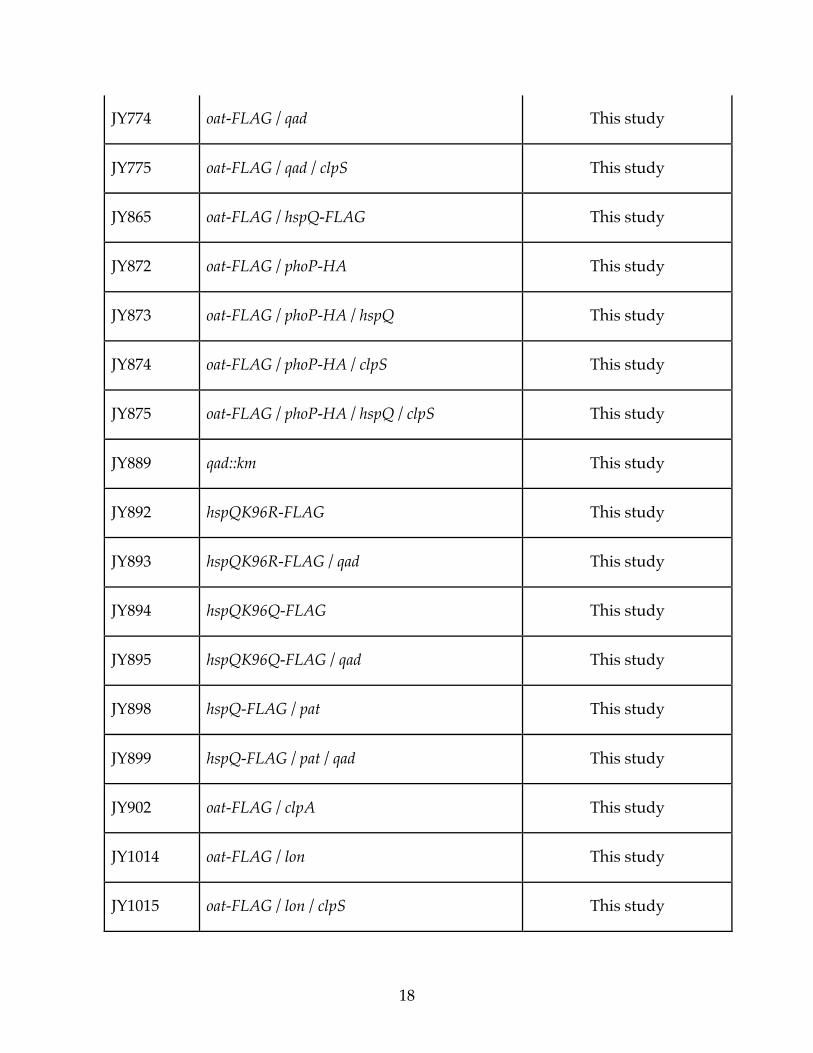
18
JY774 oat-FLAG / qad This study
JY775 oat-FLAG / qad / clpS This study
JY865 oat-FLAG / hspQ-FLAG This study
JY872 oat-FLAG / phoP-HA This study
JY873 oat-FLAG / phoP-HA / hspQ This study
JY874 oat-FLAG / phoP-HA / clpS This study
JY875 oat-FLAG / phoP-HA / hspQ / clpS This study
JY889 qad::km This study
JY892 hspQK96R-FLAG This study
JY893 hspQK96R-FLAG / qad This study
JY894 hspQK96Q-FLAG This study
JY895 hspQK96Q-FLAG / qad This study
JY898 hspQ-FLAG / pat This study
JY899 hspQ-FLAG / pat / qad This study
JY902 oat-FLAG / clpA This study
JY1014 oat-FLAG / lon This study
JY1015 oat-FLAG / lon / clpS This study

19
JY1016 oat-FLAG / lon / hspQ This study
JY1017 oat-FLAG / qad / lon This study
JY1018 oat-FLAG / lon / hspQ / qad This study
JY1019 hspQ-FLAG / lon / clpS This study
JY2000 hspQK96R-FLAG /lon This study
JY2001 hspQK96Q-FLAG /lon This study
JY2002 oat-FLAG / hspQK96R-FLAG This study
JY2003 oat-FLAG / hspQK96Q-FLAG This study
Plasmids
pKD46 reppSC101ts AmpR ParaBAD-gbexo (Datsenko and Wanner,
2000)
pKD3 repR6Kg AmpR FRT CmR FRT (Datsenko and Wanner, 2000)
pKD4 repR6Kg AmpR FRT KmR FRT (Datsenko and Wanner, 2000)
pCP20 reppSC101ts l cI857 FLP AmpR CmR (Datsenko and Wanner,
2000)
pUHE-21-2-lacIq reppMB1 lacIq AmpR (Soncini et al., 1996)
pUHE-hspQ reppMB1 lacIq AmpR Plac-hspQ This study
pUHE-qad reppMB1 lacIq AmpR Plac-qad This study
pUHE-His-lon reppMB1 lacIq AmpR Plac-his-lon This study
20
pUHE-His-hha reppMB1 lacIq AmpR Plac-his-hha This study
pUHE-ftsA-FLAG reppMB1 lacIq AmpR Plac-ftsA-FLAG (Yeom et al, 2018)
pFPV25-gfp pBR332 AmpR p-gfpmut3 (Valdivia and Falkow,
1996)
pFPV25-gfp-laa pBR332 AmpR p-gfpmut3-laa This study
pET28+a pBR332 lacI KmR pT7 Novagen
pET28+a-clpS pBR332 lacI KmR pT7- clpS (Yeom et al, 2018)
pET28+a-clpA pBR332 lacI KmR pT7- clpA (Yeom et al, 2018)
pET28+a-clpP pBR332 lacI KmR pT7- clpP (Yeom et al, 2018)
pET28+a-hspQ pBR332 lacI KmR pT7- hspQ This study
pET28+a-qad pBR332 lacI KmR pT7- qad This study
pET28+a-pat pBR332 lacI KmR pT7- pat This study

21
Supplemental Table S3. Primers used for strain or plasmid constructions or qRT-PCR
or to generate templates for in vitro transcription/translation.
No. Purpose Sequence (from 5' to 3')
1550 Amplification of gfp-laa gene for unstable GFP expression CACCTGACGTCTAAGAAACC
3071 Amplification of gfp-laa gene for unstable GFP expression
CATTAAAGCTTGCATGCCTGCAGGAGATTTAAGCTGCTAAAGCGTAGTTTTCGTCGTTTGCTGCAGGCCTTTTGTATAGTTCATCCATGC
3815 lon inactivation in Salmonella ATCTGATTACCTGGCGGACACTAAACTAAGAGAGAGCTCTTGTAGGCTGGAGCTGCTTCG
3816 lon inactivation in Salmonella TGCCAGCCCTGTTTTTATTAGCGCTATTTGCGCGAGGTCACATATGAATATCCTCCTTAG
15054 qRT-PCR of ompA gene in Salmonella GGGCTGGTCTCAGTACCATGA
15055 qRT-PCR of ompA gene in Salmonella TCATGAGTCGGGCCATCA
15949 In vitro synthesis of ClpS-HA from Salmonella
GCGAATTAATACGACTCACTATAGGGCTTAAGTATAAGGAGGAAAAAATATGGGTAAGACGAACGATTGGCTGGATTTTGACCAGTTGGTGGAA
15966 qRT-PCR of oat gene in Salmonella GGCCCTCTGGAGCTCATTTT
15967 qRT-PCR of oat gene in Salmonella GGTTTAGCGTTCGCTTCTCG
16044 Insertion FLAG tag at C-terminus of hspQ gene in
Salmonella
CAAGCAGCTTCAGGCGCCGCGACTACGTAACGACTACAAGGACGACGATGACAAGTGAGTGTAGGCTGGAGCTGCTTC
16045 Insertion FLAG tag at C-terminus of hspQ gene in
Salmonella
GTTTAAATGGCCGGATATCGCTTTCCGGCCTTTTAAATACATATGAATATCCTCCTTAGT
16046 hspQ inactivation in Salmonella
TATAAAGGGTATCTATTTCCCGGGAGGTGACTGTGTAGGCTGGAGCTGCTTC
16047 hspQ inactivation in Salmonella
AATGGCCGGATATCGCTTTCCGGCCTTTTAAATACAATATGAATATCCTCCTTAGT
22
16052 Amplification of hspQ gene
from Salmonella for expression
GCGGGATCCAATGATAGCCAGCAAATTCGGTATCG
16053 Amplification of hspQ gene
from Salmonella for expression
GCGAAGCTTTCAGTTACGTAGTCGCGGCGCCTGAAGCTG
16284 Amplification of qad gene
from Salmonella for protein purification
CGCGGATCCATGATGAAAGAGACCGATATTGCTGATGTTTTGACG
16285 Amplification of qad gene
from Salmonella for protein purification
CTCCTCGAGTTTTCGCCAGCCCCAGGCGAGGGATCTCAATCGCCG
16286 Amplification of qad gene
from Salmonella for expression
GCGGGATCCAATGATGAAAGAGACCGATATTGCTGATGTTTTGA
16287 Amplification of qad gene
from Salmonella for expression
GCGAAGCTTTTATTTCGCCAGCCCCAGGCGAGGGATCTCAATCG
16313 Amplification of pat gene
from Salmonella for protein purification
CGCGGATCCATGAGCCAGCAAGGACTGGAAGCGCTACTGCGACC
16314 Amplification of pat gene
from Salmonella for protein purification
CGCCTCGAGTCACGATTCATCACATTTGGCCAGATTCAGCGT
16629 Amplification of his-hha gene
from Salmonella for expression
GCGGGATCCAATGCATCATCACCATCACCACTCTGATAAACCATTAACTAAAACTGATTA
16630 Amplification of his-hha gene
from Salmonella for expression
GCGAAGCTTTTAACGAATGAATTTCCATACTGAAGAGGG
16924 qad inactivation in Salmonella TTTGTCGTACACTTTGCAAAACAGCCAGGAGAAAG GTGTAGGCTGGAGCTGCTTC
16925 qad inactivation in Salmonella TTAAAAGGCCGGAAAGCGATATCCGGCCATTTAAACTT TATGAATATCCTCCTTAGT
16926 pat inactivation in Salmonella GTTTAAAATTATCCGGTCACTTCTGTGTAAGGGAAACCGGT GTGTAGGCTGGAGCTGCTTC
16927 pat inactivation in Salmonella TCAGTACCCGTTAAAGTGGTCAACATTTCCAGTACATTAC TATGAATATCCTCCTTAGT
16928 Substitution of lysine with arginine at position 96 in
Salmonella HspQ
TGAACTGGCGCGTACCATTCGCCGTCAGCTTCAGGCGCCGCGACTACGTAACGACTACAAGGACGACGATGACAAGTGA GTGTAGGCTGGAGCTGCTTC
23
16929 Substitution of lysine with glutamine at position 96 in
Salmonella HspQ
TGAACTGGCGCGTACCATTCGCCAGCAGCTTCAGGCGCCGCGACTACGTAACGACTACAAGGACGACGATGACAAGTGA GTGTAGGCTGGAGCTGCTTC
16934 Amplification of hspQ gene from Salmonella for protein
purification GCGCATATGATGATAGCCAGCAAATTCGGTATCGGCCAACAGGTCCGCC
16935 Amplification of hspQ gene from Salmonella for protein
purification GCGAAGCTTTCAGTTACGTAGTCGCGGCGCCTGAAGCTGCTTGCGAATG
16936 In vitro synthesis of HspQ-FLAG from Salmonella
GCGAATTAATACGACTCACTATAGGGCTTAAGTATAAGGAGGAAAAAATATGATAGCCAGCAAATTCGGTATCGGCCAACAGGT
16937 In vitro synthesis of HspQ-FLAG from Salmonella
AAACCCCTCCGTTTAGAGAGGGGTTATGCTAGTCACTTGTCATCGTCGTCCTTGTAGTCGTTACGTAGTCGCGGCGCCTGAAGCTGCTTGCGAA
16938 In vitro synthesis of ClpS-HA from Salmonella
AAACCCCTCCGTTTAGAGAGGGGTTATGCTAGTCAAGCGTAATCTGGAACATCGTATGGGTAGGCTTTTTCCAGCGTACACAGCAACGG
16939 In vitro synthesis of RssB-HA from Salmonella
GCGAATTAATACGACTCACTATAGGGCTTAAGTATAAGGAGGAAAAAATATGACGCAGCCATTGGTCGGAAAACAGATTCTTATTGTTG
16940 In vitro synthesis of RssB-HA from Salmonella
AAACCCCTCCGTTTAGAGAGGGGTTATGCTAGTCAAGCGTAATCTGGAACATCGTATGGGTATTCCGCAGACAACATCAATCGCAGACGCCCTCCTGCGCCC
17202 qRT-PCR of hha gene in Salmonella TACAGCCAGCTCATTGTCGG
17203 qRT-PCR of hha gene in Salmonella TTTGATGCGTTTACGGCGCT
20000 clpP inactivation in Salmonella
ATGTCATACAGCGGAGAACGAGATAATTTGGCCCCTCATAGTGTAGGCTGGAGCTGCTTC
20001 clpP inactivation in Salmonella
TCAATTACGATGGGTCAAAATTGAGTCAACCAAACCGTACTATGAATATCCTCCTTAGT