achieving secrecy for images using in-vivo dna cloning techniques
TRANSCRIPT

8/13/2019 Achieving Secrecy for Images Using in-vivo DNA Cloning Techniques
http://slidepdf.com/reader/full/achieving-secrecy-for-images-using-in-vivo-dna-cloning-techniques 1/10
International Journal of Advanced Computer Science, Vol. 1, No. 6, Pp. 240-249, Dec. 2011.
ManuscriptReceived:
25, Sep., 2011
Revised:
21, Nov., 2011
Accepted:
15, Dec., 2011
Published: 15, Jan., 2012
Keywords bacteriophage
lambda,
watermarking,
Information
hiding,
DNA
computing,
E.coli
bacteria,
Data deletion
Cloning,
Mutation
Abstract Genetically engineered
machines take advantage of the
computational power of DNA and seem to be
promising in the future of computers. In this
paper a solution has been proposed and
simulated for securing images in the context
of DNA computing. A total of three
genetically engineered machines have been
proposed for hiding, watermarking and
deletion of images using cloning techniques
in genetic engineering. Our proposed
methods have numerous advantages over the
previously proposed methods. In the
proposed watermarking scheme the size of
the watermark picture, in contrast to the
previously proposed schemes has no limit in
size and takes place naturally and the
proposed in-vivo data deletion procedure
utilizes a one-sided natural process and in
spite of the case for data deletion in silicon
computers it is guaranteed to be leakage
free.
1. Introduction
Biomolecules constitute fundamental basis for
architecture of life on the earth. Their great potential can be
easily versified by observing the complexity of different
organisms on our planet. Of these molecules, DNA
(deoxyribonucleic acid) is assumed as the biochemical basis
of heredity in all organisms and in spite of being
constructed from only four kinds of nucleotides, it isconjectured as the reason for all the existing complexity and
diversity in living beings of nature.
The physical potential of DNA molecules for
computational purposes was first shown by Adleman [1]during his historical experiment of solving the Hamiltonian
path problem which is an NP-complete problem using
synthetic DNA parts and appropriate encoding and
programming these molecules to find the answer of this problem. He discovered some intrinsic properties associated
with DNA molecules such as the ability to hide information
or the massive parallelism inherent in these molecules.
Based on the work of Kari et al., the ability of DNA
This work was done in Iran University of Science and Technology and
was financially supported by the Iran Telecommunication Research Centre
(ITRC).
Arash Karimi and Hadi Shahriar Shahhoseini are with Iran University
of Science and Technology (Emails: [email protected],
molecules to do computations dates back to millions ofyears ago when a species of protozoa (ciliated protozoa)
solved a problem like finding a Hamiltonian path in a graph
[2], [3]. The outstanding work of Tom Head [4] in finding
the Turing powerful potential of operation of splicing theDNA strands supports the ideas of Adleman from his
practical experiments that it may be possible to build a
universal Turing machine based on operations and materials
constructed merely from biological parts. Based on thesetheoretical basis as well as practical and experimental
abilities of genetic engineering and biotechnological
discipline, many efforts have been accomplished to
construct finite automata and simple Turing machines from biological units and based on genetic engineering principles.
Some DNA computing models were also proposed which
simulate Turing machines. Some of the most outstanding
works in this direction can be stated as follows. In [5] somechallenges in designing a molecular computer are discussed.
In [6] mutagenesis as the basis for designing and
implementation of molecular computers is considered. The
first leap towards implementation of in-vivo finite automata
was taken in 2004 by Shapiro et al. [7] which candistinguish between strings having odd number versus even
number of input symbols. Since the initial papers and
personalities who initiated this interdisciplinary area were
cryptologists or computation theorists and because of the
similarity of the nature of genetic codes and cryptology,
providing security using biomolecules received much
attention from the first proposed papers in this area. Forinstance, implementation of the only
information-theoretically secure cipher, the Vernam
One-time pad scheme, using DNA molecules was proposed
by Gehani et al. [8]. A molecular computer scheme to breakThe Data Encryption Standard [9] based on in-vitro
synthetic DNA manipulation was proposed by Boneh et al.and afterwards by Adleman in [10] and [11] respectively.
DNA chip-based implementation of a steganographyscheme was proposed in [8]. We previously introduced
in-vivo solutions for multiclient authentication and a
watermarking scheme in [12] and [13].
In this paper, we propose in-vivo security mechanisms
which can be most suitable for securing images. The first
proposed system is an in-vivo solution for hiding
information which is generally applicable for hiding images
as well as steganography. In the second scheme, a
watermarking scheme has been introduced in which we use
the infection procedure of the E. coli bacteria with phage
lambda as our model. The third proposed scheme also deals
with annihilation of the information in-vivo which has
Achieving Secrecy for Images Using in-vivo DNA
Cloning TechniquesArash Karimi & Hadi Shahriar Shahhoseini
8/13/2019 Achieving Secrecy for Images Using in-vivo DNA Cloning Techniques
http://slidepdf.com/reader/full/achieving-secrecy-for-images-using-in-vivo-dna-cloning-techniques 2/10
Arash Karimi et al .: Achieving Secrecy for Images Using in-vivo DNA Cloning Techniques
International Journal Publishers Group (IJPG) ©
241
motifs from the Lytic cycle of infection of E. coli.
The first proposed scheme gives an in-vivo solution for
hiding messages which can be thought of as an important
secrecy primitive in DNA-based computers.
We utilize the infection procedure of E. coli bacterium as a
motive to introduce two other security mechanisms inDNA-based computers in which the state of reproduction of
viruses in E. coli defines the secrecy mechanism (Lysogeny
and Lytic cycle for watermarking and data deletion
primitives respectively).
A novel coding algorithm for encoding information into
cells of a living-being is also proposed which is based on
the multiform property of amino acids.
The rest of this paper is organized as follows. In Section 2,
the preliminary background is reviewed. Section 3 gives an
overview of the proposed schemes and the information
hiding scheme, the watermarking scheme and dataannihilation procedure are discussed respectively. Section 4
describes simulation results. Section 5 provides a securityanalysis for security of the proposed schemes and in Section
6, conclusions are drawn.
2. Preliminary Background
In this Section for conceiving the relationship betweenthe proposed schemes and their motifs in biological
phenomena we discuss the power of genetic code table for
encoding the data as well as natural processes in which E.
coli bacteria is infected with phage lambda.
A. Genetic Code
In the following, we describe an interesting property ofthe genetic code which can be considered as a fertile ground
to base the proposed scheme for encoding data in DNA
nucleotides. This interesting property which can be utilized
for encoding data in a DNA sequence can be seen in the
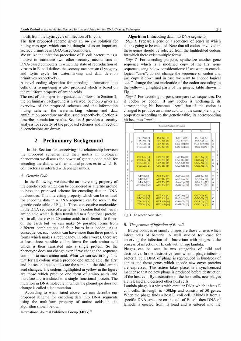
genetic code table of Fig. 1. Three consecutive nucleotides
in the DNA sequence of a gene form a codon that defines an
amino acid which is then translated to a functional protein.
All in all, there exist 20 amino acids in different life forms
on the earth but we can make 64 possible forms from
different combinations of four bases in a codon. As a
consequence, each codon can have more than three possible
forms which makes a redundancy. In other words, there areat least three possible codon forms for each amino acid
which is then translated into a single protein. So the
phenotype does not change even if we change the sequence
common to each amino acid. What we can see in Fig. 1 isthat for all codons which produce one amino acid, the first
and the second nucleotides are the same but the third amino
acid changes. The codons highlighted in yellow in the figure
are those which produce one form of amino acids andtherefore are translated to a single functional protein. The
mutation in DNA molecule in which the phenotype does not
change is called silent mutation.
According to what stated above, we can describe our
proposed scheme for encoding data into DNA segmentsusing the multiform property of amino acids in the
algorithm shown below.
Algorithm I. Encoding data into DNA segments
Step 1. Prepare a gene or a sequence of genes in which
data is going to be encoded. Note that all codons involved in
these genes should be selected from the highlighted codons
for which there exist multiple forms.
Step 2. For encoding purpose, synthesize another genesequence which is a modified copy of the first gene
sequence using below considerations: if we want to encode
logical “ zero”, do not change the sequence of codon and
just copy it down and in case we want to encode logical
“one” change the last nucleotide of the codon according to
the yellow-highlighted parts of the genetic table shown in
Fig. 1.
Step 3. For decoding purpose, compare two sequences. Do
it codon by codon. If any codon is unchanged, its
corresponding bit becomes “ zero” but if the codon is
changed to produce an amino acid with the same phenotypic properties according to the genetic table, its corresponding
bit becomes “one”.
Fig. 1 The genetic code table
B. The process of infection of E. coli
Bacteriophages or simply phages are those viruses which
infect cells of bacteria. A well studied test case for
observing the infection of a bacterium with phages is the process of infection of E. coli with phage lambda.
Phages can be seen in two categories of mild and
destructive. In the destructive form when a phage infects a bacterial cell, DNA of phage is reproduced in hundreds of
copies and those genes which encode new cover proteins
are expressed. This action takes place in a synchronized
manner so that no new phage is produced before destructionof the host cell. By destruction of the host cells, new phages
are released and destruct other host cells.
Lambda phage is a virus with circular DNA which infects E.
coli cells. Its length is ≈50kbp and consists of 50 genes.When the phage finds a host E. coli cell, it binds it from a
specific DNA structure on the cell of E. coli then DNA oflambda is ejected from its head and is entered into the

8/13/2019 Achieving Secrecy for Images Using in-vivo DNA Cloning Techniques
http://slidepdf.com/reader/full/achieving-secrecy-for-images-using-in-vivo-dna-cloning-techniques 3/10
International Journal of Advanced Computer Science, Vol. 1, No. 6, Pp. 240-249, Dec. 2011.
International Journal Publishers Group (IJPG) ©
242
interior membrane of the bacterium and then, in order to not
being destructed by exonuclease enzymes it forms a circular
structure and then the DNA molecules are linked in specific
sites existing in two sides of the linear strands. These
specific sites are shown in Fig. 2.
Fig. 2 Specific site on the end of the linear strand
After this operation, the ligase enzyme encoded by host
cell, closes the cut sites in both sides and makes a closedcircular lambda molecule. Injection of DNA of phage in
cells of E. coli is demonstrated in Fig. 3.
Fig. 3 Injection of DNA of phage in the E. coli cell
The lambda phage is a mild phage for which there are
two phases of proliferation which are called Lytic and
Lysogeny. In the Lysogeny cycle the genome of phage,
instead of proliferation, is integrated into genome of bacterium and the genes related to the cover proteins are not
expressed. This integrated and deactivated phage is called
prophage. These prophages are proliferated during cell
division procedure as a part of bacterial chromosome in the
inactive form. Therefore, each on two daughter cells is theresult of this Lysogeny cell division and this Lysogeny state
can be kept for a long duration but there is the possibility of
state change to the Lytic cycle. This state change from
Lysogeny to Lytic is called induction which is possible byejection of the prophage DNA from genome of bacteria,
proliferation and activation of required genes for generation
of cover and regulator proteins in the Lytic cycle.
Lysogeny cycle is quite stable in the normal conditions but in case the cell is exposed to destructing conditions, the
inactive phage integrated into genome of bacterium (the
prophage) can effectively change its state into the Lytic
cycle. This kind of state change from Lysogeny to Lytic is
called Lysogeny induction. Selection of everyone of these
cycles depends on the state of acceptance of Lytic or
Lysogeny gene expression programs. The programresponsible for Lysogeny cycle can be kept in the cell for
many generations of proliferation but during induction
process, this cycle is changed to the Lytic cycle with a high
efficiency. The procedure of infection of E. coli cell and the
cycles of proliferation of bacteriophages are shown in Fig.
4.
Fig. 4 Different cycles of proliferation in infection of E. coli with phages
3. The Proposed Schemes
In this section we introduce our proposed schemes for
providing secrecy in images using genetically engineered
machines.
A. An in-vivo mechanism for hiding information
In this section we show our proposed scheme for an
in-vivo system using the procedures which occur naturallyas an integral part of gene expression in all living organisms.
We define our initial setup for the hiding system as shown
below:
We utilize the silent mutation property of amino acids asdemonstrated in section (2.A) for encoding our messages
into the blocks of DNA sequences in the synthetic plasmids
conveying the information. Furthermore, we define
transmitter and receiver side information of the system as
shown in Equ. (1)-(2). The transmitter side information is a
kind of information which is added to the message at the
transmitter side and the receiver side information is added
in the receiver side to unveil the hided information.
Receiver information≡(A biochemical indirectactivator)
(Equ. 1)
8/13/2019 Achieving Secrecy for Images Using in-vivo DNA Cloning Techniques
http://slidepdf.com/reader/full/achieving-secrecy-for-images-using-in-vivo-dna-cloning-techniques 4/10
Arash Karimi et al .: Achieving Secrecy for Images Using in-vivo DNA Cloning Techniques
International Journal Publishers Group (IJPG) ©
243
)
,)(
,(
sequencemessagetheafter paddi ng for
snucleotideof sequenceknown A
genereporter a phenotype
certainawith geneknown A
inhibitor l biochemica An Informatior Transmitte
(Equ. 2)
Equ. 1 and Equ. 2 show the receiver and the transmitter side
information of our proposed data hiding system,
respectively. The first element of both of which is a
biochemical substance which can be naturally found in the bacteria we work with. As can be seen in Equ. 1, the
receiver information is a biochemical indirect activator
which indirectly activates expression of the genes which lie
downstream of the promoter of the synthetic gene sequencewhich encodes the message.
Furthermore Equ. 2 shows that the first element of the
transmitter information is a biochemical inhibitor which
effectively blocks expression of the downstream gene(s) of
the promoter of the plasmid which encodes the message ofthe proposed system, the second element of Equ. 2 is a
known gene with a specific phenotype and the third element
of it demonstrates a known sequence of nucleotides which
shows that the message data has ended. Any gene to be
expressed needs a promoter which is upstream of it and that
gene which comes after it as shown in Fig. 5.
Fig. 5 A plasmid containing its promoter and a gene
In order to provide an example to demonstrate our hiding
mechanics, we use Equ. (3)-(4) to express the
transmitter-receiver information pairs of the hiding scheme.
)(Re IPTGn Informatioceiver (Equ. 3)
)
,,(
dataafter padding for sequence
DNA A geneGFP LacI n Informatior Transmitte
(Equ. 4)
In Equ. 3, IPTG or Isopropyl β-D-1-thiogalactopyranoside
is a biochemical reagent which induces transcription of the
gene that encodes for beta-galactosidase, a hydrolase
enzyme which cooperates in catalyzing the hydrolysis of β-galactosides to monosaccharide.
Also, in Equ. 4, the transmitter information contains a
biochemical substance ( LacI protein) which inhibits
transcription of the upstream gene(s) of the promoter which belongs to the message-encoding plasmid.
IPTG molecule (with the following chemical formula
C9H18O5S), when connected to LacI , detaches it from the promoter and unblocks expression of the gene(s)
downstream of the promoter this process is shown in Fig. 6.
With this explanation at hand, we are now ready to describe
the algorithm in which Alice encrypts a message and send it
to Bob.
Algorithm II. The proposed scenario for secure
communication of Alice and BobStep 1. Alice encodes her intended message in
accordance with the silent mutation property of the genetic
code in some gene(s) which have been cloned in the
message information-bearing plasmid.
Fig. 6 Detaching LacI from the plasmid by IPTG
Step 2. Alice inserts the third element of Equ. 4 which is a
known sequence of nucleotides to the message she wishes
to send to Bob.
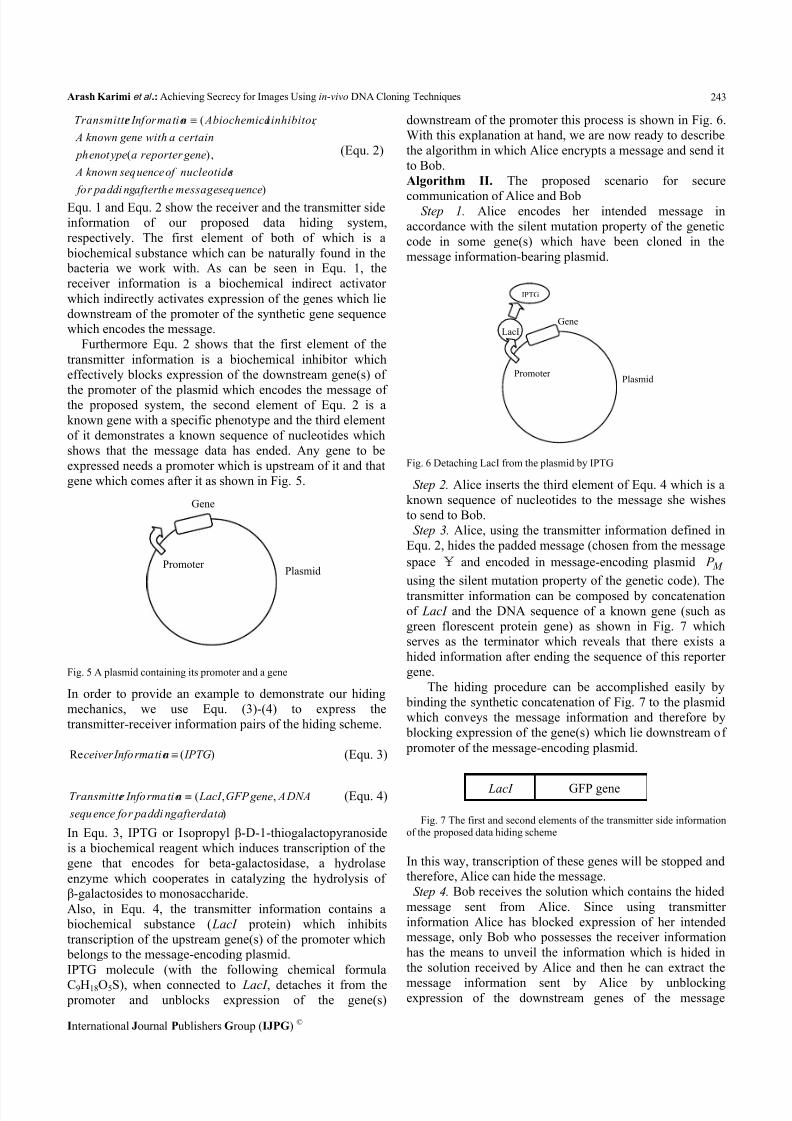
Step 3. Alice, using the transmitter information defined in
Equ. 2, hides the padded message (chosen from the message
space ¥ and encoded in message-encoding plasmid M P
using the silent mutation property of the genetic code). The
transmitter information can be composed by concatenation
of LacI and the DNA sequence of a known gene (such as
green florescent protein gene) as shown in Fig. 7 which
serves as the terminator which reveals that there exists a
hided information after ending the sequence of this reporter
gene.
The hiding procedure can be accomplished easily by
binding the synthetic concatenation of Fig. 7 to the plasmid
which conveys the message information and therefore by
blocking expression of the gene(s) which lie downstream of
promoter of the message-encoding plasmid.
Fig. 7 The first and second elements of the transmitter side informationof the proposed data hiding scheme
In this way, transcription of these genes will be stopped andtherefore, Alice can hide the message.
Step 4. Bob receives the solution which contains the hided
message sent from Alice. Since using transmitter
information Alice has blocked expression of her intendedmessage, only Bob who possesses the receiver information
has the means to unveil the information which is hided in
the solution received by Alice and then he can extract the
message information sent by Alice by unblockingexpression of the downstream genes of the message
LacI GFP gene
Promoter
Gene
Plasmid
LacI
IPTG
Promoter
Gene
Plasmid

8/13/2019 Achieving Secrecy for Images Using in-vivo DNA Cloning Techniques
http://slidepdf.com/reader/full/achieving-secrecy-for-images-using-in-vivo-dna-cloning-techniques 5/10
International Journal of Advanced Computer Science, Vol. 1, No. 6, Pp. 240-249, Dec. 2011.
International Journal Publishers Group (IJPG) ©
244
promoter. In our example, Bob by adding IPTG can remove
LacI and by removing it, the GFP gene is expressed and the
solution which contains hided and coded information turns
to green.
Step 5. By analyzing the resultant plasmid, Bob can unveil
the message sent by Alice which lies between the GFP geneand the known sequence of nucleotides which was
previously defined as a part of transmitter information.
Step 6. By decoding the sequence of nucleotides which
was derived by Bob in step 4, according to the genetic code
table shown in Fig. 1, he can find out the message Alice
sent to him.
B. A new in-vivo watermarking scheme
A remarkable difficulty that is common to all the
watermarking algorithms presented up to now is restrictionof size of the watermark picture. We have overcome this
restriction by using DNA molecules to code image
information into cells of two microbes. Our proposedmethod has the ability to encode images with a very largesize, since the total length of E. coli and phage lambda can
be used to encode the host image and the watermark picture
respectively (approximately 40004000 pixels for the
host image and 127127 pixels for the watermark image
if we use E. coli and lambda phage for encoding the hostimage and the watermark image respectively.) Furthermore
we can use larger phages to encode larger watermark
images. It is noteworthy that for implementation of thisscheme in the laboratory we should control the infection
cycle of E. coli so that it maintains the lysogenic cycle and
as shown in [14] these conditions can be achieved with a
probability of at least 90%. The proposed scheme like allthe other watermark schemes [15]-[17] includes two steps
of embedding the watermark image and extracting the
watermark image:
1) Embedding watermark: In the proposed method
the host image is first converted into a string of sequential
bits and then it is mapped to DNA sequence of the
bacterium E. coli.
The mapping of image bits and codons is based on the
concept of a Silent mutation which is a kind of mutation
that does not alter the amino acid and so does not outbreak
in the phenotype as explained in section (2.A). In the sequel,
some Algorithms are brought to explain the detailed
procedure of the proposed watermarking method.
Algorithm III. Coding of information of the host image inthe genome of E. coli:
Step 1. Selection of the host image.Step 2. Displaying the host image as a sequence of binary
bits using the halftone technology [17].Step 3. Selection of a gene from the E. coli genome to
possess the size of at least three times as big as the size of bits of the image.
Step 4. Coding of the sequence of Step 2 in the genome ofE. coli such that if its corresponding bit is zero, there will be
no change in the structure of the codon of the gene,otherwise, the codon of the corresponding gene incurs a
silent mutation.
In the next step, we should select a specific site in thegenes of E. coli and phage lambda that are regarded as the
sticky ends of them. It is substantial to note that the sticky
ends are unique. Otherwise, by doing it in the laboratory,
the circular DNA of E. coli will be patchy and will not yieldan appropriate result.
Algorithm IV. Coding of information of the watermarkimage in the genome of lambda phage:
Step 1. Selection of the watermark image.Step 2. Displaying the watermark image as a sequence of
binary bits using the halftone technique.
Step 3. Selection of a gene from the lambda phage genome
to possess the size of at least three times as big as the size of bits of the image.
Step 4. Coding of the sequence of Step 2 in the genome of
phage lambda such that if its corresponding bit is zero, there
will be no change in the structure of the codon of the gene,otherwise, the codon of the corresponding gene incurs a
silent mutation.Algorithms III and IV result in two test tubes that containthe coded DNA strands of the host image and the watermarkimage respectively. In the next algorithm, an appropriatesite must be selected for computer simulation of insertion ofthe phage DNA into the DNA of E. coli.
Algorithm V. Selection of the sticky ends of E. coli and phage lambda:
Step 1. Reception of the genes of E. coli and phage lambda produced in Algorithms III and IV.
Step 2. Finding a cos site in the phage lambda and the
corresponding site in E. coli bacterium.We have used the cos site achieved in [18] to be used in
Step 2 of Algorithm V as a secondary attachment site. In the
next algorithm, insertion of the phage lambda DNA into the
DNA of E. coli is computer simulated.
Algorithm VI. Insertion of the DNA of the phage lambda inthat of E. coli:
Step 1. Cutting the double-stranded DNA of E. coli andlambda phage from their sticky ends.
Step 2. Insertion of a piece of the lambda phage into thecutting edge of E. coli bacteria.
2) The watermark image extraction: In this stage, toextract the watermark image by the owner he should evict
lambda phage from the solution containing the infected E.coli and then he should extract the phage gene and decode it.
In Algorithm VII, stages of watermark extraction are
depicted.
Algorithm VII. Evicting phage lambda from the infected E.coli
Step 1. The owner, using the knowledge of sticky ends ofthe phage that he himself has inserted, finds two identicalsticky ends and cuts them stepwisely.
Step 2. Extraction of a shorter length sequence (phagelambda).
Step 3. Adjoining free nucleotides of the remaining
sequence (E. coli).

8/13/2019 Achieving Secrecy for Images Using in-vivo DNA Cloning Techniques
http://slidepdf.com/reader/full/achieving-secrecy-for-images-using-in-vivo-dna-cloning-techniques 6/10
Arash Karimi et al .: Achieving Secrecy for Images Using in-vivo DNA Cloning Techniques
International Journal Publishers Group (IJPG) ©
245
In laboratory Algorithm VII is implemented using
centrifuge of the resulted solution. In this regard, because
lambda phage sequence is shorter than that of E. coli, it
moves faster in the test tube and so it can be easily
extracted.
Algorithm VIII. DecodingStep 1. Comparing sequences of the extracted phage from
step 3 of the algorithm VII with the original gene of the phage to extraction of the watermark image.
Step 2. Comparing DNA sequence of the resulting E .coli
in step 4 of the algorithm VII with the original sequence
of E. coli to extract the host image.
C. An information annihilation scheme
The last scheme we introduce in this paper is a dataannihilation scheme which can be used to delete a message
in in-vivo computers. This scheme uses the concept ofinfection of E. coli with bacteriophage lambda in the Lytic
cycle. The scenario in which our wetware data annihilationscheme is useful can be stated as follows.
Assume that Alice has encoded her message into the
genome of E. coli. For a variety of reasons she may wish to
delete this message so that no one else can see or recover it.
The mechanics of this scheme can be explained in the
algorithm below.
Algorithm IX. A wetware data annihilation scheme
Step 1. Alice infects the E. coli bacterium which conveys
her encoded information (the data encoding scheme uses the
silent mutation property just as explained in section (2.A)).
Step 2. The infection procedure will be guided using a bacteriophage (we assume it to be the bacteriophagelambda). Alice controls this infection so that it goes to the
Lytic cycle.
Step 3. The E. coli bacterium containing the encoded
message is completely destroyed and therefore, her data iscompletely deleted.
As can be seen in the above algorithm, Alice is able to
delete her message so that it cannot be recyclable anymore.
The proposed scheme is similar to the deletion of a file fromrecycle bin of a usual silicon computer. The user intends to
delete any file he wishes so that it cannot be retrieved by
any means.
Our wetware scheme has advantages over the usual deletionmethod in silicon based computers since it is natural and has
no loss, and also the procedure which utilizes this scheme is
one-sided and it cannot be reversed, therefore it guarantees
deletion of the message, but in silicon computers, the
deleted data can be retrieved from the memory and it goes
to a specific address in memory even if deleted and so there
are some bits in the memory from which this deleted
information can be leaked. One example for suchinformation leakage in usual silicon-based computers can be
seen in the case for cold boot attacks introduced in [19].
4. Simulation Results
To show the performance of our proposed scheme we
have run a simulation in which we want to embed the
picture containing arm of our university of size 4040
pixels into a 200200 pixels Lenna image (Fig. 8(a)).And next, we will depict the results of our computer
simulation in two parts of embedding the watermark andextraction of it.
(b) (a)
Fig. 8 (a) Host image (b) watermark image
1) Embedding the watermark: Our proposed method ofembedding watermark is divided into three parts:
1. Coding the host image in a specific gene of E. coli
The host image will be first considered in a halftone mode.i.e. in the dark parts of the image the density of the black
pixels will be more and in bright parts of it the density of
the black pixels will be less.
00000010
00100000
00001001
00000101
01001011
00001001
01010110
11101001
Fig. 9 Host image

8/13/2019 Achieving Secrecy for Images Using in-vivo DNA Cloning Techniques
http://slidepdf.com/reader/full/achieving-secrecy-for-images-using-in-vivo-dna-cloning-techniques 7/10
International Journal of Advanced Computer Science, Vol. 1, No. 6, Pp. 240-249, Dec. 2011.
International Journal Publishers Group (IJPG) ©
246
Based on the size of the host image, we have chosen gene
leuL of E. coli to encode the host image. The part of the
abovementioned original gene which corresponds to the
selected area demonstrated in Fig. 9 is presented in matrix
of Equ. 5.
(Equ. 5)
By coding the host image in leuL , the coded matrix of the
selected area of the image is shown in Equ. 6.
(Equ. 6)
As we can see, if each pixel of the image is one, its
corresponding codon will incur a silent mutation (its amino
acid does not change) and if the pixel is zero, its
corresponding codon will not change.
2. Coding the watermark image in genome of bacteriophagelambda
We use lambda phage as the carrier of our watermark and
encode our watermark image in it. To do so, we should
mention that the specific part of the genome of lambda
phage should not contain a cos site because the image will be caught from the sticky ends.
11110001
11111001
11100001
00000011
00000011
10100001
11110001
11110001
Fig. 10 The watermark image
In the matrix of Equ. 7 part of phage lambda genome whichcorresponds to the selected area of the watermark image isshown.
AAAGGA AAC GGC TTT TCGCGATCT
AAGGAT AAGCCA AGAGAT CCGCAG
CTACAT GGT ATT GTT GTC GGT GTT
ATGGGATAGTGT CAGGCT ATACCG
ACGGGAGGGTGGCTT CGA AAG AAA
TTC CTG ATC TGG ATC AAGCTAGAT
GAC AGC AAATGC CAATTT CGC CCC
CGG ACT ATC TAT CTC CTT GAATAG
M Lam bd aSC Code _ _
(Equ. 7)
By coding the watermark image in lambda phage genome,
the coded matrix of the selected area of the image is
demonstrated in the matrix of Equ. 8.
AAGGGG AAT GGGTTT TCGCGA AGC
AAAGAC AAACCG AGGGAT CCGCAA
CTGCAC GGG ATT GTT GTC GGT GTG
ATGGGATAGTGT CAGGCT ATT CCT
ACGGGAGGGTGGCTT CGA AAA AAG
TTT CTG ATATGG ATC AAGCTAGAC GAT AGG AAGTGT CAATTT CGC CCG
AGG ACG ATATAC CTC CTT GAATAA
M Lam bd aSC Code _ _
(Equ. 8)
3. Infection of Escherichia coli with phage lambda
Bacteriophage lambda, in order to infect E. coli integrates
its genome into the TrpC gene of E. coli from its cos site
and the recombination DNA sequence of that is shown in
Fig. 11.
ATGCAAACCGTTTTAGCGAAAATCGTCGCAGACAAGGCGATTTGGGTAGAAGCCCGCAAA CAGCAGCAACCGCTGGCCAGTTTTCAGAATGAGGTTCAGCCGAGCACGCGACATTTTTAT GATGCGCTACAGGGTGCGCGCACGGCGTTTATTCTGGAGTGCAAGAAAGCGTCGCCGTCA AAAGGCGTGATCCGTGATGATTTCGATCCAGCACGCATTGCCGCCATTTATAAACATTAC GCTTCGGCAATTTCGGTGCTGACTGATGAGAAATATTTTCAGGGGAGCTTTAATTTCCTC
CCCATCGTCAGCCAAATCGCCCCGCAGCCGATTTTATGTAAAGACTTCATTATCGACCCT TACCAGATCTATCTGGCGCGCTATTACCAGGCCGATGCCTGCTTATTAATGCTTTCAGTA CTGGATGACGACCAATATCGCCAGCTTGCCGCCGTCGCTCACAGTCTGGAGATGGGGGTG CTGACCGAAGTCAGTAATGAAGAGGAACAGGAGCGCGCCATTGCATTGGGAGCAAAGGTC GTTGGCATCAACAACCGCGATCTGCGTGATTTGTCGATTGATCTCAACCGTACCCGCGAG
CTTGCGCCGAAACTGGGGCACAACGTGACGGTAATCAGCGAATCCGGCATCAATACTTAC GCTCAGGTGCGCGAGTTAAGCCACTTCGCTAACGGTTTTCTGATTGGTTCGGCGTTGATG GCCCATGACGATTTGCACGCCGCCGTGCGCCGGGTGTTGCTGGGTGAGAATAAAGTATGT GGCCTGACGCGTGGGCAAGATGCTAAAGCAGCTTATGACGCGGGCGCGATTTACGGTGGG TTGATTTTTGTTGCGACATCACCGCGTTGCGTCAACGTTGAACAGGCGCAGGAAGTGATG
GCTGCGGCACCGTTGCAGTATGTTGGCGTGTTCCGCAATCACGATATTGCCGATGTGGTG GACAAAGCTAAGGTGTTATCGCTGGCGGCAGTGCAACTGCATGGTAATGAAGAACAGCTG TATATCGATACGCTGCGTGAAGCTCTGCCAGCACATGTTGCCATCTGGAAAGCATTAAGC GTCGGTGAAACCCTGCCCGCCCGCGAGTTTCAGCACGTTGATAAATATGTTTTAGACAAC GGCCAGGGTGGAAGCGGGCAACGTTTTGACTGGTCACTATTAAATGGTCAATCGCTTGGC
AACGTTCTGCTGGCGGGGGGCTTAGGCGCAGATAACTGCGTGGAAGCGGCACAAACCGGC TGCGCCGGACTTGATTTTAATTCTGCTGTAGAGTCGCAACCGGGCATCAAAGACGCACGT CTTTTGGCCTCGGTTTTCCAGACGCTGCGCGCATATTAA
Fig. 11 The specific part of the infected E.coli genome
The specific part of the phage genome that carries the cos
site is depicted in Fig. 12.
TATTTAGCTTTCTGCTTCCTTTTGGATAACCCACTGTTATTCATGTTGCATGGTGCACTG
TTTATACCAACGATATAGTCTATTAATGCATATATAGTATCGCCGAACGATTAGCTCTTC
AGGCTTCTGAAGAAGCGTTTCAAGTACTAATAAGCCGATAGATAGCCACGGACTTCGTAG
CCATTTTTCATAAGTGTTAACTTCCGCTCCTCGCTCATAACAGACATTCACTACAGTTAT
GGCGGAAAGGTATGCATGCTGGGTGTGGGGAAGTCGTGAAAGAAAAGAAGTCAGCTGCGT
CGTTTGACATCACTGCTATCTTCTTACTGGTTATGCAGGTCGTAGTGGGTGGCACACAAA
GCTTTGCACTGGATTGCGAGGCTTTGTGCTTCTCTGGAGTGCGACAGGTTTGATGACAAA
AAATTAGCGCAAGAAGACAAAAATCACCTTGCGCTAATGCTCTGTTACAGGTCACTAATA
CCATCTAAGTAGTTGATTCATAGTGACTGCATATGTTGTGTTTTACAGTATTATGTAGTCTGTTTTTTATGCAAAATCTAATTTAATATATTGATATTTATATCATTTTACGTTTCTCGT
TCAGCTTTTTTATACTAAGTTGGCATTATAAAAAAGCATTGCTTATCAATTTGTTGCAAC
GAACAGGTCACTATCAGTCAAAATAAAATCATTATTTGATTTCAATTTTGTCCCACTCCC
TGCCTCTGTCATCACGATACTGTGATGCCATGGTGTCCGACTTATGCCCGAGAAGATGTT
GAGCAAACTTATCGCTTATCTGCTTCTCATAGAGTCTTGCAGACAAACTGCGCAACTCGT
GAAAGGTAGGCGGATCCCCTTCGAAGGAAAGACCTGATGCTTTTCGTGCGCGCATAAAAT
ACCTTGATACTGTGCCGGATGAAAGCGGTTCGCGACGAGTAGATGCAATTATGGTTTCTC
CGCCAAGAATCTCTTTGCATTTATCAAGTGTTTCCTTCATTGATATTCCGAGAGCATCAA
TATGCAATGCTGTTGGGATGGCAATTTTTACGCCTGTTTTGCTTTGCTCGACATAAAGAT
ATCCATCTACGATATCAGACCACTTCATTTCGCATAAATCACCAACTCGTTGCCCGGTAA
CAACAGCCAGTTCCATTGCAAGTCTGAGCCAACATGGTGATGATTCTGCTGCTTGATAAA
TTTTCAGGTATTCGTCAGCCGTAAGTCTTGATCTCCTTACCTCTGATTTTGCTGCGCGAG
TGGCAGCGACATGGTTTGTTGTTATATGGCCTTCAGCTATTGCCTCTCGGAATGCATCGC
Fig. 12 The specific part of the lambda bacteriophage genome
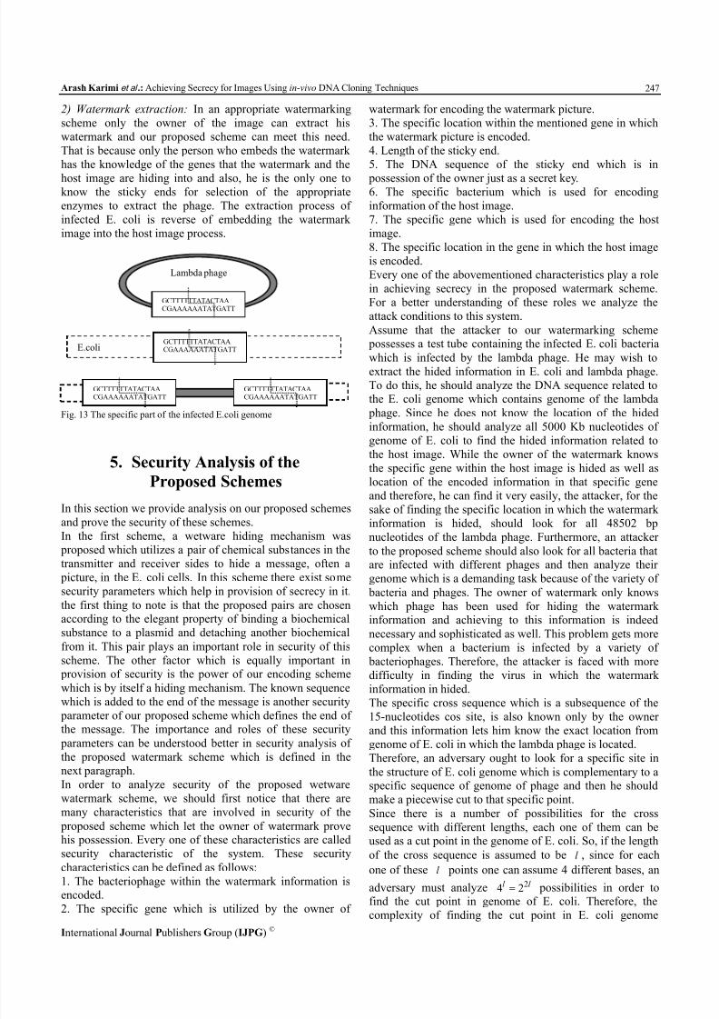
The infection procedure of bacteria E. coli with phage
lambda and the way its genome enters into the circular
DNA of E. coli is demonstrated in Fig. 13.
8/13/2019 Achieving Secrecy for Images Using in-vivo DNA Cloning Techniques
http://slidepdf.com/reader/full/achieving-secrecy-for-images-using-in-vivo-dna-cloning-techniques 8/10
Arash Karimi et al .: Achieving Secrecy for Images Using in-vivo DNA Cloning Techniques
International Journal Publishers Group (IJPG) ©
247
2) Watermark extraction: In an appropriate watermarking
scheme only the owner of the image can extract his
watermark and our proposed scheme can meet this need.
That is because only the person who embeds the watermark
has the knowledge of the genes that the watermark and the
host image are hiding into and also, he is the only one toknow the sticky ends for selection of the appropriate
enzymes to extract the phage. The extraction process of
infected E. coli is reverse of embedding the watermark
image into the host image process.
Fig. 13 The specific part of the infected E.coli genome
5. Security Analysis of the
Proposed Schemes
In this section we provide analysis on our proposed schemes
and prove the security of these schemes.
In the first scheme, a wetware hiding mechanism was proposed which utilizes a pair of chemical substances in the
transmitter and receiver sides to hide a message, often a
picture, in the E. coli cells. In this scheme there exist some
security parameters which help in provision of secrecy in it.
the first thing to note is that the proposed pairs are chosenaccording to the elegant property of binding a biochemical
substance to a plasmid and detaching another biochemical
from it. This pair plays an important role in security of this
scheme. The other factor which is equally important in provision of security is the power of our encoding scheme
which is by itself a hiding mechanism. The known sequence
which is added to the end of the message is another security
parameter of our proposed scheme which defines the end ofthe message. The importance and roles of these security
parameters can be understood better in security analysis of
the proposed watermark scheme which is defined in the
next paragraph.
In order to analyze security of the proposed wetware
watermark scheme, we should first notice that there are
many characteristics that are involved in security of the
proposed scheme which let the owner of watermark provehis possession. Every one of these characteristics are called
security characteristic of the system. These security
characteristics can be defined as follows:
1. The bacteriophage within the watermark information is
encoded.2. The specific gene which is utilized by the owner of
watermark for encoding the watermark picture.
3. The specific location within the mentioned gene in which
the watermark picture is encoded.
4. Length of the sticky end.
5. The DNA sequence of the sticky end which is in
possession of the owner just as a secret key.6. The specific bacterium which is used for encoding
information of the host image.
7. The specific gene which is used for encoding the host
image.
8. The specific location in the gene in which the host image
is encoded.
Every one of the abovementioned characteristics play a role
in achieving secrecy in the proposed watermark scheme.
For a better understanding of these roles we analyze the
attack conditions to this system.
Assume that the attacker to our watermarking scheme possesses a test tube containing the infected E. coli bacteria
which is infected by the lambda phage. He may wish toextract the hided information in E. coli and lambda phage.
To do this, he should analyze the DNA sequence related tothe E. coli genome which contains genome of the lambda
phage. Since he does not know the location of the hided
information, he should analyze all 5000 Kb nucleotides of
genome of E. coli to find the hided information related to
the host image. While the owner of the watermark knows
the specific gene within the host image is hided as well as
location of the encoded information in that specific gene
and therefore, he can find it very easily, the attacker, for the
sake of finding the specific location in which the watermark
information is hided, should look for all 48502 bp
nucleotides of the lambda phage. Furthermore, an attackerto the proposed scheme should also look for all bacteria thatare infected with different phages and then analyze their
genome which is a demanding task because of the variety of
bacteria and phages. The owner of watermark only knows
which phage has been used for hiding the watermarkinformation and achieving to this information is indeed
necessary and sophisticated as well. This problem gets more
complex when a bacterium is infected by a variety of
bacteriophages. Therefore, the attacker is faced with more
difficulty in finding the virus in which the watermark
information in hided.
The specific cross sequence which is a subsequence of the
15-nucleotides cos site, is also known only by the ownerand this information lets him know the exact location from
genome of E. coli in which the lambda phage is located.
Therefore, an adversary ought to look for a specific site in
the structure of E. coli genome which is complementary to aspecific sequence of genome of phage and then he should
make a piecewise cut to that specific point.
Since there is a number of possibilities for the cross
sequence with different lengths, each one of them can beused as a cut point in the genome of E. coli. So, if the length
of the cross sequence is assumed to be l , since for each
one of these l points one can assume 4 different bases, an
adversary must analyze
l l 2
24
possibilities in order tofind the cut point in genome of E. coli. Therefore, the
complexity of finding the cut point in E. coli genome
Lambda phage
GCTTTTTTATACTAACGAAAAAATATGATT
E.coliGCTTTTTTATACTAACGAAAAAATATGATT
GCTTTTTTATACTAACGAAAAAATATGATT
GCTTTTTTATACTAACGAAAAAATATGATT

8/13/2019 Achieving Secrecy for Images Using in-vivo DNA Cloning Techniques
http://slidepdf.com/reader/full/achieving-secrecy-for-images-using-in-vivo-dna-cloning-techniques 9/10
International Journal of Advanced Computer Science, Vol. 1, No. 6, Pp. 240-249, Dec. 2011.
International Journal Publishers Group (IJPG) ©
248
becomes of order )2( 2l O which is exponential.
There can be used a variety of different secondary cos sites
as entrance point of the phage to the E. coli genome and we
used the sequence driven in [18] as this secondary cross
sequence.
As can be seen in the abovementioned security analysis,
because of the variety of existing security parameters, the
proposed watermark model has a high level of securityagainst those attacks in which the attacker possesses the test
tube containing the infected bacteria.
The security of the proposed data deletion procedure can
also be justified using the fact that the procedure of Lyticreproduction and cell destruction takes place quite naturally
and does not need an extra control when the infection
procedure starts and therefore, it is so trustworthy and
guarantees deletion of the encoded message.
D. Considerations of implementing the proposed methods
in cloning laboratories
The proposed schemes for security assurance in images
can be easily implemented in genetic engineering
laboratories. In the first scheme, the data encoding in
plasmids can be easily implemented using UV to change thenucleotides of a codon and the usual laboratory techniques
can be utilized in adding LacI as well as IPTG to the
synthesized plasmid. In the second and third proposed
schemes, the most important task is mixing two solution
containing bacterial species and bacteriophage species and
then to control this procedure so that the E. coli bacteria is
properly infected and this solution does not go to the Lytic
cycle in the watermark scheme or goes to the Lytic cycle in
the data annihilation scheme.
The laboratory establishment for the engineered infection
process is explained in [14]. The results of this laboratory
experiment demonstrate that considering the explainedconditions, we can make sure that 99% of bacteria are
infected and also 90% of bacterial population go with the
Lysogeny cycle.
In order to make sure that the process of infection goeswith the Lysogeny cycle, there must be a control over the
experiment but if there is no control on the infection
experiment, the procedure tends to the Lytic cycle and
therefore, in practice implementing the second proposedscheme is a more demanding task.
E. The problems associated with the proposed methods
The proposed approach for providing security in images
has some constraints and drawbacks associated with it
which can be classified into two groups. The first one
contain some general drawbacks associated with allcomputational systems based on DNA molecules and the
second belongs to the proposed scheme which utilizes
cloning techniques as a model for computing with DNA.
The drawbacks of the first type belong to lack of access
to the biotechnology facilities which fades away in the light
of growing technology of genetic engineering. The other
problem of the first type also belong with the high error
rates which is tagged to all operations with DNA sequences.
These errors necessitate the need for repeating the
biotechnological experiments for achieving trusty results.
The problems of the second kind associated with the
watermarking scheme relates to the control of the infection
procedure of the phage such that it is prevented from
entering the Lytic cycle. If so, the cell containing theinformation will be lost. But as shown in the laboratory
considerations mentioned above, we are able to control this
cycle such that more that 90% of the infected cells enter the
Lysogeny cycle. The precision in experiments is the key for
achieving this goal.
6. Conclusion
In this paper three genetically engineered machines
were proposed to ensure secrecy in transmitting images in
DNA-based computers. Our proposed schemes are the first
security initiatives for images in the DNA computingcontext. They provide some improvements over the existingmethods implementable in silicon computers. The usual
data hiding mechanisms in silicon computers have practical
drawbacks in that data can be leaked but the dense medium
of DNA molecule helps us use our proposed scheme alongwith the novel data encoding and retrieval mechanisms. Our
in-vivo watermarking scheme also takes advantage of a
natural process which takes place during infection of E. coli
bacterium by bacteriophage lambda and solves the problemof size limitation of the existing watermarking schemes.
Our analysis predicts that the proposed scheme can be
implemented in laboratory with 90% probability of success.
In the proposed watermarking scheme the infection of E.coli in the Lysogeny cycle is considered and we used the
Lytic cycle of infection of E. coli for achievement of
another security assurance scheme which we called data
annihilation scheme. The process considered in this security primitive is a naturally one-sided procedure in which
bacteriophages annihilate the cells of an E. coli bacterium.
The last secrecy primitive can be used similar to the
deletion of a file from a silicon-based computer but theone-sided property of our proposed scheme guarantees
deletion of the data (such as an image file) so that it cannot
be retrieved and since it gets its motifs from a one-sided
phenomenon which occur in nature, it has no leakage in
contrast to the silicon-based computers as stated in [18] forwhich, the deleted information is recoverable and leaks
information in different locations of memory.
AcknowledgementThe authors would like to thank R. Dastanian for her
useful comments and supports in computer simulations of
this paper and Iran Telecommunications Research Centre
(ITRC) for supporting this paper financially.
References
[1] L.M. Adleman, “Molecular computation of solutions tocombinatorial problems,” (1994) Science, vol. 266, pp.1021-1024.

8/13/2019 Achieving Secrecy for Images Using in-vivo DNA Cloning Techniques
http://slidepdf.com/reader/full/achieving-secrecy-for-images-using-in-vivo-dna-cloning-techniques 10/10
Arash Karimi et al .: Achieving Secrecy for Images Using in-vivo DNA Cloning Techniques
International Journal Publishers Group (IJPG) ©
249
[2] L. Landweber & L. Kari, “The evolution of cellularcomputing: nature’s solution to a computational problem,” (1998) LNCS , vol. 2950, pp. 207-216.
[3] L. Kari, “DNA Computing: Arrival of BilogicalMathematics,” (1997) The Mathematical Intellignecer , vol.
19, No.2, pp. 9-22.[4] T. Head, “Formal Language Theory and DNA: an analysis of
the generative capacity of specific r ecombinant behaviors,”(1987) Bull. Math. Biology, vol. 49, pp. 737-759.
[5] L.M. Adleman, “On constructing a molecular computer,”
(1996) In R.J. Lipton, E.M. Baum (Eds), DNA basedComputers I, Proceedings of a DIMACS Workshop,American Mathematical Society, Providence, RI, USA,
pp.1-22.
[6] J. Khodor & D. Gifford, “Design and implementation of
computational systems based on programmed mutagenesis,”(1998) In Preliminary Proceedings of 4 th DIMACS Workshopon DNA Based Computers, pp. 101-108.
[7] Y. Benenson, B. Gil, U. Ben-Dor, R. Adar & E. Shapiro, “An
autonomous molecular computer for logical control of geneexpression,” (2004) Nature 414, pp. 430-434.
[8] A. Gehani, T. LaBean & J. Reif, “DNA-based Cryptography,
Aspects of Molecular Computing,” (2004) Springer-Verlag Lecture Notes In Computer Science, vol. 2950.
[9] National Bureau of Standards: “Data Encryption Standard,”(1977) U.S. Department of Commerce, FIPS, pub.46.
[10] D. Boneh, C. Dunworth & R. Lipton, “Breaking DEES Usinga Molecular Computer,” (1995) Princeton CS Tech-ReportCS-TR, pp.489-495.
[11] L.M. Adleman, P.W. K. Rothemund, S.Roweis & E. Winfree,
“On applying molecular computation to the Data EncryptionStandard,” (1999) 2nd annual workshop on DNA Computing,
Princeton University, Eds. L. Landweber and E. Baum, DIMACS: series in Discrete Mathematics and Theoretical
Computer Science, American Mathematical Society. pp.31-44.
[12] R. Dastanian, A. Karimi & H.Sh. Shahhoseini, “A Novel
Multi-Client Authentication Method Using Infection ofBacteria,” (2011 ) Proceedings of International Conference on
Communication and Electronics Information (ICCEI), China,1, pp. 310-314.
[13] A. Karimi, R. Dastanian & H. Sh. Shahhoseini, “A NewWatermarking Scheme for An in-vivo Computer Based onInfection of E. coli,” (2010) Proceedings of International
Conference on Computer and Electrical Engineering(ICCEE), China, 8, pp. 484-488.
[14] B.A. Fry, “Conditions for the Infection of Escherichia coliwith Lambda Phage and for the Establishment of Lysogeny,”(1959) Journal of gen. Microbiol , vol. 21, pp. 676-684.
[15] Ch.H. Huang, Sh.Ch. Chuang, Y.L. Huang & J.L. Wu,“Unseen Visible Watermarking: A Novel Methodology for
Auxiliary Information Delivery Via Visual Contents,” (2009) IEEE Trans. On Informtion Forensics and Security, vol. 4, No. 2.
[16] Y.C. Hou, “Visual cryptography for color images,” (2003) Elsevier Journal of Pattern Recognition, vol. 36, pp.1619-1629.
[17] X. Wang, Z. Xu & P. Niu, “A feature-based digital
watermarking scheme for halftone image,” (2010) AEU - International Journal of Electronics and Communications,vol. 64, no. 10, pp. 924-933.
[18] G.E. Christie & T.Platt, “A secondary attachment site for
bacteriophage lambda in trpC of E.coli,” (1979) Cell , vol.16,no. 2, pp. 407-413. [19] A. Halderman, S. Schoen, N. Heninger, W. Clarkson, W.
Paul, J. Calandrino, A. Feldman, J. Appelbaum & E. Felten.“Lest we remember: Cold boot attacks on encryption keys”
(2008) In Usenix Security Symposium. Arash Karimi Received the B.S. andM.S. degrees in the Dep. Of ElectricalEngineering from Amirkabir University
of Science and Technology(Polytechnic of Tehran) and IranUniversity of Science and Technology(IUST), Tehran, Iran, in 2008 and 2011,
respectively. His research interestsinclude cryptography, unconventional
methods in computation with a focuson cryptanalysis, Biochemicalcomputing, and formal languages and
automata.
Hadi Shahriar Shahhoseini received
B.S. degree in electrical engineeringfrom University of Tehran, in 1990,M.S. degree in electrical engineeringfrom Azad University of Tehran in
1994, and Ph.D. degree in electrical
engineering from Iran University ofScience and Technology, in 1999. Heis an assistant professor of theelectrical engineering department inIran University of Science and
Technology. His areas of research include networking,
supercomputing and reconfigurable computing. More than 130 papers have been published from his research works in scientific journals and conference proceedings. He is an executivecommittee member of IEEE TCSC and serves IEEE TCSC asregional coordinator in middle-East Countries. .