acellular pertussis vaccines protect against disease but ... · edited by rino rappuoli, novartis...
TRANSCRIPT

Acellular pertussis vaccines protect against disease butfail to prevent infection and transmission ina nonhuman primate modelJason M. Warfel, Lindsey I. Zimmerman, and Tod J. Merkel1
Division of Bacterial, Parasitic and Allergenic Products, Center for Biologics Evaluation and Research, US Food and Drug Administration, Bethesda, MD, 20892
Edited by Rino Rappuoli, Novartis Vaccines and Diagnostics Srl, Siena, Italy, and approved October 22, 2013 (received for review August 5, 2013)
Pertussis is a highly contagious respiratory illness caused by thebacterial pathogen Bordetella pertussis. Pertussis rates in theUnited States have been rising and reached a 50-y high of42,000 cases in 2012. Although pertussis resurgence is not com-pletely understood, we hypothesize that current acellular pertus-sis (aP) vaccines fail to prevent colonization and transmission. Totest our hypothesis, infant baboons were vaccinated at 2, 4, and6 mo of age with aP or whole-cell pertussis (wP) vaccines andchallenged with B. pertussis at 7 mo. Infection was followed byquantifying colonization in nasopharyngeal washes and monitor-ing leukocytosis and symptoms. Baboons vaccinated with aP wereprotected from severe pertussis-associated symptoms but notfrom colonization, did not clear the infection faster than naïve ani-mals, and readily transmitted B. pertussis to unvaccinated contacts.Vaccination with wP induced a more rapid clearance compared withnaïve and aP-vaccinated animals. By comparison, previously infectedanimals were not colonized upon secondary infection. Although allvaccinated and previously infected animals had robust serum anti-body responses, we found key differences in T-cell immunity. Pre-viously infected animals and wP-vaccinated animals possess strongB. pertussis-specific T helper 17 (Th17) memory and Th1 memory,whereas aP vaccination induced a Th1/Th2 response instead. Theobservation that aP, which induces an immune response mis-matched to that induced by natural infection, fails to prevent colo-nization or transmission provides a plausible explanation for theresurgence of pertussis and suggests that optimal control of pertus-sis will require the development of improved vaccines.
whooping cough | T-cell memory | animal models | adaptive immunity |IL-17
Pertussis is a highly contagious, acute respiratory illness causedby the bacterial pathogen Bordetella pertussis (1, 2). Infection
results in a wide spectrum of clinical manifestations ranging frommild respiratory symptoms to a severe cough illness accompaniedby marked leukocytosis and the hallmark inspiratory whoop andposttussive emesis (3). Because acellular pertussis vaccines replacedwhole-cell vaccines in the 1990s, pertussis has reemerged at a star-tling rate in theUnited States despite nationwide vaccine coverage inexcess of 95% (4). With a 50-y high of 42,000 reported cases in theUnited States in 2012, pertussis is the most common of the vaccine-preventable diseases (5). This resurgence is mirrored throughoutthe industrial world despite similar high rates of vaccination (6–9).Two common hypotheses for the resurgence have been proposed:i) current acellular pertussis vaccines (aP) vaccines are less effectivethan the whole-cell pertussis (wP) vaccines they replaced and ii)aP-induced immunity wanes more quickly than anticipated (10–13).However, pertussis resurgence is not completely understood (14, 15).Hampering our ability to counteract this resurgence is the fact
that pertussis pathogenesis and immunity to natural infectionhave not been well studied in humans because typical pertussis issporadic given high rates of vaccination in developed countries.Human challenge studies have been proposed but never con-ducted due to a variety of logistical and ethical problems in-cluding the potential for severe disease, the lack of an effective
therapeutic for established disease, and the highly contagiousnature of pertussis. Although a variety of small-animal modelshave been used to study pertussis, none of them adequately re-produce the human disease (16). To address this gap, we recentlydeveloped a nonhuman primate model of pertussis using baboons(Papio anubis) and found the disease is very similar to severe clinicalpertussis. Upon challenge, baboons experience 2 wk of heavy re-spiratory colonization and leukocytosis peaking between 30,000–80,000 cells/mL, similar to the range in pertussis-infected infants(1, 17). In addition, baboons experience a paroxysmal cough ill-ness characterized by repeated fits of 5–10 coughs. The coughingfits last on average >2 wk in the baboon, although this is less thansome severely infected children, where the cough can last up to12 wk (1, 17). We also characterized airborne transmission ofB. pertussis from infected to naïve animals, which is the route oftransmission postulated to occur between humans (18). Becausethis is the only model of pertussis to reproduce the cough illnessand transmission of the human disease, we believe it provides theunique opportunity to test our hypothesis that aP vaccines fail toprevent B. pertussis colonization, thus enabling transmissionamong vaccinated individuals.Using this model we have confirmed that, as in humans, aP
vaccines provide excellent protection against severe diseasein baboons. However, aP vaccines do not prevent colonizationfollowing direct challenge or infection by transmission. In addi-tion, aP-vaccinated animals are capable of transmitting diseaseto naïve contacts. By comparison, wP-vaccinated animals clearedinfection significantly more quickly than aP-vaccinated or naïve
Significance
Pertussis has reemerged as an important public health concernsince current acellular pertussis vaccines (aP) replaced olderwhole-cell vaccines (wP). In this study, we show nonhumanprimates vaccinated with aP were protected from severesymptoms but not infection and readily transmitted Bordetellapertussis to contacts. Vaccination with wP and previous in-fection induced a more rapid clearance compared with naïveand aP-vaccinated animals. While all groups possessed robustantibody responses, key differences in T-cell memory suggestthat aP vaccination induces a suboptimal immune responsethat is unable to prevent infection. These data provide a plau-sible explanation for pertussis resurgence and suggest thatattaining herd immunity will require the development of im-proved vaccination strategies that prevent B. pertussis coloni-zation and transmission.
Author contributions: J.M.W. and T.J.M. designed research; J.M.W., L.I.Z., and T.J.M. per-formed research; J.M.W. and T.J.M. analyzed data; and J.M.W. and T.J.M. wrote thepaper.
The authors declare no conflict of interest.
This article is a PNAS Direct Submission.
See Commentary on page 575.1To whom correspondence should be addressed. E-mail: [email protected].
This article contains supporting information online at www.pnas.org/lookup/suppl/doi:10.1073/pnas.1314688110/-/DCSupplemental.
www.pnas.org/cgi/doi/10.1073/pnas.1314688110 PNAS | January 14, 2014 | vol. 111 | no. 2 | 787–792
MED
ICALSC
IENCE
SSE
ECO
MMEN
TARY

animals. We also found that aP vaccination induces T helper 2(Th2) and T helper 1 (Th1) immune memory responses, whereasinfection and—to a lesser extent—wP vaccination induce Th17and Th1 memory. Our results suggest that in addition to thepotential contribution of reduced efficacy and waning immunityof aP, the inability of aP to prevent colonization and transmissionprovides a plausible explanation for pertussis resurgence.
ResultsAcellular Pertussis Vaccines Protect Against Disease but Fail toPrevent Infection. Several observational studies recently con-cluded that children primed with aP vaccine are at greater risk forpertussis diagnosis compared with wP-primed children (19–22).Although these data suggest aP vaccine is less effective than wPvaccine at preventing colonization, the rate of undiagnosedB. pertussis carriage in vaccinated individuals is unknown. To as-sess the ability of each vaccine to prevent colonization and clinicalpertussis symptoms, baboons were vaccinated according to the USschedule at 2, 4, and 6 mo of age with human doses of combi-nation diphtheria, tetanus, and pertussis vaccines containing aP orinactivated wP (Table 1 provides a list of the components of eachvaccine). At 7 mo of age, vaccinated, naïve, and previously infected(convalescent) animals were challenged with D420, a B. pertussisclinical isolate that causes severe infection in humans and baboons(17). Naïve animals were heavily colonized with peak levels be-tween 107–108 cfu/mL in nasopharyngeal washes (Fig. 1A). After 2wk, colonization gradually decreased, and the infection clearedafter 30 d. Consistent with our previous finding, none of the con-valescent animals were colonized (17). Compared with naïve ani-mals, aP-vaccinated animals had slightly reduced colonization forthe first 10 d but remained consistently colonized before clearingafter 35 d. In wP-vaccinated animals the initial colonization wassimilar to aP-vaccinated animals but the infection cleared after 18 d,significantly faster than naïve and aP-vaccinated animals (Fig. 1B).To assess the efficacy of the vaccines in preventing the
symptoms of severe pertussis, peripheral blood was drawn seri-ally, and complete blood counts were performed to monitorleukocytosis, a significant marker of morbidity in pertussis-infected infants (23). Compared with preinfection levels, naïveanimals had a significant increase in circulating white blood cellsat each time point, peaking at over 40,000 cells per μL, aneightfold increase over preinfection levels (Fig. 1C). In contrastto the colonization data, aP vaccination, wP vaccination, andconvalescence all prevented leukocytosis (Fig. 1C). In addition,wP-vaccinated, aP-vaccinated, and convalescent animals did notcough and showed no reduction of activity, loss of appetite, orother outward signs of disease.
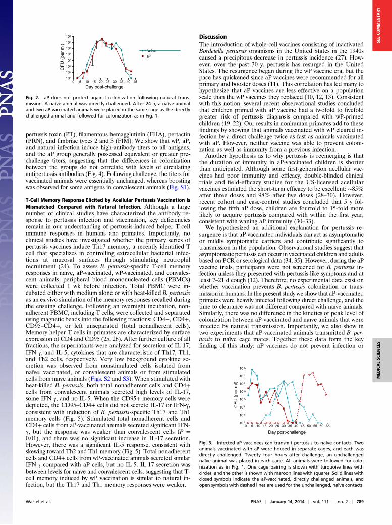
Acellular Vaccines Fail to Prevent Infection FollowingNatural Transmission.To assess the ability of vaccination to prevent pertussis infectionby transmission, two aP-vaccinated animals and one unvaccinatedanimal were cohoused with a directly challenged, unvaccinatedanimal. Similar to our previous findings (18), all animals becamecolonized 7–10 d after cohousing with the infected animal (Fig. 2).The peak levels and kinetics of colonization were indistinguishablebetween the naïve and aP-vaccinated animals.
Acellular-Vaccinated Animals Are Capable of Transmitting B. pertussisto Naïve Contacts. Because aP fails to prevent colonization wehypothesized that aP-vaccinated animals can transmit B. pertussisinfection to contacts. To test this hypothesis, two aP-vaccinatedanimals were challenged with B. pertussis and placed in separatecages. After 24 h, a naïve animal was added to each cage, and allanimals were followed for colonization. Both of the naïve ani-mals were infected by transmission from their aP-vaccinated cagemates (Fig. 3).
Vaccination and Previous Infection Induce Robust AntibodyResponses. Sera collected before vaccination or primary infectionand again at 1 wk before challenge were analyzed for IgG anti-bodies against heat-killed B. pertussis and the vaccine antigens
Table 1. Components of aP and wP vaccines used in this study
Vaccine component Daptacel Infanrix Triple antigen
Diphtheria toxoid 15 Lf 25 Lf 20–30 LfTetanus toxoid 5 Lf 10 Lf 5–25 LfWhole-cell Bordetella pertussis — — ≥4 IUInactivated pertussis toxin 10 μg 25 μg —
Filamentous hemagglutinin 5 μg 25 μg —
Pertactin 3 μg 8 μg —
Fimbriae types 2 and 3 5 μg — —
Aluminum (from aluminum phosphate) 0.33 mg ≤0.625 mg ≤1.25 mg
IU, international units; Lf, limit of flocculation units.
Naive aP wP
Conv.0
10
20
30
40
Tim
e to
cle
aran
ce(D
ay p
ost-
chal
leng
e)
BA
* †
* † ‡
‡‡
C
2/3 79/1
0 1416
/1719
/2126
/2833
/35 38 42101
102
103
104
105
106
107
108
109
wP
NaiveaP
Conv.
Day post-challenge
CFU
(per
ml)
Pre 2/3 79/1
0 1416
/1719
/21 2426
/280
10000
20000
30000
40000
50000
Naive
Conv.
aPwP
Day post-challenge
WB
C (p
er μ
l)
**
****
**
****
**
**
Fig. 1. The effect of vaccination or convalescence on colonization andleukocytosis. Naïve animals, aP-vaccinated animals, wP-vaccinated animals,and previously infected [convalescent conv.)] animals were directly chal-lenged with B. pertussis (n = 3–4 per group). (A) Colonization was monitoredby quantifying B. pertussis cfu per mL in biweekly nasopharyngeal washeswith a limit of detection of 10 cfu per mL. For each animal the time toclearance is defined as the first day that no B. pertussis cfu were recoveredfrom nasopharyngeal washes. (B) The mean time to clearance is shown foreach group (n = 3 per group). Because no B. pertussis organisms were re-covered from the conv. animals, the mean time to clearance was defined asthe first day of sampling (day 2, indicated by the dashed line). *P < 0.05 vs.Naïve, †P < 0.05 vs. aP, ‡P < 0.05 vs. wP. (C) The mean circulating white bloodcell counts before and after challenge are shown for each group of animals(n = 3–4 per group). **P < 0.01 vs. preinfection from same group.
788 | www.pnas.org/cgi/doi/10.1073/pnas.1314688110 Warfel et al.
pertussis toxin (PT), filamentous hemagglutinin (FHA), pertactin(PRN), and fimbriae types 2 and 3 (FIM). We show that wP, aP,and natural infection induce high-antibody titers to all antigens,and the aP group generally possessed equivalent or greater pre-challenge titers, suggesting that the differences in colonizationbetween the groups do not correlate with levels of circulatingantipertussis antibodies (Fig. 4). Following challenge, the titers forvaccinated animals were essentially unchanged, whereas boostingwas observed for some antigens in convalescent animals (Fig. S1).
T-Cell Memory Response Elicited by Acellular Pertussis Vaccination IsMismatched Compared with Natural Infection. Although a largenumber of clinical studies have characterized the antibody re-sponse to pertussis infection and vaccination, key deficienciesremain in our understanding of pertussis-induced helper T-cellimmune responses in humans and primates. Importantly, noclinical studies have investigated whether the primary series ofpertussis vaccines induce Th17 memory, a recently identified Tcell that specializes in controlling extracellular bacterial infec-tions at mucosal surfaces through stimulating neutrophilrecruitment (24). To assess B. pertussis-specific T-cell memoryresponses in naïve, aP-vaccinated, wP-vaccinated, and convales-cent animals, peripheral blood mononucleated cells (PBMCs)were collected 1 wk before infection. Total PBMC were in-cubated either with medium alone or with heat-killed B. pertussisas an ex vivo simulation of the memory responses recalled duringthe ensuing challenge. Following an overnight incubation, non-adherent PBMC, including T cells, were collected and separatedusing magnetic beads into the following fractions: CD4−, CD4+,CD95–CD4+, or left unseparated (total nonadherent cells).Memory helper T cells in primates are characterized by surfaceexpression of CD4 and CD95 (25, 26). After further culture of allfractions, the supernatants were analyzed for secretion of IL-17,IFN-γ, and IL-5; cytokines that are characteristic of Th17, Th1,and Th2 cells, respectively. Very low background cytokine se-cretion was observed from nonstimulated cells isolated fromnaïve, vaccinated, or convalescent animals or from stimulatedcells from naïve animals (Figs. S2 and S3). When stimulated withheat-killed B. pertussis, both total nonadherent cells and CD4+cells from convalescent animals secreted high levels of IL-17,some IFN-γ, and no IL-5. When the CD95+ memory cells weredepleted, the CD95–CD4+ cells did not secrete IL-17 or IFN-γ,consistent with induction of B. pertussis-specific Th17 and Th1memory cells (Fig. 5). Stimulated total nonadherent cells andCD4+ cells from aP-vaccinated animals secreted significant IFN-γ, but the response was weaker than convalescent cells (P =0.01), and there was no significant increase in IL-17 secretion.However, there was a significant IL-5 response, consistent withskewing toward Th2 and Th1 memory (Fig. 5). Total nonadherentcells and CD4+ cells from wP-vaccinated animals secreted similarIFN-γ compared with aP cells, but no IL-5. IL-17 secretion wasbetween levels for naïve and convalescent cells, suggesting that T-cell memory induced by wP vaccination is similar to natural in-fection, but the Th17 and Th1 memory responses were weaker.
DiscussionThe introduction of whole-cell vaccines consisting of inactivatedBordetella pertussis organisms in the United States in the 1940scaused a precipitous decrease in pertussis incidence (27). How-ever, over the past 30 y, pertussis has resurged in the UnitedStates. The resurgence began during the wP vaccine era, but thepace has quickened since aP vaccines were recommended for allprimary and booster doses (11). This correlation has led many tohypothesize that aP vaccines are less effective on a populationscale than the wP vaccines they replaced (10, 12, 13). Consistentwith this notion, several recent observational studies concludedthat children primed with aP vaccine had a twofold to fivefoldgreater risk of pertussis diagnosis compared with wP-primedchildren (19–22). Our results in nonhuman primates add to thesefindings by showing that animals vaccinated with wP cleared in-fection by a direct challenge twice as fast as animals vaccinatedwith aP. However, neither vaccine was able to prevent coloni-zation as well as immunity from a previous infection.Another hypothesis as to why pertussis is reemerging is that
the duration of immunity in aP-vaccinated children is shorterthan anticipated. Although some first-generation acellular vac-cines had poor immunity and efficacy, double-blinded clinicaltrials and field-efficacy studies for the US-licensed acellularvaccines estimated the short-term efficacy to be excellent: ∼85%after three doses and 98% after five doses (28–30). However,recent cohort and case-control studies concluded that 5 y fol-lowing the fifth aP dose, children are fourfold to 15-fold morelikely to acquire pertussis compared with within the first year,consistent with waning aP immunity (30–33).We hypothesized an additional explanation for pertussis re-
surgence is that aP-vaccinated individuals can act as asymptomaticor mildly symptomatic carriers and contribute significantly totransmission in the population. Observational studies suggest thatasymptomatic pertussis can occur in vaccinated children and adultsbased on PCR or serological data (34, 35). However, during the aPvaccine trials, participants were not screened for B. pertussis in-fection unless they presented with pertussis-like symptoms and atleast 7–21 d cough (12). Therefore, no experimental data exist onwhether vaccination prevents B. pertussis colonization or trans-mission in humans. In the present study we show that aP-vaccinatedprimates were heavily infected following direct challenge, and thetime to clearance was not different compared with naïve animals.Similarly, there was no difference in the kinetics or peak level ofcolonization between aP-vaccinated and naïve animals that wereinfected by natural transmission. Importantly, we also show intwo experiments that aP-vaccinated animals transmitted B. per-tussis to naïve cage mates. Together these data form the keyfinding of this study: aP vaccines do not prevent infection or
0 5 10 15 20 25 30 35 40 45101
102
103
104
105
106
107
108
NaiveaP
Day post-challenge
CFU
(per
ml)
Fig. 2. aP does not protect against colonization following natural trans-mission. A naïve animal was directly challenged. After 24 h, a naïve animaland two aP-vaccinated animals were placed in the same cage as the directlychallenged animal and followed for colonization as in Fig. 1.
0 5 10 15 20 25 30 35 40 45 50 55 60 65101
102
103
104
105
106
107
108
Day post-challenge
CFU
(per
ml)
Fig. 3. Infected aP vaccinees can transmit pertussis to naïve contacts. Twoanimals vaccinated with aP were housed in separate cages, and each wasdirectly challenged. Twenty four hours after challenge, an unchallengednaïve animal was placed in each cage. All animals were followed for colo-nization as in Fig. 1. One cage pairing is shown with turquoise lines withcircles, and the other is shown with maroon lines with squares. Solid lines withclosed symbols indicate the aP-vaccinated, directly challenged animals, andopen symbols with dashed lines are used for the unchallenged, naïve contacts.
Warfel et al. PNAS | January 14, 2014 | vol. 111 | no. 2 | 789
MED
ICALSC
IENCE
SSE
ECO
MMEN
TARY

transmission of Bordetella pertussis even 1 mo after completingthe primary vaccination series.We show that wP, aP, and natural infection all induce high-
antibody titers. The prechallenge titers in aP-vaccinated animalswere generally equivalent or higher than those observed inconvalescent and wP-vaccinated animals, suggesting that aP isimmunogenic in baboons and that the inability to prevent in-fection was not due to low-antibody titers. Compared with thelarge number of clinical studies that have characterized the an-tibody response to pertussis infection and vaccination, very fewhave investigated pertussis-induced helper T-cell immune re-sponses in humans. Taken as a whole, these limited data suggestthat aP vaccination induces Th2 or mixed Th2/Th1 responses,whereas wP vaccination and natural infection induce a Th1 re-sponse (13). However, none of these studies tested for Th17memory, a recently identified T cell that specializes in controllingextracellular bacterial infections at mucosal surfaces (24). Ourdata show that natural infection induced robust Th17 and Th1immunity. Animals vaccinated with wP, which cleared infectionfaster than naïve and aP-vaccinated animals, showed similar butweaker T-cell responses. wP vaccination is generally believed toinduce strong Th1 responses, but what we observed here wasrelatively weak. This observation might be explained by hetero-geneity in the manufacturing of different wP vaccines. Futurestudies will compare the immune response induced by wP vaccinesproduced by three different manufacturers. In comparison withnatural infection and wP, aP-induced immunity was mismatched,
showing a Th2 response with a weaker Th1 response and no sig-nificant Th17 response.Together, the cytokine and T-cell immunological data ob-
served in baboons are generally consistent with those observed inmice (13). We previously showed that pertussis infection inbaboons induces a mucosal immune response characterized byproduction of IL-17 and a variety of chemokines and cytokinesassociated with IL-17 signaling, including IL-6 and IL-8. Thisprimary immune response correlated with long-lived Th17 andTh1 memory responses that lasted >2 y (36). Mice infected withB. pertussis also express mucosal IL-17, IL-6, and IL-8 homologsand induce Th17 and Th1 memory (37–40). Mice vaccinated withwP also develop Th17 and Th1 memory that results in partialprotective immunity, similar to what we observed in the baboonmodel (41, 42). A recent report by Ross et al. (42) concluded thatan aP containing PT, FHA, and PRN induces Th1, Th2, andTh17 immune responses in C57BL/6 mice (42). However, a pre-vious study from the same group found Th1 and Th2 but no
Pre-immune
Pre-challenge1
10
100
1000
Ant
i-PT
IgG
(IU
/ml)
1
10
100
1000
10000
Ant
i-PR
N Ig
G (I
U/m
l)
1
10
100
1000
10000
Ant
i-FH
A Ig
G (I
U/m
l)
1
10
100
1000
10000
100000
Ant
i-FIM
IgG
(RU
/ml)*** †
**
****
***
Pre-im
mune
Pre-ch
allen
ge1
10
100
1000
10000
Naive
wPConvalescent
aP
Ant
i-B. p
. IgG
(RU
/ml)
Fig. 4. Vaccination and previous infection induce robust serum antibodyresponses. Antibody responses to the four vaccine antigens—PRN, FIM, FHA,and PT—and to heat-killed B. pertussis (B. p.) were measured by ELISA.Preimmune sera were collected from vaccinated animals before immuniza-tion and from conv. animals before initial infection (n = 3–4 per group).Because Infanrix does not contain FIM, four Daptacel-vaccinated animalswere included in the anti-FIM ELISA. Prechallenge sera were collected fromall animals 1 wk before challenge. International Units (IU) or relative units(RU) in each sample were determined by comparing the responses to theWHO international standard pertussis antiserum on each plate. ***P <0.001, **P < 0.01, *P < 0.05 vs. Convalescent. †P < 0.001 vs. wP.
NaiveConv. aP wP
0
500
1000
150025005000750010000
IL-1
7 (p
g/m
l)
Naive
Conv. aP wP0
500100015002000
CD4-CD4+CD95-CD4+
5000
15000
25000
IL-1
7 (p
g/m
l)
NaiveConv. aP wP
0
10
20
30
40
IL-5
(pg/
ml)
Naive
Conv. aP wP0
50
100
150
200
250 CD4-CD4+CD95-CD4+
IFN
- (p
g/m
l)
Naive
Conv. aP wP0
20
40
60
80
100
120
140CD4-CD4+CD95-CD4+
IL-5
(pg/
ml)
***
******A
B
C
**
***
NaiveConv. aP wP
0
50
100
150
200
250
IFN
- (p
g/m
l)
***
**
***
**
******
***
Fig. 5. Helper T-cell responses induced by vaccination and infection. PBMCcollected from naïve, aP-vaccinated, wP-vaccinated, and conv. animals 1 wkbefore infection were incubated overnight with either medium alone ormedium containing heat-killed B. pertussis (n = 3–4 per group). For eachgrowth condition, nonadherent cells were collected and either left unsep-arated (total nonadherent cells) or separated using anti-CD4 and anti-CD95magnetic particles. Total nonadherent, CD4−, CD4+, and CD95−CD4+ cellswere then cultured under the same conditions as before (with medium aloneor stimulated with heat-killed B. pertussis). After 36 h, supernatants werecollected and analyzed for IL-17 (A), IFN-γ (B), and IL-5 (C). Cytokine secretionin response to B. pertussis stimulation is presented for total nonadherentcells (Left) and separated cells (Right). ***P < 0.001, **P < 0.01, *P < 0.05 vs.same fraction from naïve animals.
790 | www.pnas.org/cgi/doi/10.1073/pnas.1314688110 Warfel et al.

significant Th17 responses in C3H/HeJ and C3H/HeN mousestrains vaccinated with an aP containing PT and FHA (41).Nevertheless, data from two clinical studies recently showednegligible Th17 recall responses (∼10 pg/mL) in PBMC isolatedfrom aP-vaccinated 4-y-old children before and after booster,suggesting aP does not induce Th17 memory in humans (43, 44).Taken as a whole, the data presented in this study suggest that
antibodies induced by aP vaccination are sufficient for prevent-ing severe pertussis symptoms but do not mitigate colonization.Inhibition of leukocytosis likely occurs through antibody-medi-ated neutralization of PT, a toxin which interferes with leukocyteextravasation by blocking chemokine receptor signaling (1). Themechanism by which aP prevents coughing despite heavy bac-terial colonization is not known but deserves further attention.On the other hand, induction of Th17/Th1 memory responsescorrelated with the ability to clear infection: convalescent andwP-vaccinated animals possessed strong Th17 responses and Th1responses and cleared infection more quickly than aP-vaccinatedanimals which lacked Th17 responses but possessed Th1/ Th2memory. Although we have not definitively shown that Th17cells are required for B. pertussis clearance, this correlation isconsistent with the role these cells play in fighting extracellularbacterial infections at mucosal surfaces by inducing neutrophilchemotaxis. The current studies were not designed to look atimmune cell recruitment to the respiratory tract, but additionalexperiments are underway to determine the role of neutrophilsin the immune response to pertussis infection and vaccination inbaboons. We are also investigating other possible mechanismsthat could prevent mucosal colonization; for example, a possiblerole for IgA and IgD which are secreted in primate lower andupper respiratory tracts, respectively (45, 46).The baboon model offers many advantages, chiefly the ability
to investigate pertussis pathogenesis, transmission, and hostimmune responses to infection and vaccination in a primatespecies that is >96% genetically similar to humans (47). How-ever, there are also several limitations associated with thismodel. There are far fewer animals available for research com-pared with smaller-animal models. In addition, there is a paucityof immunological reagents that are validated for baboons com-pared with mice and humans. Although antibodies against cellsurface markers are generally cross-reactive, anti-cytokine anti-bodies tend to be much more species-specific. For this reason wehave so far been unable to assess T-cell responses using in-tracellular cytokine staining and flow cytometry. This led us todevelop the cell separation assay as an alternative method forphenotyping the memory T-cell responses induced by pertussisinfection and vaccination (36). One limitation of our assay is thatduring the CD4+ cell purification, antigen-presenting cells suchas macrophages and dendritic cells are removed after an over-night incubation. This likely explains the low IFN-γ secretionobserved in all groups because antigen-presenting cells increaseIFN-γ secretion by antigen-specific CD4+ T cells through a pos-itive feedback loop (48). In line with this hypothesis, our previousdata showed that restimulated whole PBMC from convalescentanimals secreted much higher levels of IFN-γ. In addition,restimulation assays using human PBMC or murine splenocytesafter infection or vaccination also show higher levels of secretedIFN-γ (42, 49). Together these observations suggest that al-though our assay is valuable for phenotyping T-cell memory, itlikely underrepresents the magnitude of Th1 memory responses.We used heat-killed B. pertussis as an antigen for our restim-ulation assays because we believe this is the most relevantmethod for ex vivo simulation of T-cell memory recalled duringinfection. However, it is possible that this assay underdetectsimmune responses that would be observed had we used purifiedvaccine antigens. Another disadvantage of primate models is thatit is not feasible to directly link an immune response to pro-tection. Although protection from pertussis has been shown tobe mediated by IFN-γ and, to a lesser extent, IL-17 signalingusing knockout mouse strains lacking specific gene products (13),
the relative protection afforded by Th17 or Th1 responses invaccinated or convalescent baboons or humans is not known.Currently, a major focus of public health agencies is the pre-
vention of pertussis infection in young infants who have notcompleted their primary aP series and have considerable morbidityand mortality to pertussis infection (1). One recommendation toreduce transmission of pertussis to infants is by “cocooning,” orvaccinating people who have contact with infants (11). Our datashow that aP-vaccinated animals are infected and transmit per-tussis to naïve contacts. Consistent with these findings, seroepi-demiological studies have concluded that B. pertussis circulation isstill high in countries with excellent aP uptake (27, 50), and a cross-sectional study showed that postpartum aP vaccination of mothersdid not reduce pertussis illness in young infants (51). These datasuggest that cocooning is unlikely to be an effective strategy toreduce the burden of pertussis in infants. However, it is importantto note that our data in combination with human data show thatvaccination with aP provides excellent protection from severepertussis (52). Therefore, any short-term plan for addressing theresurgence of pertussis should include continued efforts to en-hance aP immunization. However, to protect the most vulnerablemembers of the population and achieve optimal herd immunity, itwill be necessary to develop a vaccination strategy that effectivelyblocks pertussis infection and transmission.
Materials and MethodsEthics Statement. All animal procedures were performed in a facility accreditedby the Association for Assessment and Accreditation of Laboratory Animal CareInternational in accordancewith protocols approved by the Center for BiologicsEvaluation and Research Animal Care and Use Committee and the principlesoutlined in the Guide for the Care and Use of Laboratory Animals by the In-stitute for Laboratory Animal Resources, National Research Council (53).
Bacterial Strains and Media. B. pertussis strain D420 was grown on Bordet–Gengou and Regan–Lowe plates prepared as described previously (17). Heat-killed B. pertussis was prepared by resuspending to an OD600 of 0.90 (5 × 10⁹
cfu/mL) in PBS and heating at 65 °C for 30 min.
Vaccination, Infection, and Evaluation of Baboons. Baboons obtained from theOklahoma Baboon Research Resource at the University of Oklahoma HealthSciences Center were inoculated with human doses of aP or wP administeredintramuscularly at 2, 4, and 6 mo of age. For studies using aP, equal numbersof animals were vaccinated with Daptacel (Sanofi Pasteur Ltd.) and Infanrix(GlaxoSmithKline). For wP, animals were vaccinated with Triple Antigen (Se-rum Institute of India Ltd.), which meets the World Health Organization(WHO) recommendations for potency. Naïve animals were age-matched butnot vaccinated. Previously infected animals were clear of B. pertussis infectionfor 1 to 2 mo before reinfection. Direct challenge and transmission studieswere performed as described previously (17, 18). The inoculum for each directchallenge was between 109–1010 cfu as determined by measurement of opticaldensity and confirmed by serial dilution and plating to determine the numberof cfu per mL of inoculum. Baboons were evaluated twice weekly as describedpreviously for enumeration of circulating white blood cells and serum sepa-ration (17). Nasopharyngeal washes were diluted and plated on Regan–Loweplates to quantify bacterial cell counts.
Isolation of PBMC and Cell Separation. Baboons were anesthetized, and PBMCwere isolated from peripheral blood as described previously (36) and cry-opreserved in RPMI-1640 medium supplemented with 10% (vol/vol) DMSOand 12.5% (wt/vol) BSA using Mr. Frosty containers (Nalgene). After thaw-ing, cells were washed twice and nonadherent cells were collected as de-scribed previously. For each growth condition, cells were incubatedovernight with either medium alone or medium containing heat-killed B.pertussis (50 bacteria:1 PBMC). Nonadherent cells were collected, and 2 × 10⁶
cells were left unseparated (total nonadherent cells). Using the methodpreviously described, 4 × 10⁶ cells were separated using anti-CD4 magneticparticles, and another 4 × 10⁶ cells were depleted of CD95+ cells and thenseparated with anti-CD4 magnetic particles (36). The following fractionswere collected: Total nonadherent, CD4−, CD4+, and CD95–CD4+. After in-cubation with or without heat-killed B. pertussis, cells were pelleted andsupernatants were collected for IL-17A quantitation by ELISA (Aniara) andquantitation of IFN-γ and IL-5 using the Milliplex MAP nonhuman primate kitaccording to the manufacturer’s instructions (Millipore). Data are presented as
Warfel et al. PNAS | January 14, 2014 | vol. 111 | no. 2 | 791
MED
ICALSC
IENCE
SSE
ECO
MMEN
TARY

the cytokine concentration secreted by B. pertussis-stimulated cells minus thebasal concentration secreted by cells incubated with medium alone.
Detection of Serum Antibodies to Pertussis Antigens. Nunc Maxisorp 96-wellplates were coated overnight with 0.2 μg/mL PT, 0.5 μg/mL FHA, 2 μg/mL PRN,or 0.2 μg/mL FIM (List Biologicals) as described previously (17, 54). For whole-bacteria ELISA, plates were coated overnight at 37 °C with heat-killedB. pertussis prepared as described above. Serum IgG for each antigen wasmeasured as described previously (17). Each plate contained a standardcurve from the WHO international standard pertussis antiserum (NationalInstitute for Biological Standards and Control) used to assign internationalunits for PT, FHA, and PRN and relative units for FIM and heat-killed B.pertussis by comparison with the linear portion of the standard curve. Be-cause Infanrix does not contain FIM, only Daptacel-vaccinated animals wereincluded in the anti-FIM ELISA.
Statistics. All data are reported as mean ± SEM. Statistical analyses wereperformed by ANOVA with post hoc t test using JMP (version 9) software(SAS Institute, Inc.). Antibody and cytokine data were normalized by logtransformation before analysis.
ACKNOWLEDGMENTS. We thank Dr. John Dennis, Dr. Jill Ascher, LewisShankle, Perry Altland, and Ernest Madison for technical assistance. Wethank Dr. Gary White, Dr. Roman Wolf, and Dr. James Papin for technicalassistance and critical discussions. We thank Dr. Drusilla Burns and Dr. ScottStibitz for critical reading of the manuscript. This work was funded by theFood and Drug Administration and National Institutes of Health/NationalInstitute of Allergy and Infectious Diseases through Interagency AgreementY1-AI-1727-01. Baboons were obtained from the Oklahoma Baboon Re-search Resource. The Oklahoma Baboon Research Resource was supportedby Grants P40RR012317 and 5R24RR016556-10 from the National Institutesof Health National Center for Research Resources.
1. Mattoo S, Cherry JD (2005) Molecular pathogenesis, epidemiology, and clinicalmanifestations of respiratory infections due to Bordetella pertussis and other Bordetellasubspecies. Clin Microbiol Rev 18(2):326–382.
2. Heininger U (2010) Update on pertussis in children. Expert Rev Anti Infect Ther 8(2):163–173.
3. Cherry JD, Heininger U (2009) Pertussis and other Bordetella infections. Textbook ofPediatric Infectious Diseases, eds Feigin RD, Cherry JD, Demmler-Harrison GJ,Kaplan SL (W.B. Saunders, Philadelphia), pp 1683–1706.
4. Centers for Disease Control and Prevention (CDC) (2012) National, state, and localarea vaccination coverage among children aged 19-35 months—United States, 2011.MMWR Morb Mortal Wkly Rep 61:689–696.
5. CDC (2012) Notifiable diseases and mortality tables. MMWR Morb Mortal Wkly Rep61(52):ND-719–ND-732.
6. Hozbor D, et al. (2009) Pertussis epidemiology in Argentina: Trends over 2004-2007.J Infect 59(4):225–231.
7. Quinn HE, McIntyre PB (2007) Pertussis epidemiology in Australia over the decade1995-2005–trends by region and age group. Commun Dis Intell 31(2):205–215.
8. Jackson DW, Rohani P (2013) Perplexities of pertussis: Recent global epidemiologicaltrends and their potential causes. Epidemiol Infect, available at http://dx.doi.org/10.1017/S0950268812003093.
9. Celentano LP, Massari M, Paramatti D, Salmaso S, Tozzi AE; EUVAC-NET Group (2005)Resurgence of pertussis in Europe. Pediatr Infect Dis J 24(9):761–765.
10. Cherry JD (2012) Epidemic pertussis in 2012—the resurgence of a vaccine-preventabledisease. N Engl J Med 367(9):785–787.
11. Clark TA, Messonnier NE, Hadler SC (2012) Pertussis control: Time for something new?Trends Microbiol 20(5):211–213.
12. Zhang L, Prietsch SO, Axelsson I, Halperin SA (2012) Acellular vaccines for preventingwhooping cough in children. Cochrane Database Syst Rev 3:CD001478.
13. Higgs R, Higgins SC, Ross PJ, Mills KH (2012) Immunity to the respiratory pathogenBordetella pertussis. Mucosal Immunol 5(5):485–500.
14. Friedrich MJ (2011) Research aims to boost pertussis control. JAMA 306(1):27–29.15. Poland GA (2012) Pertussis outbreaks and pertussis vaccines: New insights, new
concerns, new recommendations? Vaccine 30(49):6957–6959.16. Elahi S, Holmstrom J, Gerdts V (2007) The benefits of using diverse animal models for
studying pertussis. Trends Microbiol 15(10):462–468.17. Warfel JM, Beren J, Kelly VK, Lee G, Merkel TJ (2012) Nonhuman primate model of
pertussis. Infect Immun 80(4):1530–1536.18. Warfel JM, Beren J, Merkel TJ (2012) Airborne transmission of Bordetella pertussis.
J Infect Dis 206(6):902–906.19. Witt MA, Arias L, Katz PH, Truong ET, Witt DJ (2013) Reduced risk of pertussis among
persons ever vaccinated with whole cell pertussis vaccine compared to recipients ofacellular pertussis vaccines in a large US cohort. Clin Infect Dis 56(9):1248–1254.
20. Liko J, Robison SG, Cieslak PR (2013) Priming with whole-cell versus acellular pertussisvaccine. N Engl J Med 368(6):581–582.
21. Sheridan SL, Ware RS, Grimwood K, Lambert SB (2012) Number and order of wholecell pertussis vaccines in infancy and disease protection. JAMA 308(5):454–456.
22. Klein NP, Bartlett J, Fireman B, Rowhani-Rahbar A, Baxter R (2013) Comparative ef-fectiveness of acellular versus whole-cell pertussis vaccines in teenagers. Pediatrics131(6):e1716–1722.
23. Rowlands HE, et al. (2010) Impact of rapid leukodepletion on the outcome of severeclinical pertussis in young infants. Pediatrics 126(4):e816–e827.
24. Kolls JK, Khader SA (2010) The role of Th17 cytokines in primary mucosal immunity.Cytokine Growth Factor Rev 21(6):443–448.
25. Wei S, Zhao E, Kryczek I, Zou W (2012) Th17 cells have stem cell-like features andpromote long-term immunity. OncoImmunology 1(4):516–519.
26. Pitcher CJ, et al. (2002) Development and homeostasis of T cell memory in rhesusmacaque. J Immunol 168(1):29–43.
27. Libster R, Edwards KM (2012) Re-emergence of pertussis: What are the solutions?Expert Rev Vaccines 11(11):1331–1346.
28. Greco D, et al.; Progetto Pertosse Working Group (1996) A controlled trial of two acel-lular vaccines and one whole-cell vaccine against pertussis. N Engl J Med 334(6):341–348.
29. Gustafsson L, Hallander HO, Olin P, Reizenstein E, Storsaeter J (1996) A controlled trialof a two-component acellular, a five-component acellular, and a whole-cell pertussisvaccine. N Engl J Med 334(6):349–355.
30. Misegades LK, et al. (2012) Association of childhood pertussis with receipt of 5 doses ofpertussis vaccine by time since last vaccine dose, California, 2010. JAMA 308(20):2126–2132.
31. Klein NP, Bartlett J, Rowhani-Rahbar A, Fireman B, Baxter R (2012) Waning protectionafter fifth dose of acellular pertussis vaccine in children. N Engl J Med 367(11):1012–1019.
32. Tartof SY, et al. (2013) Waning immunity to pertussis following 5 doses of DTaP.Pediatrics 131(4):e1047–e1052.
33. Witt MA, Katz PH, Witt DJ (2012) Unexpectedly limited durability of immunity fol-lowing acellular pertussis vaccination in preadolescents in a North American out-break. Clin Infect Dis 54(12):1730–1735.
34. Deen JL, et al. (1995) Household contact study of Bordetella pertussis infections. ClinInfect Dis 21(5):1211–1219.
35. Heininger U, KleemannWJ, Cherry JD, Group SIDSS; Sudden Infant Death Syndrome StudyGroup (2004)A controlled study of the relationship betweenBordetella pertussis infectionsand sudden unexpected deaths among German infants. Pediatrics 114(1):e9–e15.
36. Warfel JM, Merkel TJ (2013) Bordetella pertussis infection induces a mucosal IL-17response and long-lived Th17 and Th1 immune memory cells in nonhuman primates.Mucosal Immunol 6(4):787–796.
37. Andreasen C, Powell DA, Carbonetti NH (2009) Pertussis toxin stimulates IL-17 pro-duction in response to Bordetella pertussis infection in mice. PLoS ONE 4(9):e7079.
38. Zhang X, et al. (2011) Interleukin-1 receptor signaling is required to overcome theeffects of pertussis toxin and for efficient infection- or vaccination-induced immunityagainst Bordetella pertussis. Infect Immun 79(1):527–541.
39. Fennelly NK, et al. (2008) Bordetella pertussis expresses a functional type III secretionsystem that subverts protective innate and adaptive immune responses. Infect Immun76(3):1257–1266.
40. Zhang X, Goel T, Goodfield LL, Muse SJ, Harvill ET (2011) Decreased leukocyte accu-mulation and delayed Bordetella pertussis clearance in IL-6-/- mice. J Immunol 186(8):4895–4904.
41. Higgins SC, Jarnicki AG, Lavelle EC, Mills KH (2006) TLR4 mediates vaccine-inducedprotective cellular immunity to Bordetella pertussis: Role of IL-17-producing T cells.J Immunol 177(11):7980–7989.
42. Ross PJ, et al. (2013) Relative contribution of Th1 and Th17 cells in adaptive immunityto Bordetella pertussis: Towards the rational design of an improved acellular pertussisvaccine. PLoS Pathog 9(4):e1003264.
43. Schure RM, et al. (2012) T-cell responses before and after the fifth consecutive acellularpertussis vaccination in 4-year-old Dutch children. Clin Vaccine Immunol 19(11):1879–1886.
44. Schure RM, et al. (2012) Pertussis circulation has increased T-cell immunity duringchildhood more than a second acellular booster vaccination in Dutch children 9 yearsof age. PLoS ONE 7(7):e41928.
45. Sørensen CH, Larsen PL (1988) IgD in nasopharyngeal secretions and tonsils fromotitis-prone children. Clin Exp Immunol 73(1):149–154.
46. Pabst O (2012) New concepts in the generation and functions of IgA. Nat Rev Im-munol 12(12):821–832.
47. Perry DL, Bollinger L, White GL (2012) The Baboon (Papio spp.) as a model of humanEbola virus infection. Viruses 4(10):2400–2416.
48. Schroder K, Hertzog PJ, Ravasi T, Hume DA (2004) Interferon-gamma: An overview ofsignals, mechanisms and functions. J Leukoc Biol 75(2):163–189.
49. Ausiello CM, Urbani F, la Sala A, Lande R, Cassone A (1997) Vaccine- and antigen-dependent type 1 and type 2 cytokine induction after primary vaccination of infantswith whole-cell or acellular pertussis vaccines. Infect Immun 65(6):2168–2174.
50. Lai FY, et al. (2012) Comparative seroepidemiology of pertussis, diphtheria and po-liovirus antibodies in Singapore: Waning pertussis immunity in a highly immunizedpopulation and the need for adolescent booster doses. Vaccine 30(24):3566–3571.
51. Castagnini LA, et al. (2012) Impact of maternal postpartum tetanus and diphtheriatoxoids and acellular pertussis immunization on infant pertussis infection. Clin InfectDis 54(1):78–84.
52. Nilsson L, Lepp T, von Segebaden K, Hallander H, Gustafsson L (2012) Pertussis vac-cination in infancy lowers the incidence of pertussis disease and the rate of hospi-talisation after one and two doses: Analyses of 10 years of pertussis surveillance.Vaccine 30(21):3239–3247.
53. Committee on Care and Use of Laboratory Animals (1985) Guide for the Care and Useof Laboratory Animals (Natl Inst Health, Bethesda), DHHS Publ No (NIH) 85–23.
54. Meade BD, et al. (1995) Description and evaluation of serologic assays used ina multicenter trial of acellular pertussis vaccines. Pediatrics 96(3 Pt 2):570–575.
792 | www.pnas.org/cgi/doi/10.1073/pnas.1314688110 Warfel et al.

Supporting InformationWarfel et al. 10.1073/pnas.1314688110
Pre Prim
e
Pre ch
allen
ge 7 1426
/281
10
100
1000
10000
Ant
i-Prn
IgG
(IU
/ml)
PrePrim
e
Pre ch
allen
ge 7 1426
/281
10
100
1000
10000
100000
Ant
i-FH
A Ig
G (I
U/m
l)
Pre Prim
e
Prech
allen
ge 7 1426
/281
10
100
1000
10000
100000
Ant
i-Fim
IgG
(RU
/ml)
Pre Prim
e
Pre ch
allen
ge 7 1426
/281
10
100
1000
10000
Ant
i-PT
IgG
(IU
/ml)
Pre Prim
e
Pre ch
allen
ge 7 1426
/281
10
100
1000
10000
100000Naive
wP
ConvalescentaP
Ant
i- B. p
. IgG
(RU
/ml)
Fig. S1. Comparison of serum antibody responses before and after challenge. Antibody responses to the four vaccine antigens—pertactin (PRN), fimbriaetypes 2 and 3 (FIM), filamentous hemagglutinin (FHA), and pertussis toxin (PT)—and to heat-killed B. pertussis (B. p.) were measured by ELISA. Data forpreimmune and prechallenge responses are reproduced from Fig. 4 for comparison with postchallenge responses. Postchallenge sera were collected from allanimals on the indicated days after challenge. International Units (IU) or relative units (RU) in each sample were determined by comparing the responses to theWHO international standard pertussis antiserum on each plate.
Warfel et al. www.pnas.org/cgi/content/short/1314688110 1 of 3
Medium Heat-killed B.p.
Naive
Conv. aP wP0
500
1000
1500
IL-1
7 (p
g/m
l)
Naive
Conv. aP wP0
500
1000
1500
5000
10000
15000
IL-1
7 (p
g/m
l)
Naive
Conv. aP wP0
50100150200250300350400450
IFN
- (p
g/m
l)
Naive
Conv. aP wP0
50100150200250300350400450
IFN
- (p
g/m
l)
Naive
Conv. aP wP0
10
20
30
40
50
60
IL-5
(pg/
ml)
Naive
Conv. aP wP0
10
20
30
40
50
60IL
-5 (p
g/m
l)
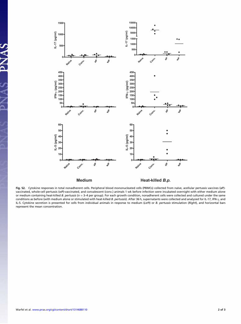
Fig. S2. Cytokine responses in total nonadherent cells. Peripheral blood mononucleated cells (PBMCs) collected from naïve, acellular pertussis vaccines (aP)-vaccinated, whole-cell pertussis (wP)-vaccinated, and convalescent (conv.) animals 1 wk before infection were incubated overnight with either medium aloneor medium containing heat-killed B. pertussis (n = 3–4 per group). For each growth condition, nonadherent cells were collected and cultured under the sameconditions as before (with medium alone or stimulated with heat-killed B. pertussis). After 36 h, supernatants were collected and analyzed for IL-17, IFN-γ, andIL-5. Cytokine secretion is presented for cells from individual animals in response to medium (Left) or B. pertussis stimulation (Right), and horizontal barsrepresent the mean concentration.
Warfel et al. www.pnas.org/cgi/content/short/1314688110 2 of 3
Naive CD4-
Naive CD4+
Naive CD95
-CD4+
Conv.CD4-
Conv.CD4+
Conv.CD95
-CD4+
aP C
D4-
aP C
D4+
aP C
D95-C
D4+
wP CD4-
wP CD4+
wP CD95
-CD4+
0
1000
2000
3000
IL-1
7 (p
g/m
l)Naiv
e CD4-
Naive C
D4+
Naive CD95
-CD4+
Conv. CD4-
Conv. CD4+
Conv. CD95
-CD4+
aP C
D4-
aP C
D4+
aP C
D95-C
D4+
wP CD4-
wP CD4+
wP CD95
-CD4+
0
1000
2000
3000
8000
24000
40000
IL-1
7 (p
g/m
l)
Naive C
D4-
Naive C
D4+
Naive C
D95-C
D4+
Conv. CD4-
Conv. CD4+
Conv. CD95
-CD4+
aP C
D4-
aP C
D4+
aP C
D95-C
D4+
wP CD4-
wP CD4+
wP CD95
-CD4+
0
100
200
300
400
IFN
- (p
g/m
l)
Naive C
D4-
Naive C
D4+
Naive C
D95-C
D4+
Conv. CD4-
Conv. CD4+
Conv. CD95
-CD4+
aP C
D4-
aP C
D4+
aP C
D95-C
D4+
wP CD4-
wP CD4+
wP CD95
-CD4+
0
100
200
300
400
IFN
- (p
g/m
l)
Naive C
D4-
Naive C
D4+
Naive C
D95-C
D4+
Conv.CD4-
Conv.CD4+
Conv.CD95
-CD4+
aP C
D4-
aP C
D4+
aP C
D95-C
D4+
wP CD4-
wP CD4+
wP CD95
-CD4+
0
25
50
75
100
125
150
175
IL-5
(pg/
ml)
Naive C
D4-
Naive C
D4+
Naive C
D95-C
D4+
Conv.CD4-
Conv.CD4+
Conv.CD95
-CD4+
aP C
D4-
aP C
D4+
aP C
D95-C
D4+
wP CD4-
wP CD4+
wP CD95
-CD4+
0
25
50
75
100
125
150
175IL
-5 (p
g/m
l)
Medium Heat-killed B.p.
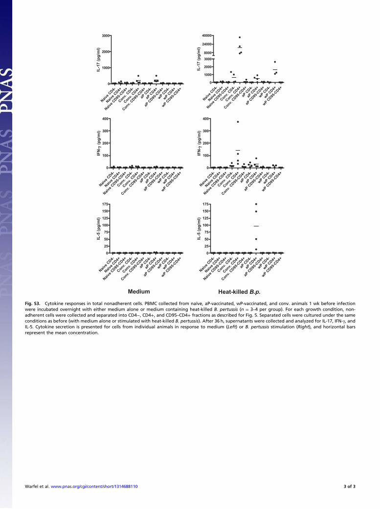
Fig. S3. Cytokine responses in total nonadherent cells. PBMC collected from naïve, aP-vaccinated, wP-vaccinated, and conv. animals 1 wk before infectionwere incubated overnight with either medium alone or medium containing heat-killed B. pertussis (n = 3–4 per group). For each growth condition, non-adherent cells were collected and separated into CD4−, CD4+, and CD95–CD4+ fractions as described for Fig. 5. Separated cells were cultured under the sameconditions as before (with medium alone or stimulated with heat-killed B. pertussis). After 36 h, supernatants were collected and analyzed for IL-17, IFN-γ, andIL-5. Cytokine secretion is presented for cells from individual animals in response to medium (Left) or B. pertussis stimulation (Right), and horizontal barsrepresent the mean concentration.
Warfel et al. www.pnas.org/cgi/content/short/1314688110 3 of 3