accidental diagnosis and management of a rare case of ... · (struma cordis). postoperative thyroid...
TRANSCRIPT

Remedy Publications LLC., | http://surgeryresearchjournal.com
World Journal of Surgery and Surgical Research
2019 | Volume 2 | Article 10861
Accidental Diagnosis and Management of a Rare Case of Intracardiac Ectopic Thyroid Mass in Premenopausal
Female Presenting with Infertility
OPEN ACCESS
*Correspondence:Reham I El-Mahdy, Department of
Medical Biochemistry, Assiut University, Egypt,
E-mail: [email protected] Date: 21 Nov 2018Accepted Date: 04 Jan 2019Published Date: 07 Jan 2019
Citation: El-Mahdy RI, Mostafa MM, Aboulhagag NA, Moustafa AM. Accidental Diagnosis
and Management of a Rare Case of Intracardiac Ectopic Thyroid Mass in
Premenopausal Female Presenting with Infertility. World J Surg Surgical Res.
2019; 2: 1086.
Copyright © 2019 Reham I El-Mahdy. This is an open access
article distributed under the Creative Commons Attribution License, which permits unrestricted use, distribution,
and reproduction in any medium, provided the original work is properly
cited.
Case ReportPublished: 07 Jan, 2019
AbstractEctopic thyroid tissue refers to the presence of thyroid tissue in positions other than normal neck region among the second and fourth tracheal cartilages. Intracardiac ectopic thyroid tissue is very rare. Here, we report a case of a 35 years old female with a history of primary infertility and irregular uterine bleeding. Echocardiography revealed large right ventricular mass during preoperative evaluation. Successful resection of the mass that originating from the right ventricular aspect of the interventricular septum and protruding into right ventricular outflow tract was done. Histological examination displayed that the mass was composed totally of thyroid tissue. According to previously reported cases of intracardiac ectopic thyroid worldwide; there is no case in Egypt reported before.
Keywords: Ectopic; Intracardiac; Thyroid
IntroductionEctopic thyroid tissue is a rare developmental malformation resulting from abnormal migration
of thyroid tissue during the embryonic descent from the floor of the primitive foregut to its final pretracheal position [1]. Major percent of ectopic thyroid tissue is located in the base of the tongue and a minority is located anteriorly near the hyoid bone [2]. Ectopic thyroid tissue in the heart is extremely rare and unanticipated [1]. Here, we present a case of intracardiac ectopic thyroid mass which was successfully surgically resected.
Case PresentationA 35 years female was presented with primary infertility and irregular uterine bleeding. She
sought gynecological counseling and abdominal sonar revealed multiple uterine leiomyomas with large submucosal one that interfere with pregnancy. On preparation for gynecological surgery and during the preoperative assessment, three-dimensional echocardiography revealed large right ventricular mass protruding in the infundibulum with an increased gradient across right ventricular outflow tract. So, we decided surgical resection of the mass.
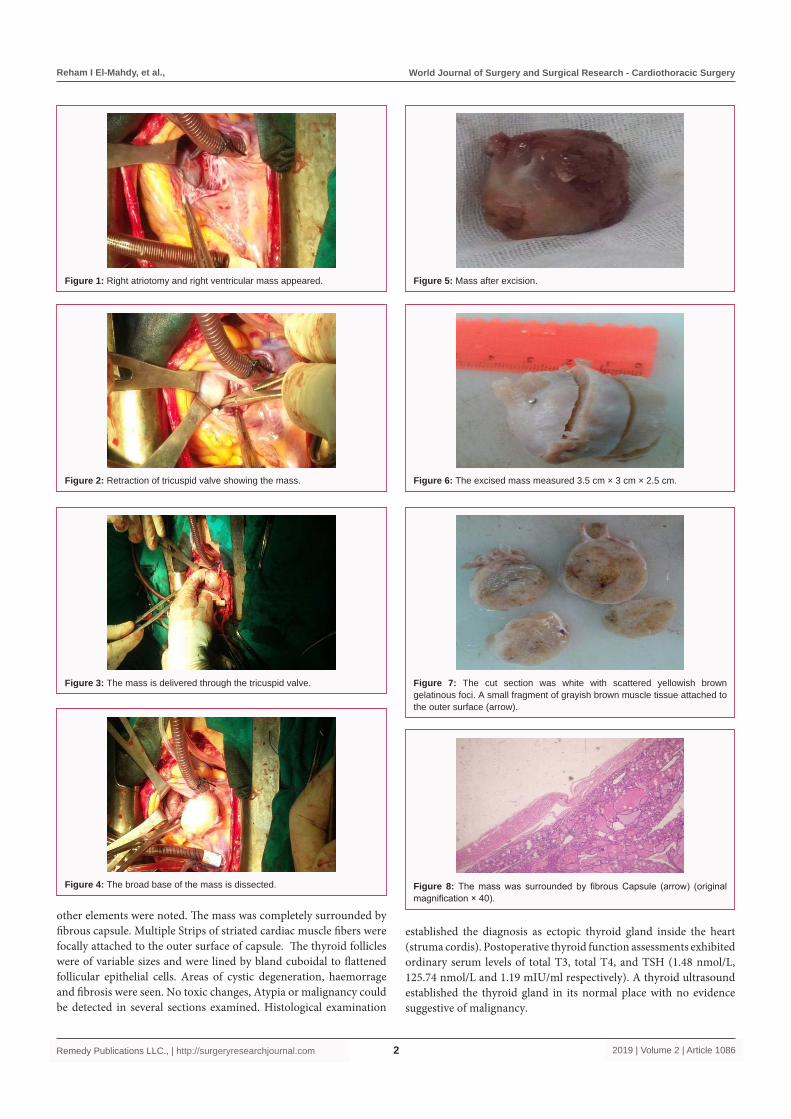
General Anesthesia, open heart surgery is done via median sternotomy with cardiopulmonary bypass aortobicaval cannulation and snaring superior and inferior vena cavae. Cold blood cardioplegia and normothermic cardiac arrest were used. Right atriotomy was done and retracting the tricuspid valve using two lid retractors and right globular ventricular mass is evident (Figure 1 and 2) and delivered digitally by index finger from the right ventricular outflow tract (Figure 3) then the broad- base of the mass is freed from below the septal leaflet of the tricuspid valve using a right-angled artery and scalpel (Figure 4). No residual ventricular septal defect. The mass after removal is shown in (Figure 5). Grossly, the excised specimen was a globular, well capsulated mass measuring 3.5 cm × 3 cm × 2.5 cm. its outer surface was smooth, the cut section was firm in consistency, whitish in color with scattered yellowish brown gelatinous foci. A small fragment of grayish brown muscle tissue was attached to the outer surface at one side of the received specimen (base of the mass) (Figure 6-10). A total of 9 sections from different parts of the mass were submitted for histopatholgical diagnosis.
Histological examination showed that the mass was composed totally of thyroid tissue and no
Reham I El-Mahdy1*, Mohammed Mahmoud Mostafa2, Noha A Aboulhagag3 and Asmaa Mahmoud Moustafa3
1Department of Medical Biochemistry, Assiut University, Egypt
2Department of Cardiothoracic Surgery, Assiut University Hospital, Egypt
3Department of Pathology, Assiut University, Egypt
Reham I El-Mahdy, et al., World Journal of Surgery and Surgical Research - Cardiothoracic Surgery
2019 | Volume 2 | Article 10862Remedy Publications LLC., | http://surgeryresearchjournal.com
other elements were noted. The mass was completely surrounded by fibrous capsule. Multiple Strips of striated cardiac muscle fibers were focally attached to the outer surface of capsule. The thyroid follicles were of variable sizes and were lined by bland cuboidal to flattened follicular epithelial cells. Areas of cystic degeneration, haemorrage and fibrosis were seen. No toxic changes, Atypia or malignancy could be detected in several sections examined. Histological examination
established the diagnosis as ectopic thyroid gland inside the heart (struma cordis). Postoperative thyroid function assessments exhibited ordinary serum levels of total T3, total T4, and TSH (1.48 nmol/L, 125.74 nmol/L and 1.19 mIU/ml respectively). A thyroid ultrasound established the thyroid gland in its normal place with no evidence suggestive of malignancy.
Figure 1: Right atriotomy and right ventricular mass appeared.
Figure 2: Retraction of tricuspid valve showing the mass.
Figure 3: The mass is delivered through the tricuspid valve.
Figure 4: The broad base of the mass is dissected.
Figure 5: Mass after excision.
Figure 6: The excised mass measured 3.5 cm × 3 cm × 2.5 cm.
Figure 7: The cut section was white with scattered yellowish brown gelatinous foci. A small fragment of grayish brown muscle tissue attached to the outer surface (arrow).
Figure 8: The mass was surrounded by fibrous Capsule (arrow) (original magnification × 40).

Reham I El-Mahdy, et al., World Journal of Surgery and Surgical Research - Cardiothoracic Surgery
2019 | Volume 2 | Article 10863Remedy Publications LLC., | http://surgeryresearchjournal.com
DiscussionHere we report a rare case of right ventricular ectopic thyroid
tissue in 32 years female patient. To the best of our knowledge, no cases of intracardiac ectopic thyroid tissue were reported in Egypt. Our histological results diagnose ectopic thyroid tissue in the heart and rule out metastatic thyroid carcinoma in the heart or cardiac teratoma. The global incidence of the ectopic thyroid is 1 per 300,000 to 400,000. The incidence of patients cured and observed for thyroid disease is 1 per 4000 to 8000 [2]. Ectopia is triggered by a trouble in initial embryonic growth. The body's endocrine glands start to grow on around the 24th day of pregnancy as a propagation of endodermal cells on the base of the primitive pharyngula. In this duration the thyroid primordium is in near proximity to adjacent constructions, comprising the embryonic heart. In the following growth, the middle anlage rages with the lateral anlage, generating and expanding what is originally the thyroid diverticulum, through which the tissue slopes to its last place in the neck. The thyroid gland receipts its last form and location in the seventh week of embryonic growth. A disappointment in this descent leads to the ectopic growth of all or part of the thyroid gland, which is drawn to an alternative organ or tissue [1].
Lingual thyroid is the most common position of ectopic thyroid accounting for 90% of cases [3]. Ectopic thyroid has been described not only in the thorax (e.g., within thymus, trachea, esophagus, lung, and ascending aorta) but also in the abdomen and pelvis (e.g., within liver, gall bladder, duodenum, pancreas, and vagina) although the occurrence of ectopic thyroid tissue in the heart is uncommon, but It has been reported [4]. This may be due to the anatomical association between thyroid primordium and the bulbus cordis of the developing heart. Due to this relationship with the bulbus cordis, the ectopic thyroid may progress in the right ventricle once the heart and great vessels slope from the neck to the chest throughout development [5].
Figure 9: Strips of striated cardiac muscle fibers (arrow) were attached to the outer surface of the capsule (original magnification × 100).
Figure 10: The thyroid follicles were of variable sizes and were lined by bland cuboidal to flattened follicular epithelial cells (original magnification × 100).
Accordingly, cases of ectopic thyroid in the heart are generally limited to the right ventricle [4]. The first case discovered was on a routine autopsy by Dosch in 1941 who reported thyroid tissue in the right ventricle [6]. The first successful resection of the intracardiac ectopic thyroid was reported by LO et al. 1984 [7].
Because of the wide range of probable sites of ectopic thyroid and its benign nature, it may be incidentally noticed on a routine heart test done for unrelated reasons. Some patients are entirely asymptomatic [8]. In our case, the patient was presented with primary infertility and irregular uterine bleeding. An echocardiography was performed during fitness and revealed a large right ventricular mass. The mass almost always initiates in the interventricular septum, is commonly largely attached, and projects into the right ventricular outflow tract, where it may cause variable grades of obstruction [8]. The present case has the same location.
The majority of patients are females (30 cases of 33), usually in middle or advanced age [2]. Also, Casanova and their colleagues [8] reported that 17 of their analyzed cases were adult female as in our case. Baykut D et al. [9] reported two ectopic thyroid masses synchronously, one in the left ventricular outflow tract and the other in the ventricular septum in the same patient [9]. Peng et al. [10] analyze summary of reported 36 cases of intrapericardial ectopic thyroid tissue with 30 cases were females and 25 cases were localized in the interventricular septum, 30 cases undergo surgery with 22 cases underwent the only excision without need patch repair [10]. Josef Besik et al. report a case of 67 years old male with gastrointestinal cancer and he was found having right ventricular outflow tract mass which was surgically removed and histopathology revealed ectopic thyroid tissue [2]. So, most reported cases were adult females and localized in the right ventricular aspect of the interventricular septum and extending to right ventricular outflow tract.
The surgical results of reported cases were satisfying and so our case. We did thyroid gland ultrasonography postoperative to ensure that the excised mass is not the only thyroid tissue in the body and it showed normal size and position of both thyroid lobes. In addition, we did thyroid function tests (T3, T4 and TSH) 2 weeks postoperative and were normal [2,7,8-10].
References1. Noussios G, Anagnostis P, Goulis D, Lappas D, Natsis K. Ectopic thyroid
tissue: anatomical, clinical and surgical implications of a rare entity. Eur J Endocrinol. 2011;165(3):375-82.
2. Altay C, Erdogan N, Karasu S, Uluç E, Sarsilmaz A, Mete B, et al. CT and MRI findings of developmental abnormalities and ectopia varieties of the thyroid gland. Diagn Interv Radiol. 2012;18(4):335-43.
3. Neinas FW, Gorman CA, Devine KD, Woolner LB. Lingual thyroid: clinical characteristics of 15 cases. Ann Intern Med. 1973;79(2):205-10.
4. Ahuja K, Bhandari T, Banait-Deshmane S, Crowe DR, Sonavane SK. Incidentally detected ectopic thyroid in juxta cardiac location-Imaging and pathology. Radiol Case Rep. 2018;13(4):909-13.
5. Fujioka S, Takatsu Y, Tankawa H, Yamanaka K, Ando F. Intracardiac ectopic thyroid mass. Chest. 1996;110(5):1366-8.
6. Dosch F. Uber einen fall von glandula thyroidea accessoria intracardialis. Beitr Pathol Anat. 1941;105:244-55.
7. Lo HM, Tseng YZ, Tseng CD, Chu SH, Chuang SM, Wu TL. Intracardiac goiter: a cause of right ventricular outflow obstruction and successful operative therapy. Am J Cardiol. 1984;53(7);976-8.

Reham I El-Mahdy, et al., World Journal of Surgery and Surgical Research - Cardiothoracic Surgery
2019 | Volume 2 | Article 10864Remedy Publications LLC., | http://surgeryresearchjournal.com
8. Casanova JB, Daly RC, Edwards BS, Tazelaar HD, Thompson GB. Intracardiac ectopic thyroid. Ann Thorac Surg. 2000;70(5):1694-6.
9. Baykut D, Fiegen U, Krian A, Thiel A. Ectopic thyroid tissue in the left ventricular outflow tract. Ann Thorac Surg. 2000;69(2);620-1.
10. Peng E, Oxenham H, Foley M, Goodwin A. Right ventricular outflow tract tumour: an unsuspected intracardiac ectopic thyroid mass. Interact Cardiovasc Thorac Surg. 2013;17(5):903-5.