accessing pseudaminic acid (pse5ac7ac) containing
TRANSCRIPT

Accessing Pseudaminic Acid (Pse5Ac7Ac)
containing Glycosides through the
Characterisation of Pse5Ac7Ac
Processing Enzymes
Emily Kate Pedley Flack
PhD
University of York
Chemistry
August 2019

2
Abstract
Cell-surface carbohydrate pseudaminic acid (Pse5Ac7Ac) is known to contribute to the
virulence of several multi-drug resistant bacterial pathogens.1 Pse5Ac7Ac and its
derivatives are not commercially available in appreciable quantities and chemical synthesis
of these molecules has proved to be challenging.2–9 Access to Pse5Ac7Ac and activated
CMP-Pse5Ac7Ac has been a hindrance in studies into the biological significance of
Pse5Ac7Ac, including Pse5Ac7Ac-processing enzymes, which may be novel therapeutic
targets.1 This project aimed to characterise enzymes which process pseudaminic acid and
to chemoenzymatically synthesise glycosides which contain pseudaminic acid.
Firstly, nucleotide-activated pseudaminic acid (CMP-Pse5Ac7Ac) was produced via a
chemoenzymatic synthesis route. Six recombinant biosynthetic enzymes which are
encoded in Campylobacter jejuni and Aeromonas caviae were purified for use in this
reaction. Purification and characterisation of the resultant CMP-Pse5Ac7Ac confirmed the
role of Aeromonas caviae PseF as an α-CMP-Pse5Ac7Ac synthetase.
With CMP-Pse5Ac7Ac in-hand, a library of bacterial sialyltransferases were assayed for
activity with CMP-Pse5Ac7Ac as donor. Success from this initial screen led to the synthesis
of glycosides containing β-linked Pse5Ac7Ac, mediated by promiscuous sialyltransferases.
Efforts were made to recombinantly produce five putative glycosyltransferases which were
predicted to use CMP-Pse5Ac7Ac or a derivative as their natural donor
(pseudaminyltransferases) however, all proteins were initially insoluble. Acinetobacter
baumannii retaining pseudaminyltransferase was solubilised through the construction of
an Im9-fusion protein. Activity studies monitored by Liquid Chromatography – Mass
Spectrometry confirmed that Im9-KpsS1 could utilise CMP-Pse5Ac7Ac as a donor. Finally,
Im9-KpsS1 was used in a seven enzyme one-pot chemoenzymatic synthesis to produce α-
2,6-Pse5Ac7Ac-pNP-β-D-Glc, confirming that KpsS1 functions as a retaining
pseudaminyltransferase. To our knowledge the work presented herein details the first
examples of chemoenzymatic synthesis of glycosides containing Pse5Ac7Ac and the first in
vivo study of a pseudaminyltransferases to provide unequivocal functional characterisation
of this novel class of enzyme.

3
Contents
1. Abstract ...................................................................................................................... 2
2. Contents ..................................................................................................................... 3
3. Acknowledgements ................................................................................................... 7
4. Author’s Declaration .................................................................................................. 9
5. Chapter 1: Introduction ........................................................................................... 10
1.1 Carbohydrates in Biology ................................................................................. 10
1.1.1 Carbohydrate Structures .................................................................................. 10
1.1.1 Biological Significance of Glycosylation and Glycans ....................................... 12
1.2 Glycosyltransferases .............................................................................................. 16
1.2.1 Glycosyltransferase classification .................................................................... 16
1.2.2 Glycosyltransferase folds ................................................................................. 17
1.2.3 Glycosyltransferase function ............................................................................ 17
1.2.3 Mechanisms of glycosyltransferases ................................................................ 18
1.3 Sialic Acids ....................................................................................................... 23
1.3.1 Sialic Acid Structures and Function in Eukaryotes ........................................ 23
1.3.2 Sialic acid biosynthesis and scavenging in bacteria ......................................... 24
1.3.3 Sialic acid in bacterial glycoconjugates ............................................................ 25
1.4 Pseudaminic Acids ................................................................................................. 26
1.4.1 Occurrence and Biological Significance of Pseudaminic Acids ........................ 26
1.4.2 Biosynthesis of Nucleotide-Activated Pseudaminic Acids ................................ 28
1.4.3 Chemical synthesis towards Pseudaminic Acids and Pseudaminic Acid
glycosides ................................................................................................................. 33
1.4.4 Chemoenzymatic synthesis of Pseudaminic Acids ........................................... 44
1.4.4 Introduction to Pseudaminic Acid Processing Enzymes ................................... 47
1.5 Project Outline ........................................................................................................ 49
6. Chapter 2: Chemoenzymatic Synthesis of CMP-Pseudaminic Acid ..................... 50

4
2.1 Introduction ............................................................................................................ 50
2.2 Experimental .......................................................................................................... 54
2.2.1 Overxpression and Purification of PseB, PseC, PseH, PseG and PseI ............ 54
2.2.2 Enzymatic synthesis of Pse5Ac7Ac with Acetyl-thiocholine Iodide ................... 56
2.2.3 Small-molecule screen for potential inhibition of PseB and PseI ...................... 56
2.2.4 Recombinant Expression and purification attempt of C. jejuni PseF in E. coli .. 58
2.2.5 Expression and purification of A. caviae PseF ................................................. 60
2.2.6 Activity assay of A. caviae PseF with Pse5Ac7Ac ............................................ 61
2.2.7 Purification of Aeromonas caviae PseF for Crystallisation trials ....................... 61
2.2.8 A. caviae PseF Protein Identification by Mass Spectrometry ........................... 62
2.2.9 A. caviae PseF Circular Dichorism ................................................................... 62
2.2.10 A. caviae PseF Size Exclusion Chromatography - Multi-Angle Laser Light
Scattering ................................................................................................................. 63
2.2.11 Crystalisation trials for A. caviae PseF ........................................................... 63
2.2.12 Large scale synthesis and purification of CMP-Pse5Ac7Ac, via a one-pot six
enzyme synthesis ..................................................................................................... 64
2.3 Results ................................................................................................................... 66
2.3.1 Overxpression and Purification of PseB, PseC, PseH, PseG and PseI ............ 66
2.2.3 Enzymatic synthesis of Pse5Ac7Ac with Acetyl-thiocholine Iodide ................... 67
2.3.3 Small-molecule screen for potential inhibition of PseB and PseI ...................... 68
2.3.4 Recombinant Expression and purification attempt of C. jejuni PseF in E. coli .. 72
2.3.5 Expression and purification of A. caviae PseF ................................................. 73
2.3.6 Activity assay of A. caviae PseF with Pse5Ac7Ac ............................................ 74
2.3.7 Purification of A. caviae PseF for Crystallisation trials ...................................... 76
2.3.8 A. caviae PseF Protein Identification by Mass Spectrometry ............................ 77
2.3.9 A. caviae PseF Circular Dichorism ................................................................... 78
2.3.10 A. caviae PseF Size Exclusion Chromatography - Multi-Angle Laser Light
Scattering ................................................................................................................. 79
2.3.11 Crystallisation trials for Aeromonas caviae PseF ........................................... 79

5
2.3.12 Large scale synthesis and purification of CMP-Pse5Ac7Ac, via a one-pot six
enzyme synthesis ..................................................................................................... 80
2.4 Discussion .............................................................................................................. 83
2.5 Conclusions and Future Work ................................................................................ 90
7. Chapter 3: Sialyltransferase mediated synthesis of glycosides containing
Pseudaminic Acid .................................................................................................... 91
3.1 Introduction ............................................................................................................ 91
3.2 Experimental .......................................................................................................... 94
3.2.1 Initial Activity assays of sialyltransferase library with CMP-Pse5Ac7Ac donor .. 94
3.2.2 PmST acceptor screen with CMP-Pse5Ac7Ac as donor .................................. 96
3.2.3 Large Scale synthesis of β-Pse5Ac7Ac-2,3-pNP-β-ᴅ-Galp using PmST .......... 96
3.3 Results ................................................................................................................... 98
3.3.1 Initial Activity assays of sialyltransferase library with CMP-Pse5Ac7Ac donor .. 98
3.3.2 PmST acceptor screen with CMP-Pse5Ac7Ac as donor ................................ 100
3.3.3 Large Scale synthesis of β-Pse5Ac7Ac-2,3-pNP-β-ᴅ-Galp using Pasteurella
multocida tPm0188Ph ............................................................................................. 102
3.4 Discussion ............................................................................................................ 104
3.5 Conclusions and Future Work .............................................................................. 107
8. Chapter 4: Characterisation of Putative Pseudaminyltransferases ................... 108
4.1 Introduction .......................................................................................................... 108
4.2 Experimental ........................................................................................................ 121
4.3.1 Sequence analysis of Putative Pseudaminyltransferases............................... 121
4.3.2 Homology Modeling of Putative Inverting Pseudaminyltransferases .............. 121
4.2.3 Putative Pseudaminyltransferase Expression Trials ....................................... 121
4.2.4 Solubilisation trials of Putative Pseudaminyltransferases Lst ......................... 123
4.2.5 Detergent Screen for Solubilisation of Putative Pseudaminyltransferases Lst
and KpsS1 .............................................................................................................. 124
4.2.6 Cloning of KpsS1 Fusion Protein Plasmids .................................................... 125
4.2.7 KpsS1 Fusion Protein Expression Trials ........................................................ 127
4.2.8 Im9-KpsS1 Large Scale Expression ............................................................... 127

6
4.2.9 Im9-KpsS1 Purification .................................................................................. 127
4.2.10 Im9-KpsS1 Protein Identification Mass Spectrometry ................................... 128
4.2.11 Im9-KpsS1 Activity Screen ........................................................................... 128
4.2.12 Chemoenzymatic synthesis of glycoside containing Pse5Ac7Ac, via a one-pot,
seven-enzyme reaction using Im9-KpsS1 ............................................................... 129
4.3 Results ................................................................................................................. 131
4.3.1 Sequence analysis of Putative Pseudaminyltranferases ................................ 131
4.3.2 Further Sequence analysis and Homology Modeling of Putative Inverting
Pseudaminyltransferases ........................................................................................ 131
4.3.3 Initial Expression trials of Putative Pseudaminyltranferases ........................... 134
4.3.4 Expression trials of Putative Pseudaminyltransferases, Lst and KpsS1 in E. coli
Tuner (DE3) ............................................................................................................ 139
4.3.5 Solubilisation trials of Putative Pseudaminyltransferase Lst ........................... 141
4.3.6 Detergent Screen for Solubilisation of Putative Pseudaminyltransferases Lst
and KpsS1 .............................................................................................................. 142
4.3.7 KpsS1 Fusion Plasmids ................................................................................. 147
4.3.8 KpsS1 Fusion Proteins Expression Trials ...................................................... 147
4.3.9 Im9-KpsS1 Purification .................................................................................. 150
4.3.10 Im9-KpsS1 Protein Identification Mass Spectrometry ................................... 150
4.3.11 Im9-KpsS1 Activity Screen ........................................................................... 151
4.3.12 Chemoenzymatic synthesis of glycoside containing Pse5Ac7Ac, via a one-pot,
seven-enzyme reaction using Im9-KpsS1 ............................................................... 157
4.4 Discussion ............................................................................................................ 161
4.5 Conclusions and Future Work .............................................................................. 165
9. Chapter 5: Concluding Remarks and Future Prospectives ................................. 167
General methods .................................................................................................... 206
10. Abbreviations ......................................................................................................... 209
11. References ............................................................................................................. 214

7
Acknowledgements
First and foremost, I would like to thank my supervisor’s Dr Martin Fascione and Professor
Gavin Thomas for giving me the opportunity to start this PhD and for your on-going
patience and encouragement that enabled me to complete it. Martin, thank you for your
efforts in teaching me chemistry and for somehow making it less daunting than it used to
be. Gavin, thank you for reminding me how fun biology is. I am grateful to you both for
allowing me to experience the best parts of both worlds throughout this PhD. Additional
thanks are owed to my TAP member Professor Jen Potts for advice on this research during
meetings.
Thank you to the technical staff in the departments of Biology and Chemistry for their
assistance in this project. I would specifically like to thank Dr Ed Bergström, for always
ensuring that a working LC-MS was available to the Fascione group.
I would like to thank Dr Kun Huang for your help with sialyltransferase work at MIB. Thanks
to Tasha for sugar synthesis and patiently helping me with chemistry. Reyme, thank you for
introducing me to crystallography. Thank you to the wonderful Darshita, Tess and Clare for
answering my stupid questions and for always being willing to help. “Lovely Darshita” thank
you for sneakily making that dissacharide. Also, thank you for your tough-love approach. It
was just what I needed. Julia, thank you for sharing your scientific wisdom and for keeping
the place running. I’m sure the fridges, freezers and gel room will be a lot emptier once I’ve
finished. (Sorry about that.) We do not deserve you!
Thank you to the Thomas lab for ensuring there was always somewhere I could go to discuss
interesting biology and for allowing me to raid the lab for supplies. Huge thanks are owed
to Martin, Alison, Lianne and Julia for their hard work in creating a brilliant lab to work in
and finding great team members to work here. To all of the Parkin-Fascione-Willems lab
members (B Block babes), thanks to each of you for making this such a weird, yet wonderful
place to work! Please do not change. Thanks for the tea-breaks (usually instigated by Mark),
numerous pub lunches (Nick, you are to blame for most of these) and many other bizzare
distractions, that helped make days at work so great.

8
I would like to thank the other three quarters of the Fascione Four: Richard, Harriet and
Robin, for five years of friendship, understanding and advice. Rich, thanks for cheering me
up with chats of home. Harriet, thank you for being the better half of “Hamily”/Team Pse.
Robin, you have been the greatest PhD buddy I could have wished to be stuck with! (I guess
we can finally stop asking one another “can we quit yet?”). This would have been
significantly harder and much less fun without you guys.
To my family and closest friends, thank you for your continued love and support. Mum and
Eddy- thank you for subtly asking Matt for an update, whenever I was being evasive. Also,
thanks for knowing that the only proper way to get me back to Baildon from York, is to
drive us over the moors! I’d like to acknowledge my Granny and Grandad for their influence
in my decision to spend seven years studying science in York. If only we’d known on our
many day trips here, that I’d end up calling it home. Finally, Matt, thank you for being my
very best friend and for always being on my team.

9
Author’s Declaration
I declare that I am the sole author of this thesis. The work presented here was neither
published before nor used previously to obtain a degree at this or another university. The
work presented was carried out by me with the exception of:
Chapter 2:
• Compounds screened as PseB and PseI inhibitors were synthesised by Joe Ferner,
University of Sheffield.
• A. caviae PseB and A. caviae PseI was purified by Joe Ferner, University of Sheffield.
• SNAc was synthesised by Dr Harriet Chidwick
• SEC-MALLS data was obtained and analysed by Dr Andrew Leech for the University
of York Technology Facility.
• PseF crystals were fished by Reyme Herman and diffraction data was collected by
Sam Hart.
Chapter 3:
• Initial sialyltransferase activity screen with LacNAc-ITag was performed with Dr Kun
Huang (KH). Mass spectrometry data for these samples was collected by KH at
University of Manchester.
Chapter 4:
• Synthesis and characterisation of β-ᴅ-Glcp-2,6-β-ᴅ-Galp-OMe was performed by Dr
Darshita Budhadev and Natasha Hatton.
• Trypsin digest and MS analysis of Im9-KpsS1 was performed by Rachel Bates for the
University of York Technology Facility
10
Chapter 1: Introduction
1.1 Carbohydrates in Biology
1.1.1 Carbohydrate Structures
All cell surfaces, without known exception, are covered with carbohydrates.10 Within
biological systems carbohydrates may be covalently linked to one-another to form oligo-
or polysaccharides (glycans) or linked to other classes of biomolecules to form
glycoconjugates, including glycolipids and glycoproteins, the latter generated by post-
translational, for instance modification of serine and threonine residues. The complexity of
carbohydrate structures must be considered in order to appreciate the challenges in
studying this large class of biomolecules. Beginning with monosaccharides, these units may
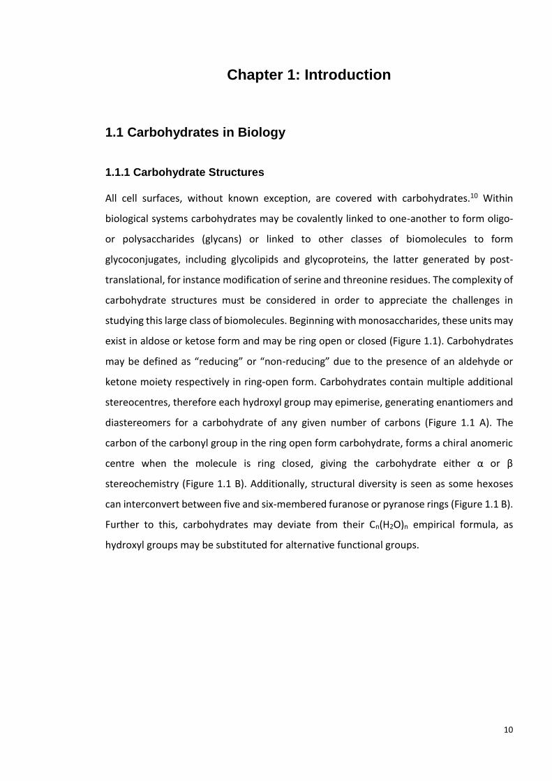
exist in aldose or ketose form and may be ring open or closed (Figure 1.1). Carbohydrates
may be defined as “reducing” or “non-reducing” due to the presence of an aldehyde or
ketone moiety respectively in ring-open form. Carbohydrates contain multiple additional
stereocentres, therefore each hydroxyl group may epimerise, generating enantiomers and
diastereomers for a carbohydrate of any given number of carbons (Figure 1.1 A). The
carbon of the carbonyl group in the ring open form carbohydrate, forms a chiral anomeric
centre when the molecule is ring closed, giving the carbohydrate either α or β
stereochemistry (Figure 1.1 B). Additionally, structural diversity is seen as some hexoses
can interconvert between five and six-membered furanose or pyranose rings (Figure 1.1 B).
Further to this, carbohydrates may deviate from their Cn(H2O)n empirical formula, as
hydroxyl groups may be substituted for alternative functional groups.

11
Figure 1.1: A: Fischer projections of hexose monosaccharides: L- and D-Glucose, an example of enantiomeric aldose carbohydrates; L- and D-Fructose, a pair of ketose enantiomers; L- and D-
Galactose, diastereomers of L- and D-Glucose. B: Ring open D-Galactose can form ring closed α- and β-Galactopyranose and α- and β-Glucofuranose (six and five membered rings respectively).

12
Carbohydrates polymerise to form glycans which are the most structurally diverse major
class of molecules. During polymerisation, glycosidic bonds are formed via condensation
reactions, usually involving the anomeric carbon. The linkage can be defined as α or β in
stereochemistry and may join the carbohydrate to its neighbouring carbohydrate at one of
several positions. Carbohydrates which form oligo- or polysaccharide may be linear or
branched structures.10
In calculating the number of possible structures for a polysaccharide of any given length,
the following factors must be accounted for: epimeric identity of each monomer, D or L
sugars, anomeric configuration of each monomer, position of glycosidic linkage, possible
branching, ring size and reducing terminal attachment. There are 1056 possible isomers for
a tetrasaccharide, increasing the oligosaccharide size to a hexasaccharide gives ~1.05 x10¹²
possible structures.11,12 Whilst many of these potential structures are not known to occur
in nature, this vast number of possible isomers adds a level of complexity when trying to
determine the structure of a chemically synthesised or biologically extracted
oligosaccharide.12 Technical difficulties in studying glycans and glycoconjugates can in-part,
account for why studies of these molecules have lagged behind other biomolecular classes.
1.1.1 Biological Significance of Glycosylation and Glycans
At the beginning of the millennium, carbohydrates were termed the “Cinderella” molecules
of biology, as the neglected, over-looked sister of the nucleic acids and proteins.13 Recently,
the ubiquity of glycosylation and glycans has become apparent and it is now accepted that
carbohydrate play significant roles beyond that of an energy source. Glycans are as
universal as nucleic acids, proteins, and lipids and essential for all living organisms.14,15 The
assembly, modification and interaction of these four biomolecular groups is crucial for cell
development and survival.14 Our understanding of the biological significance of
glycoconjugates and glycans has been predominantly gained from studies undertaken in
the past three decades.16,17
The functions of glycans, or glycosylation, may be classified into three broad groups: 1)
structural and modulatory roles, including nutrient storage; 2) recognition by other
molecules (as either extrinsic/interspecies recognition or intrinsic/intraspecies
recognition); and finally, 3) molecular mimicry of host carbohydrates.10,17

13
In addition to structural complexity, a further hindrance in the study of glycans can be
attributed to a lack of template for their biosynthesis, unlike the nucleic acids and proteins,
for which well-understood, largely universal set of instructions exist for their biosynthesis.10
A plethora of rare human diseases, collectively termed Congenital Disorders of
Glycosylation (CDG) are known to be caused by glycosylation defects. However,
heterogeneity in glycosylation and glycans makes understanding the molecular mechanism
of each CGD particularly challenging. Whilst the genetic basis of many CDG’s were known,
they were often overlooked as glycosylation patterns in laboratory-based tissue culture, do
not represent the glycosylation pattern in vivo and therefore the physiological
manifestation of the defects is altered. This not only highlights the biological significance
of eukaryotic glycosylation but indicates that many important functions of glycans are only
apparent in whole organisms.10
Despite recent advances, glycan structure and function remains unexplored in many forms
of life including bacteria, archaea, fungi and algae.17 Many classes of glycosylation were
once believed to be reserved as a eukaryotic post-translational modification. The discovery
of the Pgl pathway with Campylobacter jejuni was the first example of a general N-linked
glycosylation pathway in prokaryotic cells.18 This was a significant breakthrough in the field
of bacterial glycobiology. The types of monosaccharide found in bacteria show greater
diversity than eukaryotic monosaccharides and within bacteria carbohydrates often
undergo an extensive range of chemical modifications (including alkyl, acyl, aminoacyl,
phosphoryl, and nucleosides).19 Key cell-surface glycoconjugates in Gram-negative bacteria
include peptidoglycan, lipopolysaccharide (LPS), lipooligosaccharide (LOS), extracellular
polysaccharide (EPS), capsular polysaccharide (CPS) and glycoproteins (some of which are
depicted in Figure 1.2). Gram-positive bacteria do not contain LPS or LOS but do have
glycosylated lipotechoic acids and wall techoic acids (depicted in Figure 1.3).19
Perhaps one of the most intriguing biological roles of carbohydrates is the aforementioned
“molecular mimicry” that microbial pathogens engage in, by covering their surfaces with
glycans that are similar to host glycans, with the aim of evading host immune response or
increasing host tolerance towards the pathogen.10 This is observed with the nonulosonic
acids (NulOs), a group of nine-carbon carbohydrates, some of which are found as terminal
residues in human glycoconjugates and often in bacterial glycoconjugates.20 Interestingly,

14
phylogenetic analysis of NulO biosynthesis and N-glycosylation genes revealed that NulOs
and N-glycosylation pathways may have originated in bacteria and archaea.21,22
Figure 1.2: Depiction of Gram-Negative cell wall and examples of cell surface glycoconjugates, LPS and CPS.*
* Adapted from figure originally produced by Dr Harriet Chidwick

15
Figure 1.3: Depiction of Gram-Positive cell wall and examples of cell surface glycoconjugate and CPS.†
† Adapted from figure originally produced by Dr Harriet Chidwick

16
1.2 Glycosyltransferases
1.2.1 Glycosyltransferase classification
Around two thirds of all biological carbon occurs in the form of carbohydrates.23 Taking in
to consideration the range of potential structures, described above, it can be reasoned that
a vast number of enzymes exist to build and degrade carbohydrates.23 Structural and
kinetic studies of these enzymes are important for gaining insights into their function. A
molecular level understanding of their function can help to identify enzymes involved in
disease, or that could be engineered to synthesize biologically or therapeutically relevant
molecules.24 Glycosyltransferases (GTs) are a class of enzymes which transfer an activated
donor, to an acceptor moiety, forming a glycosidic bond with either inverted or retained
stereochemistry at the anomeric centre.23 Around 65% of all donor carbohydrates are
activated with a nucleotide.25 The identity of the acceptor can include other carbohydrates,
proteins, and lipids (Figure 1.4 depicts the mechanism of a GT transferring an activated
carbohydrate to a general carbohydrate donor). It should be noted that whilst
glycosyltransfer usually occurs at the oxygen of a hydroxyl, it can also occur to nitrogen
(e.g. N-linked glycoproteins), sulfur (e.g. the formation of thioglycosides), and carbon (e.g.
C-glycoside antibiotics).26
Figure 1.4: General mechanism of glycosyltransferases. Stereochemistry of the anomeric centre may be either inverted or retained with respect to that of the activated donor carbohydrate
when a GT forms a glycosidic bond between the donor and acceptor moieties.

17
In recent years, there has been an exponential growth in the number of gene sequences
available that encode GTs.24 The Carbohydrate-Active EnZymes (CAZy) database is curated
to describe the abundance of known GTs based on their sequences, structures and
functions. CAZy classifies GTs into sequence-based families, of which there are currently
107 (July 2019).27 GTs may take one of three overall three-dimensional folds and a GT family
generally contain proteins with shared fold.26 The polyspecificity seen within some GT
families means that this classification alone can make it difficult to predict GT function and
therefore empirical evidence is valuable in correctly assigning the function of novel
GTs.24,27,28
1.2.2 Glycosyltransferase folds
Structurally characterised GTs typically display one of three overall folds. All structurally
characterised GTs which used nucleotide-activated donor substrates have either GT-A or
GT-B folds. The catalytic domain of a GT-A structure contains a sequence of around 120
residues which resembles the Rossmann fold of nucleotide-binding proteins. This region of
the GT interacts with the nucleotide of the activated donor sugar. Almost all GT-A fold GTs
contain a characteristic DXD motif, which co-ordinates a metal-ion (significance described
below in 1.2.2). GTs classed as GT-B fold in architecture, have two distinct domains both of
which are similar to the Rossman type fold and the active-site is found in a cleft between
domains. The C-terminal domain interacts with the nucleotide moiety of the donor
substrate. These enzymes lack a DXD motif and are metal-ion independent.10 Unlike the
GT-A and GT-B fold GTs, GT-C enzymes use lipid-linked donor substrates. GT-C is a more
recently defined GT fold, with only a few structures available. GT-C fold enzymes are
integral membrane proteins and it is predicted that GT-C members are related due to
similarities in transmembrane region, may not have similar catalytic domain structure.10,26
1.2.3 Glycosyltransferase function
An initial challenge in studying GTs, is often their insolubility. The nature of GT function
means that they are often membrane bound or membrane associated, therefore
production of high-yielding, recombinant, pure protein can be challening.29 To combat this,
GT activity may be assayed from crude cell lysate, to eliminate the requirement for pure
protein.30 Secondly, their functional characterisation requires identification of both

18
acceptor and donor molecules, which may not be commercially available and can be
challenging molecules to synthesise.24 Given the instability of nucleotide-activated sugars,
their in-situ generation is a routine approach when studying GT function.30–32 Additionally,
substrate analogues may be used if native substrates cannot be accessed with ease.30,33
Once the GT and potential substrates, or analogues are in hand, several assays may be used
to study their function. Initially, thin layer chromatography (TLC) may be used to monitor
the depletion of substrates and the formation of products.28,30 Beyond this more
quantitative assays such as spectrophotometric and fluorescence based methods, may be
used to measure the depletion of the acceptor or the nucleotide donor or the accumulation
of the free nucleotide or the glycosylated product can be monitored. Similarly,
radiochemical assays may be used quantitative monitoring of GT activity if radiolabelled
substrates can be accessed. These assays can be now used in small volumes, be applied in
high throughput and assay membrane bound GTs.28,34 Immunological approaches may be
used, particularly in assays of bacterial GTs, where antibodies or lectins may identify the
reaction products.35
More sophisticated chromatography techniques such as high-performance liquid
chromatography (HPLC) may be used to monitor GT reactions, where quantitative data can
be obtained.36–40 Alternatively with mass spectrometry (MS) is frequently used or
chromatography and MS techniques may be paired, therefore two readouts are obtained
for each sample.36,37,41,42 Finally, chemical analysis of products, through techniques such as
NMR, is often required to confirm the identity, stereochemistry and linkage of GTs, such
that their function may be unequivocally assigned.21,29,32,33
1.2.3 Mechanisms of glycosyltransferases
As depicted above, glycosyltransferases catalyse the formation of glycosidic bonds in which
the resulting configuration of the anomeric centre in the product is either inverted or
retained in comparison to that of the donor carbohydrate (Figure 1.4).23 In order to
rationally design inhibitors of GTs as therapeutics, the precise mechanism by which these
enzymes perform these nucleophilic substitution reactions must be understood.24 As
mentioned above, the insolubility of GTs may preclude the production of high-yielding
recombinant protein, which is required for structural studies.29 Beyond these initial

19
hurdles, GTs may be difficult to crystallise as they are often multidomain proteins which
undergo significant conformational changes in the presence of substrates. Probing the
mechanisms of these enzymes not only relies upon being able to obtain structures of these
proteins, it also requires site-direct mutagenesis and chemical probes, which again can be
challenging to synthesise.24
Mechanisms of inverting glycosyltransferases
Experimental data has revealed that inverting GTs use a single-displacement, SN2
mechanism. A catalytic base residue within the active-site, deprotonates the nucleophilic
acceptor moiety, thus enabling an SN2-like displacement of leaving group on the activated
donor. This reaction proceeds via an oxocarbenium ion-like transition state (Figure 1.5). A
divalent cation, typically Mg2+ or Mn2+, which is coordinated by the conserved DXD motif,
aids departure of the leaving group by stabilising the negative charge of the transition
state.26 However, structural data of some inverting GTs suggests that an alternative
mechanism must be employed, due to the lack of a catalytic base within the active site.24
Figure 1.5: SN2 mechanism of inverting GTs. Blue molecule: activated donor moiety; Red molecule: acceptor moiety; R: a nucleoside, nucleoside monophosphate, lipid phosphate, or
phosphate; R’: carbohydrate or protein.
Proposed mechanisms of retaining glycosyltransferases
There is more uncertainty surrounding the mechanisms used by retaining GTs. Much of the
knowledge on the mechanisms on retaining GTs stems from an understanding of the
mechanism used by retaining glycosylhydrolases (GHs). Koshland noted that in
biomolecules where stereochemistry was retained it was likely that enzymatic reaction
proceeded via a double-displacement reaction, requiring an enzymatic nucleophile.44 In the
first step, a nucleophile active-site residue forms a covalent-enzyme intermediate with the

20
anomeric carbon of a substrate, with inversion of stereochemistry in the intermediate. A
second inverting displacement occurs when a second nucleophile, H2O for GHs, performs a
back-side attack of the anomeric carbon, resulting in overall retention of the original
configuration. This mechanism is widely accepted as it proceeds with lower free-energy
than the alternative SN1-like mechanism, in which it was proposed that the enzyme shields
the opposite face of the substrate, such that it may not be attacked from the shielded face,
after a covalent-enzyme intermediate has formed.26
In the absence of experimental data, it was assumed from studies of GHs that retaining GTs
use a double-displacement mechanism, forming covalent-enzyme intermediate, initiated
by enzymatic nucleophilic attack of the anomeric carbon (Figure 1.6). In comparison to
inverting GTs, divalent metal cations (coordinated by aforementioned enzymatic DXD
motifs) or helix-dipoles, stabilise negative charge of the leaving group. Additionally, the
phosphate moiety of the leaving group is proposed the act as the base to deprotonate the
acceptor moiety.26 In retaining GTs, the deprotonated acceptor acts as the non-catalytic
nucleophile, in place of H2O, which is used in GHs. However, of the structurally
characterised GTs only the GT6 family has a suitably placed residue, which may act as the
catalytic nucleophile residue.45
Figure 1.6: Proposed double-displacement mechanism of retaining GT. Blue molecule: activated donor moiety; Red molecule: acceptor moiety; R: a nucleoside, nucleoside monophosphate,
lipid phosphate, or phosphate; R’: carbohydrate or protein.
In response to these experimental findings, an alternative mechanism was proposed,
termed SNi-like (substitution internal nucleophilic or internal-return mechanism). A front-
face SNi mechanism is seen in solution based glycosyltransfer and has been confirmed to
occur during solvolysis of glycosyl fluorides in ethanol and chloroethanol solution, using

21
kinetic isotope effect measurements.45 In SNi-like reactions facilitated by retaining GTs, the
donor carbohydrate acts as the nucleophile as the leaving group decomposes. The leaving
group and acceptor are held as an ion pair on the same face of the donor. This intermediate
breaks down and nucleophilic attack occurs, so that the resulting product has retained
stereochemistry at the anomeric centre (Figure 1.7).46,47 Structural data obtained for some
GTs now supports an SNi-like mechanism.37,48 However, for many retaining GTs, SNi-like
mechanisms have been proposed due to a lack of experimental evidence to support an
alternative mechanism, for example when an enzymatic nucleophile has not been
identified.26
Figure 1.7: SNi mechanism of inverting GTs. Blue molecule: activated donor moiety; Red molecule: acceptor moiety; R: a nucleoside, nucleoside monophosphate, lipid phosphate, or
phosphate; R’: carbohydrate or protein.
When studying retaining GHs, substrate analogues may be used, in which hydroxyls are
substituted with fluorine at the C2 or C5 position in pyranose sugars, to capture the
intermediate formed during a double displacement mechanism.26 The increased
electronegativity of the fluoro-sugar destabilises the oxocarbenium ion that forms during
the double displacement mechanism, resulting in a decreased reaction rate. Secondly, a
good leaving group can be used in the substrate analogue, so that the enzyme can perform
step one of the hydrolysis mechanism, but the glycosyl-enzyme intermediate will
accumulate. This approach allows the long-lived intermediate to be captured via analytical
techniques such as X-ray crystallography.49 However, implementing similar approaches for
the examination of retaining GTs, proves challenging due to the enzymes specificity for
leaving groups, where phosphates or nucleotides are required. Activated-donor sugars are
metabolically expensive to biosynthesise; therefore, it can be anticipated that the cleavage
of the activating group from the carbohydrate is rate limiting, preventing the accumulation
of an intermediate. Additionally, for GHs substrate analogues may be used along with
mutagenesis of the predicted acid/base catalyst within the active site to decrease reaction

22
rate and capture intermediate. However, for GTs the leaving group is predicted to act as a
base, in place of an enzymatic base residue, therefore the suggested mutagenesis strategy
cannot be implemented. In the absence of strategies to manipulate the rate of
intermediate formation, structural studies must be used to predict the mechanism of these
enzymes.26
A challenge with inferring mechanism from apo-structure alone is that this does not
account for conformational changes in the enzyme that may occur when substrate binds.26
Similarly, in structures with substrate or substrate analogue in the active site, a residue in
suitable proximity to the anomeric centre cannot be assumed to be the enzymatic
nucleophilic. Site-directed mutagenesis can be used to probe the function of active site
residues.50–52
Due to a lack of conclusive experimental data on retaining GT mechanisms, theoretical
approaches have been used.26,45 Early work was focused on modelling the active site of
retaining GTs. The modelling suggested that SNi reactions in these enzymes were feasible if
the nucleophile and leaving group were both on one face of the sugar, with the mechanism
using a single transition state. However, a similar model indicated that a double-
displacement mechanism would occur if a nucleophile was near the anomeric carbon. A
major caveat of both studies was that only the active site residues were modelled.
Therefore, the protein environment and dynamics could not be accounted for.26 More
recently, models of full retaining GTs have been used.45
GTs are clearly challenging proteins to study. However, with perseverance, the functional
structural and mechanistic characterisation of novel GTs is able to guide further studies
such as the design of inhibitors for clinically relevant GTs, or the engineering of these
enzymes, so that a greater range of biological relevant glycosides may be accessed via
enzymatic methods.24,53,54

23
1.3 Sialic Acids
1.3.1 Sialic Acid Structures and Function in Eukaryotes
The term nonulosonic acid (NulO) refers to a class of carbohydrates defined as nine carbon
α-keto acid sugars.1 A well-studied sub-class of NulOs are the sialic acids (Figure 1.6), of
which over 50 naturally occurring derivatives have been identified.55 Sialic acids can be
found in all three domains of life, and include ubiquitous -keto-3-deoxy-5-acetamido-D-
glycero-D-galacto-nonulosonic acid (N-acetyl-neuraminic acid, Neu5Ac 1.1), as well as those
found exclusively in one species.56,57 All sialic acids have D-glycero-D-galacto
stereochemistry and most sialic acids are derivatives of Neu5Ac 1.1 or 2-keto-3-deoxy-D-
glycero-D-galacto-nonulosonic acid (KDN 1.2).58 The most common derivatives are O-
acetylated at carbon positions 4, 7, 8, or 9 (Figure 1.8 depicts carbon numbering).59
Figure 1.8: Structures of nonulosonic acids Neu5Ac 1.1 and KDN 1.2
Within mammals, sialic acids are prevalent in cell-surface glycoconjugates of epithelial cells
throughout the gastrointestinal tract and mucosal surfaces. Sialylated glycans in humans
range from di- to polysaccharides, where they may be linked to various other
carbohydrates and sialylglycoconjugates including a range of glycoproteins and
glycolipids.60 Studies in mice have demonstrated that sialic acids are essential for
embryonic development, where embryonic death occurred in those with mutations in sialic
acid biosynthesis genes.61 As the most abundant terminal sugar on eukaryotic surface
glycoconjugates, sialic acids are important in many cell-cell interactions and recognition
processes.61,62
In eukaryotes, sialic acids may perform the following (but not exhaustive) functions:
stabilisation of glycoconjugates and cell membranes due to charge-charge repulsion;
mediating cell-cell regulation; act as chemical messengers; regulation of transmembrane
receptor function; affecting membrane transport; controlling the half-lives of cells.63

24
Additionally, sialic acids may be involved in communication between host and pathogen
and in the immune response.64
1.3.2 Sialic acid biosynthesis and scavenging in bacteria
Sialic acids may be synthesised de novo within bacteria (Figure 1.9), including Neisseria
meningitidis and Campylobacter jejuni, where the biosynthesis pathway begins with uridine
5′-diphosphate-N-acetyl-D-glucosamine (UDP-GlcNAc 1.3), The dual function enzyme,
NeuC, hydrolyses this nucleotide-activated sugar and epimerises carbon 2 to yield, N-
acetyl-D-mannosamine (ManNAc 1.4). The resultant hexose is then converted into the NulO
Neu5Ac, via a condensation reaction with phosphoenol pyruvate (PEP), facilitated by sialic
acid synthase (NeuB). CMP-sialic acid synthetase (NeuA) then nucleotide-activates Neu5Ac
1.1 using cytidine-5'-triphosphate (CTP) to produce using cytidine-5'-monophosphate-
Neu5Ac (CMP-Neu5Ac 1.5) (Figure 1.9) which may be utilised by CMP-sialic acid
glycosyltransferases (sialyltransferases, (SiaTs)) to produce a range of sialylated
glycoconjugates.58
Figure 1.9: CMP-Neu5Ac 1.5 biosynthesis pathway in bacteria.
Alternatively, sialic acid may be scavenged by bacteria from their environment. In the case
of pathogenic bacteria, including Pasteurella multocida, Haemophilus influenzae and
Neisseria gonorrhoeae, sialic acids may be scavenged from their host.65–67 Some scavengers
secrete a glycosylhydrolase (sialidase) to cleave sialic acid from host glycoconjugates.
Bacteria must then uptake environmental sialic acid, through a range of sialic acid
transporters.68 However, some bacteria are unable to synthesise sialic acids, yet do not

25
secrete sialidases. Therefore, they are reliant on free sialic acids, released by the sialidases
of other pathogens or cleaved by the host. These pathogens typically possess high-affinity
sialic acid transporters.69 Alternatively, free CMP-sialic acids, found with the host, can be
used by cell-surface SiaTs to directly sialylate the bacterial cell-surface.70
Even though sialic acids are an important virulence factor for many pathogenic bacteria,
some scavengers can use environmental sialic acid in catabolic as well as anabolic
pathways.65 Sialic acid can be used as a carbon and nitrogen source, where a series of
enzymes convert the NulO into fructose-6-phosphate which can be used in glycolysis, in
addition to pyruvate and ammonia.69 A second function of some NeuA proteins is that they
may de-acetylate intracellular sialic acid monomers.67 This deacetylase function may be
crucial in enabling scavenged sialic acids to be used in catabolism.67 Alternatively,
scavenged sialic acids may be used as a source of amino-carbohydrate for cell wall
biosynthesis.63 There exist bacteria that can scavenge sialic acid, yet do not use sialic acid
in glycoconjugates, therefore, it appears that sialic acid is solely utilised in catabolic
pathways within these cells.60 Experimental evidence using a mouse model confirmed that
catabolising scavenged host sialic acid, liberated by the sialidase activity of commensal
bacteria, enhanced colonization of bacterial pathogens Salmonella typhimurium and
Clostridium difficile.71
1.3.3 Sialic acid in bacterial glycoconjugates
Sialic acids are present on the cell surfaces of both commensal and pathogenic bacteria.63
Due to their abundance on cell surfaces of vertebrates, sialic acids are often used in
pathogenic bacteria as an aforementioned form of “molecular mimicry”, in order to evade
host immune systems.65,72,73 Sialic acids enable pathogens to dampen the innate immune
response, via interaction with the sialic acid-binding lectins (siglecs).74,75 Sialic acids also
provide bacterial pathogens a mask to escape the adaptive immune system.76
Whether scavenged or biosynthesised, sialic acid must be nucleotide-activated through the
action of NeuA, for use in anabolic pathways, to build glycoconjugates. It may be
incorporated into bacterial capsular polysaccharide which mimics host cells molecules,
conferring low immunogenicity to these bacteria.63 In E. coli O-acetyltransferases may
modify Neu5Ac, at carbon 7 or 9, which is incorporated into the polysialic acid CPS.67

26
Additionally, sialic acid may be incorporated into the LPS O-antigen repeat unit, or
lipooligosaccharide of a number of bacteria.77 The sialic acid in some LPS may also be O-
acetylated. Therefore, a diverse range glycan structures containing sialic acid can be found
on bacterial cell surfaces. Several bacterial SiaTs are well-characterised and have been
shown to be able to biosynthesise glycans containing sialic acid that is attached to several
different carbohydrates, through various different linkages.41,43,78–80
1.4 Pseudaminic Acids
1.4.1 Occurrence and Biological Significance of Pseudaminic Acids
A less common, more recently discovered, group of NulOs which are of particular interest
are the sugars generally termed pseudaminic acids as these “sialic acid-like” structures are
not present in mammalian cells, but have been found in many bacterial species.81 The
general term “pseudaminic acid” (Pse) arose from the initial discovery of α-5,7-
diacetamido-3,5,7,9-tetradeoxy-L-glycero-L-manno-non-2-ulosonic acid (Pse5Ac7Ac 1.6)
(Figure 1.10 A), within the LPS O-antigen of Pseudomonas aeruginosa O7 and O9.81
Pse5Ac7Ac varies from Neu5Ac in stereochemistry at carbons 5, 8 and 9 and functionality
at several positions (Figure 1.10 A). Derivatives of Pse5Ac7Ac have since been characterised
in which N-linked substituents at C5 and C7 can show wide diversity (Figure 1.8 B).1 The
Bacterial Carbohydrate Structure Database currently lists 149 structures in which
Pse5Ac7Ac or a derivative is present (http://csdb.glycoscience.ru/bacterial/) (May 2019).

27
Figure 1.10: A: Structures of nonulosonic acids α-Pse5Ac7Ac 1.F and β-Neu5Ac 1.1. Green highlights differing stereochemistry and/or functionality. B: Structures of prevalent pseudaminic acids: Pse5Ac7Ac 1.6, Pse5Ac7Fm 1.7, Pse5Am7Ac 1.8, Pse5Ac7Hb 1.9.1
Pse glycoconjugates have also been identified in human gut archaea, Methanobrevibacter
smithii, where Pse5Ac7Ac is present in the CPS.82,83 To date this remains the sole reported
example of Pse in archaea.21,82 Although putative nonulosonic acid biosynthesis genes have
been found in several other species of archaea, complete pathways within these genomes
are yet to be assigned.82
Pse and its derivatives are widespread in a range of bacterial cell-surface glycans, where
they are known to be present in both Gram-negative and Gram-positive species. Unlike
sialic acids which are most commonly terminal glycan residues, Pse is predominantly found
as an internal component of a glycoconjugate, although there are examples of terminal Pse
residues.64,84 In addition to Pse being found in LPS O-antigen and CPS of bacteria, serine
and threonine residues of flagella and pili may be O-glycosylated with Pse-derivatives,
present in both α and β forms.81,85–87

28
Most notable is the presence of Pse-containing glycoconjugates within pathogenic bacteria,
where Pse-derivatives are known to play a crucial role in the virulence of many clinically
relevant, drug resistant strains.88–90 For example, Helicobacter pylori flagella are exclusively
glycosylated with Pse5Ac7Ac and experimental evidence showed that a disruption to
flagellin glycosylation resulted in a loss of bacterial motility, due to a lack of detectable
flagella.91 Campylobacter jejuni flagellin has been suggested to be an immunoprotective
antigen of this human pathogen.92 In Campylobacter jejuni, a pathogen which is the leading
cause of gastroenteritis, the FlaA1 flagellin protein is glycosylated with Pse5Ac7Ac or a
derivative at multiple residues, where all but one are surface-exposed in the assembled
flagellin.93 Mutations to the C. jejuni flagellin serine residues which are glycosylated with
Pse5Ac7Ac resulted in truncated flagella and reduced motility.94,95 Motility has previously
been shown to be a key factor in the ability of these two bacteria to colonize the host,
therefore novel therapeutics could target Pse biosynthesis or flagellin glycosylation.96,97
It is generally believed that terminal sequences, rare structures and modifications of
glycosides may mediate more specific biological functions.10 Therefore, as Pse5Ac7Ac and
derivatives are relatively rare carbohydrates, which are biochemically expensive for cells to
produce, it can be assumed that they play significant biological roles. Although the
biological relevance of Pse-derivatives within pathogens is not fully understood, based
upon structural similarities to eukaryotic sialic acids, it has been noted that immune
responses to bacteria may be dampened by the presence of cell surface Pse-based
moieties.98 Pse derivatives on the flagella of C. jejuni have been shown to bind to the human
immune-modulatory receptor siglec-10, resulting in increased murine dendritic cell
interleukin-10 expression. This evidence demonstrates the direct role that Pse-derivatives
play in host-pathogen interaction and immune response to infection.20
1.4.2 Biosynthesis of Nucleotide-Activated Pseudaminic Acids
Given the importance of Pse-derivatives in several multidrug resistant pathogens the
biosynthetic pathways of nucleotide-activated α-CMP-Pse5Ac7Ac 1.10 and its derivatives
have been proposed in multiple organisms (all pathways described in this section are
depicted in Figure 1.11).86,99,100 These organisms include the aforementioned C. jejuni and
H. pylori, another gastric pathogen and the only known bacterial species associated with

29
cancer, where the α-CMP-Pse5Ac7Ac biosynthetic pathways have been well characterised,
in vitro (Figure 1.11).99,101,102 The biosynthesis of Pse has a number of common features
that are seen in the biosynthesis of all CMP-NulOs. Firstly, an activated hexose is utilised
in all CMP-NulO biosynthesis pathways, in Pse biosynthesis UDP-GlcNAc 1.3. Varying
numbers of steps, including hydrolysis of the nucleotide are required to convert the
activated hexose into the desired hexose, before a condensation with a three-carbon
substrate, phosphoenol pyruvate (PEP 1.11) for 1.6, yields a NulO. Finally, all NulOs are
nucleotide activated, usually CMP-activated, as is seen in 1.6 biosynthesis, such that they
may be incorporated into glycoconjugates.1,58
Typically, within Gram-negative bacteria, biosynthesis of α-CMP-Pse5Ac7Ac and its
derivatives is performed in six enzymatic steps.99 In C. jejuni and H. pylori, 1.3 is dehydrated
and epimerised by PseB to give UDP-2-acetamido-2,6-dideoxy-β-L-arabino-hexos-4-ulose
1.12, the substrate for L-glutamine dependent aminotransferase PseC, which produces
UDP-4-amino-4,6-dideoxy-β-L-AltNAc 1.13.103 N-acetyltransferase, PseH, then uses 1.13
and acetyl-coenzyme A (Ac-CoA) to yield UDP-2,4-diacetamido-2,4,6-trideoxy-β-L-
altropyranose 1.14, which undergoes hydrolysis of UDP, facilitated by nucleotidase PseG,
producing 2,4-diacetamido-2,4,6-trideoxy-L-altropyranose (6-deoxy-AltdiNAc 1.15).
Pseudaminic acid synthase, PseI, utilises PEP to convert 1.15 to the nonusonic acid
Pse5Ac7Ac 1.6.103 The sixth enzyme installs the CMP-moiety at the C1 hydroxyl, using CTP
in produce CMP-Pse5Ac7Ac 1.10 (Figure 1.11).1 This varies in the human pathogen
Aeromonas caviae, where only five enzymes are required due to the dual-functionality of
FlmD which performs the acetyl-transferase and UDP-hydrolase roles which are carried out
by PseH and PseG, respectively within C. jejuni and H. pylori.104
Within the Gram-positive bacterium Bacillus thuringiensis, the biosynthesis of α-CMP-
Pse5Ac7Ac is carried out by seven enzymatic reactions, where the UDP-GlcNAc 5-
epimerase and 4,6-dehydratase roles of Gram-positive PseB are performed by the enzymes
“Pen” and “Pal” respectively.99,105 This yields the PseC or Pam substrate 1.12, from which
point the remainder of the pathway is analogous to that in C. jejuni and H. pylori (Figure
1.11). These seven enzymes are encoded in one operon.
The putatively assigned α-CMP-5-acetamido-7-N-(3-hydroxybutanyl)-3,5,7,9-tetradeoxy-L-
glycero-L-manno-non-2-ulosonic acid (CMP-Pse5Ac7Hb 1.16) biosynthetic pathway can be

30
found within Sinorhizobium fredii HH103, where 5-acetamido-7-N-(3-hydroxybutanyl)-
3,5,7,9-tetradeoxy-L-glycero-L-manno-non-2-ulosonic acid (Pse5Ac7Hb 1.9) makes up a
homopolysaccharide K-antigen.106 The biosynthetic pathway begins with two enzymes,
homologous to that of C. jejuni PseB and PseC. The pathways diverge at the third enzyme,
where in S. fredii the primary amine of C7 has a hyroxybutanol group installed 1.17, by the
transferase RkpP, in place of an N-acetylation that occurs in Pse5Ac7Ac biosynthesis. The
remainder of the CMP-Pse5Ac7Hb pathway, comprises of three subsequent enzymes,
homologous to those of the CMP-Pse5Ac7Ac pathway.99,100
Most recently the α-CMP-5-acetamido-7-N-formyl-3,5,7,9-tetradeoxy-L-glycero-L-manno-
non-2-ulosonic acid (CMP-Pse5Ac7Fm 1.18) biosynthesis pathway from Gram-positive
Anoxybacillus kamchatkensis has been partially elucidated in vitro (Figure 1.9).107 This
pathway requires six-enzyme, demonstrating that a six-enzyme pathway is not an exclusive
feature of Gram-negative species. The activities of the first three enzymes, PseB, PseC and
PseJ were confirmed in vitro. PseB and PseC were shown to function similarly to their Gram-
negative homologues to produce 1.14 from 1.3. In a one-pot three enzyme synthesis, the
formyl donor N10-formyltetrahydrofolate (N10-fTHF, 1.19) was used to formylate the C4
amine of 1.14 to produce UDP-4,6-dideoxy-4-formamido-L-AltNAc 1.20.107 Three
downstream enzymes have been putatively assigned as those required to produce CMP-
Pse5Ac7Fm from 1.18 in steps which are analogous to those used in C. jejuni CMP-
Pse5Ac7Ac 1.10 biosynthesis.107 Currently there are no other examples of in vitro
characterisation of enzymes involved in biosynthesis of Pse-derivatives.
In addition to Pse5Ac7Ac, the derivative 5-acetamido-7-acetamidino-3,5,7,9-tetradeoxy L-
glycero-L-manno-non-2-ulosonic acid CMP-Pse5Ac7Am 1.20, is found on the flagella of C.
jejuni. It is currently unclear at which point in the pathway PseA converts the acetyl group
to an acetamidino group and it has been suggested that Pse or CMP-Pse5Ac7Ac is the
substrate.108 CMP-Pse5Ac7Am was isolated for C. jejuni lysate, indicating that the
modification is made prior to glycosylation of the flagellin protein.108
The elucidation of the CMP-Pse5Ac7Ac biosynthetic pathway has been complemented by
structural characterisation some of the individual enzymes from H. pylori and C. jejuni. As
Pse can be considered a key virulence factor for many pathogens, it follows that these
enzymes are therefore valid therapeutic targets for inhibitor development

31
studies.99,103,109,110 The absence of Pse in eukaryotes increases their suitability as targets for
novel therapeutic compounds.

32
Figure 1.11: Biosynthesis pathways for CMP-Pse5Ac7Hb 1.16, CMP-Pse5Ac7Ac 1.10 and CMP-Pse5Ac7Fm 1.18. C. jejuni and H. pylori enzymes in blue, S. fredii HH103 enzymes in purple, B. thuringiensis enzymes in green, A. caviae enzymes in pink and A. kamchatkensis enzymes in
orange.

33
1.4.3 Chemical synthesis towards Pseudaminic Acids and Pseudaminic Acid
glycosides
Many studies on the biological significance of Pse are reliant on a supply of Pse, Pse-
derivatives and Pse-based chemical probes. Currently Pse-derivatives are not commercially
available in appreciable quantities. Whilst Pse5Ac7Ac 1.6 is structurally similar to
commercially available Neu5Ac 1.1, synthesis of Pse5Ac7Ac has been much more
demanding and very low yielding, and indeed limitations can be found in each of the
published methods.2–9 Many of the complications in synthesis of Pse-based molecules can
be attributed to the epimeric stereochemistry at the C5 position and the propyl chain (C7-
9) and different desired functionalities at several positions. Generally, all current literature
describes one of three broad approaches: synthesis begins with a NulO acid, which requires
inversion of several stereocentres, achieved with varying levels of success; alternatively, a
four-carbon amino acid is used as starting material; or thirdly, synthesis of the biosynthetic
precursor 6-deoxy-AltdiNAc 1.14 (or similar) and subsequent condensation reaction to
attach the functionalised propyl chain, via chemical or biological routes.2–9 Whilst routes to
Pse5Ac7Ac and derivatives have required much optimisation, more recent reports also use
Pse-based donors in chemical glycosylation, yielding Pse-based glycosides.3,6,7,9
The initial publication reporting the synthesis of Pse5Ac7Ac came from Knirel and co-
workers in 2001, 17 years after his reported discovery of Pse5Ac7Ac.2,81 They used an
approach that was analogous to the synthesis of the sialic acid KDN, where a six-carbon
carbohydrate, undergoes a condensation reaction with oxaloacetic acid, followed by
decarboxylation, to yield nine-carbon product.111 In the synthesis employed by Knirel and
colleagues, 2,4-diacetamido-2,4,6-trideoxy-L-allose 1.21 was as used the six-carbon
intermediate, an epimer of naturally occurring biosynthetic intermediate 1.15 (Scheme
1.1).2 A 12 step synthesis beginning with 3,4-di-O-benzoyl-β-L-rhamnopyranoside 1.22 was
required to produce 1.21. The condensation step produced three nonulosonic acids, 1.23,
1.6 and 1.24, in 8%, 3% and 1% yields respectively. Knirel et al. remarked that the
stereoselectivity of the condensation reaction was unexpected. As Pse5Ac7Ac was a very
low-yielding by-product, in a multistep synthesis there was clear scope for improvement of
synthetic routes to Pse5Ac7Ac.2

34
Scheme 1.1: Synthesis of nonulosonic acids 1.23, 1.6 and 1.24 via biosynthetic intermediate 1.21, as reported by Knirel and co-workers.2 Reagents and conditions: i: Bu2SnO, benzene, reflux; ii: BnBr, Bu4NBr, benzene, reflux; iii: MeONa, MeOH; iv: trimethyl orthoacetate, PTSA, MeCN, rt; v: Ac2O, pyridine, rt; vi: 80% aq AcOH, rt; vii: Tf2O, pyridine, CH2Cl2 ,0 °C; viii: NaN3, DMF, rt; ix: NaN3, NH4Cl, aq EtOH, reflux; x: H2, Pd(OH)2/C, MeOH, 30°C; xi: Ac2O, MeOH, rt; xii: oxaloacetic acid, Na2B4O7, pH 10.5.
Ito and co-workers reported synthesis of Pse5Ac7Ac via production of biosynthetic
intermediate 6-deoxy-L-AltdiNAc 1.15 (the naturally occurring product of PseG enzyme)
using an inexpensive starting material, N-acetyl-glucosamine (GlcNAc 1.25) starting
material. The route to 1.15 was not trivial and required nine steps (Scheme 1.2), as the
initial strategy for introducing a C4 amino moiety to 1.15 precursor was unsuccessful.
Therefore, an alternative four step strategy was employed to obtain 1.15 in 15% yield,
which was converted to the desired Pse5Ac7Ac over an additional three steps (Scheme 1.2).
Conversion of 1.15 to 1.6 involved the synthesis of the ethyl-ester 1.26, α:β = 5:1. However,
an undesired C4 epimer 1.27 was produced, lowering the potential yield of Pse5Ac7Ac. 1.15
and 1.27 were saponified to produce 4-epi-Pse5Ac7Ac 1.28 and Pse5Ac7Ac 1.6, in a low
overall yield of 4%.

35
Scheme 1.2: Synthesis of Pse5Ac7Ac 1.6 and 4-epi-Pse5Ac7Ac 1.28 using 1.15 as reported by Ito and coworkers.3 Reagents and conditions: i: as reported by Sharma 1990112; ii: I2, PPh3, imidazole, THF, 0 °C, 2 h, 88%; iii: TIPSOTf, 2,6-lutidine, CH2Cl2, 12 h, 92%; iv: t-BuOK, THF, 70 °C, 10 h, then TBAF, THF, 2 h, 96%; v: H2, RhCl(PPh3)3, benzene, EtOH, 3 h, 84%; vi: Dess–Martin periodinane, NaHCO3,CH2Cl2, 17 h, 94%; vii: MeONH2.HCl, NaHCO3, MeOH, 65 °C, 17 h, 95%, major/minor = 2:1; viii: SmI2, MeOH, THF, 12 h, then Ac2O, pyridine, 6 h, 66%; ix: H2, Pd(OH)2, EtOH, 4 h, 94%, α:β= 2:1; x: In, 0.1 N HCl–EtOH (1:6), 40 °C, 12 h, 77%; xi: O3, MeOH, -78 °C, 30 min, followed by 30% H2O2, H2O, HCO2H, 90 min, for 1.28: 85%, α:β = 5:1; for 1.31: 86%, α:β = 3:1; xii: Et3N–H2O (1:3), 0 °C, 2 h, for 1.6: 96%, for 1.32: 92%
36
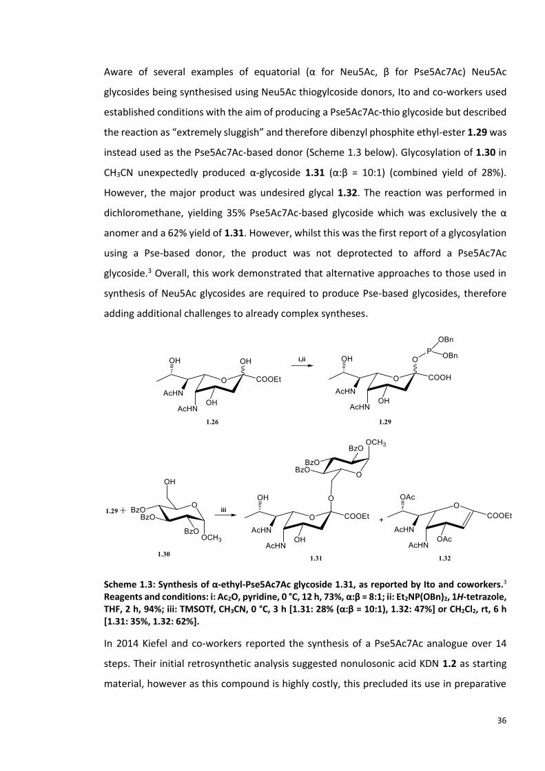
Aware of several examples of equatorial (α for Neu5Ac, β for Pse5Ac7Ac) Neu5Ac
glycosides being synthesised using Neu5Ac thiogylcoside donors, Ito and co-workers used
established conditions with the aim of producing a Pse5Ac7Ac-thio glycoside but described
the reaction as “extremely sluggish” and therefore dibenzyl phosphite ethyl-ester 1.29 was
instead used as the Pse5Ac7Ac-based donor (Scheme 1.3 below). Glycosylation of 1.30 in
CH3CN unexpectedly produced α-glycoside 1.31 (α:β = 10:1) (combined yield of 28%).
However, the major product was undesired glycal 1.32. The reaction was performed in
dichloromethane, yielding 35% Pse5Ac7Ac-based glycoside which was exclusively the α
anomer and a 62% yield of 1.31. However, whilst this was the first report of a glycosylation
using a Pse-based donor, the product was not deprotected to afford a Pse5Ac7Ac
glycoside.3 Overall, this work demonstrated that alternative approaches to those used in
synthesis of Neu5Ac glycosides are required to produce Pse-based glycosides, therefore
adding additional challenges to already complex syntheses.
Scheme 1.3: Synthesis of α-ethyl-Pse5Ac7Ac glycoside 1.31, as reported by Ito and coworkers.3 Reagents and conditions: i: Ac2O, pyridine, 0 °C, 12 h, 73%, α:β = 8:1; ii: Et2NP(OBn)2, 1H-tetrazole, THF, 2 h, 94%; iii: TMSOTf, CH3CN, 0 °C, 3 h [1.31: 28% (α:β = 10:1), 1.32: 47%] or CH2Cl2, rt, 6 h [1.31: 35%, 1.32: 62%].
In 2014 Kiefel and co-workers reported the synthesis of a Pse5Ac7Ac analogue over 14
steps. Their initial retrosynthetic analysis suggested nonulosonic acid KDN 1.2 as starting
material, however as this compound is highly costly, this precluded its use in preparative
37
scale synthesis. They also reported that attempts to use KDN had resulted in both pyranose
and furanose products with both α and β anomers and therefore deemed this wasteful.
Alternatively, methyl ester β-methyl glycoside of Neu5Ac 1.33 was used to produce β-
methyl glycoside methyl ester of KDN 1.34 in 57% yield, an improvement on previously
reported yields. Installation of the N-acetyl groups was problematic and required many
optimisations (Scheme 1.4). A 5,7-diol 1.35 was produced on route to installing the key 5,7-
di-N-acetyl functionalities of Pse5Ac7Ac. Desired 1.36 was isolated in 80% yield after diols
were converted to azido groups, hydrogenated and acetylated. However, initial
hydrogenation and acetylation attempts produced 1,5-lactam 1.37. Serendipitously, this
undesired compound may allow for selective acylation of the two amine groups, therefore
potentially enabling access to naturally occurring Pse-derivatives.4 However, no further
work on this route has been reported.
Further steps were required to produce a deprotected Pse5Ac7Ac analogue 1.38. However,
in the final compound the stereochemistry of C8 matches that of the sialic acids, therefore
1.38 is termed an 8-epi-Pse5Ac7Ac analogue (Scheme 1.4).113

38
Scheme 1.4: Synthesis of 8-epi-Pse5Ac7Ac 1.38, reported by Kiefel co-workers. 113 Reagents and conditions: i: NaNO2, Ac2OH-AcOH (2:1), 0 °C, 1 hr, then 50 °C, 6 hrs; ii: MeONa, MeOH; iii: 2,2-dimethoxypropane, PTSA, acetone, r.t.; iv: imidazole, TBDMS-Cl, DMF, r.t, 16 hrs; v: NaN3, DMF, 4 °C; vi: H2 Pd on C, MeOH; vii: Ac2O py; viii: p-TSOH.H2O, Pd(OH)2/C, H2, MeOH, r.t, 2 hrs; ix: Ac2O, pyridine, r.t, 12 hrs; x: TFA, THF-H2O (4:1), r.t, 30 mins; xi: NaOMe, MeOH, r.t, 2 hrs. xii I2, PPh3, imidazole, THF, 60 °C, 2 hrs. xiv iPr2EtN, Pd(OH)2/C, H2, MeOH, r.t, 16 hrs; xiv: aq. NaOH (1 M), 40 °C, 1 hr; xv: Dowex-50WX8(H+), 80 °C, 36 hrs.
2 years after reporting the synthesis of 8-epi-Pse5Ac7Ac 1.38 Kiefel, Payne and colleagues
were able to produce Pse5Ac7Ac in a 17-step synthesis. This was achieved using the
aforementioned intermediate 1.34, where the stereochemistry of the C8 hydroxyl was
inverted using an oxidation then subsequent reduction.5
Xuechen and colleagues began synthesis of Pse5Ac7Ac 1.6 with the cheap starting material,
L-threonine 1.39 which required a 28-step synthesis to yield a ring closed sugar 1.40 with
the desired stereochemistry which was converted to Pse5Ac7Ac 1.6 over five additional
steps (Scheme 1.5). This work is a significant improvement in the synthesis of Pse5Ac7Ac
1.6 due higher yields, lower cost of materials and higher control of stereochemistry.6
Xuechen and colleagues not only produced Pse5Ac7Ac but also, published the first reported
39
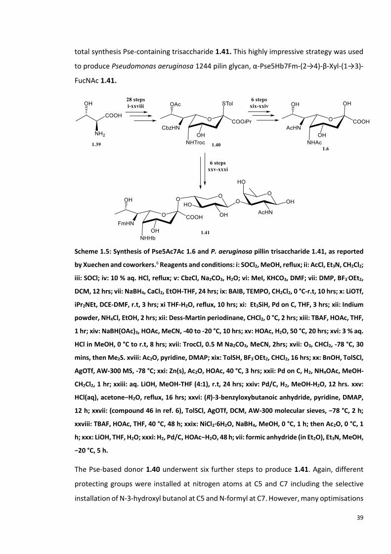
total synthesis Pse-containing trisaccharide 1.41. This highly impressive strategy was used
to produce Pseudomonas aeruginosa 1244 pilin glycan, α-Pse5Hb7Fm-(2→4)-β-Xyl-(1→3)-
FucNAc 1.41.
Scheme 1.5: Synthesis of Pse5Ac7Ac 1.6 and P. aeruginosa pillin trisaccharide 1.41, as reported
by Xuechen and coworkers.6 Reagents and conditions: i: SOCl2, MeOH, reflux; ii: AcCl, Et3N, CH2Cl2;
iii: SOCl; iv: 10 % aq. HCl, reflux; v: CbzCl, Na2CO3, H2O; vi: MeI, KHCO3, DMF; vii: DMP, BF3.OEt2,
DCM, 12 hrs; vii: NaBH4, CaCl2, EtOH-THF, 24 hrs; ix: BAIB, TEMPO, CH2Cl2, 0 °C-r.t, 10 hrs; x: LiOTf,
iPr2NEt, DCE-DMF, r.t, 3 hrs; xi THF-H2O, reflux, 10 hrs; xi: Et3SiH, Pd on C, THF, 3 hrs; xii: Indium
powder, NH4Cl, EtOH, 2 hrs; xii: Dess-Martin periodinane, CHCl2, 0 °C, 2 hrs; xiii: TBAF, HOAc, THF,
1 hr; xiv: NaBH(OAc)3, HOAc, MeCN, -40 to -20 °C, 10 hrs; xv: HOAc, H2O, 50 °C, 20 hrs; xvi: 3 % aq.
HCl in MeOH, 0 °C to r.t, 8 hrs; xvii: TrocCl, 0.5 M Na2CO3, MeCN, 2hrs; xvii: O3, CHCl2, -78 °C, 30
mins, then Me2S. xviii: Ac2O, pyridine, DMAP; xix: TolSH, BF3.OEt2, CHCl2, 16 hrs; xx: BnOH, TolSCl,
AgOTf, AW-300 MS, -78 °C; xxi: Zn(s), Ac2O, HOAc, 40 °C, 3 hrs; xxii: Pd on C, H2, NH4OAc, MeOH-
CH2Cl2, 1 hr; xxiii: aq. LiOH, MeOH-THF (4:1), r.t, 24 hrs; xxiv: Pd/C, H2, MeOH-H2O, 12 hrs. xxv:
HCl(aq), acetone−H2O, reflux, 16 hrs; xxvi: (R)-3-benzyloxybutanoic anhydride, pyridine, DMAP,
12 h; xxvii: (compound 46 in ref. 6), TolSCl, AgOTf, DCM, AW-300 molecular sieves, −78 °C, 2 h;
xxviii: TBAF, HOAc, THF, 40 °C, 48 h; xxix: NiCl2·6H2O, NaBH4, MeOH, 0 °C, 1 h; then Ac2O, 0 °C, 1
h; xxx: LiOH, THF, H2O; xxxi: H2, Pd/C, HOAc−H2O, 48 h; vii: formic anhydride (in Et2O), Et3N, MeOH,
−20 °C, 5 h.
The Pse-based donor 1.40 underwent six further steps to produce 1.41. Again, different
protecting groups were installed at nitrogen atoms at C5 and C7 including the selective
installation of N-3-hydroxyl butanol at C5 and N-formyl at C7. However, many optimisations

40
were required to introduce the desired group at the nitrogen of C5 before the
monosaccharide was used in glycosylation. The trisaccharide was fully deprotected to
obtain the desired 1.41 (Scheme 1.5).
The reducing carbohydrate at the end of the trisaccharide gives the possibility for installing
chemical handles, via established methodologies, which could be used be used as reporters
or in the synthesis of glycoconjugates. Access to 1.41 is perhaps the most significant
achievement in chemical synthesis of Pse-glycosides, and this naturally occurring product
may be used in studies the biological function and significance of Pse glycosylation in P.
aeruginosa and in pathogenesis and development of novel therapeutics.6 An additional
benefit of this overall scheme is that it allows for the incorporation of various N-linked
functionalities at C5 and C7, where naturally occurring Pses vary in functionality.1,6
Crich and co-workers recently described both the synthesis of a Pse-donor 1.42 (Scheme
1.6 A) and Pse5Ac7Ac 1.6 (Scheme 1.6 B). Beginning with Neu5Ac 1.1 the Pse-donor 1.6
was synthesised in 5% yield over 18 steps. In contrast to the α-Pse-based disaccharide
produced by Ito et al. this work reports the total synthesis of β-Pse-based disaccharides.
Di-azido donor 1.43 was used to obtain disaccharides, which were regioselectively Boc-
protected at either the C5 or C7 azide, allowing selective conversion of the remaining azide
to N-acetyl. Crich noted that this strategy would allow for installation of different N-
functionalities at C5 and C7, using a single glycosyl-donor, producing 1.44-1.47. The β-
donor (equatorial OH at C2) was remarked to have “exquisite equatorial selectivity”,
predicted to be due to the stronger electron withdrawing effects of the conformation of
the C7-9 side chain in the Pse-based molecule vs that of the Neu5Ac side chain. Synthesis
of these β-Pse-based glycosides is impressive and selective installation of desired functional
groups at N5 and N7, where many Pse5Ac7Ac derivatives vary from that of the parent
molecule, would greatly benefit studies into the biological significance of naturally
occurring β-Pse-glycosides.7 However, a reoccurring theme in the Pse total synthesis
literature, is that after the challenges of producing the Pse-skeleton have been overcome,
the final deprotection steps are not reported.3,7 Additionally, Pse5Ac7Ac was synthesised
was synthesised from 1.47 in three steps, as mixture of α- and β- anomers, in an overall
yield of 2.6%.

41
Scheme 1.6: A: Synthesis of Pse-based glycosylation donor 1.43 and production of glycosides 1.44-1.47. B: Synthesis of Pse5Ac7Ac using 1.43. A and B described by Crich and co-workers.7 Reagents and conditions: Reagents and conditions: A: i-iii: as reported114,115; iv: NaOMe; v: Me2C(OMe)2, CSA; vi: AcCl, py; vii: TBSOTf, Et3N; NaH, NaBr, DMF; viii: NOBF4, py; ix: TFA, H2O, TPSCl, Et3N, Bu2SnO; x: Tf2O, py, xi: Bu4NNO2, xii: NaI, Me2CO; H2, Pd/C, EtOAc, Et3N; xiii: Ac2O, DMAP; xiv: DDQ; xv: NH2NH2.H2O, AcOH, py; xvi: Tf2O, py; xvii: NaN3, DMF; xviii: 4Å AW molecular sieves, CH2Cl2:MeCN 2:1, NIS, TfOH. B: i: AcSH, py; ii: Ba(OH)2; iii: H2, Pd on C, H2O, dioxane.
Most recently, Xuechen and co-workers expanded on their initial work on Pse synthesis,
reporting a total synthesis strategy for stereocontrolled α- and β-glycosylation using a
single glycosyl donor 1.48, over 16 steps (Scheme 1.7). The donor was prepared on gram
scale and contains orthogonal N5 and N7 protecting groups, allowing for selective
introduction of N-acyl groups at each position, an analogous feature to the donor prepared
by Crich.7,9 Acceptors were selected for screening based on the known structures of Pse-

42
containing oligosaccharides (1.49-1.52) and glycopeptides (1.53), or due to its potential for
use in glycoconjugate preparation (1.54). Protected Pse-based glycosides 1.55-1.66 were
produced with each acceptor in yields from 56-80%. Under different glycosylation
conditions the donor showed predominantly α- or β-selectivity, regardless of the chemistry
of the acceptor. None of the glycosides were synthesised with complete anomeric
selectivity however, the synthesis of these products is a great advance on previous efforts
towards Pse-based biologically relevant glycosides. Efforts were made to produce a β-
linked Pse-based trisaccharide 1.67, which has the same the same skeleton as the repeating
unit of LPS O-antigen from Pseudomonas chlororaphis UCM B-106. Whilst this is a great
achievement, further deprotection and modification of N5 and N7 is required to yield the
native LPS O-antigen structure.9

43
Scheme 1.7: Synthesis of α- and β-Pse-based glycosides using donor 1.48, with acceptors 1.49-1.67 and synthesis of 1.67 trisaccharide as reported by Xuechen and co-workers.9 i-xi equivalent to i-xv in scheme 1.5; xvi: 3% HCl in MeOH, 0 °C to rt, 8 hr; xvii TfN3, 0.5 M Na2CO3, MeCN, 2 hrs; xviii: O3, DCM, −78 °C, 0.5 hr; then Me2S; xix: Ac2O, Py, DMAP, 0 °C to rt, 95%; xx: AdSH, BF3·OEt2, DCM, 16 hrs; xxi: NIS/TfOH (2.4/0.1 equiv.), AW-300 molecular sieves, DCM/MeCN (5 equiv.), -78 °C; xxii: NIS/TfOH (2.4/0.1 equiv.) AW-300 MS DCM/DMF (5 equiv.) -40 °C.

44
1.4.4 Chemoenzymatic synthesis of Pseudaminic Acids
Notably, several of the total synthesis schemes involved installation of azido groups. 5,7,113
These functional groups may be used as bioorthogonal handles on Pse-derivatives, as
demonstrated by the work of Tanner and colleagues who used a chemoenzymatic
approach to azido-modify C. jejuni flagella with Pse5Ac7NHAcN3 1.68 (Scheme 1.8).116
Enzymes PseB and PseC were used in vitro to produce 1.13 from UDP-GlcNAc 1.3. 1.13 was
chemically acetylated with chloroacetic anhydride, to produce PseH substrate analogue,
UDP-2-acetamido-4-chloroacetamido-2,4,6-trideoxy-β-L-Alt (UDP-6-deoxy-AltNAc4NAcCl)
1.69. The UDP group was hydrolysed by PseG yielding 1.70 and treated with NaN3 to yield
2-acetamido-4-azidoacetamido-2,4,6-trideoxy-β-L-Alt (6-deoxy-AltNAc4NAcN3 1.71) which
C. jejuni then transported into their cells from the growth media. A biotin-linked Staudinger
reagent was used to detect Pse5Ac7NHAcN3 on the flagella of C. jejuni. This indicated that
PseI produced 1.70, PseF nucleotide-activated to yield 1.72, that served as the donor for
inverting glycosyltransferase to glycosylate flagellin subunits.116

45
Scheme 1.8: In vitro chemoenzymatic synthesis of 6-deoxy-AltNAc4NAcN3 1.71, for in vivo production of Pse5Ac7AcN3-modified flagella of C. jejuni. Biotin-linked Staudinger reagent
reacts with azido-moiety of Pse5Ac7AcN3 1.68 which is detected by Western Blot analysis with Streptavidin HRP. As reported by Tanner and co-workers.116
Xuechen and co-workers built upon methodology used in their 2017 total synthesis paper,
by using L-allo-threonine 1.39 as the starting material for synthesis 1,3-di-O-acetyl-6-deoxy-
AltNAc4NAcN3 1.73 and 1,3-di-O-acetyl-6-deoxy-AltNAc2NAcN3 1.74, via intermediate
1.75, with the aim of producing Pse5Ac7AcN3 1.77 and Pse5AcN37Ac 1.78 in vivo (Scheme
1.9). Rich growth media for P. aeruginosa, A. baumannii and Vibrio vulnificus was
supplemented with 1.73 and 1.74. Following a 48 hr incubation period cells underwent a

46
Cu-free click reaction to fluorescently label any resulting cell-surface azido-modified Pse,
via the azide moiety of Pse. Studies confirmed that within some of the P. aeruginosa
studied, the LPS had been labelled, indicating that a Pse-derivative was present in the O-
antigen.8 Further examination confirmed that only Pse5Ac7AcN3 1.70 the downstream
product of 1.73, could be labelled in all organisms tested. This suggests that within the P.
aeruginosa, A. baumannii and V. vulnificus strains and serotypes tested, an enzyme
downstream of PseG is unable to utilise 1.74. In the case of P. aeruginosa 1244 it was
suggested that it may be PseI which cannot turnover 1.74 as the native substrate contains
an N-hydroxybutanol group at C2. However, further studies are required to find the
enzymatic bottleneck in each of the organisms studied.8 This demonstrates that whilst
metabolic engineering can provide valuable information in the biological relevance of Pse,
the strategy is not infallible.
Scheme 1.9: Chemical synthesis of 1.73 and 1.74, from L-allo-threonine 1.39. Synthesised with the intended use in in vitro synthesis of Pse5AcN37Ac and Pse5Ac7AcN3, as reported by Xuechen and colleagues.8 Reagents and conditions: i: equivalent to steps i-xi in Scheme 1.X; ii: Pd/C, Et3SiH, THF; iii: Ph3P=CHCOOBn, DCM; iv: TFA/H2O 10/1, 0 °C, 30 min; v: 3% HCl (aq) in MeOH, 0 °C to r.t., 8 hrs; vi: Ac2O, Na2CO3, MeCN/H2O; vii: O3, DCM, −78 °C, then Me2S; viii: TBSOTf, 2,6-lutidine, DCM, ix: Pd/C, H2, MeOH; x: 2-azidoacetic acid, HATU, iPr2NEt, DMF; xi: TBAF, HOAc, THF; xii: Ac2O, pyridine, DMAP; xiii: TFA/H2O 10/1, 0 °C, 30 min; xiv: 3% HCl (aq) in MeOH, 0 °C to r.t., 8 hrs; xv: Ac2O, Na2CO3, MeCN/H2O; xvi: FmocCl, iPr2NEt, DCM; xvii: O3, DCM, −78 °C, then Me2S; xviii: TBSOTf, 2,6-lutidine, DCM; xix Pd/C, H2, EtOAc; xx: Ac2O, pyridine; Et2NH, DCM, DMF; xxi: 2-azidoacetic acid, HATU, iPr2NEt, DMF; xxii: TBAF, HOAc, THF; xxiii: Ac2O, pyridine, DMAP.

47
The work of both Tanner et al. and Xuechen et al. demonstrates the benefits of utilising
enzymes in synthesis due to their inherent regio- and stereoselectivity, factors which are
challenging to control using traditional chemical methods for carbohydrate synthesis.8,116
It also demonstrates that a degree of enzyme promiscuity may be exploited, allowing
substrate analogues to be converted in to desired products. A caveat to this methodology
was made apparent when results suggested that 1.74 was not utilised in vivo to produce
the desired Pse5AcN37Ac-containing glyococojugates.8 This work may have benefitted from
the precautionary step taking by Tanner et al. where the promiscuity of enzymes was tested
in vitro with substrate analogues before the analogues were used for in vitro
experiments.116 However, both studies have shown that chemoenzymatic synthesis has the
potential to access products ranging from naturally occurring Pse5Ac7Ac derivatives, to
Pse-derivative chemical probes, which may be used to further the biological relevance of
Pse and the mechanisms used by Pse processing enzymes.8,116
1.4.4 Introduction to Pseudaminic Acid Processing Enzymes
There is currently very limited knowledge of GTs which utilise CMP-Pse
(Pseudaminyltransferases, PseTs).117 A few genes had putatively been assigned as PseTs
based upon sequence homology to SiaTs, or other structurally related α-keto sugars.
However, none of the proteins encoded by the putative PseTs had been functionally
characterised in vitro. Pse-glycoconjugates can be found in both α and β linkages, as
opposed to sialic acid glycoconjugates which exclusively contain α-anomers, due to the
inverting mechanism of SiaTs.55 Therefore, unlike SiaTs, both inverting and retaining PseTs
exist, which utilise CMP-Pse5Ac7Ac 1.10 or a similar CMP-Pse-derivative.1,55
In 2018 the first crystal structure of a putative PseT, Maf, from Magnetospirillum
magneticum AMB-1 was solved in an apo form.117 Whilst the recombinant expression,
purification and successful crystallisation of this enzyme, from a potentially novel GT class,
was an impressive feat, the identity of the Pse-derivative from M. magneticum AMB-1
could not be determined. Additionally, lack of access to CMP-Pse and Pse-derivatives
precluded functional studies of Maf required to confirm its identity as a PseT.117
An additional class of Pse processing enzymes that are yet to be studied are
glycosylhydrolases. In general, this class of enzyme hydrolyses glycosidic bonds from
48
glycoconjugates, to yield free carbohydrates. To date, the sole example of a putative Pse
GH (pseudaminidase) is PA2794 from P. aeuringosa.118 Studies showed that in mice
infected with pa2794 knockout P. aeruginosa PA01 (Δ2794), there was a decrease in biofilm
formation, in comparison to infection with wild type P. aeruginosa PA01 and that Δ2794
strains were unable to colonize the mouse respiratory tract.119 PA2794 was originally
assigned as a neuraminidase, however, in vitro studies showed that the enzyme did not
cleave Neu5Ac.118 The crystallographic structure suggest that the active site is unable to
accept Neu5Ac as a substrate and in-silico docking of Pse5Ac7Ac suggested that this may
be the product of the hydrolysis reaction. However, lack of access to a Pse5Ac7Ac-based
probe precluded further functional studies of PA2794.118
P. aeruginosa is the most prevalent cause of lung infection in patients with cystic fibrosis,
where PA2794 is highly overexpressed.119 The role of PA2794 in biofilm formation makes it
an attractive therapeutic target. Additionally, if PA2794 is shown to utilise Pse substrates
then this enhances its suitability as a therapeutic target as Pse-based inhibitors could be
designed which could avoid off-target effects on human neuraminidases.118
The biological mechanism behind the increased biofilm formation and ability of P.
aeruginosa strains expressing pa2794 to colonize a host remains curious. It is currently
unclear where the proposed PA2794 Pse5Ac7Ac-substrate is found. Whilst the full
composition of PAO1 biofilm is unknown, Pse5Ac7Ac is known to be present in the
glycoconjugates of many P. aeruginosa strains.120,121 Based upon knowledge of sialic acid
scavenging by bacterial pathogens, it may be speculated that following hydrolysis of
Pse5Ac7Ac, P. aeruginosa could utilise Pse5Ac7Ac as either a carbon source, or to build
Pse5Ac7Ac-based glycoconjugates. As with sialic acid scavenging, this would require
PA2794 to contain a transporter, currently no putative Pse transporters have been
identified.69 A desire to examine genomes for putative pseudaminidases, and potential
transporters may be initiated by improved access to Pse-based molecules.
49
1.5 Project Outline
This project aimed to characterise enzymes which process pseudaminic acid and to
chemoenzymatically synthesise glycosides which contain pseudaminic acid.
This was achieved through three specific aims:
1. Chemoenzymatic synthesis, purification and characterisation of nucleotide-activated pseudaminic acid, CMP-Pse5Ac7Ac (described in Chapter 2).
2. Synthesis of glycosides containing Pse5Ac7Ac, mediated by promiscuous sialyltransferases (described in Chapter 3).
3. In vitro functional characterisation of putative glycosyltransferases for pseudaminic acid (pseudaminyltransferases) (described in Chapter 4).

50
Chapter 2: Chemoenzymatic Synthesis of CMP-
Pseudaminic Acid
2.1 Introduction
In 2006 Schoenhofen et al. reported the first enzymatic synthesis of α-cytidine-5’-
monophosphate-Pse5Ac7Ac (CMP-Pse5Ac7Ac 1.10) facilitated by elucidation of the
enzymatic pathway found in within Helicobacter pylori and Camplyobacter jejuni (Figure
2.1).99 This pathway begins with uridine diphosphate-N-acetyl-glucosamine (UDP-GlcNAc
1.3) and the first of six enzymes, dehydratase/epimerase PseB, producing UDP-2-
acetamido-2,6-dideoxy-β-L-arabino-hexos-4-ulose 1.12. 1.12 is the substrate for the L-
glutamine dependent aminotransferase PseC, which produces UDP-4-amino-4,6-dideoxy-
β-L-AltNAc 1.13.103 N-acetyltransferase, PseH, then uses 1.13 and acetyl-coenzyme A (Ac-
CoA) to yield UDP-2,4-diacetamido-2,4,6-trideoxy-β-L-altropyranose 1.14, which
undergoes hydrolysis of UDP, faciltated by nucleotidase PseG, producing 2,4-diacetamido-
2,4,6-trideoxy-L-altropyranose (6-deoxy-AltdiNAc 1.15). Pseudaminic acid synthase, PseI,
ultilises phosphoenol pyruvate (PEP) to convert 1.15 to the nonulosonic acid Pse5Ac7Ac
1.6.103 This condensation reaction between a hexose and PEP is analogous to that seen in
the biosynthesis of Neu5Ac.58 Finally, CMP-Pse5Ac7Ac synthetase, PseF, nucleotide
activates Pse5Ac7Ac using cytidine-5’-triphosphate (CTP) to produce CMP-Pse5Ac7Ac 1.10.
1.10 was found to be a comptetive inhibitor of the first enzyme in the pathway, PseB.103
Therefore, whilst the enzymatic synthesis may be perfomed in an elegant one-pot system
where purification of reaction intermediates is not required, PseF must not be added to
the reaction mixture until succificent conversion of UDP-GlcNAc to 1.12 has been
achieved.103

51
Whilst impressive, the enzymatic synthesis of CMP-Pse5Ac7Ac described by
Schoenhofen et al. was not conducive to large-scale (~100mg) production. Firstly, as is true
for many nucleotide activated carbohydrates, the starting material UDP-GlcNAc is costly
(£913 per gram, Sigma). Secondly, the third step of the pathway performed by
acetlytransferase PseH, requires 1.5 equivalents of co-factor Ac-CoA. The quantity of
acetyl-CoA required for a reaction with 1g UDP-GlcNAc currently costs £8850 (Sigma).
Therefore, there was a clear need to optimise this enzymatic synthesis in order to reduce
cost.
These enzymes are also potential therapeutic targets as non-eukaryotic Pse5Ac7Ac and
derivatives are key virulence factors in several multi-drug resistant bacterial pathogens.
The strong negative feedback inhibition of PseB by CMP-Pse5Ac7Ac, indicates
concentrations of that CMP-Pse5Ac7Ac are regulated within bacteria, highlighting the
potential of these enzymes for therapetic intervention.122
Access to Pse5Ac7Ac biosynthesis enzymes has enabled studies into their inhibition.103,110
Over 96,000 compounds were screened against Pse5Ac7Ac biosynthesis enzymes from H.
pylori and C. jejuni. The study found 320 compound which each inhibited one or more
enzymes of the pathway. Five compounds showed concentration-dependent inhibition of
Figure 2.1: CMP-Pse5Ac7Ac biosynthesis pathway in C. jejuni and H. pylori, starting with UDP-GlcNAc, using enzymes PseB, PseC, PseH, PseG, PseI and PseF.

52
H. pylori and C. jejuni PseB, where 50% inhibitory concentration (IC50) values ranged from
12 µM to 72 µM. Docking of these compounds onto the structure of H. pylori PseB,
suggested that these inhibitors bind to the the active site. Three of these compounds met
Lipinski’s rule of five and the other two were predicted to have good oral bioavailability.110
In addition to a role in the CMP-Pse5Ac7Ac biosynthesis pathway, PseB has also been
shown to display C5 epimerase activity towards 1.12 producing 2.1 which may be
hydrolysed to produce 2.2 (Figure 2.2). Within C. jejuni 2.1 is a substrate for the pgl
pathway, which is used in the biosynthesis of 2,4-diacetamido-2,4,6-trideoxy-D-glucose
(N,N′-diacetylbacillosamine or diNAcBac) 2.3. Many bacterial proteins in C. jejuni and
Neisseria gonorrhoeae are known to be N- and O-glycosylated with diNAcBAc.122–124
Additionally, diNAcBAc has been reported as a component of the capsular polysaccharide
for drug-resistant Acinetobacter baumannii.125 Therefore, the value of PseB as a
therapeutic target is enhanced by its role in two enzymatic pathways which each produce
bacterial carbohydrate that play a role in bacterial pathogenicity.26
Figure 2.2: Pgl and CMP-Pse5Ac7Ac biosynthesis pathways within C. jejuni.
At the commencement of this study, knowledge of enzymes which process Pse5Ac7Ac 1.6
or CMP-Pse5Ac7Ac 1.10 was lacking. This was in-part hindered by a lack of access to

53
appreciable quantities of 1.6 and 1.10. Enzymes that process CMP-Pse5Ac7Ac such as
Leloir glycosyltransferases, may also be novel therapeutic targets. Therefore, for these
enzymes to be the studied as novel drug targets, their function must first be unequivocally
characterised. These functional studies are reliant on a source of CMP-Pse5Ac7Ac.
Structural studies of these enzymes may also require Pse5Ac7Ac and Pse-based probes.
The overall aim of this chapter was the large-scale production of CMP-Pse5Ac7Ac. Firstly,
this required the purification of five recombinant enzymes, (PseB, PseC, PseH, PseG and
PseI) for use in a reaction producing Pse5Ac7Ac.
Secondly, recombinant PseF was required to convert Pse5Ac7Ac to CMP-Pse5Ac7Ac. This
was achieved using Aeromonas caviae PseF which was uncharacterised at the
commencement of this study, therfore at this point, biochemical characterisation of the
enzyme was undertaken. The reaction to produce CMP-Pse5Ac7Ac was scaled to allow
purification and characterisation of 1.10. The production of 1.10 facilitated the studies
described in Chapter 3 and 4.
Additionally, small-molecules were screened as potential inhibitors of PseB and PseI. The
screening of compounds as potential PseB inhibitors was reliant upon access to active PseB
and commercially available UDP-GlcNAc. The screening of compounds as potential PseI
inihbitors, active PseI and its substrate, 6-deoxy-AltdiNAc 1.15 was required, which was
produced enzymatically during this study.
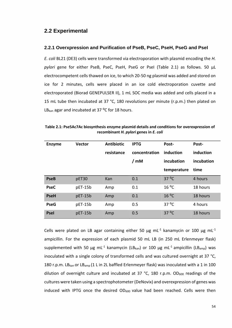
54
2.2 Experimental
2.2.1 Overxpression and Purification of PseB, PseC, PseH, PseG and PseI
E. coli BL21 (DE3) cells were transformed via electroporation with plasmid encoding the H.
pylori gene for either PseB, PseC, PseH, PseG or PseI (Table 2.1) as follows. 50 µL
electrocompetent cells thawed on ice, to which 20-50 ng plasmid was added and stored on
ice for 2 minutes, cells were placed in an ice cold electroporation cuvette and
electroporated (Biorad GENEPULSER II), 1 mL SOC media was added and cells placed in a
15 mL tube then incubated at 37 °C, 180 revolutions per minute (r.p.m.) then plated on
LBkan agar and incubated at 37 ⁰C for 18 hours.
Table 2.1: Pse5Ac7Ac biosynthesis enzyme plasmid details and conditions for overexpression of recombinant H. pylori genes in E. coli
Cells were plated on LB agar containing either 50 µg mL-1 kanamycin or 100 µg mL-1
ampicillin. For the expression of each plasmid 50 mL LB (in 250 mL Erlenmeyer flask)
supplemented with 50 µg mL-1 kanamycin (LBkan) or 100 µg mL-1 ampicillin (LBamp) was
inoculated with a single colony of transformed cells and was cultured overnight at 37 °C,
180 r.p.m. LBkan or LBamp (1 L in 2L baffled Erlenmeyer flask) was inoculated with a 1 in 100
dilution of overnight culture and incubated at 37 °C, 180 r.p.m. OD600 readings of the
cultures were taken using a spectrophotometer (DeNovix) and overexpression of genes was
induced with IPTG once the desired OD600 value had been reached. Cells were then
Enzyme Vector Antibiotic
resistance
IPTG
concentration
/ mM
Post-
induction
incubation
temperature
Post-
induction
incubation
time
PseB pET30 Kan 0.1 37 ⁰C 4 hours
PseC pET-15b Amp 0.1 16 ⁰C 18 hours
PseH pET-15b Amp 0.1 16 ⁰C 18 hours
PseG pET-15b Amp 0.5 37 ⁰C 4 hours
PseI pET-15b Amp 0.5 37 ⁰C 18 hours

55
incubated at 16 or 37 °C, 180 r.p.m. for 4 or 18 hours (Table 2.1). Cell pellets were harvested
via centrifugation (6000 ×g, 30 min, 4 °C) and stored at -80 °C.
Cell pellets from 2 L cultures lysed on ice resuspended in 50 mL lysis buffer (50 mM sodium
phosphate, 400 mM NaCl, 10 mM β-mercaptoethanol, pH 7.4) supplemented with protease
inhibitor, lysozyme, and Benzonase. Cells were lysed via sonication in ice (30 seconds, 30
second pause, 25 repeats) and centrifuged (37000 ×g, 30 min, 4 °C). Supernatant was
applied to a 5 mL HisTrap HP column (GE Healthcare) pre-equilibrated with lysis buffer, the
column was washed with 10 column volumes (C.V.), lysis buffer and protein was eluted
with a gradient of elution buffer (lysis buffer + 490 mM imidazole, pH 7.4) over 40 C.V. as 4
mL fractions were collected. 15 μL samples of insoluble material, soluble material,
flowthrough wash and fractions were analysed via SDS-PAGE.
SDS-PAGE analysis was performed using 10 or 12% polyacrylamide gels. Samples were
mixed with 5x reducing buffer (2% SDS, 2 mM 2-mercaptoethanol, 4% glycerol, 40 mM Tris-
HCl pH 6.8, 0.01% bromophenol blue) and were heated to 95 °C for 4 minutes. 6 µL
PageRuler Plus Prestained Protein Ladder (Thermo Scientific) molecular weight marker was
used per gel. Gels were run at 200 volts for 45-75 min. For Coomassie stain experiments,
the gel was washed with fixing solution (40% (v/v) MeOH, 10% AcOH), stained with 0.1%
(w/v) Coomassie Brilliant Blue R-250 (50% (v/v) MeOH, 10% AcOH), and repeatedly washed
with destain solution (50% (v/v) MeOH, 10% AcOH).
Following SDS-PAGE analysis fractions containing the desired protein were pooled and
applied to desalting column (HiPrep 26/10 desalting column, GE Healthcare) to exchange
into reaction buffer (50 mM sodium phosphate, 25 mM NaCl, pH 7.4). Protein was collected
as it eluted from the column and concentration of protein was recorded using a nanodrop
spectrophotometer. Protein was aliquoted in 500 µL aliquots, glycerol was added to a final
concentration of 25% (v/v) and samples were flash frozen and stored at – 80 °C. In a later
optimisation of this protocol, no glycerol was added prior to flash freezing the enzymes.

56
2.2.2 Enzymatic synthesis of Pse5Ac7Ac with Acetyl-thiocholine Iodide
1 mM UDP-GlcNAcp 1.3 (200 mg), 0.15 mM coenzyme-A, 2 mM pyridoxal 5′-phosphate, 10
mM L-glutamic acid, 1.5 mM phosphoenolpyruvate, 60 mM acetylthiocholine iodide, 0.1
mg mL-1 PseB, 0.1 mg mL-1 PseC, 0.1 mg mL-1 PseH, 0.1 mg mL-1 PseG and 0.1 mg mL-1 PseI
(enzymes purified as described in 2.3.1, containing glycerol), 50 mM 25 mM sodium
phosphate pH 7.4 in 33 10 mL aliquots. The reaction mixtures were incubated at 37 °C for
6 hours and were analysed via –ESI LC-MS, to monitor the production of Pse5Ac7Ac.
Reactions were pooled, enzymes were removed from the reaction mixtures using
molecular weight cut off filters (MWCO) (30 kDa and then the subsequent filtrate was
passed through a 10 kDa MWCO). The filtrate was lyophilised and resuspended in 50 mL
dH2O and 5 mL was applied to a 500 mL column packed with LH20 resin (GE Healthcare) in
HPLC-grade H2O. 3 mL fractions were collected for 40 hours and analysed via LC-MS for the
presence of Pse5Ac7Ac. The column was repeated with the remaining lyophilised material
and all fractions containing Pse5Ac7Ac were pooled and lyophilised and analysed by NMR
(D2O, 500MHz). The resulting material was purified by anion exchange (Dowex), with a 0-2
M gradient of formic acid. TLC plates were used to analyse fractions for the presence of
carbohydrates (TLC plate soaked in sugar stain: MeOH, 5% H2SO4 and charring) and
fractions which appeared to contain carbohydrate was analysed by LC-MS (details in
Appendix-General methods.) Fractions containing Pse5Ac7Ac were pooled, lyophilised and
analysed by NMR (D2O, 500MHz).
2.2.3 Small-molecule screen for potential inhibition of PseB and PseI
A library of small-molecules which were synthesised as potential inhibitors of PseB and PseI
were made available to this study (Figure 2.3).‡
‡ Small-molecule library was synthesised by Joseph Ferner, Shaw Laboratory, University of Sheffield.

57
Figure 2.3: Compounds 2.4-2.8 tested for inhibition of C. jejuni or A. caviae PseI. 2.4 was tested for inhibition of C. jejuni PseB.
To test whether 2.4 is an inhibitor of H. pylori PseB the following reaction mixtures
containing 0.1 mg mL-1 PseB, 0.1 mg mL-1 PseH, 0.1 mg mL-1 PseG, 0.1 mg mL-1 PseI (all
purified as described in 2.2.1) and 0.2 mg mL-1 AcPseC, in addition to 1 mM UDP-GlcNAcp
1.3, 1.5 mM coenzyme-A, 2mM pyridoxal 5′-phosphate, 10 mM L-glutamic acid, 1.5 mM
PLP, 60 mM acetylthiocholine iodide, 1 mM PEP, in plus 0 mM 0.1 mM, or 5 mM 2.4 in 50
mM 25 mM sodium phosphate pH 7.4 to a final volume of 50 µL. The reactions were
performed in duplicate and incubated overnight at 30 °C. Enzyme precipitate was removed
by centrifugation (37,000 ×g, 10 min, 4 °C) and supernatant was analysed by -ESI LC-MS, as
described in 2.2.2.
To test inhibition of PseI and Aeromonas caviae PseI (AcPseI) the substrate 6-
deoxyAltDiNAc 1.15 was produced via chemoenzymatic synthesis. To facilitate this the
activity of pure, recombinantly expressed Aeromonas caviae PseC (AcPseC)§ was used in
combination with C. jejuni enzymes PseB, PseH, and PseG, expressed and purified as
§ AcPseC and AcPseI were expressed and purified by by Joseph Ferner, The University of Sheffield.

58
described in 2.2.1. A reaction mixture containing 0.114 mg mL-1 PseB, 0.114 mg mL-1 PseH,
0.114 mg mL-1 PseG (all purified as described in 2.2.1) and 0.228 mg mL-1 AcPseC, in addition
to 1.14 mM UDP-GlcNAcp 1.3 (2.4 mg), 1.71 mM coenzyme-A, 2.228 mM pyridoxal 5′-
phosphate, 11.1 mM L-glutamic acid, 1.71 mM PLP, 68.4 mM acetylthiocholine iodide, 50
mM 25 mM sodium phosphate pH 7.4. The reaction mixture incubated overnight at 30 °C.
Enzyme precipitate was removed by centrifugation (37,000 ×g, 10 min, 4 °C). To 87.5 µL of
supernatant PseI or AcPseI, PEP and dH2O were added to give a final sample volume of 100
µL (such that final concentrations would be equivalent to those in 2.2.2 after PEP and PseI
or AcPseI were added) and the samples incubated at 30 °C and incubated for 2 hours. The
sample was analysed by LC-MS (details in Appendix-General methods) to check for the
presence of Pse5Ac7Ac, to confirm that the initial reaction had produced 6-deoxyAltDiNAc
1.15.
To test PseI inhibition, 87.5 µL of supernatant, PseI, PEP and an inhibitor compound 2.4-2.8
were added to give a final sample volume of 100 µL (such that final concentrations would
be equivalent to those in 2.2.2 after PseI and PEP were added) and inhibitor concentrations
of 0 mM, 0.1 mM, 1 mM and 5 mM. The screen was repeated with AcPseI in place of PseI.
Reactions were performed in duplicate and incubated at 30 °C and incubated for overnight.
The sample was analysed by LC-MS to check for the presence of Pse5Ac7Ac, to assess
inhibition of PseI.
2.2.4 Recombinant expression and purification attempt of C. jejuni PseF in E.
coli
Plasmid encoding N-terminal hexahistidine tagged C jejuni PseF, codon optimised for E. coli
(Genscript), in a pET-15b vector (CjPseF) (plasmid details in Appendix) was transformed into
E. coli BL21 (DE3) cells (via electroporation- as described in 2.2.1). A single colony was used
to inoculate 60 mL 2 x yeast tryptone, supplemented with 50 μg mL-1 kanamycin (2×YTkan)
that was incubated at 30 °C, 180 rpm, overnight, before being used to inoculate 4 L of
2×YTkan (six 2 L baffled Erlenmyer flasks each contained 1 L LBkan). The cultures were
incubated at 37 °C, 180 rpm. At OD600 0.6, 0.1 mM Isopropyl-β-D 1-thiogalactopyranoside
(IPTG) was added to induce expression of CjPseF. Cultures were incubated for 2.75 h post-
induction and cells were harvested via centrifugation (6,000 ×g, 30 minutes, 4 °C). Two cell
59
pellets, each harvested from 2 L culture, were stored at -80 °C. A single cell pellet,
corresponding to 2 L culture was thawed on ice. Cells were resuspended in 20 mL lysis
buffer (50 mM sodium phosphate, pH 7.3, 400 mM NaCl, 10 mM β-mercaptoethanol, 10
mM imidazole, 1 mM MgCl2) supplemented with protease inhibitor (1 tablet per 50 mL,
Pierce) and Benzonase nuclease (25 units mL -1), before being lysed on ice via sonication
(30 seconds, 30 second pause, 12 repeats). Lysed cells were centrifuged (17,700 ×g, 35 min,
4 °C) and the resultant supernatant was retained for purification. The supernatant was
applied to a 5 mL HisTrap HP column (GE Healthcare), prewashed with lysis buffer.
Unbound material was washed through the column with 10 column volumes (C.V.) of lysis
buffer. In order to elute bound material from the column, 30 C.V. of 0-50% buffer B (50 mM
sodium phosphate, pH 7.3, 400 mM NaCl, 10 mM β-mercaptoethanol, 500 mM imidazole)
was mixed with lysis buffer, in a linear gradient, followed by 10 C.V. of buffer B. The eluent
was collected as 3 mL fractions, which were analysed by 12 % SDS-PAGE (as described in
2.2.1).
Following unsuccessful purification of PseF, due to protein insolubility, expression trials
were carried out to find conditions that produced soluble CjPseF. CjPseF plasmid was
transformed into E. coli Tuner (DE3) cells that were then used in expression trials alongside
E. coli BL21 (DE3) encoding PseF plasmid. Overnight cultures of both cell strains in 2×YTkan
were incubated at 37 °C, 180 rpm. Each overnight culture was used to inoculate seven 250
mL Erlenmyer flasks, each containing 50 mL 2×YTkan to give an initial OD600 of 0.02.
Additionally, as a control a pET-15b vector (vector for the CjPseF gene) was cultured in the
same conditions. All cultures were incubated at 37 °C, 180 rpm, until expression was
induced, at OD600 measurements of 0.4, 0.6 and 0.8, using either 0.1 mM or 0.5 mM IPTG
for each of the three OD600 values. At this point flasks were incubated at 30 °C, 180 rpm.
Three 1 mL samples were taken from each flask at 1 or 2, 4 and 24 hrs post-induction. An
uninduced sample was also cultured as a control. The samples were centrifuged (6,000 ×g,
10 minutes, 4 °C) and cells pellets were stored at −20 °C. This process was repeated, with
induced cells incubated at 25 °C and 16 °C. Cell pellets were resuspended in 50 μL lysis
buffer supplemented with 1 mg mL-1 lysozyme and incubated at 37 °C for 45 minutes to
lyse cells. Pellets were centrifuged (6,000 ×g, 10 minutes, 4 °C), following this soluble and
insoluble material was separated. The insoluble material was resuspended in 50 μL dH2O
60
and 15 μL of both soluble and insoluble fractions, from each sample, were mixed with 3 μL
5× reducing dye heated to 95 °C for 3 minutes and analysed by 12% SDS-PAGE (as described
in 2.2.1).
2.2.5 Expression and purification of A. caviae PseF
E. coli BL21 (DE3) were transformed via electroporation (as described in 2.2.1) with plasmid
encoding N-terminal hexahistidine tagged PseF (PseF) from A. caviae (pET-28a vector)
(PseF)** as follows. Then plated on LBkan agar and incubated at 37 ⁰C for 18 hours. A single
colony of E. coli BL21 (DE3) of transformed cells was used to inoculate 60 mL LBkan, that was
incubated at 30 °C, 180 rpm, overnight. The cultured cells were used to inoculate seven 250
mL Erlenmyer flasks, each containing 50 mL 2× YTkan to give an initial OD600 0.02. All cultures
were incubated at 37 °C, 180 rpm, until expression was induced, at OD600 measurements
of 0.3, 0.4, 0.6 and 0.8, using either 0.1 mM, 0.5 mM or 1 mM IPTG for each of the four
OD600 values. At this point flasks were incubated at 30 °C, 180 rpm. Three 1 mL samples
were taken from each flask at 2, 4 and 24 hrs post-induction. The samples were centrifuged
(6,000 x g, 10 minutes, 4 °C) and cells pellets were stored at −20 °C. This process was
repeated, with induced cells incubated at 30 °C, 25 °C and 16 °C. Cell pellets were
resupended in 50 μL Bugbuster supplemented with protease inhibitor (1 tablet per 50 mL,
Pierce) plus 1 mg mL-1 lysozyme and incubated at room temperature for 45 minutes to lyse
cells. Pellets were centrifuged (6,000 ×g, 10 minutes, 4 °C), following this soluble and
insoluble material was separated. The insoluble material was resuspended in 50 μL dH2O
and 15 μL of both soluble and insoluble fractions from each sample were analysed via SDS-
PAGE (as described in 2.2.3 and 2.2.1).
A single colony of E. coli BL21 (DE3) of transformed cells encoding PseF was used to
inoculate 60 mL LBkan, that was incubated at 30 °C, 180 rpm, overnight, before being used
to inoculate 3 L of LBkan (five 2 L baffled Erlenmyer flasks each contained 600 mL LBkan). The
cultures were incubated at 37 °C, 180 rpm. At OD600 0.6, 0.1 mM IPTG was added to induce
expression of PseF. Cultures were incubated 30 °C, 180 rpm for 3 hrs post-induction and
** Plasmid was gifted by Dr Jonathon Shaw, The University of Sheffield

61
cells were harvested via centrifugation (6,000 × g, 30 minutes, 4 °C). Two cell pellets were
obtained and stored at -80 °C.
Cell pellets were thawed on-ice and resuspended in 50 mL lysis buffer (50 mM sodium
phosphate, pH 7.4, 400 mM NaCl, 10 mM β-mercaptoethanol, 1 mM MgCl2, 10 mM
imidazole and Benzonase nuclease (25 units mL-1) (Sigma-Aldrich), before being lysed on
ice via sonication (30 seconds, 30 second pause, 10 repeats). Lysed cells were centrifuged
(17,700 ×g, 40 min, 4 °C) and the resultant supernatant was retained for purification. The
supernatant was filtered (0.45 μm syringe filter) and applied to a 5 mL HisTrap HP column,
prewashed with lysis buffer. Unbound material was washed through the column with 10
column volumes (C.V.) of lysis buffer. Bound material was eluted from the column, with a
linear gradient of 0-100% elution buffer (50 mM sodium phosphate, pH 7.3, 400 mM NaCl,
10 mM β-mercaptoethanol, 1 mM MgCl2, 500 mM imidazole) over 40 C.V. The eluent was
collected as 5 mL fractions, which were analysed by 12 % SDS-PAGE. Fractions containing
pure PseF were pooled and dialysed (50 mM sodium phosphate, 50 mM NaCl, 1 mM MgCl2)
overnight at 4 °C. PseF was then either used in enzyme assays or flash frozen and stored at
-80 °C (PseF was aliquoted and stored with and without 20% glycerol).
2.2.6 Activity assay of A. caviae PseF with Pse5Ac7Ac
100 μL reactions were performed with 130 μg mL-1 PseF, 0.5 mM Pse5Ac7Ac (Sussex
Research), 1.5 mM CTP, 1 mM MgCl2, 25 mM sodium phosphate, pH 7.4, 50 mM NaCl and
incubated at 25 °C. Methodology was based upon that used by Schoenhofen et al. Control
reactions were performed with reaction mixture as described without either Pse5Ac7Ac,
CTP or PseF. Upon addition of PseF to reaction mixture samples were analysed by -ESI LC-
MS (details in Appendix-General methods) every 15 minutes for 2 hours and after 6.5 hours.
Freshly purified PseF, thawed PseF containing 20% glycerol and thawed PseF without
glycerol was used in these reactions.
2.2.7 Purification of Aeromonas caviae PseF for Crystallisation trials
Fractions containing pure PseF (as described in 2.2.4) were dialysed (25 mM Tris, 50 mM
NaCl, 2 mM MgCl2) overnight at 4 °C.

62
3 L of E. coli BL21 (DE3) encoding BL21 were cultured as described in 2.24. Thawed cell
pellet was purified as described with Tris buffers in place of sodium phosphate buffers (Lysis
buffer- 50mM Tris, pH 7.65, 400 mM NaCl, 10 mM β-mercaptoethanol, 2 mM MgCl2, 500
mM imidazole. Elution buffer- 50mM Tris, pH 7. 65, 400 mM NaCl, 10 mM β-
mercaptoethanol, 2 mM MgCl2, 500 mM imidazole.) Fractions containing pure PseF were
pooled and dialysed (Tris dialysis buffer- 25 mM Tris, 50 mM NaCl, 2 mM MgCl2) overnight
at 4 °C. Dialysed protein was centrifuged (6,000 ×g, 40 minutes, 4 °C) to remove precipitant
and analysed via 12% SDS-PAGE. Sample was spin concentrated (6,000 ×g, 40 minutes, 4
°C, 10 kDa MWCO tube) and loaded onto a S-200 size-exclusion column (GE Healthcare),
pre-equilibrated with Tris dialysis buffer. PseF was eluted from the column in Tris dialysis
buffer and collected in 1 mL samples, which were stored on ice. 12% SDS-PAGE was used
to analyse the purity of these fractions (as described in 2.2.1).
2.2.8 A. caviae PseF Protein Identification by Mass Spectrometry
Protein was extracted from two bands found in SDS-PAGE of PseF following SEC (2.2.7) and
subject to trypsin digest. The resultant peptides were analysed by MALDI-MS and MS/MS
using a Bruker ultraflex III MALDI-TOF/TOF. Spectral data was compared to the Mascot
database to identify the protein contained in each SDS-PAGE band.
2.2.9 A. caviae PseF Circular Dichorism
Circular dichroism data was collected for 0.2 mg mL-1 Pure PseF was dialysed into in 25 mM
sodium phosphate buffer pH 7.4, for 16 hours at 4 ⁰C, using 2 x 2L buffer and dialysis tubing
(6-8 kDa MWCO, Fisherbrand). Protein concentration was determined using molecular
extinction co-efficient (41,940 M-1 cm-1) and nanodrop spectrophotometer (DeNovix) and
protein was diluted with dialysis buffer to a final concentration of 0.2 mg mL-1. Circular
dichroism data was collected at 30 °C from 180-230 nm, using a Jasco J810 CD
Spectrophotometer. A quartz cuvette with a path length of 1 mM was used, a 400 µL of
dialysis buffer was used to obtain a blank spectrum, after which 400 µL of PseF was used
and a spectrum was recorded. Secondary structure predictions were made from CD data
using K2D3 (http://cbdm-01.zdv.uni-mainz.de/~andrade/k2d3/).

63
2.2.10 A. caviae PseF Size Exclusion Chromatography - Multi-Angle Laser
Light Scattering
For the collection of size exclusion chromatography - multi-angle laser light scattering (SEC-
MALLS) data pure PseF (prepared as described in 2.2.4, without glycerol) was dialysed in to
running solvent (20 mM Tris, 50 mM NaCl, 2 mM MgCl2, pH 7.8). Experiments were
conducted on a system comprising a Wyatt HELEOS-II multi-angle light scattering detector
and a Wyatt rEX refractive index detector linked to a Shimadzu HPLC system (SPD-20A UV
detector, LC20-AD isocratic pump system, DGU-20A3 degasser and SIL-20A autosampler).
Work was conducted at room temperature (20 ±2°C). Running solvent was 0.2 µm filtered
and at least 2 column volumes were used to equilibrate Superdex S200 (10/300 GL
#0805015 (G.E. Healthcare)) size-exclusion column, running buffer was applied until for UV,
light scattering and refractive index detectors were all stable. PseF was supplied at a
nominal concentration of 10 mg mL-1 and was diluted with the running buffer to 4 mg mL-1
and 1 mg mL-1, then microfuged for 3 mins before use. 100 µL samples were applied to a
Superdex S200 size-exclusion column (G.E. Healthcare), ran at flow rate 0.5 mL min-1.†† A
2.5 mg/mL BSA sample was run as a standard. Blank buffer injections were used as
appropriate to check for carry-over between sample runs. Data were analysed using Astra
V software.
2.2.11 Crystalisation trials for A. caviae PseF
Following purification, PseF was spin concentrated (6,000 ×g, 50 minutes, 4 °C, 10 kDa
MWCO tube) resulting in two 200 µL samples at 10 and 6.5 mgmL-1. 2 mM DTT was added
to 100 µL of each sample. The samples were stored on ice before being used in initial
crystallisation screens. 98-well sitting drop plates were set up, using a Mosquito robot, to
screen both concentrations of PseF, with and without DTT. Two commercially available
screens were used, PEG/ion and PBD minimal set (Hampton research). Above each well a
150 nL drop of PseF was mixed with 150 nL of well mother-liquor, this was repeated with
the PseF sample which contained DTT. Trays were stored at room temperature for
approximately four weeks and examined for crystals. Regular crystals were fished from the
†† SEC-MALLS data was collected by Dr Andrew Leech, Technology facility, Department of Biology, University of York

64
solution, flash frozen in N2 (l) and stored in N2 (l) prior to diffraction testing. X-ray diffraction
was carried out using in-house facilities; a Rigaku MicroMAx 007HF generator, RAXIS IV++
imaging plate detector and an Actor robotic sample changer.
Two 24-well hanging drop trays were set up based upon successful conditions found in the
initial screen (Conditions in Appendix Table A.1). 0.3% (v/v) Pentaethylene Glycol
Monooctyl Ether (C8E5) detergent was added to mother liquor in 24 well trays with the aim
of slowing the rate of crystal formation. Trays were stored at room temperature for
approximately four weeks and were analysed by ultraviolet (UV) light in the Xtal imager.
2.2.12 Large scale synthesis and purification of CMP-Pse5Ac7Ac, via a one-
pot six enzyme synthesis
2 mM UDP-GlcNAc 1.3 (90 mg), 0.3 mM coenzyme-A, 4 mM pyridoxal 5′-phosphate, 20 mM
L-glutamic acid, 3 mM phosphoenolpyruvate, 20 mM N-acetyl-S-acetylcysteamine (SNAc)‡‡,
0.2 mg mL-1 PseB, 0.4 mg mL-1 PseC, 0.2 mg mL-1 PseH, 0.2 mg mL-1 PseG and 0.2 mg mL-1
PseI . The reaction mixture was incubated at 37 °C for 12 hours and was monitored via –ESI
LC-MS, to monitor the production of Pse5Ac7Ac. After 12 hours, 0.2 mg mL-1 PseF, 4 mM
CTP and 20 mM MgCl2 were added to facilitate the production of CMP-Pse5Ac7Ac 1.10, as
confirmed by –ESI LC-MS analysis. The reaction incubated at 37 °C for 4 hours and was
analysed via –ESI LC-MS. The mixture was lyophilised and resuspended in 10 mL dH2O and
10 mL EtOH and stored at 4 °C for 30 minutes to precipitate enzymes, the solution was
centrifuged (38,759 ×g, 1 hours, 4 ⁰C). The supernatant was diluted in dH2O to a final
volume of 200 mL before lyophilisation. The lyophilised material was resuspended in 7 mL
dH2O and passed through a 45 µM Millex® syringe filter (Merck) to remove any remaining
enzymes before being applied to a 500 mL column packed with Bio-Gel® P-2 resin (Biorad)
in HPLC-grade H2O. 4 mL fractions were collected for 24 hours and analysed via LC-MS
(details in Appendix-General methods) for the presence 1.10 of and all fractions containing
1.10 were pooled and lyophilised. Following NMR analysis of the resulting material (D2O,
500MHz).
‡‡ Synthesised and characterised by Matthew Best (MChem student- Fascione laboratory)

65
1H NMR (500 MHz, D2O) δ 8.02 (d, J = 7.5, 1H, H2’), 6.14 (d, J = 7.6 Hz, 1H, H1’), 6.00 (d, J =
4.1 Hz, 1H, H3’), 4.37 – 4.32 (m, 2H, H4’, H6’), 4.32 – 4.22 (m, 6H, H4, H5, H6, H5’, H7a’,
H7b’), 4.12 (dd, , J =6.5, 5.1 Hz, 1H, H8), 4.03 (dd, J =9.9, 5.0 Hz, 1H, H7), 2.23 (dd, J =13.4,
4.5 Hz, 1H, H3eq), 2.00 (s, 3H, 5NHAc), 1.97 (s, 3H, 7NHAc), 1.61 (ddd, J = 13.4, 12.0, 5.2 Hz,
1H, H3ax), 1.20 (d, J = 6.5 Hz, 3H, 9CH3). 13C NMR (126 MHz, D2O) δ 174.5, 173.2, 165.8,
157.3, 141.7, 100.0, 96.5, 89.2, 82.9, 74.1, 72.7, 69.2, 68.6, 64.9, 64.38, 53.7, 48.77, 36.0,
22.0, 21.9, 17.2. HR-MS data was collected with –ESI MS: Expected [M-H]- m/z: 638.1789,
measured [M-H]- m/z: 638.1727. ATR-FTIR Vmax: 3227, 2941, 2888, 1592, 1403, 1075 cm-1

66
2.3 Results
2.3.1 Overxpression and Purification of PseB, PseC, PseH, PseG and PseI
To facilitate the enzymatic production of Pse5Ac7Ac E. coli BL21 (DE3) were transformed
with plasmid containing one of the PseB, PseC, PseH, PseG, or PseI genes from C. jejuni.
Transformed cells were each cultured on a 4 L scale and overexpression of the gene was
induced with IPTG, such that the resultant His-tagged protein could be purified by nickel-
affinity chromatography. The conditions for expression and purification has been
previously optimised§§ therefore, as anticipated each purification yielded high levels of
protein, which was sufficiently pure for use in chemoenzymatic synthesis (Figure 2.4). The
yield for each enzyme were as follows: PseB- 14 mg L-1, PseC- 9 mg L-1, PseH- 18 mg L-1,
PseG- 12 mg L-1, PseI- 20 mg L-1.
Following purification proteins were mixed with 25% glycerol, flash frozen and stored at -
80 °C. Glycerol was used as a cryoprotectant for the enzymes, to ensure that proteins
retained activity after a freeze-thaw cycle. However, as an aim of this study was to purify
§§ Optimisation of expression and purification of PseB, PseC, PseG, PseH and PseI was performed by Dr Harriet Chidwick and Emily Flack (BSc project).
Figure 2.4: 4-20% SDS-PAGE for Pse5Ac7Ac biosynthesis enzymes following nickel-affinity purification. Lanes loaded as follows: 1-PAGE Ruler MW marker, 2- PseB (39.5 kDa), 3- PseC (44.6
kDa), 4- PseH (20.9 kDa), 5-PseG (33.4 kDa), 6- PseI (40.8 kDa).

67
CMP-Pse5Ac7Ac, from a complex reaction mixture, it was decided that the removal of
glycerol from enzymes stocks may aid CMP-Pse5Ac7Ac purification. Therefore, expression
and purifications were repeated, and enzymes were flash frozen without glycerol.
2.2.2 Enzymatic synthesis of Pse5Ac7Ac with Acetyl-thiocholine Iodide
To test the activity of purified enzymes described in 2.3.1 a one pot reaction was performed
which contained enzymes PseB, PseC, PseH, PseG and PseI along with UDP-GlcNAc starting
material, co-factors and the acetyl-CoA regeneration co-factor acetylthiocholine iodide
(reaction summarised in Figure 2.5, A).*** The reaction incubated at 37 °C and LC-MS
analysis showed that over 2 hours there was a decrease in intensity of a peak corresponding
to UDP-GlcNAc 1.3 (Figure 2.5, B and C. [M-H]-, m/z 606.0). The reaction was successful as
a peak corresponding to Pse5Ac7Ac was observed in LC-MS data after 2 hours (Figure 2.5,
D. [M-H]-, m/z 333.0). As all enzymes were active and this reaction was successful it was
determined that efforts should focus on the expression and purification of C. jejuni PseF
(CjPseF), with the aim of using it to produce CMP-Pse5Ac7Ac 1.10.
*** Acetylthiocholine iodide was previously shown to be an acetyl-CoA regeneration co-factor by Dr Harriet Chidwick, Fascione Laboratory.

68
2.3.3 Small-molecule screen for potential inhibition of PseB and PseI
A library of small-molecules synthesised as potential inhibitors for the Pse5Ac7Ac synthase,
PseI were made available to this study.††† It has previously been shown that 2.4 is an
inhibitor of H. pylori PseB. A reaction containing recombinantly express C. jejuni enzymes
PseB, PseC, PseH, PseG, PseI, UDP-GlcNAc 1.3 and all corresponding co-factors, plus 0 mM
0.1 mM, or 5 mM 2.4 incubated overnight and were analysed by -ESI LC-MS. Results showed
that there was a decrease in peak corresponding to Pse5Ac7Ac 1.6 molecular ion ([M-H]-
††† Synthesis, characterisation and purification performed by Joseph Ferner, The University of Sheffield.
1.6 1.3
Figure 2.5: Chemoenzymatic synthesis of Pse5Ac7Ac. A: Summary of reaction- starting UDP-GlcNAc, 1.3 was mixed with enzymes PseB, PseC, PseH, PseG and PseI, plus co-factors to produce Pse5Ac7Ac 1.6. B- LC-MS peak corresponding to UDP-GlcNAc, 0 hours. C- LC-MS
peak corresponding to UDP-GlcNAc, 2 hours. D- LC-MS peak corresponding to Pse5Ac7Ac, 2 hours.

69
m/z 333.2) as the concentration of 2.4 increased, suggesting that there was an inhibitory
effect (extracted ion chromatograph (EIC) for m/z 333.2 is shown in Figure 2.6)
Figure 2.6 Inhibition studies for PseB with compound 2.4. Extracted ion chromatograms for Pse5Ac7Ac, m/z 333.2 [M-H]-.
For monitoring inhibition of PseI, the substrate 6-deoxy-AltdiNAc 1.15 and active PseI were
required. In addition to C. jejuni PseI which was produced as described in 2.2.1, Aeromonas
caviae PseI (AcPseI) was also tested.‡‡‡ To produce 1.15 a reaction mixture containing
enzymes PseB, PseC, PseH, and PseG along with the required starting material 1.3 and co-
factors was set up and incubated for 12 hours at 30 °C. LC-MS analysis was used to monitor
the reaction. As a peak corresponding to 6-deoxyAltdiNAc was not visible in LC-MS spectra,
PseI and PEP were added to a sample of reaction mixture to give final volumes of 100 µL,
which incubated for 2 hours at 30 °C, before being analysed via LC-MS for the presence of
Pse5Ac7Ac. A peak at 333.2 m/z in LC-MS spectra confirmed that Pse5Ac7Ac was being
produced (data in Appendix Figure A.1), therefore it was concluded that the mixture
containing enzymes PseB-G had produced 6-deoxyAltdiNAc and that PseI was active.
‡‡‡ AcPseI produced by Joseph Ferner, The University of Sheffield.

70
Following overnight storage at 4 °C, enzyme precipitate was removed via centrifugation
and the resultant supernatant was used in an inhibition assay. Compounds 2.4-2.8 were
added at 0.1 mM, 1 mM or 5 mM to reaction mixture along with PEP and PseI or AcPseI to
give final sample volumes of 100 µL. Reactions were performed in duplicate and a control
without potential inhibitor compound (2.4-2.8) was also performed for both PseI and
AcPseI. Reactions incubated at 30 °C overnight and were subsequently analysed via LC-MS.
EICs for ions with m/z 333.2 ([M-H]- Pse5Ac7Ac) for each sample was compared to that of
the control, to determine the extent of PseI inhibition (Figure 2.7).
This data showed a clear decrease in the intensity of the EIC (and MS peak) for ions with
m/z 333.2, concentration of potential inhibitor increased, in samples containing
compounds 2.4, 2.5 and 2.7 (Figure 2.7 A-D, G and H), suggesting that these compounds
cause an inhibitory effect on both PseI and AcPseI. Samples containing 2.4 showed a
greater reduction in Pse5Ac7Ac, for both PseI and AcPseI, than equivalent samples
containing 2.5 (Figure 2.7 A vs C and B vs D), suggesting that the addition of the hydroxyl
moiety contributed towards PseI inhibition. Compound 2.4 (Figure 2.7 A and B) appeared
to be most effective concentration-dependent inhibitor of PseI and AcPseI, at 5 mM no
Pse5Ac7Ac was detected in the samples containing C. jejuni PseI.
In samples containing compounds 2.6 and 2.8 the EIC peaks for ions with m/z 333.2 also
show a decrease in intensity as concentrations of 2.6 and 2.8 increase, suggesting some
inhibition of PseI and AcPseI; however, the concentration-dependent effect is less apparent
than in other samples. This suggests that that 2.6 and 2.8 may not have as greater inhibitory
effects on PseI and AcPseI as compounds 2.4-2.8. A phosphate release assay may be used
as a more quantitative assay for Pse5Ac7Ac production, as phosphate is released when PEP
is used by PseI in the condensation reaction with 1.6. All compounds could be rescreened
in the assay, in particular 2.6 and 2.8, to confirm their function as PseI inhibitors.

71
Figure 2.7: Inhibition studies for PseI and AcPseI with compounds 2.4-2.8. Extracted ion chromatographs for Pse5Ac7Ac, m/z 333.2 [M-H]-. A, C, E, G and I- C. jejuni PseI, B, D, F, H and J-
A. caviae PseI. Compounds added to each sample is depicted.

72
2.3.4 Recombinant Expression and purification attempt of C. jejuni PseF in E.
coli
The α-CMP-Pse5Ac7Ac synthase, CjPseF was recombinantly produced in E. coli BL21 (DE3).
A cell pellet collected from 2 L of cultured cells was lysed and purification of the His6-tagged
protein was attempted using nickel-affinity chromatography, following a published
protocol outlined by Schoenhofen et al. The lack of a peak at A254 recorded on the
chromatogram throughout the procedure suggested that PseF had not been successfully
purified (chromatogram in Appendix). Further inspection of the SDS-PAGE showed that the
CjPseF was insoluble, accounting for the lack of success in purification (Figure 2.8).
Figure 2.8: 12% SDS-PAGE for CjPseF purification. Lanes loaded as follows: 1- NEB prestained broad range MW Marker, 2- uninduced control, 3- lysate, 4- supernatant, 5- pellet, 6- flowthrough, 7- wash. PseF (MW 28.0 kDa) can be seen in lysate and pellet samples.
With the aim of yielding soluble CjPseF a series of expression trials were undertaken.
However, SDS-PAGE analysis indicated that soluble CjPseF was not obtained in any of the
conditions tested.
The plasmid used in this work was codon optimised for E. coli (gene sequence in Appendix-
Plasmid information, gene sequences and recombinant protein sequences- CjPseF Codon
optimised), unlike that used by Schoenhofen et al.99 Therefore plasmid encoding CjPseF
which was not codon optimised for E. coli (gene sequence in Appendix- Plasmid
information, gene sequences and recombinant protein sequences- CjPseF Non-codon
optimised) was used in subsequent attempts to recombinantly express CjPseF and purify
the resultant protein. However, no peak was present at A254 on the chromatogram
recorded as the elution buffer passed through the column (Appendix Figure A.20) the SDS-

73
PAGE analysis confirmed that CjPseF remained insoluble (Figure 2.8, lane 2) and purification
attempt via nickel-affinity chromatography failed to yield pure CjPseF (Figure 2.9, lane 6).
It was concluded that studies should focus on a PseF homologue from A. caviae, which was
yet to be shown to be active in vitro as a CMP-Pse5Ac7Ac synthetase.§§§
Figure 2.9: 12% SDS-PAGE for CjPseF purification, from plasmid which was not codon-optimised for E. coli. Lanes loaded as follows: 1- PAGE Ruler MW Marker, 2- pellet, 3- supernatant, 4- flowthrough, 5- wash, 6- representative fraction. PseF (MW 28.0 kDa) can be seen in pellet
sample.
2.3.5 Expression and purification of A. caviae PseF
The putative CMP-Pse5Ac7Ac synthetase, PseF from A. caviae (referred to as PseF,
throughout), was recombinantly overexpressed in E. coli BL21 (DE3) (30 °C, OD600 0.6, 0.1
mM IPTG, 3 hr post-induction incubation) based upon the results of small-scale expression
trials (data not shown). A cell pellet collected from 3 L of cultured cells was lysed and the
His6-tagged protein was purified using nickel-affinity chromatography, following a
published protocol outlined by Schoenhofen et al. SDS-PAGE analysis of fractions obtained
from chromatography showed that PseF had been successfully purified (Figure 2.10),
yielding approximately 13 mg L-1 of E. coli culture. It was concluded that the protein was
sufficiently pure for use in activity assays.
§§§ A. caviae PseF (and resultant protein AcPseF) is referred to as NeuA (and resultant protein NeuA) in previous literature, which is concurrent with sialic acid biosynthesis nomenclature.104

74
Figure 2.10: 12% SDS-PAGE for PseF purification. Lanes loaded as follows: 1- PAGE Ruler MW Marker, 2- pellet, 3- supernatant, 4- flowthrough, 5- wash, 6- pure PseF (MW 28.0 kDa) can be
seen in pellet, supernatant and pure PseF samples.
2.3.6 Activity assay of A. caviae PseF with Pse5Ac7Ac
Pure PseF was used in enzymatic synthesis of CMP-Pse5Ac7Ac. Reactions were monitored
via negative ESI LC-MS. CMP-Pse5Ac7Ac was produced as indicated by the presence of [M-
H-] peak, at m/z 638.2 (Figure 2.11, E). In all spectra for reactions [M-H]- peaks for both
CMP-Pse5Ac7Ac and Pse5Ac7Ac (m/z 333) (Figure 2.11 D shows Pse5Ac7Ac) indicating that
whilst the reaction was successful, either complete conversion to product was not
occurring or that CMP-Pse5Ac7Ac was hydrolysing to give CMP and Pse5Ac7Ac. The
reactions were performed using both fresh and thawed PseF, in both cases PseF was shown
to be active. Control reactions were performed with reaction mixture as described without
either Pse5Ac7Ac, CTP or PseF. In control experiments peaks corresponding to CMP-
Pse5Ac7Ac cannot be seen in mass-spectrometry data (Figure 2.11, B- shows extracted ion
count chromatographs for CMP-Pse57Ac7Ac [M-H]-) and a peak corresponding to
Pse5Ac7Ac can be seen in the no CTP control can be observed (Figure 2.11, C). This data
confirms that A. caviae PseF functions as a CMP-Pse5Ac7Ac synthetase.

75
Figure 2.11: LC-MS analysis of activity assay for A. caviae PseF with Pse5Ac7Ac. A- Summary of reaction. PseF converts Pse5Ac7Ac to CMP-Pse7Ac5Ac using CTP, releasing pyrophosphate (PPi).
B- Extracted Ion Chromatographs for m/z 638.2 (CMP-Pse5Ac7Ac [M-H]-) Red: PseF reaction, Dark green: No CTP control, Light green: No Pse5Ac7Ac control. C- No CTP control, peak at m/z
333.1 (Pse5Ac7Ac [M-H]-), D- PseF reaction, peak at m/z 333.1 (Pse5Ac7Ac [M-H]-), E- PseF reaction, peak at m/z 638.2 (CMP-Pse5Ac7Ac [M-H]-)

76
2.3.7 Purification of A. caviae PseF for Crystallisation trials
To our knowledge, the functional activity study of CjPseF, described by Schoenhofen et al.
was the sole in vitro study on any PseF homologue, before this study.99 Therefore, it was
determined that further studies, including structural studies should be pursued with pure
PseF. To screen crystallisation conditions for PseF, purified protein (as described in 2.3.4)
was dialysed in Tris dialysis buffer at 4 °C overnight, however protein precipitated during
this step. With the aim of combating precipitation, PseF was purified using Tris buffer in
place of sodium phosphate-based lysis and elution buffers and fractions were analysed by
SDS-PAGE (Figure 2.12 A). This analysis showed the presence of potential contaminant
proteins and truncated or degraded PseF, indicated by a band of lower molecular weight in
pure PseF fractions (Figure 2.12 A, lanes 6 and 7). With the aim of further purifying PseF by
size-exclusion chromatography, fractions containing pure PseF were pooled and dialysed
overnight into Tris buffer, where protein precipitation occurred once more. The sample
was centrifuged, and supernatant separated from pelleted precipitant and analysed via
SDS-PAGE (data not shown). The presence of a strong band of around 28 kDa in SDS-PAGE
showed that the supernatant still contained soluble PseF, truncated PseF and contaminant
proteins. Therefore, the sample was subjected to size-exclusion chromatography (SEC)
where the protein was eluted from the column in Tris dialysis buffer. SDS-PAGE analysis
revealed that fractions containing PseF, indicated by strong band at around 28 kDa also still
contained a less-prominent band of a slightly lower molecular weight (Figure 2.12 B, lane
2), this may be due to proteolysis of PseF. It was concluded that this protein sample should
be used in crystals trials, in order to gather preliminary data which may indicate whether
the protein was likely to be amenable to crystallisation (results of crystal trials discussed in
2.3.11).

77
Figure 2.12: SDS-PAGE analysis of PseF purification in Tris buffer for crystallisation trials. A: 12% SDS_PAGE analysis following HisTrap Purification. Gel loaded as follows: 1-PAGE Ruler MW marker, 2- pellet, 3- supernatant, 3- flowthrough, 4- wash, 5 and 6- Pure PseF. Truncated/ degraded PseF (28.0 kDa) can be seen in lanes 5 and 6. Pooled PseF fractions (28.0 kDa). B:
Purification of PseF using Sephacryl S-400 column and Tris buffer. Gel loaded as follows: 1-PAGE Ruler MW marker, 2-Pooled PseF fractions (truncated/degraded PseF can be seen).
2.3.8 A. caviae PseF Protein Identification by Mass Spectrometry
As described in 2.3.6, following SEC of PseF, SDS-PAGE showed the presence of two protein
bands at the approximate MW of PseF. As the protein sample was to be used for
crystallisation trials, it was crucial to establish the identity of the proteins. Protein was
extracted from the bands, subjected to trypsin digest and the resultant peptides were
analysed by mass-spectrometry, which identified both bands as PseF. Peptides were found
for both proteins which corresponded to the ninth residue from the C-terminus (Figure 2.13
A and B), suggesting that the protein was not truncated during translation and may be of
lower MW due to proteolysis. Despite unsuccessful attempts to separate the two species

78
via SEC, it was concluded that crystallisation trials should be attempted with this protein
sample.
Figure 2.13: Protein Identification Mass Spectrometry for PseF. A- Analysis following trypsin digest and MS of PseF band seen in figure 2.9, lane 2. B- Analysis following trypsin disgest and MS of Trucated/degraded PseF band, seen in figure 2.9, lane 2. Peptides found are shown in
blue text.
2.3.9 A. caviae PseF Circular Dichorism
Circular dichroism data was collected for 0.2 mg mL-1 PseF, in 25 mM sodium phosphate
buffer pH 7.4 (Figure 2.14). Secondary structure predictions were made using from data at
190-240 nm. Results showed that under these conditions PseF was predicted to be 85.21
% α-helical and 1.33 % β-strands. Therefore 86.54 % of the protein is predicted to have a
fixed secondary structure, in the absence of ligands. This suggests that PseF should contain
sufficient secondary structure to be amenable to crystallisation.

79
Figure 2.14: CD spectrum for PseF for 0.2 mg mL-1 PseF, in 25 mM sodium phosphate buffer pH
7.4.
2.3.10 A. caviae PseF Size Exclusion Chromatography - Multi-Angle Laser
Light Scattering
SEC-MALLS data was obtained for PseF to determine the oligomeric state of PseF. PseF was
used at a concentration of 4 mg mL-1 and 1 mg mL-1. SEC-MALLS data (Appendix Figure A.3)
revealed that a protein of around 54 kDa was present, indicating that 28 kDa PseF was
present as a homodimer. This is consistent with literature reports that homologous
proteins CMP-sialic acid synthetase and cytidine-monophosphate-3-deoxy-d-manno-oct-2-
ulosonic acid (CMP-Kdo) synthetase both exist as homodimers.126,127
2.3.11 Crystallisation trials for A. caviae PseF
PseF purified as described 2.2.6 in was used in a series of initial crystallisation trials. Two
commercially available crystallisation screens, PEG/ion and PBD minimal set (Hampton
research) were used to set up 96-well sitting drop plates in which each of the 98 buffer
conditions contains two concentrations of PseF, each with and without 1mM DTT. Crystals
formed in several of the conditions tested (examples in Figure 2.15), these crystals varied
in shape, many of which were hollow needle-like structures. The larger crystals were fished
from these trays and diffracted in-house but did not produce high quality diffraction data.

80
Figure 2.3: Examples of crystals formed in 98 well tray with PEG ion screen, for PseF
With the aim of producing larger, higher quality PseF crystals, 24 well hanging drop trays
were set up based upon conditions where crystals were found in the initial screen. As some
crystals in the initial screen appeared hollow C8E8 detergent was added to mother liquor
in 24 well trays with the aim of slowing the rate of crystal formation. There appeared to be
crystals present in several wells. However, analysis using the Xtal imager showed that the
crystals were not visible under UV light, indicating they were not protein crystals and were
likely to be crystallised salts at which point no further attempts were made to crystallise
PseF.
2.2.3.12 Large scale synthesis and purification of CMP-Pse5Ac7Ac, via a one-
pot six enzyme synthesis
A large scale (90 mg UDP-GlcNAc) chemoenzymatic synthesis of CMP-Pse5Ac7Ac was
performed with C. jejuni enzymes PseB, PseC, PseH, PseG and PseI to produce Pse5Ac7Ac,
followed by A. caviae PseF and CTP to afford CMP-Pse5Ac7Ac. This reaction was performed
with the aim of purifying CMP-Pse5Ac7Ac, to unequivocally characterise the product of A.

81
caviae PseF which had not previously been used in vitro. Additionally, the product of this
reaction was required to facilitate further work discussed in Chapters 3 and 4.
The reaction mixture containing enzymes PseB-I was incubated at 30 °C and LC-MS was
used to monitor the progress of the reaction. After 12 hours CTP and PseF were added to
the reaction to facilitate the conversion of Pse5Ac7Ac to CMP-Pse5Ac7Ac. LC-MS analysis
of the reaction confirmed that the reaction was successful, observed by a decrease in
intensity of peak corresponding to Pse5Ac7Ac ([M-H]-, m/z 333.1) and a peak
corresponding to CMP-Pse5Ac7Ac ([M-H]-, m/z 637.6) (Appendix Figure A.2). After 5 hours,
the reaction was stopped by precipitating enzymes with EtOH. EtOH also appeared to
remove any remaining PLP, which is yellow in appearance. The initially yellow reaction
mixture was separated to clear colourless supernatant and yellow pellet containing
enzymes and PLP following centrifugation. The enzyme-free supernatant was lyophilised
and subsequently resuspended in dH2O and applied to a size exclusion column. Fractions
were collected and analysed by LC-MS for the presence of CMP-Pse5Ac7Ac. Fractions
containing 1.10 were pooled and lyophilised before being analysed by NMR (D2O, 500
MHz).
NMR analysis showed that the purification was partially successful in removing some by-
products and excess starting materials. Peaks corresponding to both Pse5Ac7Ac and the
CMP moiety were assigned (1H NMR in Figure 2.16). Crucially, the stereochemistry of the
product was assigned. NMR analysis confirmed that the product contained α-CMP-
Pse5Ac7Ac, the sterochemistry was determined by the the 0.58 p.p.m. difference between
δ values for H-3ax and H-3eq peaks in the 1H NMR spectrum (reported as ~0.6 p.p.m. for
equatorial carboxy group and ~0.9 p.p.m for axial carboxy group).106,128 However, the NMR
data showed that a contaminant remained, and integration of peaks suggested that the
mixture was 4:1 CMP-Pse5Ac7Ac to contaminant. A yield of 64% (61 mg) was achieved.

82
Figure 2.16: 500 MHz 1H NMR for α-CMP-Pse5Ac7Ac 1.10

83
2.4 Discussion
A crucial first step of this work was to successfully purify recombinant PseB-PseI. As
conditions for this had been previously optimised, all enzymes were obtained in high yield.
With the eventual aim of purifying CMP-Pse5Ac7Ac, it was determined that the enzymes
remained activity after being frozen without cryoprotectant.
As demonstrated in Figure 2.2, PseB has a secondary function as a C5 epimerase of its initial
product 1.12.122 Therefore, to limit the formation of this product it was crucial to use an
excess of PseC to drive the conversion of 1.12 to 1.13. It has recently been demonstrated
that Anoxybacillus kamchatkensis PseC can function in vitro without the addition of PLP to
reaction mixtures, indicating that endogenous PLP is sufficient for function.107 Future
enzymatic syntheses which use PseC should omit PLP.
Previous work performed within the Fascione laboratory had optimised the third step in
enzymatic synthesis of Pse5Ac7Ac, making the process more economically viable****. As
described in 2.3.2 it was determined that in place of 1.5 equivalents of Ac-CoA which were
required for the PseH step in the work of Schoenhofen et al. the Ac-CoA could be reduced
to 0.01 equivalents when 60 equiv. acetyl-thiocholine iodide was used to regenerate Ac-
CoA in-situ. Further work performed in the Fascione laboratory focused on an alternative
acetyl-donor, SNAc. This compound was used in place of acetyl-thiocholine iodide, in the
large-scale synthesis of CMP-Pse5Ac7Ac, described in 2.2.12 and 2.3.12.
This methodology was further utilised within the Fascione laboratory in the
chemoenzymatic synthesis of CMP-5-acetamido-3,5,7,9-tetradeoxy-7-formyl-L-glycero-L-
manno-nonulosonic acid (CMP-Pse5Ac7Fm 1.18),†††† a naturally occurring carbohydrate in
several bacteria including drug-resistant P. aeruginosa.1,107 This was achieved through the
synthesis of N-formyl-cysteamine (SNFm 2.9) which was used in place of SNAc, and
exploited the substrate flexibility of C. jejuni PseH, PseG and PseI, in addition to A. caviae
PseF. However, SNFm appeared to undergo an undesirable reaction with a PseC co-factor.
Therefore, to improve yield, a two-step synthesis was performed, beginning with the UDP-
GlcNAc, PseB and PseC reaction. Subsequent addition of PseH, PseG and PseI (and co-
**** Work performed by Dr Harriet Chidwick †††† Work performed by Matthew Best, MChem student.

84
factors including SNFm) produced Pse5Ac7Fm 2.10 from the PseC product 1.13. Finally,
PseF and CTP were added to afford CMP-Pse5Ac7Fm (Figure 2.17). This reaction was
performed on a 30 mg scale and the product was successfully purified and characterised,
following procedures in line with those described in this chapter.
Figure 2.17: Summary of reaction for chemoenzymatic synthesis of CMP-Pse5Ac7Fm 1.18.
This strategy may be used to produce a range of N-R-cysteamine compounds where
bioorthogonal groups may be installed as the R group, to be used as chemical handles in
Pse5Ac7R and CMP-Pse5Ac7R probes. The success of this strategy is dependent upon
substrate promiscuity of PseH and the further three enzymes of the CMP-Pse5Ac7Ac
pathway. PseH will need to be able to utilize R-CoA in place of Ac-CoA.99 In a
crystallographic structure of C. jejuni PseH with Ac-CoA in the active-site, it appears there
is space around the acetyl moiety of Ac-CoA, therefore larger groups may be
accommodated.129 It is also suggested acetyl transfer occurs via nucleophilic attack of Ac-
CoA directly from the amino group of the carbohydrate substrate.129,130 This may provide a
mechanistic explanation as to how reactions with Ac-CoA derivatives are possible, as the
reaction is initiated without the formation of a covalent Ac-CoA enzyme intermediate.
Structural data for the fourth enzyme, PseG, suggests a large binding pocket exists for the
carbon 4 group of the substrate 1.15.109 Therefore bulky C4 substituents of 1.15 derivatives,
which will become carbon 7 in Pse-derivatives should be accommodated by PseG.
Unnatural Pse5Ac7R has previously been synthesized via a similar approach. In the work of
Lui et al., PseG and PseI utilized unnatural substrates, to produce azido-modified
Pse5Ac7Ac derivative (Pse5Ac7AcN3), further highlighting the desired promiscuity of these
enzymes.116 The resulting Pse5Ac7AcN3, was used to label and visualize the flagella of C.
jejuni.116 Producing a range of derivatives may aid studies on the biological roles of Pse in
clinical relevant bacteria.

85
Following successful synthesis of Pse5Ac7Ac, several small-molecules were screened for
their ability to inhibit C. jejuni PseB and PseI in addition to A. caviae PseI. The results of this
study were in line with those described by Ménard et al. where 2.4 was found to inhibit C.
jejuni PseB.110 PseB is an attractive target in the inhibition of Pse5Ac7Ac biosynthesis for
several reasons. Firstly, as stated previously, C. jejuni PseB plays a role in both the CMP-
Pse5Ac7Ac and pgl pathways. Secondly, PseB has been structurally characterised, therefore
the structure-activity relationship of PseB and inhibitors may be examined via in-silico
docking.110,131
The small-molecule screen described in 2.2.3 was a successful initial assay to test whether
compounds 2.4-2.8 inhibit PseI and AcPseI. It is crucial to note that a large excess of
inhibitor compound (0.1-5 mM concentrations) was used in comparison to that of PseI
(2.45 µM) and AcPseI (2.3 µM), yet in samples where apparent PseI inhibition was
observed, Pse5Ac7Ac production was not fully inhibited (in all samples excluding those
shown in Figure 2.13 A 5 mM and C 5mM). One of the five compounds screened (2.4) has
been previously screened against both H. pylori and C. jejuni PseI and was shown to be
inhibitory.110 The results described in 2.3.3, where C. jejuni PseI inhibition was monitored
validated the results of Ménard et al.110 Additionally, to my knowledge, this is the first
small-molecule screen for AcPseI inhibition where promising results were observed.
As PseI converts 6-deoxyAltDiNAc 1.15 and PEP into Pse5Ac7Ac, phosphate is produced.99
To obtain more quantitative data on the degree of PseI inhibition caused by compounds
2.4-2.8, spectrophotometric phosphate release assays may be performed, in which µM
phosphate concentrations can be detected. This assay would and was the approach used
in the high-throughput screen for inhibitors described by Mérnard et al., where HpPseI was
screened for inhibition.110
Future work on inhibitors of PseI should examine their in vivo efficacy in C. jejuni and A.
caviae. A suitable study should analyze Pse5Ac7Ac content and survival of cells treated with
these compounds. This assay is crucial as when Ménard et al. et al. used 2.4 in a cell-based
assay, there was no impact on the growth of C. jejuni (monitored by OD600 values).110 This
compound meets Lipinski’s rule of five and has drug-like physicochemical properties for
oral bioavailability.110 However, it was suggested that the negative charge of the
carboxylate moiety, resulted in poor membrane permeability.110 As all the compounds

86
tested in this screen also contain a carboxylate moiety, similar results may be anticipated
from cell-based assays.
PseI is currently not structurally characterized. Structural data for this enzyme could greatly
aid inhibition studies, particularly the design of competitive inhibitors which bind to the
enzymes active site.122 Additionally, structural studies may provide insight into the mode
of inhibition used by the compounds screen in 2.2.3 and account for variations in the extent
of inhibition observed between PseI and AcPseI.
Efforts were made to solubilise CjPseF, using conditions which matched those described by
Schoenhofen et al. and a range of alternative conditions.99 However, all attempts failed to
obtain soluble, pure CjPseF. Work then focused on AcPseF, which was purified and
obtained in a yield of 13 mg L-1 of E. coli culture. Despite success with AcPseF, it remains
curious that pure CjPseF was not obtained and that it appeared to be insoluble.
Computational analysis and modelling of H. pylori PseF (HpPseF) were reported; the grand
average of hydropathy (GRAVY) index of the enzymes amino acid content suggests that this
enzyme will also be insoluble.132 GRAVY calculations for each of the three PseF homologues
suggest that A. caviae PseF is most likely to be soluble, with the least negative GRAVY index
of -0.13 compared to values of -0.23 for CjPseF and -0.24 for HpPseF.133
CjPseF and PseF share 47.8% protein identity and HpPseF shares 36.7% sequence identity
with PseF (sequences aligned using Clustal Omega). Whilst all three proteins lack apparent
trans-membrane regions (sequences analysed using TOPCONS) and are predicted to be
cytoplasmic (sequences analysed by PSORTb), it is worth considering that their function as
carbohydrate-activating enzymes may suggest that they are membrane associated.134,135
Pse5Ac7Ac may be activated by PseF in the cytoplasm, near the inner membrane, before
being using in glycosylation reactions and transported through the periplasm to the cell
surface, where it constitutes cell-surface glycans and glycoproteins.
Serendipitously, the insolubility of CjPseF in this study allowed for the characterisation of
the previously putatively assigned PseF from A. caviae. In previous studies PseF (referred
to as NeuA, throughout this literature) knockout mutant A. caviae strains showed a loss of
motility, caused by a lack of polar-flagellin and an increase in the LD50, attributed to the
loss of Pse5Ac7Ac from flagellin proteins and LPS in the mutants compared to wild type A.

87
caviae. A loss of Pse5Ac7Ac was observed in the PseF mutant strain.104 However, unlike
enzymes earlier in the biosynthesis pathway PseF, was previously biochemically
uncharacterised, perhaps due to a lack of commercially available Pse5Ac7Ac substrate and
associated challenges in synthesis.
Analysis of all PseF reactions showed the presence of the substrate, Pse5Ac7Ac and the
product, CMP-Pse5Ac7Ac. To determine whether the presence of Pse5Ac7Ac in reaction
mixtures is due to lack of conversion to product or due to hydrolysis of the CMP group from
the product 18O-labelled H2O could be added to reaction mixtures. In negative ion ESI mass
spectrometry an m/z peak at 335 would indicate product is hydrolysed to Pse5Ac7Ac by
18O-labelled H2O.
Additional biochemical studies may be carried out, including quantifying kinetic parameters
for the reactions facilitated by this enzyme. As pyrophosphate is released from CTP when
Pse5Ac7Ac is nucleotide-activated by PseF, a spectrophotometric assay may be used to
quantify the release of pyrophosphate in reactions of PseF, to quantify kinetic parameters
of this enzyme. This assay has been used previously the study enzymatic production of
CMP-activated carbohydrates which utilize CTP, including CMP-sialic acid production.127
Before commencing structural trials with PseF, CD data was obtained to ensure that the
protein was folded and may be amenable to crystallisation. For CD measurements the
buffer composition must be carefully considered, as many common buffer components
absorb strongly at the wavelength which are generally useful for protein CD (~180- 230
nm). The NaCl and MgCl2 components of the PseF reaction buffer were omitted from the
sample and buffer used for CD as chloride absorbs strongly at X nm. Therefore, the values
for predicted secondary structure, may show variation from the overall extent of secondary
structure that PseF has in reaction buffer or crystallisation buffer. However, as the data
predicted that contained over 86% secondary structure, it is can be assumed that a large
proportion of the enzyme maintains secondary structure when buffer is exchanged. This is
consistent with computational data obtained for the homologous HpPseF which predicted
that this homologue shows α/β fold.132
PseF from A. caviae shows 27% sequence identity to NmCNS, for which a 2 Å resolution X-
ray crystal structure has been solved. Substrate analogue CDP was present in crystals and

88
the binding of Neu5Ac 1.1 was docked into the structure.136 Notably, NmCNS was shown
to be homodimeric with residues from each monomer comprising the active site.137 SEC-
MALLS data obtained for PseF indicated that this enzyme is also homodimeric.
Amino acid sequences of NmCNS and PseF, along with several other CMP-Neu5Ac 1.5
synthetase homologues and PseF homologues, including CjPseF and HpPseF and also CMP-
2-keto-3-deoxy-manno-octonic acid (CMP-Kdo) synthetase homologues were aligned.
Structural data is available for several of the enzymes included in this alignment.126,127,137–
139 Alignment shows the conservation of several key residues where anticipated, such as
those involved in binding the cytosine moiety of CTP. Other conserved residues include
those interacting with regions of Neu5Ac where the structure matches that of Pse5Ac7Ac,
for example residues interacting with Neu5Ac O4. However, the carbohydrate substrates
for these enzymes show several structural differences, such as at the C5 position where the
NHAc substituent is equatorial in Neu5Ac and axial in Pse5Ac7Ac (Figure 2.18 Pse5Ac7Ac
1.6 and Neu5Ac 1.1). NeuA Glu-104 forms a hydrogen bond to the nitrogen of C5 in docked
Neu5Ac and also interacts with O8, another position with opposing stereochemistry in
Pse5Ac7Ac. Additionally Glu-104 is predicted to discriminate between the C6 propyl chain
(C6 to C9) which can vary between sialic acids and is structurally different between Neu5Ac
and Pse5Ac7Ac. The Glu-104 residue of NeuA is not well conserved in PseF where Tyr is
present.137 This difference in sequence may in-part account for altered specificity for
carbohydrate substrate between these enzymes.
Mutations of key residues in PseF may guide biochemical characterisation and confirm how
PseF confers specificity for Pse5Ac7Ac over structurally similar NulOs, including Neu5Ac, as
PseF was not seen to turnover Neu5Ac in this study. Additionally, structural data and
mutations may provide insight into the range of Pse5Ac7Ac derivatives that PseF can utilise.
Mutagenesis may expand the promiscuity of PseF, allowing for a range of CMP-Pse
derivatives to be chemoenzymatically produced, using methodology described in this work.

89
Figure 2.18: Structures of nonulosonic acids α-Pse5Ac7Ac 1.6 and β-Neu5Ac 1.5. Variations in structures are highlighted in green
NMR analysis of CMP-Neu5Ac synthesised using E. coli NeuA shows that the enzyme utilises
β-Neu5Ac (equatorial carboxylic acid group) and retains stereochemistry of the anomeric
carbon to produce β-CMP-Neu5Ac 1.5.138 NMR data confirmed that α-Pse5Ac7Ac
(equatorial carboxylic acid group) is retained by PseF to synthesise α-CMP-Pse5Ac7Ac . This
suggests that there will be similarities in the mechanism used by PseF, compared to that
used by NeuA.
Perhaps the most valuable achievement of the work described in this chapter was the large
scale chemoenzymatic synthesis of CMP-Pse5Ac7Ac. The synthesis described here shows
improvement on that previously described by Schoenhofen et al. as the use of SNAc in place
of stoichiometric quantities of Ac-CoA to acetylate 1.13, greatly reduced the cost
associated with synthesis of CMP-Pse5Ac7Ac (~£800 Ac-CoA would be required for the 90
mg reaction described in 2.2.12).99 Further to this, the use of S-cysteamine derivatives
provides a facile synthesis for CMP-Pse5Ac7R derivatives.

90
2.5 Conclusions and Future Work
The chemoenzymatic synthesis of CMP-Pse5Ac7Ac described in this chapter was crucial in
facilitating further work of this study (Chapter 3 and 4). Access to Pse5Ac7Ac and CMP-
Pse5Ac7Ac has been a hindrance in studies into the biological significance of Pse.
This study has provided, to my knowledge the first in vitro study of A. caviae PseF
(previously NeuA), providing unequivocal evidence that this enzyme functions as an αCMP-
Pse5Ac7Ac synthetase. NMR characterisation confirmed the stereochemistry of this
carbohydrate, revealing that α-CMP-PseAc7Ac was produced, this data was crucial for
subsequent conclusions of this study (Chapter 3 and 4).
Future work may revisit structural characterisation of PseF, where conditions used to
crystallise the homologous NeuA may be screened.127 The Pse5Ac7Ac and derivatives
described in this work may be used in crystallisation of this enzyme. Additionally, PseF
mutants may be produced in order to probe the mechanism and substrate selectivity of
this enzyme.
Additionally, PseI and AcPseI may be the focus of crystallisation structures to further aid
inhibition studies and the substrate selectivity of the enzymes

91
Chapter 3: Sialyltransferase mediated synthesis of
glycosides containing Pseudaminic Acid
3.1 Introduction
The biological roles of diacetamido-3,5,7,9-tetradeoxy-L-glycero-L-manno-non-2-ulosonic
acid (Pse5Ac7Ac) 1.6 and its derivatives remain relatively elusive, in part due to lack of
access to Pse, glycosides containing Pse and Pse-based probes. Isolation of appreciable
quantities of these pure sugars from bacterial isolates is challenging. As discussed in
Chapter 1, the production of Pse5Ac7Ac or derivatives by fully synthetic methods is also
rare, and although impressively elegant, all reported strategies are low yielding.5–9 Current
literature details one example of a trisaccaride containing Pse5Ac7Ac, produced by total
synthesis.6 When producing glycosides through chemical synthesis, an additional challenge
occurs in controlling the stereochemistry of Pse-glycosidic linkages during glycosylation.141
Typically in the synthesis of aldose carbohydrates, protecting group chemistry is used at C2
of a glycosyl donor to selectiviely drive either inversion or retention of stereochemistry at
the anomeric centre, during glycosylation.142 However, ketose sugars such as Neu5Ac and
Pse5Ac7Ac are generally glycosylated at the C2 hydroxyl and would therefore rely on the
C3 moiety to direct the formation of either α or β glycosides. However for nonulosonic
acids, this is not achievable with traditional protecting groups as C3 is deoxy and therefore
lacks neighboring functionality. Further to this, the presence of electron-withdrawing
carboxylic acid at the anomeric positionmakes these donors prone to elimination, resulting
in undesirable 2,3-glycal formation. As such, glycosylation of nonulosonic acids usually
requires multi-step syntheses, which must be adapted depending on the identity of the
acceptor carbohydrate and the desired stereo- and regiochemical outcome.6,141
Conversely, the highly stereo- and regioselective nature of enzymatic glycosylation make it
an attractive route to Pse-glycosides. Glycosyltransferases (GTs) proposed to utilise
cytidine 5′-monophosphate Pse (CMP-Pse) donors (pseudaminyltransferases) have been
have tentatively identifed in a range of bacteria through in vivo studies and assignments of

92
genes based upon homology (see Chapter 4).89,104,143,144 However, at the commencement
of this study, no direct in vitro enzymatic transfer of CMP-Pse had been reported.
In vitro studies of a number of well-characterised GTs which use CMP-Neu5Ac 1.5
(sialyltranferases (SiaTs)) (general reaction scheme shown in Figure 3.1 A) have been shown
to display promiscuity in their utilisation of donor carbohydrates, ranging from CMP-
Neu5Ac and a range of natural and bioorthogonal derviatives 3.1-3.6 to bacterial
carbohydrate CMP-5,7-diacetamido-3,5,7,9-tetradeoxy-D-glycero-D-galacto-non-2-
ulosonic acid (CMP-legionaminic acid CMP-Leg5Ac7Ac) 3.7 (Figure 3.1 B). Leg5Ac7Ac is an
isomer of Pse5Ac7Ac that posesses the D-glycero-D-galacto stereochemistry of
Neu5Ac.81,128
Given the structural similaries between CMP-Neu5Ac, CMP-Leg5Ac7Ac and CMP-
Pse5Ac7Ac (structures all seen in Figure 3.1), we reasoned that SiaTs may be used in the
chemoenzymatic synthesis of Pse5Ac7Ac-based glycosides (proposed general reaction
scheme shown in Figure 3.1 C). With access to a library of SiaTs, including those which show
promiscuity towards donor, and access to CMP-Pse5Ac7Ac (synthesis described in Chapter
2) we were able to address the lack of practical syntheses of Pse-glycosides through
chemoenzymatic methodology, concluding with the enzymatic transfer of CMP-Pse5Ac7Ac
to a range of glycosyl acceptors using four promiscuous bacterial SiaTs to afford β-2,3/2,6-
linked Pse5Ac7Ac di- and trisaccharides. 36,40,145,146‡‡‡‡
‡‡‡‡ Library available in Flitsch laboratory, MIB. Originally provided by Prozomix.

93
Figure 3.1: A: General reaction scheme for glycosyltranfer facilitated by sialyltransferases. B: CMP-Nonulosonic acids utilised by promiscuous SiaTs. CMP-Neu5Ac 1.5, CMP-KDN 3.7, CMP-9-N3-Neu5Ac 3.2, CMP-Neu5NHCOR 3.3-6 and CMP-Leg5Ac7Ac 3.7. C: Proposed general reaction scheme for pseudaminyltransfer facilitated by sialyltransferases, using CMP-Pse5Ac7Ac 1.10 as
donor substrate.

94
3.2 Experimental
3.2.1 Initial Activity assays of sialyltransferase library with CMP-Pse5Ac7Ac
donor
SiaTs were provided by ProZomix as ammonium sulphate precipitate stocks (Table 3.1 lists
enzymes screened). 150 µL of enzyme stock was centrifuged (15,000 × g, 5 min, 4 °C),
supernatant discarded and pellet resuspended in 40 or 15 µL dH2O, material was
centrifuged (15,000 × g, 5 min, 4 °C) and the resultant supernatant contained SiaT.
Concentrations were recorded using a nanodrop spectrophotometer.
For the activity screen of the SiaT library 10 µL reactions containing SiaT (Table 1 for
concentrations), 1 mM CMP-Pse5Ac7Ac 1.10, 0.25 mM imidazolium-tagged N-Acetyl-D-
lactosamine (LacNAc-ITag) 3.8, 10 mM MgCl2 and 0.1 U Pyrophosphatase (ThermoFisher)
in 50 mM Tris HCl pH 7.5 were incubated at 37 °C for 18 hours and then analysed via matrix-
assisted laser desorption/ionization time of flight mass spectrometry (MALDI-TOF MS) for
the presence of β-Pse5Ac7Ac-2,3-LacNAc-ITag 3.9 or β-Pse5Ac7Ac-2,6-LacNAc-ITag 3.10
(M+ m/z 984.5).§§§§ Samples containing Photobacterium leiognathi α-2,6-SiaT (PlST),
Pasteurella dagmatis α-2,3-SiaT (PdST), Pasteurella multocida α-2,3-/α-2,6-SiaT (PmST) and
Photobacterium sp. JT-ISH-224 α-2,3-SiaT (PJT-ISH-224ST) were subsequently analysed by
negative ion mode electrospray ionisation liquid chromatography-mass spectrometry (-ESI
LC-MS (LC-MS)).
§§§§ MALDI-TOF MS data was collected by Dr Kun Huang, Manchester institute of Biotechnology, The University of Manchester
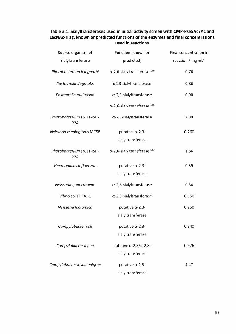
95
Table 3.1: Sialyltransferases used in initial activity screen with CMP-Pse5Ac7Ac and LacNAc-ITag, known or predicted functions of the enzymes and final concentrations
used in reactions
Source organism of
Sialyltransferase
Function (known or
predicted)
Final concentration in
reaction / mg mL-1
Photobacterium leiognathi α-2,6-sialyltransferase 146 0.76
Pasteurella dagmatis α2,3-sialyltransferase 0.86
Pasteurella multocida α-2,3-sialyltransferase
α-2,6-sialyltransferase 145
0.90
Photobacterium sp. JT-ISH-
224
α-2,3-sialyltransferase 2.89
Neisseria meningitidis MC58 putative α-2,3-
sialyltransferase
0.260
Photobacterium sp. JT-ISH-
224
α-2,6-sialyltransferase 147 1.86
Haemophilus influenzae putative α-2,3-
sialyltransferase
0.59
Neisseria gonorrhoeae α-2,6-sialyltransferase 0.34
Vibrio sp. JT-FAJ-1 α-2,3-sialyltransferase 0.150
Neisseria lactamica putative α-2,3-
sialyltransferase
0.250
Campylobacter coli putative α-2,3-
sialyltransferase
0.340
Campylobacter jejuni putative α-2,3/α-2,8-
sialyltransferase
0.976
Campylobacter insulaenigrae putative α-2,3-
sialyltransferase
4.47
96
3.2.2 PmST acceptor screen with CMP-Pse5Ac7Ac as donor
PmST was provided by Prozomix as ammonium sulphate precipitate and prepared as
described in 3.2.1. To screen the activity of PmST with various acceptors and CMP-
Pse5Ac7Ac donor, 25 µL reactions contained 0.130 mg mL-1 PmST, 2 mM CMP-Pse5Ac7Ac
1.10 and 8 mM acceptor in 50 mM Tris HCl pH 7.5. One of the following acceptors was
added to each reaction: N-acetyl-ᴅ-lactosamine (LacNAc) 3.11, β-D-galactopyranosyl-1,4-D-
glucose (lactose (Lac)) 3.12, ᴅ-Galactopyranose (Galactose (Galp)) 3.13, methyl β-ᴅ-
galactopyranoside (Me β-ᴅ-Galp) 3.14, 6-deoxy-6-fluoro-ᴅ-Galactose (6-F-Galp) 3.15, 4-
nitrophenyl-β-D-galactopyranoside (pNP-β-ᴅ-Galp) 3.16. Reactions were performed in
duplicate and control reactions without either PmST, CMP-Pse5Ac7Ac or acceptor were
also performed. All samples incubated at 37 °C for 4 hours and were analysed via LC-MS
(details in Appendix-General methods).
3.2.3 Large Scale synthesis of β-Pse5Ac7Ac-2,3-pNP-β-ᴅ-Galp using PmST
PmST was prepared from ammonuim sulphate precipitate as described in 3.2.1. 2 mM
CMP-Pse5Ac7Ac (9.5 mg), was added to a solution containing 50 mM Tris HCl pH 7.5, 8
mM pNP-β-ᴅ-Galp, 110 μg mL-1 PmST, 100 μg mL-1 PseF (prepared as described in 2.2.X), 2
mM cytidine-5’-triphosphate (CTP) and 10 mM MgCl2, in a final volume of 7.5 mL. The
reaction mixture was incubated at 37 °C for 18 hours, and the reaction was monitored by -
ESI LC-MS as described for samples in 3.2.2. The reaction mixture was centrifuged (37,000
× g, 4 °C, 30 minutes) to remove enzyme precipitate and the resultant supernatant was
lyophilised and resuspended in 10 mL dH2O and cotton wool filtered to remove enzyme
precipitate. The filtrate was then mixed with 10 mL EtOH and stored at 4 °C for 30 minutes
to precipitate any remaining enzyme. The mixture was cotton wool filtered, and the filtrate
was diluted in dH2O to a final volume of 200 mL before lyophilisation. The lyophilised
material was resuspended in 5 mL dH2O and applied to a 500 mL column packed with Bio-
Gel® P-2 resin (Biorad) in HPLC-grade H2O. 3 mL fractions were collected for 40 hours and
analysed via LC-MS for the presence of β-Pse5Ac7Ac-2,3-pNP-β-ᴅ-Galp 3.17 ([M-H]- m/z:
616.2). Fractions containing Pse5Ac7Ac-pNP-β-D-Galp were pooled and lyophilised.
Following NMR analysis of the resulting material (D2O, 700MHz), the material was dry
loaded onto silica and purified via flash chromatography using EtOH:MeOH:dH2O (5:2:1). 2
mL fractions were collected and analysed by TLC (sugar stain: MeOH, 5% H2SO4 and

97
charring) and LC-MS (details in Appendix-General methods) for the presence of Pse5Ac7Ac-
pNP-β-D-Galp. Solvent was evaporated from desired fractions which were then pooled and
analysed by NMR (D2O, 700 MHz).
1H NMR (700 MHz, Deuterium Oxide) δ 8.29 (d, J = 9.3 Hz, 2H, ArH), 7.28 (d, J = 9.4 Hz, 2H,
ArH), 5.32 (d, J = 7.9 Hz, 1H, H1’), 4.26-4.24 (m, 2H, H3’, H6), 4.19-4.18 (m, 1H, H5), 4.12 (t,
J = 3.1 Hz, 1H, H8), 4.05-4.02 (m, 2H, H4’, H7), 3.91 (m, 1H, H2’), 3.91-3.90 (m, 1H, H5’),
3.88-3.86 (m, 1H, H4), 3.78 (d, J = 6.1 Hz, 2H, H6’), 2.58 (dd, J = 12.6, 4.3 Hz, 2H, H3eq), 2.01
(s, 3H, C5-NHAc), 1.95 (s, 3H, C7-NHAc), 1.69 (t, J = 12.7 Hz, 1H, H3ax), 1.33 (d, J = 6.9 Hz,
3H). 13C NMR (176 MHz, D2O) δ 174.8, 173.6, 173.5, 161.7, 142.6, 126.1, 116.4, 100.1, 99.7,
75.5, 75.1, 73.5, 69.0, 68.8, 68.5, 64.8, 60.7, 53.7, 48.3, 35.85, 22.1, 21.9, 20.0. [α]D25 = 208.4
(c 0.08, H2O). HR-MS data was collected with –ESI MS: Expected [M-H]- m/z: 616.1995,
measured [M-H]- m/z: 616.2008. ATR-FTIR Vmax: 3354, 2971, 2891, 1591, 1406, 1062 cm-1.
2.1 mg. β-Pse5Ac7Ac-2,3-pNP-β-ᴅ-Galp 3.17 was obtained as an off-white solid (23% yield).

98
3.3 Results
3.3.1 Initial Activity assays of sialyltransferase library with CMP-Pse5Ac7Ac
donor
A library of bacterial SiaTs were screened for their ability to utilise CMP-Pse5Ac7Ac 1.10 as
as donor, with LacNAc-ITag 3.8 used as the acceptor as the postively charged ITag has
previously been used to identify low level product formation in enzymatic reactions
monitored by MALDI-TOF-MS (Scheme in Table 3.2).148 Following overnight incubation the
reactions were analysed by MALDI-TOF-MS for the presence of peaks corresponing to β-
2,3-Pse5Ac7Ac-LacNAc-ITag 3.9 or β-2,6-Pse5Ac7Ac-LacNAc-ITag 3.10. It was concluded
that four of the 13 SiaTs assayed were active under these conditions, due to the presence
of peaks at m/z 985.4 in the MALDI data for these samples, which corresponds to either
3.9 or 3.10 (results summarised in Table 3.2- green text denotes SiaT activity was observed,
red text denotes SiaT activity was not observed) (MALDI spectra in Appendix). MALDI
spectra for all thirteen samples showed that LacNAc-ITag 3.8 remained in the samples, seen
as a positively charged molecular ions peak (M+) at m/z 669.3. As this was the limiting
reagent (4:1 equivalents of 1.10 to 3.8) this suggests that successful reactions did not go to
completition. MALDI data also revealed ITag-GlcNAc (m/z 507.2), suggesting that each of
the SiaT prepartions also contained β-galactosidase, which resulted in the hydrolysis of Gal
from LacNAc-ITag. Samples which contained 3.9 or 3.10 were also subjected to –ESI LC-MS
analysis, which revealed that no detectable CMP-Pse5Ac7Ac remained in these samples.
Pse5Ac7Ac was present in each sample, which may have been the hydrolysis product of
CMP-Pse5Ac7Ac or due to hydrolysis of β-2,3- or β-2,6-Pse5Ac7Ac-LacNAc-ITag. It was
conlcuded that PlST, PdST, PmST and PJT-ISH-224ST were able to function as pseudo-
pseudaminyltransferases.

99
Table 3.2: Results of Initial Sialyltransferase activity screen with α-CMP-Pse5Ac7Ac 1.10 and
LacNAc-ITag 3.8, for the production of β-2,3-Pse5Ac7Ac-LacNAc-ITag 3.9 or β-2,6-Pse5Ac7Ac-
LacNAc-ITag 3.10, as shown in reaction scheme. Green enzymes: active; Red enzymes: no
activity observed.
Sialyltransferase source organism Predicted Product Reference
Pasteurella multocida 3.9 145
Photobacterium sp. JT-ISH-224 3.9 149
Photobacterium leiognathi 3.10 150
Pasteurella dagmatis 3.10 151
Haemophilus influenzae 3.9 N/A
Neisseria lactamica 3.9 N/A
Campylobacter jejuni 3.9 N/A
Campylobacter insulaenigrae 3.9 N/A
Neisseria meningitidis MC58 3.9 N/A
Vibrio sp. JT-FAJ-1 3.9 39
Campylobacter coli 3.9 N/A
Neisseria gonorrhoeae 3.10 NA
Photobacterium sp. JT-ISH-224 3.10 78

100
3.3.2 PmST acceptor screen with CMP-Pse5Ac7Ac as donor
Following the positive results of the initial SiaT actiivty screen PmST was used with CMP-
Pse5Ac7Ac 1.10 to screen a range of galactose derivative acceptors including LacNAc 3.11,
Lac 3.12, D-Gal 3.13, Me-β-D-Galp 3.14, 6F-Gal 3.15 or pNP-β-D-Galp 3.16, producing
Pse5Ac7Ac galactosides. Reactions at pH 7.5, incubated at 37 °C for 4 hours and were
analysed via LC-MS. Standard curves of Pse5Ac7Ac and CMP-Pse5Ac7Ac on -ESI LC-MS and
were used to calculate conversion of donor and acceptor to β-2,3-Pse5Ac7Ac-glycoside
product. As each reaction contained an initial concentration of 2 mM CMP-Pse5Ac7Ac, the
standards curves were used to determine the remaining concentration of CMP-Pse5Ac7Ac
and the concentration of any Pse5Ac7Ac, the conversion to Pse5Ac7Ac-glycoside was
decuded from these values.
Conversions of CMP-Pse5Ac7Ac to β-2,3-Pse5Ac-β-D-galactoside di- or tri-saccharide
products ranged from 57%-91% (results summarised in Table 3.3). (Standard concentration
curves of CMP-Pse5Ac7Ac and Pse5Ac7Ac, used to calculate conversions can be found in
Appendix.) LC-MS analysis showed that, with the exception of the no donor sample, each
of the samples contained free Pse5Ac7Ac, observed as a 332.9 m/z ([M-H]-). Previous
studies of PmST revealed that the enzyme shows sialidase activity towards α-2,3-sialyl-
galactosides, therefore some of the Pse5Ac7Ac found in these samples may be due to
hydrolysis of β-2,3-Pse5Ac-β-D-galactoside product.149 However, the reactions were
performed above the optimal sialidase activty of PmST (optimal activity at pH 5.0-5.5,
reactions were performed at pH 7.5) therefore, it is likely that a large proportion of the
Pse5Ac7Ac present in these samples was due to hydrolysis of CMP-Pse5Ac7Ac.149
A third function of PmST is that it displays α-2,6-SiaT activity with CMP-Neu5Ac 1.5 at pH
4.5- 7.0 with less efficiency than the α-2,3-SiaT activity of the enzyme.149 As LC-MS analysis
reveal that PmST was able to utilise CMP-Pse5Ac7Ac donor and Gal acceptor at pH 7.5,
where literature suggests that the enzyme affords 2,3 linked Pse5Ac7Ac galactosides, 6F-
Gal was used in this acceptor screen to provide evidence in support of this. No product
would be observed in the sample containing 6F-Gal, if PmST was producing 2,6 linked
Pse5Ac7Ac galactosides. As anticipated, a peak at 497.0 m/z, corresponding to the
molecular ion of Pse5Ac7Ac-6F-Gal was present in LC-MS data, suggesting that C6 hydroxyl

101
of galactose-based acceptors was not used in glyosidic bond formation under these
conditions.
Table 3.3: Acceptor Screen (compounds 3.11-3.16) of P. multocida SiaT (PmST) with CMP-Pse5Ac7Ac as donor. Samples analysed by LC-MS and conversion to β-2,3-Pse5Ac7Ac galactoside calculated using EIC of Pse5Ac7Ac and CMP-Pse5Ac7Ac remaining in samples.
Acceptor Conversion to β-2,3-Pse5Ac7Ac galactoside
91%
90%
78%
80%
57 %
91%

102
3.3.3 Large Scale synthesis of β-Pse5Ac7Ac-2,3-pNP-β-ᴅ-Galp using
Pasteurella multocida SiaT
Following the results of the acceptor screen, in order to unequivocally confirm the
stereochemistry and linkage of the products of reactions mediated by PmST, the reaction
between CMP-Pse5Ac7Ac 1.10 and pNP-β-ᴅ-Galp 3.16 was performed using 9.5 mg CMP-
Pse5Ac7Ac, such that the product could be purified and analysed by NMR. To drive the
formation of Pse5Ac7Ac-glycoside product, CMP-Pse5Ac7Ac synthetase, PseF was added
to the reaction so that hydrolysed Pse5Ac7Ac could be reactivated with a CMP group. The
reaction incubated for 18 hours and progress was monitored via LC-MS. LC-MS analysis
showed conversion to β-Pse5Ac7Ac-2,3-pNP-β-ᴅ-Galp 3.17 was achieved but that CMP-
Pse5Ac7Ac remained however, enzyme prepitation was observed so the reaction was
stopped at this time . Enzymes were removed from the reaction by lyophilisation, ethanol
preciptation and filtration, and the enzyme free material was purified by gel-filtration and
subsequent silica flash columnchromatography.
1H-NMR analysis confirmed that the product did indeed contain a β-Pse5Ac7Ac linkage
with the sterochemistry determined by the the 0.9 p.p.m. difference between δ values for
H-3ax and H-3eq peaks in the 1H NMR spectrum (Figure 3.2 -reported as ~0.6 p.p.m. for
equatorial carboxy group and ~0.9 p.p.m for axial carboxy group).106,128 As PmST is known
to function as a inverting GT, this result was anticipated as the CMP-Pse5Ac7Ac starting
material shows α-stereochemistry (Chapter 2.3.9).145

103
Figure 3.2: 700 MHz 1H NMR Spectrum of β-Pse5Ac7Ac-2,3-pNP-β-ᴅ-Galp 3.17, in D2O. 1H peaks for H9, H3ax, H3eq, 5NHAc, 7NHAc shown (blue labels). pNP-β-ᴅ-Galp H1, H7
and H8 shown (orange labels).

104
3.4 Discussion
Galan and co-workers previously demonstrated that ionic liquid ITag substrates could be
used in sensitive enzymatic reactions and subsequently analysed by MS.148 On the basis of
these results ITag-LacNAc was used with two commericially available SiaTs to produce α-
2,3- and α-2,6-linked Neu5Ac-LacNAc-ITag trisaccharide.152 For the initial SiaT screen of this
study we used LacNAc-ITag as accpetor as ILs have been shown to give greater peak
intensities and lower limits of detection in MS, therefore low level turnover of CMP-
Pse5Ac7Ac to Pse5Ac7Ac-LacNAc-ITag product should be detectable in MALDI analysis.153
The work of Sittel and Galan had confirmed that PmST and PdST were able to ulitilse
LacNAc-ITag with CMP-Neu5Ac 1.5 as donor.152 Both of these enzymes showed activity with
CMP-Pse5Ac7Ac in the initial screen (3.2.1, results described in 3.3.1). However, during this
initial acitvity screen, no positive control for SiaT activity was collected. Unlike the PmST
and PdST SiaTs, some of the enzymes assayed had not been shown to be active with CMP-
Neu5Ac (activity listed as putative in Table 3.1), therefore some of the negative hits from
this assay shuld be treated with caution. For the screen described in 3.2.1, CMP-Neu5Ac
could have been used as donor to confirm that these enzymes were active and able to
utilise LacNAc-ITag as an acceptor. Further to this, the concetrations of enzymes used in
this initial screen ranged from 0.15 to 4.47 mg mL-1, and activity was seen in samples with
0.76 to 2.89 mg mL-1 of enzymes. Therefore, a lack of activity in some of the samples may
be due to the sample not containing sufficient enzyme to produce a detectable quantity of
Pse5Ac7Ac-LacNAc-ITag in the assay. The pH (7.5) and temperature (37 °C) may not have
been optimal for some of these SiaTs, therefore this could also be varied if the screen were
to be repeated. Additionally, alternative ITagged acceptor carbohydrates may have been
screened. However, as four positive hits were obtained with CMP-Pse5Ac7Ac, work with
these enzymes was pursued and no further efforts were made to confirm the activity of
the remaining SiaTs with CMP-Neu5Ac or CMP-Pse5Ac7Ac under alternative conditions.
CMP-sugars are known to be unstable and rapidly hydrolyse, releasing the CMP group,
therefore this can in part account for the free Pse5Ac7Ac observed in samples. However,
three of the four SiaTs which ultiised CMP-Pse5Ac7Ac have previously been shown to
display sialidase activity, cleaving Neu5Ac from glycosides. Pse5Ac7Ac seen in the reaction

105
containing PlST (performed at pH 7.5) may in part be due to the enzymes sialidase activity,
which had previously been shown to display around 20-30% activity at pH 7.0 and pH
8.0.146 PmST sialidase activty is not reported to have been observed at the pH of this
reaction (pH 7.5) with optimal activity reported at pH 5.0-5.5 therefore, the Pse5Ac7Ac
found in these samples is therefore likely as a result of CMP-Pse5Ac7Ac hydrolysis.43
Similarly PdST sialidase is optimal at pH 4.5, given that the reaction with CMP-Pse5Ac7Ac
were performed at pH 7.5, the majority of the Pse5Ac7Ac in the sample is againlikely due
to starting material hydrolysis.36 Further to this, in the large scale synthesis of β-2,3-
Pse5Ac7Ac-β-D-Galp mediated by PmST, PseF and CTP were used to drive the conversion
of Pse5Ac7Ac to CMP-Pse5Ac7Ac. Whilst hydrolysis of starting material and product may
be a caveat of these enzymatic syntheses, this methodology resulted in the synthesis of a
number of potentially biologically relevant compounds, where acceptor moiety can easily
be altered, unlike in traditional chemical synthesis.141
The carbohydrate structure database, currently lists 27 unique, naturally occuring
oligosaccharides that contain β-Pse5Ac7Ac or β-Pse-derivative which are linked to
galactose via a 2,3- or 2,6- glycosidic bond.154 These structures range from cell surface
glycans suchs as lipopolysaccharide and exopolysaccharide to the teichuronic acid
compenent of Kribbella cell walls.144,155,156 Cell surface oligosaccharides containing β-
Pse5Ac7Ac-2,3-Galp and β-Pse5Ac7Ac-2,6-Galp have been found to occur in drug-resistant
bacteria, from clinical isolates.144,157,158 Therefore, the novel β-Pse5Ac7Ac-2,3-Galp and β-
Pse5Ac7Ac-2,6-Galp based carbohydrates synthesised in this study may be valuable tools
in work towards new therapeutics.
Sialic acids within bacteria are only known to exist as α-linked glycosides however, CMP-
sialic acids are biosynthesised as β-anomers. All sialic acid GTs SiaTs can be classified as
inverting.69 Each of the SiaTs in this study therefore produces α-linked Neu5Ac glycosides
and where possible, β-linked Pse5Ac7Ac-glycosides (equatorial glycosidic bonds in both
Neu5Ac glycosides and Pse5Ac7Ac glycosides). A number of multi-drug resistant bacterial
strains from clinical isolates are known to contain α-Pse-glycosides in cell surface
structures, which are crucial for virulence.159–161 Therefore a more comprehensive toolkit
for Pse-glycoside synthesis should ideally be able to produce Pse-glycosides in which the
sterochemistry of C1 is retained with respect to CMP-Pse5Ac7Ac, yielding α-linked Pse

106
products. This could be achieved through the use of retaining GTs, however to date no
retaining SiaTs have been shown to be active in vitro. This issue of access to α-Pse-
glycosides, via chemoenzymatic routes is adressed in Chapter 4.
This work has demonstrated the ability of four promicious SiaTs to ultilise non-natural
substrate CMP-Pse5Ac7Ac to generate β-2,3- and β-2,6-Pse5Ac7Ac-glycosides. The
products of this screen bear a reactive ITag handle which has been shown to be a useful
tool in the studies of the biological roles of carbohydrates.162 Further to this, the highly
promiscuous, well characterised, stable, PmST was shown to utilise a range of acceptors
with CMP-Pse5Ac7Ac 1.10.43 Structural data is available for three of the four enzymes
(PmST, PdST and PJT-ISH-224ST) which displayed activity with CMP-Pse5Ac7Ac.36,38,147,163
Structural analysis of these enzymes revealed that they all have overall GT-B topology.
36,38,147,163 It has previously been noted that GT-B family enzymes generally have greater
promiscuity for subsrates than GT-B family transferases.38
Crystal structure data of PmST and Photobacterium sp. JT-ISH-224 α-2,6-SiaT has previously
been used to rationally redesign the active site of PdST such that the enzyme formed α-
Neu5Ac-2,6-β-D-galactosides instead of the α-2,3- wild type activity. This provides
precedent for the redesign of activity sites of these well-characterised, highly-stable SiaTs,
it is feasible that structural data and site-driected mutagenesis may be used to further
engineer these enzymes, enabling a broader range of NulO-glycosides to be enzymatically
synthesised.
Given the diversity of CMP-nonulosonic acids that PmST has been previously shown to
utilise, it is highly feasible that this enzyme may be used to synthesise a potentially vast
range of glycosides incorporating Pse5Ac7Ac or Pse derivatives. Notably, PmST has shown
activity for CMP-sialic acids with a range of N-linked functionalities at C5, this is promising
for the use of this enzyme with CMP-Pse-derivatives which show a range of N-linked
functionalities at this position.2,145 In addition the crystal structure of this enzyme has been
solved with substrate analogues bound in the active-site, this data could potentially be
used to engineer the active site to turnover acceptor substrates not currently
accommodated by the enzyme or to alter the regioselectivity of the glycosidic bond
produced, expanding the library of Pse-based glycosides that can be generated using this
approach.164
107
3.5 Conclusions and Future Work
This work presented here provides the first examples of chemoenzymatically synthesised
Pse5Ac7Ac containing glycosides. Considering that at the commencement of this study no
native pseudaminyltransferases had yet been shown to be active in vitro, these well-
characterised, highly stable, promiscuous SiaTs, which display activity with CMP-Pse5Ac7Ac
provide a promising route to synthesis of a range of novel and biologically relevant Pse-
glycosides.
Crystal structure data of PmST with various ligands in the active site are available, including
a structure with Lac acceptor and non-hydrolyzable donor substrate analogue, CMP-3-
deoxy-3-fluoro-Neu5Ac (CMP-3F-Neu5Ac) both present in the active site.38,164 Future work
could focus on crystallisation of PmST with CMP-3F-Pse5Ac7Ac to gain insight into the
molecular basis of donor substrate promiscuity that has been demonstrated in previous
studies and expanded on in this work with CMP-Pse5Ac7Ac.
Chapter 2 described a strategy developed in the Fascione laboratory for the
chemoenzymatic synthesis of CMP-Pse5Ac7R derivatives. Future work may examine
whether any of the SiaT which were shown to be activty with CMP-Pse5Ac7Ac are able to
utilise these CMP-Pse derivatives as donor substrates, potentially expanding the range of
Pse-glycosides which can be synthesised using the approach decribed in this study.

108
Chapter 4: Characterisation of Putative
Pseudaminyltransferases
4.1 Introduction
In contrast to the detailed characterisation of PseB, C, G, H, and I enzymes in the
biosynthesis of 5,7-diacetamido-3,5,7,9-tetradeoxy-L-glycero-L-manno-non-2-ulosonic acid
(Pse5Ac7Ac 1.6 Figure 4.1), knowledge of pseudaminic acid (Pse) processing enzymes is
lacking. Glycosyltransferases (GTs) for which cytidine 5′-monophosphate Pse5Ac7Ac (CMP-
Pse5Ac7Ac 1.10 Figure 4.1) or a similar CMP-linked Pse derivative are the substrate, are
necessary for the incorporation of Pse into glycoconjugates, but have yet to be
unequivocally characterised.89 The major limitation in the study of
pseudaminyltransferases (PseTs) is the lack of available Pse-based molecules, most
crucially nucleotide activated CMP-Pse5Ac7Ac and derivatives.
Figure 4.1: Structures of Ulosonic Acids: Pse5Ac7Ac 1.6, β-Kdo 4.1.
PseTs are responsible for facilitating the formation of a glycosidic bond between Pse and a
range of acceptors. These acceptors include proteins where Pse can be O-glycosylated to
serine and threonine residues of cell surface proteins, or carbohydrates, where linkage may
be via an oxygen or nitrogen atom of Pse.1

109
When considering the diversity of PseTs, it is also important to account for the differences
in the mechanisms of these enzymes. PseTs facilitate the formation of glycosidic bonds
formed between Pse and an acceptor with either a retention or inversion of the
configuration of the Pse anomeric centre, in comparison to that of the CMP-Pse substrate
(Figure 4.2). Whilst a wealth of experimental evidence demonstrates that inverting GTs use
a single-displacement SN2 mechanism, uncertainty remains over the mechanism employed
by retaining GTs.26 Currently, knowledge of the mechanism used by retaining ulosonic acid
GTs is limited. The 3-deoxy-d-manno-oct-2-ulosonic acid (Kdo 4.1 Figure 4.1) transferase
WbbB, from Raoultella terrigena is the first structurally characterised retaining GT to use a
ketose donor sugar. Whilst further studies are needed to definitively decipherer the
mechanism of WbbB2-401, structural studies suggest that an internal-return mechanism (SNi)
mechanism is not feasible for this enzyme and there is evidence to support an SN2-like
double displacement mechanism.31
Figure 4.2: Mechanism of retaining and inverting pseudaminyltransferases. A carbohydrate acceptor and the donor α-CMP-Pse5Ac7Ac 1.10 are used in a PseT catalysed glycosylation
reaction where CMP is the leaving group. Anomeric configuration of the donor is either inverted or retained dependent on the mechanism of the PseT.
To date, the best-studied putative PseTs are those of the motility accessory family (Maf),
which are predicted to be involved in O-glycosylation of flagellin proteins in a number of
bacterial species.89,104,108,165,166 Knockout mutational studies of Motility associated factor-1
(Maf1) from the opportunistic pathogen Aeromonas caviae have shown that it is likely
responsible for the Pse5Ac7Ac glycosylation of flagellin subunits in the cytoplasm. The

110
glycosylation of flagellin subunits in A. caviae is required for flagella assembly, influencing
cell motility. 89,104,165 However, despite this promising initial evidence, no direct biochemical
data has been obtained to confirm that Maf1 has been correctly classified as a PseT. In vitro
characterisation of Maf1 is likely to be hindered by the insolubility of the Maf1 acceptor
substrates, FlaA and FlaB flagellin proteins, in addition to lack of access to CMP-
Pse5Ac7Ac.165
Promisingly, a crystal structure has been solved for Maf from Magnetospirillum
magneticum AMB-1, putatively assigned as a PseT, which shares 29% sequence identity
with A. caviae Maf1.143 MS analysis of the glycans present on the M. magneticum flagella
revealed that a mass corresponding to Pse5Ac7Ac was present, further to this the genome
contains homologues for the six CMP-Pse5Ac7Ac biosynthesis enzymes. Deletion of the
maf gene resulted in a lack of glycosylation of the flagella, providing further evidence that
Maf functions as a putative PseT. Structural analysis showed that Maf shares similarities
with inverting sialyltransferases from GT42 and GT29.166 However, co-crystallisation
attempts of Maf with CMP-N-acetyl neuraminic acid (CMP-Neu5Ac 1.5 Figure 4.1) were
unsuccessful. Due to a lack of access to CMP-Pse5Ac7Ac and some remaining uncertainty
regarding the exact identity of the CMP-nonulosonic acid which this enzyme is predicted to
utilise, this enzyme has not been shown to be functional in vitro.166 The literature currently
contains no other evidence of biochemical studies of putative PseTs; therefore no member
of this enzyme family has yet been unequivocally assigned.
The location of the maf gene often distinct to that of the other genes required for flagella
formation, however, maf genes within A. caviae are in close proximity to putative CMP-Pse
biosynthesis genes.166 This association with CMP-Pse biosynthesis genes is also seen for
putative PseTs that utilise carbohydrate acceptor moieties, rather than protein acceptors.
These potential PseTs are commonly found within genomic islands containing genes for the
biosynthesis of capsular polysaccharide (CPS) or O-antigen subunit of the
lipopolysaccharide (LPS). Further analysis of these loci revealed the presence of GTs
required for repeat unit assembly, including putative PseT genes (Figure
4.3).104,144,159,160,167,168
Five putative PseTs that were to be studied in this work (Table 1) are found within four
Gram-negative pathogenic bacterial species including the aforementioned A. caviae, two

111
Acinetobacter baumanii strains, Shigella boydii and Pseudomonas
aeruginosa.104,144,159,160,167,168 These transferases are predicted to incorporate Pse5Ac7Ac
or a Pse-derivative into either the CPS or LPS O-antigen in the cytoplasm, before subunits
are transported across the inner membrane, assembled in the periplasm and presented on
the cell surface. 104,144,159,160,167,168 Table 4.1 summarises source organism, Pse-containing
glycoconjugate, CMP-Pse substrate and predicted mechanism and GT homologue for each
putative PseT.

112
Figure 4.3: Genetic islands encoding cell surface carbohydrate synthesis and assembly genes. Islands contain genes for CMP-Pse5Ac7Ac or CMP-Pse5Ac7Hb (green box). Putative
pseudaminyltranferase genes are shown (bold red text).

113
Table 4.1: Putative Pseudaminyltransferases used in this study
Putative PseT
Source Organism
Predicted Pse-containing
glycoconjugate
Predicted CMP-Pse substrate
Predicted Mechanism
GT homologue (Sequence identity)
Lst A. caviae Sch3N
LPS O-antigen α-CMP-Pse5Ac7Ac Inverting NST (25%)
WfdL S. boydii B7 LPS O-antigen (4.2) α-CMP-Pse5Ac7(R)Hb
Inverting NST (25%)
Gtr16 A. baumanii 16562-2
CPS (4.3) α-CMP-Pse5Ac7Ac Inverting NST (25%)
KpsS1 A. baumanii ACICU
CPS (4.4) α-CMP-Pse5Ac7Ac Retaining KpsS (30%)
Orf14 P. aeruginosa 1244
LPS O-antigen (4.5) α-CMP-Pse5(R)Hb7Fm
Retaining KpsS (19%)
Lst: In addition to maf1, the A. caviae Sch3N genome also contains lst, a putative
LPS O-antigen PseT gene.104 The LPS O-antigen repeat of A. caviae Sch3N is known to
contain Pse5Ac7Ac, alongside other as yet incompletely characterised hexose sugars 2 (Dr
Jon Shaw, personal communication).104 It is predicted that Lst, the product of the lst gene,
along with other GTs assemble O-antigen in the cytoplasm, before the Wzx-like flippase,
Lsg transports O-antigen repeat units across the cytoplasmic membrane. Mutational
studies, carried out alongside characterisation of Maf1, demonstrate that Lst is not
involved in flagellin glycosylation. However, mutation of lst does result in the loss of O-
antigen, as observed by SDS-PAGE analysis of LPS.104 Although the stereochemistry of the
Pse5Ac7Ac linkage in the LPS of A. caviae is unknown, it is has been shown in this study
(Chapter 2.2.12) that A. caviae PseF produces α-CMP-Pse5Ac7Ac, therefore this is the likely
substrate for Lst to act as an inverting enzyme, resulting in the synthesis of a β-linked
Pse5Ac7Ac glycoconjugate.104,169 The unknown identity of the acceptor moiety for Lst
demonstrates a common hindrance in the study of GTs. Deciphering the identity of two
substrates for putative GTs often requires challenging structural analysis of glycans, which
may be difficult to perform.24
Whilst no direct evidence of the role of Lst has been collected, the functional prediction of
Lst as a putative PseT was based upon homology to several inverting sialyltransferases
(SiaTs).104 Notably, studies of Lst may be aided by its homology with Neisseria meningtidis

114
lipooligosaccharide-specific SiaT (NST), which transfers N-acetyl neuraminic acid (Neu5Ac
1.5) to carbohydrate acceptors.32 Crystallographic data of NST shows the protein exists as
a novel domain swapped homodimer, with a GT-B fold. Typical of GT-B fold enzymes NST
operates with a metal ion independent SN2-type mechanism, characteristic of inverting
GTs.24,32 The Carbohydrate Active EnZYme (CAZY) database, lists NST and currently 531
other α-2,3-SiaTs and α-glucosyltransferases, in the GT52 family (www.cazy.org). CAZY also
includes Lst in the GT52 family, based upon sequence homology, however if functional
studies confirm that Lst is an inverting PseT this enzyme will likely be the first member of a
new GT family.
WfdL: Two additional putative inverting PseTs with homology to NST were to be
studied. The first of which is WfdL, a putative PseT predicted to catalyse the incorporation
of 5-N-acetyl-7-N-[(R)-3-hydroxybutanoyl]-Pse (Pse5Ac7Hb) into S. boydii B7 LPS O-antigen
repeat unit 4.2 (Figure 4.4).168 Liu et al. assigned WfdL as a GT based upon sequence
homology to the Salmonella enterica LPS α-1,2-glucosyltransferase, WaaH.168 Further
analysis of WfdL shows that it has 25% sequence identity to NST and 27% sequence identity
with Lst (observation of this study). We assign WfdL as a putative PseT based upon this
homology in addition to its occurrence in the O-antigen gene cluster along with the six psb
genes needed for α-CMP-Pse5Ac7(R)Hb biosynthesis, several other glycosyltransferases
and transporters (Figure 4.3) required for O-antigen pentasacharride repeat unit 4.2 (Figure
4.3) assembly and export. Additionally, CAZY currently includes WfdL in the family GT52.
The presence of an α-linked Pse5Ac7Hb at the non-reducing end of the O-antigen suggests
that this carbohydrate is likely to play a role in S. boydii B7 immunospecificity within the
human host.168,170

115
Figure 4.4: Shigella boydii B7 LPS O-antigen repeat unit 4.2:
8)-β-Pse5Ac7(R)Hb-(2→6)-α-ᴅ-Galp-(1→6)-α-ᴅ-Glcp-(1→2)-β-ᴅ-Galf-(1→3)-α-ᴅ-GlcpNAc-(1→
Putative PseT WfdL is predicted to incorporate β-Pse5Ac7Hb.
Gtr16: Within clinically relevant, drug-resistant A. baumannii genomes, the capsule
biosynthesis loci (KL) have been assigned, and contain genes required for carbohydrate
biosynthesis, assembly and transport of CPS or LPS. KL6 A. baumannii 1656-2 includes
putative α-CMP-Pse5Ac7Ac biosynthetic genes and the putative inverting PseT Gtr16, a
homolog of NST. Based upon 30-40% sequence identity with NulO GTs, Gtr16 is predicted
to incorporate Pse5Ac7Ac into CPS. The tetrasaccharide CPS repeat unit 4.3 of KL6 A.
baumannii 1656-2 contain β-Pse5Ac7Ac, linked to both galactopyranose (Galp) and 2-
Acetamido-2-deoxy-ᴅ-galactopyranose (ᴅ-GalNAcp) (Figure 4.5).144 Gtr16 is the final
putative PseT of this study to show homology to NST. As with Lst and WfdL, CAZY
(www.cazy.org) currently lists Gtr16 as a GT52 member, however as with Lst, functional
studies could deem assignment incorrect as inverting PseTs represent a distinct GT
family.171 In addition to KL6, two other A. baumannii K loci also encode putative inverting
PseTs. The CPS repeat unit produced by the enzymes of the KL16 gene cluster, varies from
that of KL6 tetrasaccharide only at the 2,4 linkage of Pse5Ac7Ac to Gal.172 KL16 encodes
Gtr37 which shares 53% sequence identity with Gtr16. Secondly, A. baumannii with K93
CPS repeat unit, encode Gtr167 which is predicted to form a 2,6 linkage between Pse
derivative Pse5Ac7Hb and Galp as β-Pse5Ac7Hb is the terminal residue, in the CPS repeat

116
unit.173 Gtr167 shares 41% sequence identity with Gtr16, this may be due to difference in
the CMP-Pse substrate.
Figure 4.5: A. baumannii 1656-2 CPS repeat unit 4.3:
4)-β-Pse5Ac7Ac-(2→6)-β-ᴅ-Galp-(1→6)-β-ᴅ-Galp-(2→3)-α-ᴅ-GalpNAc-(1→
Putative PseT Gtr16 is predicted to incorporate β-Pse5Ac7Ac.
KpsS1: As highlighted previously, vast differences can be expected in the
mechanism used by inverting and retaining PseTs, thus it is of interest to characterize
enzymes in both classes. KpsS1, the putative retaining PseT from A. baumanii ACICU, is
found in the KL2 locus that encodes genes for CPS carbohydrate biosynthesis, assembly and
transport (Figure 4.3). The CPS repeat unit 4.4 of A. baumannii ACICU is made up of
Pse5Ac7Ac and three monosaccharides (Figure 4.6), and has also been proposed to
constitute a protein glycosylation motif in A. baumannii and is also present in the
exopolysaccharide of A. baumannii 54149 (Ab-54149).156,167 The CPS repeat unit
biosynthesised by enzymes in the A. baumannii KL2 locus is structurally similar to that
produced by A. baumannii KL6 locus 4.3, by the aforementioned Gtr16 in A. baumannii.
However structural characterisation suggests that these repeat units vary in the
stereochemistry linking Pse5Ac7Ac to a hexose. Inverting PseT Gtr16 facilitates the
formation of an 2,6 glycosidic linkage between β-Pse5Ac7Ac and Galp, whereas retaining

117
KpsS1 is predicted to facilitate formation of a α-2,6 linkage between Pse5Ac7Ac and
glucopyranose (Glcp 4.6).144,167
Figure 4.6: A. baumannii ACICU CPS repeat unit 4.4:
……………………………………………………α-Pse5Ac7Ac-(2→6)-β-ᴅ-Glcp-(1 ……………………………………….……………………..………………………………… ↓ ……………………………………………..……………………..……………………………6) ……………………………………………..………………………………………[3)-β-ᴅ-Galp-(2→3)-β-ᴅ-GalpNAc-(1→]
Putative PseT KpsS1 is predicted to incorporate α-Pse5Ac7Ac.
KpsS1 is named based upon homology to Escherichia coli KpsS (30% sequence identity), the
first retaining Kdo transferase characterised.30,160 Biochemically characterised KpsS has
been assigned as a member of the GT99 family. Recently WbbB was the first member of
this family to be structurally characterized. WbbB is a multidomain β-Kdo transferase that
is involved in biosynthesis of the OPS in Raoultella terrigena.31 WbbB2-401 shows 21%
sequence identity to A. baumannii KpsS1, likely due to differences in substrate. However,
an invariant motif in β-Kdo GTs, which is also highly conserved in CMP-sialic acid GTs, is the
active site HP motif, which is also present in KpsS1.31 Difficulties in complete
characterization of GTs are perhaps highlighted by the fact that there is only one
structurally characterized protein in the family of KpsS1 homolog, KpsS. The structural
characterization of WbbB may facilitate the studies of retaining other GTs that use CMP-
activated donors, including KpsS1.
In addition to KL2, several other A. baumannii K loci also encode the psaABCDEF genes
required for the synthesis of CMP-Pse5Ac7Ac or homologous genes, for the biosynthesis of
CMP-Pse derivatives.144,160,174 A GT for the turnover of CMP-Pse5Ac7Ac or a CMP-Pse
derivative can also be found within each of these K loci. KL23 encodes a putative GT which

118
shares 84% sequence identity with KL2 kpsS1 and has therefore also been named kpsS1
(kpsS1KL23).174 K loci KL33 also contains a putative retaining PseT, which Is predicted to form
the same α-Pse5Ac7Ac-1,6-β-ᴅ-Glcp linkage as KL2 KpsS1. The remainder of the KL33 CPS
repeat unit of the KL33 CPS varies from the tetrasaccharide repeat unit in A. baumannii
containing KL2, which may account for differences in sequence.161,175 Arbatsky et al.
annotated this KpsS1 homologue as KpsS2KL33.161 A. baumannii LUH5550 KL42 cluster
encodes a putative retaining PseT KpsS2KL42, required for incorporating Pse5Ac7Hb into the
CPS repeat unit. KpsS2KL42 shows 51% identity to KpsS1 and is a distant E. coli KpsS
homologue.176 A more distant KpsS1 homolog occurs in the KL12 locus of A. baumannii
which contains Grt59, a putative retaining GT which uses CMP-Pse5Ac7Ac epimer CMP-5,7-
di-N-acetylacinetaminic acid (CMP-Aci5Ac7Ac) for CPS repeat unit assembly.177 Structural
analysis of A. baumannii polysaccharides has aided the annotation of K loci genes, however,
biochemical characterisation to confirm these proposed functions of these genes is lacking.
Ultimately, unequivocal characterization of KpsS1 and other putative PseTs hinges on the
production of CMP-Pse5Ac7Ac.
The A. baumannii strains that synthesise 4.4 are antibiotic resistant isolates.156,167,175 A
recent study used bacteriophage ΦAB6 tailspike protein which depolymerize Ab-54149 EPS
into tetrasaccharide units. This strategy was used in place of alternative chemical methods
to access homogenous samples of 4.4, which was conjugated to a carrier protein for use as
a vaccine. Boosted sera from rabbits injected with this glycoconjugate vaccine could
recognize both Ab-54149 EPS and the glycoconjugate vaccine. The sera showed bactericidal
activity towards Ab-54149, only when the Pse5Ac7Ac present in the carbohydrate moiety
of the vaccine and the sera was shown to selectively bind Pse5Ac7Ac over sialic acid. This
study has demonstrated the therapeutic potential of 4.4 against an antibiotic resistant A.
baumannii strain, where Pse5Ac7Ac was shown to be a crucial epitope.178
Orf14:
Pseudomonas aeruginosa is responsible for multidrug resistant infection within
immunocompromised hosts. Despite P. aeruginosa LPS being considered a virulence factor,
crucial in host immune responses, the genes for LPS biosynthesis are relative under studied.
179 Analysis of the P. aeruginosa O7 LPS O-antigen biosynthesis locus (Figure 4.3) shows the
presence of genes which may putatively be assigned as those required for α-CMP-5-N-[(R)-

119
3-hydroxybutanoyl]-7-N-formyl-Pse (CMP-Pse5Hb7Fm) synthesis. The O7 O-antigen
biosynthesis locus also contains the gene annotated orf14 (accession: AAM27840.1), which
we predict encodes the putative retaining PseT.159 Orf14, which shows sequence homology
to KpsS1, is predicted to incorporate α-Pse5(R)Hb7Fm into the LPS O-antigen repeat unit
4.5 (unpublished observation of this study), a trisaccharide comprised of xylose (Xyl), and
N-acetyl fucosamine (FucNAc) (Figure 4.7), producing compound 5.159
Figure 4.7: P. aeruginosa O7 O-antigen repeat unit 4.5:
4)-α- Pse5(R)Hb7Fm-(2→4)-β-ᴅ-Xylp-(1→3)-β-ᴅ-FucpNAc-(1→
Putative PseT KpsS1 is predicted to incorporate α-Pse5Fm7Hb.
There is evidence to suggest that the enzymes involved in biosynthesis of this trisaccharide,
including Orf14, are also required for pilin glycosylation, as trisaccharide 1.41 (which
constitutes one monomer of the trisaccharide repeat unit 4.5) is present on at the C-
terminal Ser148 residue through a β-FucNAc-(1,3)-β-Seer linkage, where Pse5Hb7Fm is the
terminal carbohydrate.180 This pilin glycosylation has been shown to play a role in P.
aeruginosa 1244 virulence. Castaic et al. produced a P. aeruginosa 1244 strain with a
deletion of the pilO gene (ΔpilO), encoding the oligosaccharyltransferase that transfers 4.5
to the pilin and confirmed that this mutant strain produced lacked pilin glycosylation. In
mouse model studies, wild type P. aeruginosa 1244 had greater respiratory pathogenicity,
than the ΔpilO strain.181
In a mouse model study, when injected with pilin fragments glycosylated with 5 mice were
able to induce a specific immune response when challenged with P. aeruginosa 1244
infection, resulting in higher survival rate. It was shown that the immune response arose
from the production of antibodies that targeted 4.5 when present on both the pili and LPS
O-antigen. This study showed promise that a glycoconjugate vaccine containing 4.5, may
be used in the treatment of P. aeruginosa 1244 infection.182

120
Recently, the total synthesis of the trisaccharide 1.41 has been reported by Lui et al.6 The
product of this highly impressive synthesis is not only the first example of a naturally
occurring Pse-derivative to be produced by chemical synthesis but is also the first reported
synthesis of a Pse-based glycoside. However, as discussed in Chapter 1, the multi-step
synthesis required a vast amount of optimisation to yield 8.2 mg of 1.41.6
As discussed in Chapter 2, enzymatic synthesis of Pse-based glycosides may offer a valuable
alternative to chemical routes. Access to PseTs will increase the repertoire of Pse-based
molecules, which may aid studies into the biology roles of Pse. Additionally, in vitro studies
of PseTs can provide insight in to the functionality and mechanism of these enzymes,
enabling them to be studied as new therapeutic targets.
The aims of this chapter were to study of the putative PseTs Lst, WfdL Gtr16, KpsS1 and
Orf14, in vitro. Therefore, our initial aim was to develop a suitable strategy for the
recombinant expression of these targets. Following completion of this, the second aim was
to functionally characterise the enzymes, facilitated by access to CMP-Pse5Ac7Ac 1.10 and
a strategy for the synthesis of CMP-Pse derivatives (as discussed in Chapter 2).

121
4.2 Experimental
4.3.1 Sequence analysis of Putative Pseudaminyltransferases
The sequence of each putative PseT was analysed for the presence of transmembrane
helices, using TMHMM Server v. 2.0 (www.cbs.dtu.dk). The sequences were further
analysed for the presence of secretion signals, using SignalP-5.0 (www.cbs.dtu.dk). BLASTp
was performed using each putative PseT as the query sequence, to identify homologues.
Sequence alignments for Gtr16, WfdL, Lst, NST were performed using Clustal Omega
(www.ebi.ac.uk).
4.3.2 Homology Modeling of Putative Inverting Pseudaminyltransferases
A multiple sequence alignment of Nst, Gtr16, Lst and WfdL was produced using Clustal
Omega. This was then edited manually to improve the quality of the alignment for use in
homology modeling. Models were then produced using MODELER automodel through
Discovery Studio 3.5, using the apo structure of NST (PBD entry: 2YK4).
In order to prevent fixed body domain movements, the coordinates of some main chain
atoms were copied from the template. The residues chosen, in NST, were Gln60, Tyr92,
Tyr139, Tyr212, Gly255, Tyr276 and Tyr318. The actual coordinates in the model vary
slightly due to post-building refinement. Some secondary structure restraints were also
placed at one of the alpha helices, namely the helix from the conserved Glu260, as there
are lots of gaps in the sequence alignment at this region. A restraint was used from Glu210-
Ile226 in Gtr16, Glu209-Phe224 in Lst and Glu204-Tyr219 in WfdL. Twenty models were
produced for each of the proteins. Both the total PDF energy and the DOPE energy metrics
were used in choosing the best quality model.*
4.2.3 Putative Pseudaminyltransferase Expression Trials
Each of the putative PseT encoding genes (WfdL, Lst, Gtr16, KpsS1 and Orf14) were
obtained in plasmids constructed from either pET-15b or pET-28a(+) vector, to produce N-
terminal His6 tagged PseT, with genes of interest inducibly expressed using isopropyl β-D-
1-thiogalactopyranoside (IPTG) (Table 4.2) (plasmids were obtained from Genscript).
* Modeling was performed by Paul Bond, YSBL, University of York.

122
Plasmids for the five putative PseTs were initially transformed into chemically competent
E. coli BL21 (DE3) cells, by heat shock or into electrocompetent E. coli BL21 (DE3) cells, by
electroporation (as described in 2.2.1). Transformed colonies were selected for using the
selectable antibiotic resistant marker, encoded in the pET plasmid of choice (Table 4.2). As
controls the empty vectors pET-28a(+) and pET-15b were also transformed into both E. coli
BL21 (DE3) cells.
Table 4.2: Putative PseTs Plasmids and Molecular Weights of Recombinant Putative PseTs
Putative PseT Plasmid Vector Antibiotic Resistance of Plasmid MW of His6PseT
(kDa)
Lst pET-28a(+) Kanamycin 38.3 WdfL pET-15b Ampicillin 38.9 Gtr16 pET-15b Ampicillin 40.4 KpsS1 pET-15b Ampicillin 59.4 Orf14 pET-15b Ampicillin 59.5
Transformed E. coli cells were then used in expression trials. Throughout these initial trials,
the following four parameters were studied in combination: temperature, OD600 at
induction, IPTG concentration and incubation time post-induction. Transformed cells were
cultured overnight at 37 °C, grown aerobically at 180 revolutions per minute (r.p.m). in LB
supplemented with 50 μgmL-1 kanamycin (LBKan) or 100 μgmL-1 ampicillin (LBAmp). For each
sample, 50 mL LBKan or LBAmp was inoculated with a 1 in 100 dilution of overnight culture of
E. coli BL21 (DE3), in a 250 mL Erlenmyer flask, and grown at 37°C, 180 r.p.m. until OD600
reached 0.4 and 0.8 for Lst and 0.4, 0.6 and 0.8 for all other WfdL, Gtr16, KpsS1 and Orf14
at which point cultures were induced with either 0.1 mM or 1 mM IPTG. Post induction
cultures were incubated at 30 °C and 37 °C for Gtr16, WfdL and Orf14 and at 25 °C, 30 °C
and 37 °C for Lst and KpsS1. 2 mL Samples were taken at either 4 and 24 hours post
induction for Lst, WfdL, Gtr16 and Orf14 samples and 1, 4 and 24 hours post induction for
KpsS1 samples. The samples were centrifuged (6,000 × g, 10 minutes, 4 °C), supernatant
was removed, and cells pellets were stored at −20 °C. An uninduced control was also
collected, for each putative PseT, to assess basal expression of the genes and vector only
(pET-15b) control sample was taken to analyse alongside KpsS1 samples. To lyse cells,
pellets were resuspended in 100 μL BugBuster mix (BugBuster® Protein Extraction Reagent
(Merck), supplemented with 1 Pierce Protease Inhibitor Tablet (Thermo Scientific) per 50

123
mL and 25 units mL-1 Benzonase® Nuclease (Sigma)), incubated at room temperature for 20
minutes and centrifuged (17,000 ×g, 10 min, 4 °C). Supernatant and insoluble material was
separated. The insoluble material was resuspended in 100 μL dH2O. 20 μL samples of both
the soluble and insoluble material were mixed with 5 μL reducing dye (10% SDS, 10 mM β-
mercaptoethanol, 20% glycerol 0.2 M Tris HCl pH 6.8, 0.05 % bromophenol blue) heated to
95 °C (with the exception of Lst which was not heated) then run on 10%, 12% or 4-20% SDS-
PAGE (Described in 2.2.1). 5 μL PageRuler™ Plus Prestained Protein Ladder (Thermo
Scientific) or 6 μL Blue Prestained Protein Standard, Broad Range marker (New England
Biolabs) or both were used as molecular weight markers in SDS-PAGE.
Following these initial expression trials, the plasmids for KpsS1 and Lst were transformed
into chemically competent E. coli Tuner (DE3) cells. The expression trial was repeated, as
described for E. coli BL21 (DE3) cells, however, two additional post-induction temperatures
of 16 °C and 25 °C were studied. Samples were collected and lysed as described above and
analysed via 10 or 12% SDS-PAGE.
4.2.4 Solubilisation trials of Putative Pseudaminyltransferases Lst
E. coli BL21 (DE3) which were transformed with plasmid encoding Lst were cultured
overnight at 37 °C, 180 rpm in LBkan. The overnight Lst culture was used to inoculate 250
mL Erlenmeyer flasks, each containing 50 mL LBkan to give an initial OD600 0.02. All cultures
were incubated at 37 °C, 180 rpm, until expression was induced, at OD600 measurements
of 0.6, using either 0.1 mM or 1 mM IPTG for each OD600 value. Post-induction Lst was
incubated at 25 °C, 180 rpm. Three 1 mL samples were taken from each flask at 4 and 24
hrs post-induction. The samples were centrifuged (6,000 × g, 10 minutes, 4 °C) and cells
pellets were stored at −20 °C.
Cell pellets from each 1 mL cell culture sample were thawed on ice and resuspended in 50
μL Bugbuster mix, supplemented with either 8 M urea, 6 M Guanidine HCl or 1 M NaCl.
Cells were then incubated with shaking for 20 minutes at room temperature to facilitate
lysis. Lysed cells were then centrifuged (6,000 × g, 10 minutes, 4 °C), and the supernatant
was separated from pellet, which was resupended in 50 μL dH2O. 15 μL of each sample
were analysed using 12% SDS-PAGE (as described in 2.2.1, with the exception that Lst
samples were not heated before being applied to gels).

124
4.2.5 Detergent Screen for Solubilisation of Putative Pseudaminyltransferases
Lst and KpsS1
A 2 L Erlenmeyer flask, containing 500 mL LBkan was inoculated with Lst overnight culture
to give an initial OD600 0.02 and incubated at 37 °C, 180 r.p.m. Lst expression was induced,
at an OD600 measurement of 0.6, using 0.1 mM IPTG. Cells were centrifuged (6,000 ×g, 10
minutes, 4 °C) and cells pellets were stored at −20 °C. Cells were thawed on ice and
resuspended in 25 mL lysis buffer (50 mM sodium phosphate, pH 7.3, 400 mM NaCl, 10
mM β-mercaptoethanol, 10 mM imidazole, 1 mM MgCl2 supplemented with 1 Pierce
Protease Inhibitor Tablet (Thermo Scientific) per 50 mL and 25 units mL-1
Benzonase® Nuclease (Sigma). 250 μL samples were taken and were each supplemented
with one of five detergents, at various concentrations (Table 4.3) and additional lysis buffer
to give a final volume of 500 μL. A no detergent control sample was prepared, using lysis
buffer only. Cells underwent 10 freeze-thaw cycles to facilitate lysis. Lysed cells were then
incubated at 25 °C for 30 minutes, centrifuged (17,000 × g, 30 minutes, 4 °C), and the
supernatant was separated from pellet, which was resupended in 500 μL dH2O. 15 μL of
each sample, was mixed with 5× SDS reducing buffer and analysed using 12% SDS-PAGE (as
described in 2.2.1). This experiment was repeated with KpsS1 from cells cultured at 30 °C,
180 rpm LBamp. KpsS1 expression was induced, at an OD600 measurement of 0.8, using 0.1
mM IPTG. Post-induction KpsS1 was incubated at 30 °C, 180 r.p.m. for 4 hours.
Table 4.3: Detergents screened for ability to solubilise KpsS1 and Lst
Detergent Concentration
n-Dodecyl β-D-maltoside (DDM)
15 mM 30 mM 75 mM
Tween 20 0.06 mM 0.3 mM 0.6 mM
CHAPS 6 mM 16.2 mM (1%)
Triton X-100 0.5% 2% 5%
SDS 100 mM 50 mM

125
4.2.6 Cloning of KpsS1 Fusion Protein Plasmids
Three pET-based plasmids each containing commonly used fusion proteins for the
solubilisation of recombinantly expressed protein were obtained (University of York,
Biology Technology Facility) (plasmid sequences in Appendix) in order to in generate fusion
plasmids encoding either, glutathione-S-transferase (GST) or colicin E9 immunity protein
(Im9), maltose binding protein (MBP) with A. baumannii KpsS1 fused to the C-terminal end
of each of these proteins. CleR and CleF primers (Table 4.4) were used in gradient PCR to
linearize the vectors, in 50 μL reactions, using Q5 DNA polymerase (DNAP) (Table 4.5 and
4.6). 5 μL of each PCR sample was analysed via gel electrophoresis, using a 0.6% agarose
gel where DNA was visualised with SYBR Safe stain. Reaction tubes containing the desired
product (Gst: 6050 bp, Im9: 5651 bp) were run on 0.6 % agarose gel and purified via gel
extraction (illustra GFX PCR DNA and Gel Band Purification Kit, GE Healthcare). Linearised
vector was stored at –20 °C.
Table 4.4 PCR Primers for KpsS1 Fusion Plasmids
Primer Sequence
CleR 5'-TTGCTGGTCCCTGGAACAGAACTTCC-3' CleF 5'-CGCGCCTTCTCCTCACATATGGCTAGC-3' KpsS1 Forward 5'-TCCAGGGACCAGCAATGAACTTTCTGATCCTGAT- 3' KpsS1 Reverse 5'-TGAGGAGAAGGCGCGTTACTCAATGTCGGTGTT-3'
Table 4.5 PCR Protocol
Step Temperature Time
Initial Denaturation 98 °C 30 seconds
35 Cycles 98 °C 5 seconds
55–72 °C (gradient over 9 temperatures) 10–30 seconds
72 °C 30 seconds/kb
Final Extension 72 °C 2 minutes
Hold 4 °C /
The KpsS1 insert was amplified via PCR from the pET-15b KpsS1 (Genscript) plasmid used
for initial expression trials. Primers KpsS1 Forward and KpsS1 Reverse (Table 4.4) were used

126
in PCR to create regions of homology at either end of the KpsS1 gene. PCR was carried out
using Q5 DNAP (Table 4.5 and 4.6) as described for vectors, before being analysed via 0.6
% agarose gel electrophoresis and purified by gel extraction and KpsS1 product was stored
at –20 °C. A Quick-Fusion cloning kit (Biomake) was used to create two plasmids encoding
the KpsS1 fusion proteins from the linearised vector and KpsS1 insert (10 μL reactions were
set up containing 1 μL fusion enzyme, 2 μL fusion buffer, [0.01 × bp of vector] ng vector, 30
ng KpsS1 insert, dH2O to 10 μL). A control reaction using DNA provided in the kit was also
performed. Reactions were incubated at 37 °C for 30 minutes. 5 μL of each reaction was
then transformed into 50 μL XL-1 Blue Supercompetent cells, using heat-shock. Following
transformation, the cells were plated on LBkan plates containing a Blue-White Screen for
the selection of recombinant cells and were incubated at 37 °C overnight.
Table 4.6 Components for Q5 High-Fidelity DNAP mediated PCR
Component 50 µL Reaction 15 µL Reaction (Colony PCR)
Final Concentration
Q5 High-Fidelity 2X Master Mix
25 µL 7.5 µL 1X
10 µM Forward Primer 2.5 µL 0.75 µL 0.5 µM 10 µM Reverse Primer 2.5 µL 0.75 µL 0.5 µM Template DNA 1µL pET15b-KpsS1 1.6µL dH2O containing
DNA 20 ng pET-15b KpsS1
Nuclease-Free Water to 50 µL to 15 µL
White colonies were screened for the presence of KpsS1 fusion plasmid using colony PCR.
Colonies were selected, spotted onto LBkan agar and placed in 20 μL DNase free dH2O, which
was then heated to 98 °C for 5 minutes. 1.6 μL of the dH2O containing DNA was added to
Q5 reaction mixes to generate 15 μL reactions (Table 4.6), PCR was then performed (Tables
4.5 and 4.6, annealing temperature: 72 °C) with KpsS1 Forward and KpsS1 Reverse primers.
Gel electrophoresis was used to analyse reactions (0.6 % agarose). Colonies corresponding
to positive colony PCR results were incubated in 10 mL LBkan overnight (37 °C, 180 r.p.m.)
before plasmid DNA was extracted via miniprep (QIAGEN) and sequenced (GATC) using T7
and T7 reverse primers. DNA encoding each of the desired KpsS1 fusion plasmids was
transformed into chemically competent E. coli BL21 (DE3) via electroporation (as described
in 2.2.1).

127
4.2.7 KpsS1 Fusion Protein Expression Trials
Following the transformation of either Im9-KpsS1 or GST-KpsS1 plasmid into chemically
competent E. coli BL21 (DE3) single colonies were picked to inoculate LBkan, for overnight
cultures incubated at 37 °C, 180 rpm. 24 deep-well blocks were inoculated with the culture
to give an initial OD600 0.02, in a final volume of 2 mL LBkan in each 10 mL well. The cultures
were incubated at 37, 30 °C and 16 °C, at 180 r.p.m. and expression of recombinant KpsS1-
fusion protein was induced with the addition of either 0.1 mM, 0.5 mM or 1 mM IPTG, at
OD600 values of 0.4 and 0.8, resulting in 2 wells of cells per condition studied. Cells were
harvested from each condition 4 hrs and 20 hrs post-induction. OD600 values of the cultures
were recorded at the point of harvesting and cell pellets were obtained (6,000 ×g, 10
minutes, 4 °C) and stored at −20 °C. Pellets were resuspended in Bugbuster mix (without
lysozyme) and shaken at room temperature for 45 minutes to lyse cells. Cells were
centrifuged (6,000 × g, 10 minutes, 4 °C); following this soluble and insoluble material was
separated. The insoluble material was resuspended in an equal volume of dH2O and 15 μL
of both soluble and insoluble fractions, from each sample, were analysed via 10 % SDS-
PAGE (as described in 2.2.1).
4.2.8 Im9-KpsS1 Large Scale Expression
E. coli BL21 (DE3) was transformed with Im9-KpsS1 plasmid. LBkan (4 x 600mL, in baffled 2
L Erlenmyer flasks) was inoculated with a 1 in 100 dilution overnight of culture of E. coli
BL21 (DE3) transformed with Im9-KpsS1 plasmid (overnight culture: 50 mL LBkan, in 250 mL
Erlenmeyer flask, cultured at 37 °C, 180 r.p.m.). Cells were cultured at 37 °C, 180 r.p.m. and
protein expression was induced at an OD600 value of 0.8 with 0.1 mM IPTG. Cells were
incubated at 30 °C, 180 r.p.m. for 4 hrs post-induction before cell pellets were harvested
via centrifugation (6000 × g, 30 min, 4 °C) and stored at -80 °C.
4.2.9 Im9-KpsS1 Purification
Cell pellet from a 2.4 L culture was resuspended in 40 mL lysis buffer (50 mM sodium
phosphate, 400 mM NaCl, 10 mM β-mercaptoethanol, 10 mM MgCl2, pH 7.4)
supplemented with protease inhibitor, lysozyme, and Benzonase. Cells were lysed via
sonication in ice (30 seconds, 30 second pause, 25 repeats) and centrifuged (37000 ×g, 30
min, 4 °C). Supernatant was applied to a 1 mL HisTrap HP column (GE Healthcare) pre-

128
equilibrated with lysis buffer, the column was washed with 10 column volumes (C.V.), lysis
buffer and protein was eluted with a gradient of elution buffer (lysis buffer + 490 mM
imidazole, pH 7.4) over 40 C.V. as 1 mL fractions were collected. 15 μL samples of insoluble
material, soluble material, flowthrough wash and fractions were analysed via 10 % SDS-
PAGE. Following SDS-PAGE analysis (as described in 2.2.1) fractions containing Im9-KpsS1
(68.9 kDa, fractions 11-14) were pooled and applied to desalting column (HiPrep 26/10
desalting column, GE Healthcare) to exchange Im9-KpsS1 into reaction buffer (50 mM
sodium phosphate, 25 mM NaCl, pH 7.4).
4.2.10 Im9-KpsS1 Protein Identification Mass Spectrometry
Band which appeared to correspond to Im9-KpsS1 in pure fractions of SDS-PAGE (4.2.9)
was subjected to protein identification mass spectrometry, as described in 2.2.8 for PseF.
4.2.11 Im9-KpsS1 Activity Screen
40 µL reaction mixtures containing 20 mM acceptor (either ᴅ-Glucose (ᴅ-Glc) 4.6, methyl
β-ᴅ-glucopyranoside (Me β-ᴅ-Glcp) 4.7, 4-nitrophenyl β-ᴅ-glucopyranoside (pNP β-ᴅ-Glcp)
4.8, β-ᴅ-glucopyranosyl-2,6-methyl-β-D-galactopyranoside (β-ᴅ-Glcp-2,6-β-ᴅ-Galp-OMe)
4.9,***** 4-nitrophenyl β-D-galactopyranoside (pNP β-ᴅ-Galp) 3.16, 4-nitrophenyl β-ᴅ-
xylopyranoside (pNP β-ᴅ-Xylp) 4.10, or 4-methoxyphenyl β-ᴅ-glucopyranoside (pMeP β-ᴅ-
Glcp) 4.11), 5 mM CMP-Pse5Ac7Ac 1.10, 10 mM MgCl2, 50 mM sodium phosphate, 25 mM
NaCl, pH 7.4 and 12.2 µL partially purified Im9-KpsS1 or cell lysate containing Im9-KpsS1
were prepared and incubated at 30 °C for 12-18 hours. Reactions were performed in
duplicate. Control reactions were set up as a described above without the addition of one
of the following: pure or crude Im9-KpsS1, acceptor or CMP-Pse5Ac7Ac. Reactions were
also performed as described above with pNP-β-ᴅ-Glcp acceptor 4.6 and CMP-Neu5Ac 1.5,
in place of CMP-Pse5Ac7Ac and Im9-KpsS1 lysate. Additionally, a reaction was set up with
pNP-β-ᴅ-Glcp acceptor, containing both CMP-Neu5Ac and CMP-Pse5Ac7Ac and Im9-KpsS1
lysate. All samples were analysed via negative-ion mode electrospray ionisation liquid-
chromatography mass spectrometry (-ESI LC-MS) (details in Appendix-General methods).
***** 4.9 was synthesized and characterized by Dr Darshita Budhadev and Natasha Hatton, detailed in Appendix Scheme A.1
129
4.2.12 Chemoenzymatic synthesis of glycoside containing Pse5Ac7Ac, via a
one-pot, seven-enzyme reaction using Im9-KpsS1
2 mM UDP-GlcNAc 1.3 (30 mg), 0.3 mM coenzyme-A, 4 mM pyridoxal 5′-phosphate, 20 mM
L-glutamic acid, 3 mM phosphoenolpyruvate, 20 mM N-Acetyl-S-Acetylcysteamine, 0.2 mg
mL-1 PseB, 0.4 mg mL-1 PseC, 0.2 mg mL-1 PseH, 0.2 mg mL-1 PseG and 0.2 mg mL-1 PseI
(enzymes purified as described in Chapter 2.2.1). The reaction mixture was incubated at 37
°C for 6 hours and was monitored via –ESI LC-MS, to monitor the production of Pse5Ac7Ac.
Reaction was stored at 4 °C overnight, before enzyme precipitate was removed via
centrifugation (37,000 × g, 30 min, 4 °C). Supernatant was retained and 0.2 mg mL-1 PseF
(purified as described in Chapter 2.2.7), 4 mM CTP and 20 mM MgCl2 were added to
facilitate the production of CMP-Pse5Ac7Ac 1.10, as confirmed by –ESI LC-MS analysis. The
reaction incubated at 30 °C for 3 hours. Cell pellet containing Im9-KpsS1 from 1.8 L culture
(conditions as described for large scale expression) was lysed as described above. The
resulting supernatant was added to the CMP-Pse5Ac7Ac reaction mixture to a final volume
of 60 mL to give 1 mM CMP-Pse5Ac7Ac. 5 mM pNP-β-ᴅ-Glcp was then added to facilitate
the formation of Pse5Ac7Ac-pNP-β-ᴅ-Glcp 4.12. The reaction incubated at 30 °C for 72
hours and was analysed via –ESI LC-MS. The mixture was lyophilised and resuspended in
10 mL dH2O and cotton wool filtered to remove enzyme precipitate. The filtrate was mixed
with 10 mL EtOH and stored at 4 °C for 30 minutes to precipitate any remaining enzyme.
The mixture was cotton wool filtered, and the filtrate was diluted in dH2O to a final volume
of 200 mL before lyophilisation. The lyophilised material was resuspended in 10 mL dH2O
and 5 mL was applied to a 500 mL column packed with Bio-Gel® P-2 resin (Biorad) in HPLC-
grade H2O. 3 mL fractions were collected for 40 hours and analysed via LC-MS (details in
Appendix-General methods) for the presence of Pse5Ac7Ac-pNP-β-ᴅ-Glcp. The column was
repeated with the remaining lyophilised material and all fractions containing Pse5Ac7Ac-
pNP-β-ᴅ-Glcp were pooled and lyophilised. Following NMR analysis of the resulting
material (D2O, 700MHz), the material was dry loaded onto silica and purified via flash
chromatography using EtOH:MeOH:dH2O (5:2:1). 2 mL fractions were collected and
analysed by TLC (sugar stain: MeOH, 5% H2SO4 and charring) and LC-MS for the presence of
Pse5Ac7Ac-pNP-β-ᴅ-Glcp. Solvent was evaporated from desired fractions that were pooled
and analysed by NMR (D2O, 700 MHz).

130
1H NMR (700 MHz, Deuterium Oxide) δ 8.32 – 8.27 (m, 2H, ArH), 7.31 – 7.26 (m, 2H, ArH), 5.31 (d, J = 7.8 Hz, 1H, H1’), 4.27 (ddd, J = 12.2, 5.0, 4.0 Hz, 1H, H4), 4.18 (dd, J = 4.3, 1.8 Hz, 1H, H5), 4.17 – 4.05 (m, 4H, H7, H8, H4’ H5’), 4.04 – 3.96 (m, 2H, H6’), 3.84 – 3.80 (m, 2H, H6, H3’), 2.61 – 2.55 (m, 1H, A3eq), 2.00 (s, 3H, A5-NHAc), 1.95 (s, 3H, A7-NHAc), 1.69 (t, J = 12.7 Hz, 1H, A3ax), 1.19 (d, J = 6.4 Hz, 3H, A9).13C NMR (176 MHz, D2O) δ 175.5, 174.2, 174.0, 135.0, 126.1, 118.9, 116.2, 100.2, 99.1, 68.6, 68.1, 67.1, 61.5, 60.3, 59.5, 53.4, 48.8, 39.76, 35.8, 30.7, 29.0, 24.4, 24.1, 22.0, 21.9, 20.8, 19.8, 17.9. HR-MS data was collected with –ESI MS: Expected [M-H]- m/z: 616.1995, measured [M-H]- m/z: 616.2012. ATR-FTIR Vmax: 3236, 2974, 2897, 1588, 1575, 1562, 1407, 1112, 1078 cm-1

131
4.3 Results
4.3.1 Sequence analysis of Putative Pseudaminyltranferases
To aid the design of plasmids for overexpression, the amino acid sequences of the five
putative PseTs (Lst, WfdL, Gtr16, KpsS1 and Orf14) were analysed for the presence of
transmembrane helices. As these putative PseTs are predicted to be involved in the
synthesis of cell-surface glycoconjugates, there is a likelihood that these enzymes may be
membrane bound. As demonstrated with the Lst, WfdL and Gtr16 homologue NST,
truncation of proteins to remove membrane helices can aid protein solubility, which is
crucial for in vitro characterisation.32 Sequence analysis found that none of the putative
PseTs were predicted to contain transmembrane helices. Therefore, plasmids encoding
KpsS1, Orf14, Gtr16 and WdfL were all designed with full-length gene sequences. However,
a Δ9Lst (referred to as Lst throughout) construct was designed based on NST sequence
similarity to the membrane helix of NST, with the aim of producing soluble Lst.††††† GTs may
often be membrane associated and are therefore often insoluble when produced
recombinantly.
To determine a suitable expression strategy for the putative PseTs, the protein sequences
were analysed for the presence of predicted Gram-negative secretion signals, which may
direct the proteins across the cytoplasmic membrane, to the periplasm or out of the cell.183
None of the target proteins were predicted to contain Sec secretion signals (Appendix-
Table A.2) suggesting that these enzymes are cytoplasmic, therefore it was decided that
these recombinant proteins should be produced in the cytoplasm, therefore E. coli BL21
(DE3) was chosen as the first expression strain to be used in expression trials.
4.3.2 Further Sequence analysis and Homology Modeling of Putative Inverting
Pseudaminyltransferases
NST catalyses the formation of an α2,3/α2,6 glycosidic bond between, the nonulosonic acid
(NulO) Neu5Ac 1.1 and a galactosyl containing moiety.32 NST is the first enzyme of the GT52
family to be structural characterized and has been shown to possess a GT-B fold.184 Due to
††††† Lst plasmid was designed by Dr Glyn Hemsworth.

132
a lack of functional and structural characterization, the NST homologs Gtr16 and Lst are
currently listed as GT52 family enzymes (cazy.org), based on sequence similarity to this
family. Gtr16, WdfL and Lst are the three putative PseTs that show homology to NST and
are predicted to transfer a CMP-Pse donor sugar to a hexose acceptor sugar. Therefore,
due to similarities in the substrates of these GTs, it can be expected that homology will be
seen between the substrate binding sites and active site. The amino acid sequences of NST
and the three retaining putative PseTs were aligned (Figure 4.8) and used in combination
with NST structural data to build homology models.
The protein data bank (PDB) contains four structures of Nst (2YK4, 2YK5, 2YK6, 2YK7) that
have various ligands bound. The apo structure (2YK4) has the best resolution at 1.94 Å.
2YK4 was used as the template for building models of Lst, WfdL and Gtr16. The target
proteins all show less than 30% sequence identity with NST, which is below the ideal
threshold for homology modeling. However, in the absence of an alternative structurally
characterised homologue and based upon genomic evidence, and experimental evidence
for Lst, that these proteins function as PseTs it was decided that NST be used as the
template for modeling. Models of this standard may be used to predict overall fold of the
enzymes and may cautiously be used to predict the location of key conserved residues
within the enzyme.
The GT-B fold of the enzymes can be seen in the NST structure and the putative PseT
models (Figure 4.9). GT-B fold enzymes are typically metal-ion independent, NST and its
homologues, with the exception of Lst, do indeed lack the candidate DXD motif required
for metal-ion co-ordination in GT-A fold enzymes.171 Sequence alignment shows a
conversation of the PHPR motif, which has been characterized as a key motif in NST
catalytic activity.32 The conserved PHPR motif can be seen on what appears to be an
equivalent loop on models for all four proteins. Within SiaTs the histidine residue of this
conserved motif acts as an acid in the active site, to protonate the leaving group.32,38 The
second proline residue of this motif in NST interacts with the donors cytidine ring. Finally,
the arginine is proposed to form a hydrogen bond with the C-4 hydroxyl of Neu5Ac.32,185
These residues interact with regions of CMP-Neu5Ac 1.5 that are common to both CMP-
Neu5Ac and CMP-Pse5Ac7Ac 1.10.

133
Structural data shows that NST forms a domain-swapped homodimer, which is believed to
increase protein stability and be required for protein activity and specificity.32 Based on
sequence similarity and predicted structural homology of putative PseTs to NST there is
likelihood that these putative inverting PseTs may also form dimers. Regions of
conservation can be seen in alignments of the four sequences, between residues 49 and
130, which correspond to the domain-swapped region of NST homodimers (Figure 4.9).32
SDS-PAGE analysis of NST showed the presence of two bands, with molecular weights
corresponding to both the NST monomer and homodimer.41 The homology of NST to Lst,
Gtr16 and WfdL suggests that if present, dimerization caused by domain swapping may be
visible on SDS-PAGE. However, a hydrophobic cavity is present in NST at the point of the
domain-swap. Data suggests that a fatty acyl chain of the cell membrane may fill the
hydrophobic cavity.32 As the putative PseTs are not predicted to be a membrane-associated
protein the fatty acyl filled cavity may not be present.104
Figure 4.8: Multiple sequence alignments of NST and putative PseTs Gtr16, Lst and WfdL, used to build homology models of putative PseTs. Green lines: highlight residue 49-130, domain-
swapped region of NST homodimer. Red box: conserved PHPR motif.

134
Figure 4.9: Structure of NST from crystal data and homology models of putative inverting PseTs. A: NST structure from PBD file 2YK4. B: WfdL homology model. C: Lst homology model. D: Gtr16
homology model.
4.3.3 Initial Expression trials of Putative Pseudaminyltranferases
Plasmids encoding the five putative PseTs were constructed from pET vectors, which
produce target proteins with a cleavable N-terminal hexahistidine tag (His-tag). The
plasmids contain T7-lac promoters and expression of the gene insert is therefore induced
by the addition of isopropyl β-D-1-thiogalactopyranoside (IPTG). Plasmids encoding His-
tagged genes for each of the five putative PseTs to be studied were transformed into E. coli
BL21 (DE3) to be used in expression trials, with the aim of obtaining soluble protein, which
A
D
B
C

135
may be purified by affinity chromatography using the N-terminal His-tag, for use in
characterisation studies.
Throughout these trials the following parameters were studied for their impact on the level
of protein expression: temperature, OD600 of cells when expression was induced, IPTG
concentration used for induction and the time cells were cultured for post-induction.
Samples of cells were lysed and analysed by SDS-PAGE to assess whether the putative PseT
was present and soluble. SDS-PAGE analysis showed that all proteins were either not
overexpressed or insoluble when produced under the conditions studied (Figure 4.10
shows Lst, which was insoluble when incubated at 25 °C (B) and Figure 4.11 shows KpsS1
which was insoluble when incubated at 30 °C (B)).
E. coli BL21 (DE3) encoding Lst were cultured for expression trials, however cells cultured
at 20 and 30 °C post-induction did not show expression of Lst, after cell pellets were lysed
in Bugbuster mix and analysed via SDS-PAGE. Lst was shown to be expressed in E. coli BL21
(DE3) cultured at 25 °C post-induction. However, the 38.3 kDa recombinant protein was
insoluble when cells were lysed in Bugbuster protein extraction reagent (Figure 4.10, B,
Lanes 4-10). No band corresponding to the approximate MW of Lst (38.3 kDa) can be seen
in the soluble fractions.
Recombinant KpsS1 was not present in samples from expression trials which were
performed at 20 or 25 °C (determined by the lack of a band at the approximate MW of
KpsS1 in both soluble and insoluble fractions on SDS-PAGE). However, SDS-PAGE analysis
revealed that KpsS1 was highly overexpressed in E. coli BL21 (DE3) cultured at 30 °C post-
induction (Figure 4.11, B, Lanes 2-7. KpsS1 MW: 59.4 kDa). Some insoluble KpsS1 can be
seen in the uninduced sample, suggesting basal or leaky expression occurred (Figure 4.11,
B, Lane 8). There is no band visible at the approximate MW of KpsS1, either the insoluble
or soluble samples of cells containing pET-15b vector, further indicating that the band seen
at approximately 59 kDa in the other insoluble samples is KpsS1. However, no bands
corresponding to KpsS1 can be seen in the soluble samples (Figure 4.11, A, Lanes 2-8).
Similarly, Gtr16, Orf14 and WfdL were found to be overexpressed but insoluble, in the
conditions screened (SDS-PAGE in Appendix).

136
Due to the availability of CMP-Pse5Ac7Ac to this study (described in Chapter 2), it was
concluded that putative PseTs that utilise this donor should be studied further. On this
basis, as Orf14 and WfdL are predicted to use alternative CMP-Pse derivatives as donors
(CMP-Pse Pse5Hb7Fm and CMP-Pse5Ac7Hb respectively) these target proteins were not
studied further. Given the mechanistic differences of inverting and retaining GTs, it was
decided that both an inverting and retaining candidate protein should be studied further.
Therefore, efforts were focused on the solubilisation of the putative inverting PseT, Lst and
putative retaining PseT, KpsS1.

137
Figure 4.10: 12% SDS-PAGE of expression trials for Lst (MW: 38.3 kDa) in E. coli BL21 (DE3) cultured at 25 °C. A: Cells lysed in Bugbuster mix, soluble fractions. B: Cells lysed in Bugbuster
mix, insoluble fractions. Lanes loaded as follows: 1-PAGE-RULER MW marker 2- Uninduced sample; 3-OD600: 0.4, 0.1 mM IPTG, 4 h; 4- OD600: 0.4, 0.1 mM IPTG, 24 h; 5- OD600: 0.4, 1 mM
IPTG, 4 h; 6- OD600: 0.4, 1 mM IPTG,2 4h; 7- OD600: 0.6, 0.1 mM IPTG, 4 h; 8- OD600: 0.6, 0.1 mM IPTG, 24 h; 9- OD600: 0.6, 0.1 mM IPTG, 4 h Uninduced sample; 10- : 0.6, 1 mM IPTG, 24 h.

138
Figure 4.11: 12% SDS-PAGE of expression trial for KpsS1 (MW: 59.4kDa) in E. coli BL21 (DE3) cultured at 30 °C. A: Cells lysed in Bugbuster mix, soluble fractions. B: Cells lysed in Bugbuster mix, insoluble fractions. Lanes loaded as follows: 1-Broad range MW marker 2- OD600: 0.6, 0.1
mM IPTG, 1 h; 3- OD600: 0.6, 1 mM IPTG, 1 h; 4- OD600: 0.6, 0.1 mM IPTG, 4 h; 5- OD600: 0.6, 1 mM IPTG, 4h; 6- OD600: 0.6, 0.1 mM IPTG, 24 h; 7- OD600: 0.6, 1 mM IPTG, 24 h; 8- Uninduced sample;
9- pET-15b vector (no KpsS1 insert) 10- PAGE Ruler MW marker.

139
4.3.4 Expression trials of Putative Pseudaminyltransferases, Lst and KpsS1
in E. coli Tuner (DE3)
As expression trials using E. coli BL21 (DE3) had yielded highly overexpressed, insoluble
protein, an alternative expression cell line was used in expression trials. E. coli Tuner™
(DE3) cells vary from BL21 (DE3) as they contain a lacZY gene knockout. Therefore, this
expression strain was chosen with the aim of reducing basal expression of the putative
PseTs, in the hope that lower level expression may promote solubility of putative PseTs. E.
coli Tuner™ (DE3) encoding Lst or KpsS1, were grown at 20, 25 and 30 °C post-induction.
SDS-PAGE analysis of expression trial samples revealed KpsS1 was not overexpressed in E.
coli Tuner™ (DE3), under the conditions tested. SDS-PAGE analysis showed that for cells
containing Lst plasmid, Lst was only potentially overexpressed in one condition and this
protein was insoluble (Figure 4.12- B: Lane 1. Lst MW: 38.3 kDa.). Therefore, no soluble Lst
was produced in E. coli Tuner™ (DE3) during these expression trials (shown by a lack of
band in Figure 4.12- A: Lanes 1-8, corresponding to the approximate MW of Lst.) As this cell
line failed to yield the proteins of interest, it was decided that work should focus on
solubilisation of putative PseTs expressed in E. coli BL21 (DE3).

140
Figure 4.12: 12% SDS-PAGE of expression trials for Lst (MW: 38.3 kDa) in E. coli Tuner™ cultured at 25 °C. A: Cells lysed in Bugbuster mix, soluble fractions. B: Cells lysed in Bugbuster mix,
insoluble fractions. Lanes loaded as follows in A: 1- PAGE Ruler MW marker; 2- OD600: 0.4, 0.1 mM IPTG, 4 h; 3- OD600: 0.4, 0.1 mM IPTG, 24 h; 4- OD600: 0.4, 1 mM IPTG, 4 h; 5- OD600: 0.4, 1
mM IPTG, 24 h; 6- OD600: 0.6, 0.1 mM IPTG, 4 h; 7- OD600: 0.6, 0.1 mM IPTG, 24 h; 8- OD600: 0.6, 1 mM IPTG, 4 h; 9- OD600: 0.6, 1 mM IPTG, 24 h; 10- Uninduced sample. B: 1- OD600: 0.4, 0.1 mM IPTG, 4 h; 2- OD600: 0.4, 0.1 mM IPTG, 24 h; 3- OD600: 0.4, 1 mM IPTG, 4 h; 4- OD600: 0.4, 1 mM
IPTG, 24 h; 5- OD600: 0.6, 0.1 mM IPTG, 4 h; 6- OD600: 0.6, 0.1 mM IPTG, 24 h; 7- OD600: 0.6, 1 mM IPTG, 4 h; 8- OD600: 0.6, 1 mM IPTG, 24 h; 9- PAGE Ruler MW marker; 10- Uninduced sample.
250 130 100
70
55
35
25
15
kDa 1 2 3 4 5 6 7 8 9 10
A
Un
ind
uced
OD600
: 0.4 OD600
: 0.6
250 130 100 70
55
35
25
15
1 2 3 4 5 6 7 8 9 10 kDa
B
Un
ind
uced
OD600
: 0.4 OD600
: 0.6
Lst
141
4.3.5 Solubilisation trials of Putative Pseudaminyltransferase Lst
Following the initial expression trials, it was concluded that further efforts should be made
to soluble Lst from cells cultured at 25 °C post-induction. With the aim of solubilising Lst,
several additives were used alongside Bugbuster during cell lysis. Additives included 1 M
NaCl, 8 M urea, or 6 M guanidine HCl. The chaotropic agents, urea and guanidine HCl were
both added to lysis buffer in concentrations that are frequently used to denature protein,
in order to solubilise the protein. Following treatment with urea or guanidine HCl,
solubilized proteins should then be refolded. However, the success of refolding to yield
active protein is often limited.186,187 NaCl was added to lysis buffer with the aim of
overcoming any electrostatic interactions that were preventing Lst from solubilizing.
The guanidine HCl supplemented lysis buffer caused precipitation of proteins in the sample,
therefore these samples were discarded. Neither NaCl nor urea was able to solubilise Lst
(Figure 4.13 shows samples with urea. 4.13-A shows a lack of soluble Lst in all samples. Lst
can be observed in the insoluble material, Figure 4.13 B, red box, Lanes 2-7). As these three
additives were unsuccessful in solubilising Lst, it was concluded that detergents should be
screened for their ability to yield soluble Lst.

142
Figure 4.13: 12% SDS-PAGE of expression trials for Lst (MW: 38.3 kDa) in E. coli BL21 (DE3) cultured at 25 °C. Cells lysed in Bugbuster mix, plus 1 M NaCl. A: soluble fractions. B: insoluble
fractions. Lanes loaded as follows: 1-Broad range MW marker 2- OD600: 0.6, 0.1 mM IPTG, 1 h; 3- OD600: 0.6, 1 mM IPTG, 1 h; 4- OD600: 0.6, 0.1 mM IPTG, 4 h; 5- OD600: 0.6, 1 mM IPTG, 4h; 6-
OD600: 0.6, 0.1 mM IPTG, 24 h; 7- OD600: 0.6, 1 mM IPTG, 24 h; 8- Uninduced sample; 9- pET-28a vector (no Lst insert) 10- PAGE Ruler MW marker.
4.3.6 Detergent Screen for Solubilisation of Putative Pseudaminyltransferases
Lst and KpsS1
Lst: As discussed previously, initial expression trials had found that Lst was insoluble
when expressed E. coli BL21 (DE3) (Figure 4.10 B lanes 2-10). Based upon the results of

143
initial expression trials of Lst a number of detergents were screened for their ability to
solubilize Lst. It was shown that for Lst homologue NST, Triton X-100 was required to
stabilize the truncated protein during purification and structural characterization.32
However, it is worth proceeding with caution as the addition of detergent such as Triton X-
100, may interfere with protein function, impact structural studies and can be problematic
to remove from samples.
Each of the chosen detergents were added to lysis buffer in various concentrations, where
necessary the detergent concentration was limited to that compatible with HisTrap
chromatography to facilitate downstream purification of His6-tagged Lst. SDS-PAGE
analysis showed that in the presence of SDS (Figure 4.14, A, lanes 7 and 8) a limited amount
of Lst appeared to have solubilised. The use of SDS to solubilise protein that retains function
is highly unfeasible as SDS denatures proteins and attempts at refolding Lst may not yield
the functional protein. For all other detergents Lst appeared to remain in the insoluble
fraction. Due to the inability to solubilise functional Lst from the strategies used, in
combination with challenges that would likely be encountered in activity assays due to the
undetermined identity of the predicted carbohydrate acceptor, no further studies with Lst
were performed.

144
Figure 4.14: Lst Detergent Screen: A and B: soluble material. C and D: insoluble material. Lanes loaded as follows: A, B, C, D: 1- PAGE Ruler MW marker; A and C: 2, 3, 4-DDM; 5, 6- CHAPS; 7, 8-
SDS, 9- no detergent; B and D: 2, 3, 4- Tween 20, 5, 6, 7- Triton X-100; 8- no detergent; A: 10- Broad Range MW; B: 9- Broad Range MW marker; C: 10- Blank; D: 9-Blank. Concentrations of
each detergent increase as lane number increases. (see Table 4.3 for concentrations of detergents.)
KpsS1: As previously discussed, expression trials for the production of recombinant
KpsS1 had shown no expression in E. coli Tuner cells and had yielded insoluble KpsS1 in E.
coli BL21 (DE3). With the aim of solubilising KpsS1 from E. coli BL21 (DE3) cell lysate, five
detergents were trialled for their ability to solubilise the putative retaining PseT KpsS1, as
described for Lst. SDS-PAGE showed that SDS (Figure 4.15, A, lanes 13 and 14) appeared to
have solubilised a limited amount of KpsS1. In both cases a large proportion of KpsS1
remained within the insoluble fraction (Figure 4.15, B, lanes 13 and 14). However, the use

145
of SDS to solubilise KpsS1 is unfavourable as this detergent will denature KpsS1 and
refolded KpsS1 may not retain function. It appeared that some KpsS1 was solubilised by
the addition of Triton X-100 to cell lysate (Figure 4.15, A, lanes 10-12, MW: 59.4 kDa) as
SDS-PAGE analysis of soluble material showed the presence of a band appearing to
correspond to KpsS1 (Figure 4.15, A, lanes 10-12) but the majority of this protein remained
insoluble (Figure 4.15, A: lanes 2-15). 5% Triton X-100 yielded more soluble protein than
the lower concentrations, however concentrations greater than 2% are not compatible
with purification of His-tagged KpsS1, as they should not be applied to HisTrap columns.
Additionally, as LC-MS was the preferred method to analyse activity screens of KpsS1, no
further work was performed with KpsS1 and Triton X-100, as the presence detergent is not
suitable for LC-MS analysis.188

146
Figure 4.15: SDS-PAGE analysis of detergent screen with KpsS1. A: soluble material. B: insoluble
material. Lanes loaded as follows: A and B: 2, 3, 4-DDM; 5, 6, 7- TWEEN 20; 9, 10- CHAPS, SDS,
10, 11, 12 Triton X-100 13, 14-SDS; 15- no detergent. Concentrations of each detergent increase
as lane number increases. (See Table 4.3 for concentrations of detergents.)

147
4.3.7 KpsS1 Fusion Plasmids
A common strategy for solubilising a recombinant insoluble protein of interest (POI) is to
create a construct in which the POI is fused to a highly soluble protein. The highly soluble
protein may aid the solubilisation of the POI, however the underlying mechanism for this
enhanced solubility is unclear. It is likely that a fusion protein will act as a chaperone to the
POI to promote proper folding, reducing the likelihood of POI aggregation and inclusion
body formation.189 As the addition of detergents to cell lysates containing KpsS1 had shown
limited success in solubilising KpsS1 for use in functional studies, pET-based vectors each
encoding a protein commonly used as fusion-proteins were used in the construction of
KpsS1 fusion-protein plasmids. The use of MBP fusion to solubilise KpsS from E. coli
provides precedent for the use of this strategy for the homologous KpsS1.30
The MBP, GST and Im9 vectors were linearised by PCR, generating overhangs at both the
3’ and 5’ ends of the DNA. PCR was also used to amplify kpsS1 from the pET-15b plasmid,
where complementary overhangs were added to either end, to facilitate cloning (PCR
products analysed by gel electrophoresis. Following the cloning reaction XL-1 Blue
Supercompetent cells were transformed with product, and cells were screened for the
presence of the desired plasmid. Colony PCR and sequencing confirmed that Im9-KpsS1
and GST-KpsS1 had been successfully transformed into E. coli XL-1 Blue Supercompetent
cells, however no MBP-KpsS1 was obtained. The MBP-KpsS1 fusion reaction and
transformation of product was repeated. However, no positive results were obtained,
suggesting that the fusion reaction was unsuccessful. Im9-KpsS1 and GST-KpsS1 plasmid
was transformed into E. coli BL21 (DE3) for use in expression trials.
4.3.8 KpsS1 Fusion Proteins Expression Trials
With the aim of producing soluble KpsS1-fusion proteins, the Im9-KpsS1 and Gst-KpsS1
plasmids were each transformed into E. coli BL21 (DE3) cells. The transformed cells were
used in expression trials where growth temperature, OD600 at induction, IPTG
concentration and incubation period after induction were all varied.
Cells were lysed in Bugbuster mix, as described for initial expression trials, and subjected
to SDS-PAGE analysis. Under many conditions KpsS1-fusion proteins were insoluble,
however low levels of soluble Im9-KpsS1 (MW: 68.9 kDa) was recombinantly expressed in

148
cells induced at an OD600 value of 0.4, 0.6 or 0.8 with 0.1 mM IPTG, and incubated at 30 °C
for 4 hrs post-induction (Figure 4.16, A, lanes 2, 4 and 6). All cultures analysed after
overnight incubation show a lack of overexpressed Im9-KpsS1 (Figure 4.16, A, lanes 3, 5
and 7), suggesting that soluble protein may aggregate or may be degraded over time. Cells
were cultured on a larger scale, using conditions that had appeared to yield soluble protein
during the initial Im9-KpsS1 expression trial (OD600 value of 0.6 when induced with 0.1 mM
IPTG, incubated at 30 °C for 4 hrs post-induction) with the aim of purification for functional
studies.

149
Figure 4.16: SDS-PAGE analysis of Im9-KpsS1 Expression Trial. Samples from E. coli BL21 (DE3) cells cultured at 30 °C, induced with 0.1 mM IPTG. A: soluble material. B: insoluble material. Lanes loaded as follows: 1- PAGE Ruler MW marker, 2- OD600 0.4, 4 h post-induction (PI), 3-
OD600 0.4, 20 h PI, 4- OD600 0.6, 4 h PI, 5- OD600 0.6, 20 h PI, 6- OD600 0.8, 4 h PI, 7- OD600 0.8, 20 h PI, 8- Uninduced.

150
4.3.9 Im9-KpsS1 Purification
As expression trials had shown that the fusion of KpsS1 to Im9 did appear to yield limited
amounts of soluble protein, E. coli BL21 (DE3) cells containing Im9-KpsS1 plasmid were
cultured on a 2.4 L scale, with the aim of purifying Im9-KpsS1. SDS-PAGE analysis showed
that His-tagged Im9-KpsS1 appeared to be purified from the supernatant of cell lysate using
nickel-affinity chromatography (Figure 4.17, lanes 6-10, fractions 10-14, Im9-KpsS1 MW:
69.9 kDa). However, the majority of Im9-KpsS1 had remained insoluble and was found in
the pellet after cells were lysed and centrifuged (Figure 4.17, lane 2). Fractions 10-14 were
pooled and applied to a desalting column to exchange the protein into buffer that was
suitable for use in activity assays.
4.3.10 Im9-KpsS1 Protein Identification Mass Spectrometry
Following purification, to confirm the identity of the band that appeared to be Im9-KpsS1,
a gel extraction was performed to obtain protein from the band (From SDS-PAGE, following
desalting column). The protein was subjected to a trypsin digest and resulting peptide
fragments were analysed mass spectrometry (MS), the data obtained was then analysed
using a database to compare MS data to a database of peptide MS, in order to identify the
protein. Results confirmed that Im9-KpsS1 had been purified, as peptides corresponding to
1 2 3 4 5 6 7 8 9 10
pellet
kDa 250130 100
72
55
35
25
15
sup
ernatant
flow
thro
ugh
wash
fraction
10
fraction
11
fraction 12
fraction
13
fraction
14
Im9-KpsS1
Figure 4.17: 10% SDS-PAGE analysis of Im9-KpsS1 purification. Lanes loaded as follows: 1: PAGE-RULER MW marker, 2: pellet, 3: supernatant, 4: flowthrough, 5: wash, 6: fraction 10, 7: fraction
11, 8: fraction 12, 9: fraction 13, 10: fraction 14.

151
both Im9 and KpsS1 were identified in the protein ID MS analysis (Figure 4.18, blue text).
Peptide was found which corresponded to residues 552-563 of this 587 amino acid protein,
suggesting that full-length protein was being purified. Whilst SDS-PAGE results showed that
the Im9-KpsS1 was not entirely pure, no further purification steps were attempted due to
the low yield. This level of purity of Im9-KpsS1 was sufficient for use in functional activity
screens.
4.3.11 Im9-KpsS1 Activity Screen
As Im9-KpsS1 had been successfully solubilised and purified, to test whether Im9-KpsS1
functions as an active PseT, reactions were performed using the predicted donor CMP-
Pse5Ac7Ac and a range of acceptor carbohydrates. Initial activity assays were performed
using crude lysate of cells that were overexpressing Im9-KpsS1 and partially purified Im9-
KpsS1. Reaction samples were subjected to LC-MS analysis, where the presence of starting
material CMP-Pse5Ac7Ac and/or Pse-based products determined whether Im9-KpsS1 had
activity towards these substrates, under the conditions tested (results summarised in Table
4.7 and Figure 4.19). Control reactions were also performed which lacked either Im9-KpsS1,
CMP-Pse5Ac7Ac or no acceptor.
1 MGSSHHHHHH SSMELKHSIS DYTEAEFLQL VTTICNADTS SEEELVKLVT
51 HFEEMTEHPS GSDLIYYPKE GDDDSPSGIV NTVKQWRAAN GKSGFKQGLE
101 VLFQGPAMNF LILINSAPNY KYFFYELAKE IESRGHNIYF AIDSHRSKYL
151 EPLPELDNNQ NSFFFDSYLE KNFDKNLSVS HNNNQEYWGD YFYSDYDRFL
201 THDFNLNKDK NYWLNVKVSL DSFFEDIIKD KQIDFVLYEN ISNSFAYAAY
251 LQCTKLGKKY IGLMGSRLPN HFEIQNSIVE EELKKLEILA QKPITQDEME
301 WFENYKKSIV DIQPDYMKQN GLDNVAISRI VKLNKFLKAL RLLTIGFKYK
351 HYYDYQFGNP FMVPIKAIRV NIKRYLNTKK SQKFYINNDE LEICSSKERF
401 YIYPIHFHPE SSTSVLAPEY TNEYSNIINI ANNLPFGTYL YVKDHKSAKG
451 VQSYEFYKKV SSLPNVRLVN FDVNIKRLIL KSLGVITVNS TAGYEALLLG
501 KPVYLLGRVF YENFNNVYNL KSFRDIRDIR DILDFQFLDV KKDFIAYKKY
551 VYKGVIFIDY GNRVNDKKRY FSELVDSIFL NINTDIE
Figure 4.18: Im9-KpsS1 amino acid sequence. Protein-ID MS found peptides shown in blue to confirm identity of protein extracted from gel.

152
Given the structure of the A. baumannii CPS repeat unit 4.4 (Figure 4.4) where Pse5Ac7Ac
is bound to β-ᴅ-Glcp; ᴅ-Glcp 4.6 was the first acceptor to be screened in this activity assay.
LC-MS analysis of this sample did not show a peak corresponding to Pse5Ac7Ac-ᴅ-Glcp ([M-
H]-: 495) and a peak corresponding to CMP-Pse5Ac7Ac ([M-H]-: 638.1) was present. A CMP-
Pse5Ac7Ac peak was also observed in the no enzyme and no acceptor controls but was not
present in the sample without donor. From these results it was concluded that enzymatic
activity was not observed with Im9-KpsS1 and ᴅ-Glcp acceptor, suggesting that ᴅ-Glcp
monosaccharide is not the native acceptor substrate for KpsS1.
Based upon this result and the structure of the KL2 CPS repeat unit structure (Figure 4.6),
a non-reducing glucoside Me-β-ᴅ-Glcp 4.7, was the next substrate to be screened with Im9-
KpsS1 as described for Glc. LC-MS analysis showed a lack of Im9-KpsS1 activity with Me-β-
Glcp, a molecular ion peak for CMP-Pse5Ac7Ac was observed at m/z 638.1, however, no
peak corresponding to a Pse5Ac7Ac-Me-β-Glc ([M-H]-: 509.2) was present. Again, this
result suggested that in CPS biosynthesis within A. baumannii, KpsS1 utilises a di-, tri- or
tetrasaccharide acceptor. As the disaccharide β-ᴅ-Glcp-2,6-β-ᴅ-Galp is not commercially
available, pNP-β-ᴅ-Glcp 4.8 was used as a β-ᴅ-Glc-based disaccharide mimic. LC-MS analysis
subsequently showed a peak corresponding to Pse5Ac7Ac-pNP-β-ᴅ-Glcp 4.12 ([M-H]-:
616.2) (Figure 4.19, B) and a lack of CMP-Pse5Ac7Ac 1.10, observed with full conversion of
CMP-Pse5Ac7Ac to disaccharide product 4.12 (summarised in Table 4.7).
As Im9-KpsS1 showed activity with a disaccharide mimic pNP-β-ᴅ-Glcp 4.8, the disaccharide
β-ᴅ-Glcp-2,6-β-ᴅ-Galp-OMe 4.9 was produced in-house via a de novo synthesis. This
compound was chosen as potential acceptor as 4.9 is structurally closer to that of the A.
baumannii CPS repeat unit 4.8 than pNP-β-ᴅ-Glcp. Whilst LC-MS analysis of the Im9-KpsS1
reaction suggested that 9 % turnover of 4.9 was observed, due to the presence of a peak
corresponding to the molecular ion of Pse5Ac7Ac-β-ᴅ-Glcp-2,6-β-ᴅ-Galp-OMe 4.13 ([M-H]-
m/z 671.3) (Figure 4.19, C), Pse5Ac7Ac ([M-H]-: 332.8) peaks were observed and CMP-
Pse5Ac7Ac remained in the samples.
The low level of turnover may be due to the presence of the Im9 tag on KpsS1, this may
alter the folding of KpsS1 compared to that of the wild type enzyme. Alternatively, the low
activity observed for KpsS1 with this substrate, maybe due to the effect of the C1 methyl
substituent on Galp. The structure of the CPS repeat unit in A. baumannii 4.4 suggests that

153
KpsS1 may require an acceptor closer to the natural tri- or tetrasaccharide moiety was the
natural acceptor. The low level of activity of Im9-KpsS1 towards disaccharide 4.9 suggests
that the third and fourth sugars of the acceptor may contribute significantly towards
decreasing the Km of binding. Kenyon et al. noted that GTs in Acinetobacter baumannii KL
gene clusters are often distributed in inverse order to function.172,190 KpsS1 is the first GT
in the KL6 gene cluster, upstream from the remain putative GTs (Figure 4.3), this genetic
evidence supports the hypothesis that the terminal Pse5Ac7Ac residue is the final
carbohydrate to be installed into the tetrasaccharide repeat unit 4.4.
To test substrate specificity towards the Glcp moiety of the acceptor, pNP-β-ᴅ-Glcp was
replaced as the acceptor by pNP-β-ᴅ-Galp 3.16, 21 % conversion of to Pse5Ac7Ac-pNP-β-ᴅ-
Galp 4.14 was observed ([M-H]-: 616.2) via LC-MS however (Figure 4.19, D), CMP-
Pse5Ac7Ac remained. This suggests that Im9-KpsS1 has selectivity for Glc moieties, over
Gal moieties.
Structural characterisation of the CPS repeat unit 4.4 suggested that KpsS1 forms a 2,6
linkage between Pse5Ac7Ac and Glcp, therefore pNP-β-ᴅ-Xylp 4.10 was screened due to a
lack the of a sixth carbohydrate carbon atom.174 The results of this screen provided further
evidence in support of KpsS1 forming a 2,6 linkage, as turnover of 4.10 was not observed
and CMP-Pse5Ac7Ac remained (summarised as no conversion observed, Table 4.7).
Finally, to examine the effect of the nitro group on the phenyl ring, pMeP-β-ᴅ-Glcp 4.11,
was used as an acceptor, therefore the nitro group of pNP-β-ᴅ-Glcp was substituted with a
methyl group. A lack of CMP-Pse5Ac7Ac or Pse5Ac7Ac peaks and the presence of a peak
corresponding to Pse5Ac7Ac-pMeP-β-ᴅ-Galp molecular ions 4.15 ([M-H]-: 601.2) in LC-MS
spectra indicated near-full conversion of CMP-Pse5Ac7Ac to glycoside product (Figure 4.19,
E). No CMP-Pse5Ac7Ac remained, however, some Pse5Ac7Ac was present likely due to the
inherent instability of CMP-Pse7Ac5Ac. This result was comparable to conversion with pNP-
β-ᴅ-Glcp as acceptor, suggesting that the presence of a substituted phenyl ring on the Glc-
based acceptor aids turnover. Structural characterisation of KpsS1 or Im9-KpsS1 may
provide further insight into why high levels of conversion are observed with this class of
acceptor.

154
Table 4.7: Acceptors screened for activity with Im9-KpsS1, CMP-Pse5Ac7Ac used as donor
Acceptor Conversion to Pse glycoside
Not observed
Not observed
100 %
9 %
21 %
Not observed
99 %

155
Figure 4.19: LC-MS data for positive results of Im9-KpsS1 acceptor screen. A: General
reaction scheme, B: [M-H]- peak for β-2,6-Pse5Ac7Ac-pNP-β-D-Glcp 4.12 (m/z 616), C: [M-H]- peak for β-2,6-Pse5Ac7Ac-β-D-Glcp-1,6-β-D-GalpOMe 4.13 (m/z 671), D: β-2,6-Pse5Ac7Ac-
pNP-β-D-Galp 4.14 (m/z 616), E: β-2,6-Pse5Ac7Ac-pOMeP-β-D-Glcp 4.15 (m/z 601).

156
In addition to screening acceptors, the selectivity of KpsS1 towards an alternative CMP-
NulO was studied. CMP-Neu5Ac 1.5 was used in place of CMP-Pse5Ac7Ac in the Im9-KpsS1
activity screen. In these reactions pNP-β-ᴅ-Glcp was used as acceptor, due to the positive
results obtained in the test with CMP-Pse5Ac7Ac. LC-MS analysis showed that CMP-Neu5Ac
remained in the sample (Figure 4.20, [M-H]-: 613.1) and no peak corresponding to Neu5Ac-
pNP-β-ᴅ-Glcp 4.16 was present. Therefore, under the conditions tested, no turnover of
CMP-Neu5Ac 1.5 by Im9-KpsS1 was observed. This lack of activity with CMP-Neu5Ac was
further validated by a reaction that contained CMP-Pse5Ac7Ac as an internal positive
control. In these samples Pse5Ac7Ac-pNP-β-ᴅ-Glcp molecular ion peaks were present,
Neu5Ac-pNP-β-ᴅ-Glcp molecular ion peaks were absent and CMP-Neu5Ac remained
(Appendix- Figure A.8).

157
4.3.12 Chemoenzymatic synthesis of glycoside containing Pse5Ac7Ac, via a
one-pot, seven-enzyme reaction using Im9-KpsS1
To validate the results of the initial activity screen and characterise the stereochemistry of
the glycosidic bond formed by Im9-KpsS1, the reaction of CMP-Pse5Ac7Ac and pNP-β-ᴅ-
Glcp, catalysed by Im9-KpsS1 was performed on a larger scale (Scheme 4.1). α-Pse5Ac7Ac-
2,6-pNP-β-ᴅ-Glcp 4.12 was produced in a seven enzyme one-pot reaction, starting with
UDP-GlcpNAc 1.3, in combination with the five C. jejuni enzymes (PseB, C, H, G and I) and
reagents required to produce Pse5Ac7Ac. Once LC-MS analysis had confirmed that
Figure 4.20: Im9-KpsS1 activity screen with CMP-Neu5Ac. A: Scheme of reaction tested between CMP-Neu5Ac and pNP-Glcp with Im9-KpsS1. B: LC-MS analysis shows CMP-Neu5Ac molecular ion
peak, no peak corresponding to Neu5Ac-2,6-pNP-β-ᴅ-Glcp 4.16 product was observed.

158
Pse5Ac7Ac was present in the reaction mixture PseF, Im9-KpsS1 and the required reagents
for the final enzymatic steps were added and were used to convert Pse5Ac7Ac to
Pse5Ac7Ac-pNP-β-ᴅ-Glcp, via CMP-Pse5Ac7Ac. In this reaction cleared cell lysate
containing Im9-KpsS1 was used as it had been previously demonstrated that purification
was low yielding and the Im9-KpsS1 obtained was not completely pure. This reaction was
monitored via LC-MS and did not show full conversion to product, however over time
enzymes precipitated therefore they were removed, and the reaction mixture was
lyophilised (LC-MS data in Appendix- Figure A.9).
The product of the reaction was partially purified via gel-filtration and subsequent flash
chromatography, using a process which has proved successful for the purification of α-
Pse5Ac7Ac-2,3-pNP-β-ᴅ-Galp (results described in Chapter 3.3.3).106,128 NMR analysis
confirmed that crude α-Pse5Ac7Ac-2,6-pNP-β-ᴅ-Glcp had been produced. Key peaks have
been highlighted in Figure 4.21, demonstrating the presence of the Pse5Ac7Ac, Glcp and
pNP moieties of the product. The stereochemistry of Pse5Ac7Ac was assigned as α, based
on the 0.54 p.p.m. difference between δ values for H-3ax and H-3eq peaks in the 1H NMR
spectrum (Figure 4.21) (reported as ~0.6 p.p.m. for the equatorial carboxy group and ~0.9
p.p.m for axial carboxy group (as shown in Chapter 3.3.3).106,128 Therefore, NMR
characterisation of α-Pse5Ac7Ac-2,6-pNP-β-ᴅ-Glcp provides unequivocal evidence that A.
baumannii ACUCI KpsS1 functions as a retaining PseT.

159
Scheme 4.1: Chemoenzymatic synthesis of α-Pse5Ac7Ac-2,6-pNP-β-ᴅ-Glcp 4.12. Enzymes in blue (PseB, PseC, PseH, PseG and PseI) are from Campylobacter jejuni. PseF is from A. caviae. Im9-
KpsS1 contains KpsS1 from A. baumannii.

160
Figure 4.21: 700 MHz 1H NMR Spectrum of α-Pse5Ac7Ac-2,6-pNP-β-ᴅ-Glcp 4.12, in D2O. 1H peaks for H9, H3ax, H3eq, 5NHAc, 7NHAc shown (blue labels). pNP-β-ᴅ-Glcp H1, H7
and H8 shown (orange labels).

161
4.4 Discussion
The overall aim of this chapter was to characterize putative PseTs, which to our knowledge
had previously not been studied in vitro. The successful solubilisation and purification of
KpsS1, through the construction of a fusion protein, facilitated functional characterisation
of this protein confirming its function as a retaining PseT. Results of the activity screen,
where no turnover of Glcp acceptor and low turnover of β-ᴅ-Glcp-2,6-β-ᴅ-Galp-OMe were
observed, suggest that KpsS1 likely acts as the final GT in the assembly of A. baumannii
ACICU CPS tetrasaccharide repeat unit synthesis using a trisaccharide acceptor. This idea is
supported by the order of putative GTs in the KL6 genetic island, where kpsS1 is the fourth
GT and additionally supported by CPS structural data which shows that Pse5Ac7Ac is a
terminal carbohydrate.174 As predicted by examining the CPS structure, Im9-KpsS1 showed
enhanced selectivity towards Glcp in pNP-β-ᴅ-glycosides, over Galp. Additionally, the lack
of turnover of with CMP-Neu5Ac provides insight into the enzyme’s selectivity for its
glycosyl donor.
The results of this chapter highlight some of the many challenges faced when studying GTs.
Given the abundance of genomic sequence data, genes may be putatively assigned GT
functions based upon sequence homology with the limited empirically characterised
examples, which are curated in the CAZY database (cazy.org). Structural characterisation
of glycoconjugates can aid in putative assignment of GT function, as demonstrated for the
proteins of interest in this chapter.144,159,168,174 For the target putative PseTs this study,
there is a distinct lack of functionally and structurally characterised homologues. As GTs
are a relatively under-characterised class of proteins this is a reoccurring issue.24 Structural
data has been obtained for one putative PseT, M. magenticum Maf. However, this enzyme
is predicted to pseudaminylate a protein acceptor, as opposed to the carbohydrate
acceptors that are used by the target PseTs in this study.143,144,159,168,174 This may account
for the low sequence identity (18-22%) between Maf and the PseTs studied in this work.
Therefore, whilst the Maf structure may be useful for modeling other putative PseTs, which
act on protein substrates, it has not been used to model Lst, Gtr16, WfdL, KpsS1 or Orf14.
After identifying target proteins, the role of GTs in glycosylation cell surface
glycoconjugates means they are frequently membrane bound or membrane associated
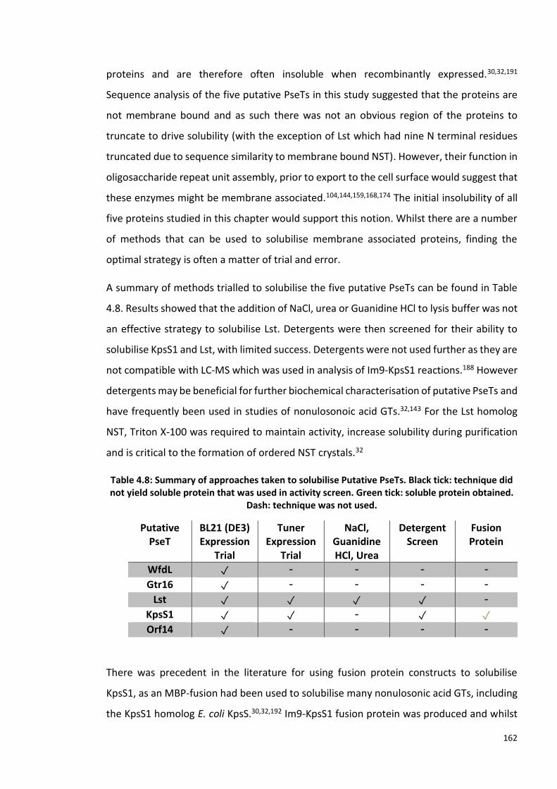
162
proteins and are therefore often insoluble when recombinantly expressed.30,32,191
Sequence analysis of the five putative PseTs in this study suggested that the proteins are
not membrane bound and as such there was not an obvious region of the proteins to
truncate to drive solubility (with the exception of Lst which had nine N terminal residues
truncated due to sequence similarity to membrane bound NST). However, their function in
oligosaccharide repeat unit assembly, prior to export to the cell surface would suggest that
these enzymes might be membrane associated.104,144,159,168,174 The initial insolubility of all
five proteins studied in this chapter would support this notion. Whilst there are a number
of methods that can be used to solubilise membrane associated proteins, finding the
optimal strategy is often a matter of trial and error.
A summary of methods trialled to solubilise the five putative PseTs can be found in Table
4.8. Results showed that the addition of NaCl, urea or Guanidine HCl to lysis buffer was not
an effective strategy to solubilise Lst. Detergents were then screened for their ability to
solubilise KpsS1 and Lst, with limited success. Detergents were not used further as they are
not compatible with LC-MS which was used in analysis of Im9-KpsS1 reactions.188 However
detergents may be beneficial for further biochemical characterisation of putative PseTs and
have frequently been used in studies of nonulosonoic acid GTs.32,143 For the Lst homolog
NST, Triton X-100 was required to maintain activity, increase solubility during purification
and is critical to the formation of ordered NST crystals.32
Table 4.8: Summary of approaches taken to solubilise Putative PseTs. Black tick: technique did not yield soluble protein that was used in activity screen. Green tick: soluble protein obtained.
Dash: technique was not used.
Putative PseT
BL21 (DE3) Expression
Trial
Tuner Expression
Trial
NaCl, Guanidine HCl, Urea
Detergent Screen
Fusion Protein
WfdL ✓ - - - -
Gtr16 ✓ - - - -
Lst ✓ ✓ ✓ ✓ -
KpsS1 ✓ ✓ - ✓ ✓ Orf14 ✓ - - - -
There was precedent in the literature for using fusion protein constructs to solubilise
KpsS1, as an MBP-fusion had been used to solubilise many nonulosonic acid GTs, including
the KpsS1 homolog E. coli KpsS.30,32,192 Im9-KpsS1 fusion protein was produced and whilst

163
a large proportion of Im9-KpsS1 was found within the pellet of lysed cells, a sufficient
amount of soluble protein was produced to facilitate functional characterisation. As
purification of Im9-KpsS1 was low yielding, and a large amount of protein remained
insoluble, further solubilisation of KpsS1 would be beneficial to future characterisation of
this PseT. The use of detergents with Im9-KpsS1 may achieve this. Alternatively, there are
a number of additional fusion partners that may be trialled, specifically the construction of
MBP-KpsS1 should be reconsidered.189
As it was confirmed that Im9-KpsS1 had been produced, no further efforts were made to
solubilise the other putative PseTs. For these remaining target proteins, fusion protein
constructs may be a viable approach to accessing soluble enzymes. Alternatively,
truncations may be made to enhance solubility. If present, transmembrane regions of the
putative PseTs would have been the first residues to remove. It has been shown for initially
insoluble GTs that truncations can be made to facilitate the production of soluble protein.
For example the sequence of Campylobacter jejuni SiaTs Cst-I and Cst-II were aligned, to
reveal that Cst-II lacked the C-terminal domain of Cst-I, therefore soluble, functional Cst-I
was produced which lacked the C-terminal domain.79 Truncations of the putative PseTs
should be made cautiously, to avoid loss of enzymatic function.
Obtaining soluble protein is one of several challenges faced in studying GTs, however the
undetermined identity of the acceptor sugar for Lst highlights a further barrier in functional
characterisation of GTs, where both donor and acceptor sugar identity must be
elucidated.24 Before any soluble target protein was produced, it was determined that Lst
should not be studied further as the unknown identity of the acceptor was likely to hinder
functional studies. For many GTs including the remaining putative PseTs studied in this
work the donor and acceptor moieties can be predicted from empirical evidence, where
the resultant glycoconjugate structure has been characterised.144,159,168,174 As
demonstrated in this work, this knowledge can direct the choice of appropriate substrates
to screen during preliminary functional studies. However, if the desired substrates are not
commercially available functional studies may be impeded.
To perform the desired activity screen of Im9-KpsS1 both the donor CMP-Pse5Ac7Ac and a
potential acceptor mimic 4.9, were synthesised in house. However, only 9% conversion was
seen with Im9-KpsS1 and 4.9, further characterisation of the enzyme is required to provide

164
an informed explanation of this result. LC-MS analysis showed that Pse5Ac7Ac was present
in this sample; this may be as a result of CMP-Pse5Ac7Ac hydrolysis. Ideally to assess this,
the reaction could be performed with PseF present to convert any hydrolysed Pse5Ac7Ac
to CMP-Pse5Ac7Ac, driving the glycosylation reaction (as shown in Chapter 3.3.3 with
PmST1 reaction). Reactions could be analysed at several time points to monitor Pse5Ac7Ac-
glycoside formation and potential hydrolysis. In addition to altering incubation time, it is
possible that other reaction conditions, such as pH and temperature could be optimised to
increase turnover of 4.9.
In contrast to the Im9-KpsS1 activity screen result with 4.9, full conversion or near-full
conversion to Pse5Ac7Ac-glycoside was seen when pNP-β-ᴅ-Glcp 4.8 and pOMeP-β-ᴅ-Glcp
4.11 were used as acceptors. If aromatic residues are present in the KpsS1 active site, a
possible explanation for the high level of turnover observed with pNP-β-ᴅ-Glcp and
pOMeP-β-ᴅ-Glcp is that the aromatic moieties of these substrates are π stacking with
aromatic residues in the active site. Aromatic residues are common in carbohydrate-
protein interactions, where tryptophan is the most abundant residue within 5 Å of β-ᴅ-Glcp
moieties in structurally characterised carbohydrate binding proteins.193 Ultimately, an
explanation of the different levels of conversion of the substrates screened relies on
structural data for KpsS1.
Without structural data or a close homolog, a definitive GT fold cannot be assigned for
KpsS1. A characteristic “DXD” motif can be found in many GT-A members, where a metal-
ion co-ordinates with the residues side chains and two phosphate oxygen atoms of
nucleotide donor.194 Residues 88-90 of KpsS1 are DYD and therefore could be a candidate
DXD motif. GT-B enzymes are also often metal-ion dependent, but a motif for coordination
is yet to be identified.143 Whilst NulO GTs are not all metal dependent, divalent metal ions
have been shown to enhance activity for many NulO GTs.79,145 WbbB and the homologous
KpsS were both functionally assayed with 10 mM MgCl2, therefore this was included in all
functional assays of KpsS1.30,31 Additionally, Mg2+ was required in the one-pot reaction for
PseF activity. However, the metal-ion dependence of KpsS1 should be examined, in future
mechanistic studies.
The exact mechanism used to pseudaminylate the acceptor is perhaps the most intriguing
feature of KpsS1 that is yet to be determined. Debate surrounds mechanism of retaining

165
GTs. Structural data of WbbB2-401, the KpsS1 distant homolog, suggests that this retaining
β-Kdo transferase uses an SN2-like mechanism. This conclusion was made based on the role
of histidine residue from the HP motif, which coordinates the cytosine ring.30–32 In WbbB
this histidine is predicted to protonate the CMP leaving group.31 This enzymatic acidic
proton would not be required in the alternative SNi mechanism as it would be donated by
the acceptor.37,48 HP motifs are conserved in both inverting and retaining nonulosonic acid
in transferases, therefore the presence of these residues alone cannot be used to speculate
whether KpsS1 uses an SNi or SN2-like mechanism.31,38 Elucidation of the full KpsS1
mechanism likely requires protein structural data, mutagenesis and Pse-based probes.
4.5 Conclusions and Future Work
The work performed in this chapter has unequivocally confirmed that A. baumannii ACICU
KpsS1 is a retaining PseT. To my knowledge, this is the first example of a PseT that has been
shown to be active in vitro and one of a few examples of a retaining nonulosonic acid
transferase. Additionally, as a result of the functional studies on Im9-KpsS1 the novel
compound α-Pse5Ac7Ac-pNP-β-ᴅ-Galp has been synthesised via a chemoenzymatic route.
Future work on KpsS1 should focus on further biochemical characterisation. The first step
towards this would ideally require cleavage of a fusion tag. Structural studies of KpsS1
should be pursued, given that this enzyme will be the first in its family and that the
mechanism of retaining GTs currently remains elusive. With access to large quantities of
Pse5Ac7Ac and CMP-Pse5Ac7Ac within the Fascione laboratory, Pse-based probes could be
designed for co-crystallisation studies, to help in determining the mechanism of this
enzyme. Additionally, in the absence of structural data, the function of the HP motif could
begin to be examined by mutagenesis.
For the other putative PseTs studied in this chapter, which are yet to solubilised, fusion
protein constructs could be produced with the aim of improving protein solubility. Soluble
protein could then be studied in activity assays, using the general protocol followed for
Im9-KpsS1. For the putative inverting PseT Gtr16, structural characterisation of the LPS O-
antigen can be used to infer the identity of the putative acceptor and the predicted donor

166
CMP-Pse5Ac7Ac is available. Therefore, this may be the best of the remaining target
proteins to focus future efforts on.
Work carried out within the Fascione laboratory on acetyl-CoA regeneration factors
through the synthesis of N-acetyl-cysteamine (SNAc) provides a promising potential route
to accessing CMP-Pse derivatives that vary at the C7 substituent. This methodology could
be employed to synthesis N-hydroxybutanol-cysteamine (SNHb) for the chemoenzymatic
synthesis of the predicted donor moiety for the putative PseTs WfdL, CMP-Pse5Ac7Hb.
Additionally, if Orf14 were to be solubilised, functional studies may be performed with
CMP-Pse5Ac7Fm 1.18, previously synthesised in house. This CMP-Pse derivative is
structurally closer to the predicted Orf14 donor CMP-Pse5Hb7Fm, than CMP-Pse5Ac7Ac,
and so may be useful in studies of this enzyme.
Finally, CMP-Pse5Ac7Ac produced in this study was gifted to the Sulzenbacher laboratory
to be used in crystal studies of M. magneticum Maf with the aim of obtaining a structure
of Maf with CMP-Pse5Ac7Ac bound to the active site

Chapter 5: Concluding Remarks and Future Prospectives
Non-eukaryotic, cell-surface carbohydrate pseudaminic acid (Pse5Ac7Ac) is known to
contribute to the virulence of several multi-drug resistant bacterial pathogens.1 Chemical
synthesis of Pse5Ac7Ac and its derivatives has proved challenging.2–9 Access to Pse5Ac7Ac
and activated CMP-Pse5Ac7Ac has been a hindrance in studies into the biological
significance of Pse5Ac7Ac, including Pse5Ac7Ac-processing enzymes, which may be novel
therapeutic targets. This project aimed to characterise enzymes which process
pseudaminic acid and to chemoenzymatically synthesise glycosides which contain
pseudaminic acid.
This was achieved through three specific objectives. Firstly, nucleotide activated CMP-
Pse5Ac7Ac 1.10, was required. This was achieved through a chemoenzymatic synthesis
described in Chapter 2. Access to CMP-Pse5Ac7Ac was crucial in facilitating further work of
this study, described in Chapters 3 and 4, where it was the starting material for
glycosyltransferase mediated reactions.
Six biosynthetic enzymes (Campylobacter jejuni PseB, PseC, PseH, PseG and PseI, plus
Aeromonas caviae PseF) were recombinantly produced for the synthesis of 1.10. This study
has provided unequivocal evidence that A. caviae PseF functions as an α-CMP-Pse5Ac7Ac
synthetase. NMR characterisation confirmed the stereochemistry of this carbohydrate,
revealing that α-CMP-PseAc7Ac was produced, this data was crucial for subsequent
conclusions of this study (Chapter 3 and 4).
Future work may revisit structural characterisation of PseF, where conditions used to
crystallise the homologous NeuA may be screened.127 Additionally, PseF mutants may be
produced in order to probe the mechanism and substrate selectivity of this enzyme. The
Pse5Ac7Ac and derivatives produced by methodology established within the Fascione
laboratory, may be used in crystallisation of this enzyme.
Additionally, small-molecules were assayed as inhibitors of C. jejuni PseB and PseI plus A.
caviae PseI, where some of the compounds tested showed a concentration dependent
decrease in Pse5Ac7Ac 1.6 production. Future work could aim to obtain structural data for
PseI to further aid inhibition studies and probe the substrate selectivity of this enzyme.

168
The work presented in this thesis, to my knowledge, provides the first examples of
chemoenzymatically synthesised Pse5Ac7Ac containing glycosides, thus achieving the
second objective of this thesis. As there are currently no published reports of in vitro
activity assay of native pseudaminyltransferases, the use of well-characterised, highly
stable, commerically available, promiscuous SiaTs, which ultilise CMP-Pse5Ac7Ac as a
donor, provides a promising route to synthesis of a range of novel and biologically relevant
Pse-glycosides.
Crystal structure data of PmST with various ligands in the active site are available, including
a structure with Lac acceptor and non-hydrolyzable donor substrate analogue, CMP-3-
deoxy-3-fluoro-Neu5Ac (CMP-3F-Neu5Ac) both present in the activty site.38,164 Future work
may co-crystallise PmST with CMP-3F-Pse5Ac7Ac to gain insight into the molecular basis of
donor substrate promiscuity that has been demonstrated in previous studies and expanded
on in this work, with CMP-Pse5Ac7Ac.
Chapter 2 described a strategy developed in the Fascione laboratory for the
chemoenzymatic synthesis of CMP-Pse5Ac7R derivatives. Future work may examine
whether any of the SiaT which were shown to be activty with CMP-Pse5Ac7Ac are able to
utilise these CMP-Pse derivatives as donors, potentially expanding the range of Pse-
glycosides which can be synthesised using the approach decribed in this study.
Finally, this project set out to characterise putative pseudaminyltransferases (PseTs)
(Chapter 4). The work presented unequivocally confirmed that A. baumannii ACICU KpsS1
is a retaining PseT. To my knowledge, this is the first example of a PseT that has been shown
to be active in vitro and one of a few examples of a retaining nonulosonic acid transferase.
Additionally, as a result of the functional studies on Im9-KpsS1 the novel compound α-
Pse5Ac7Ac-pNP-β-ᴅ-Galp was synthesised via a chemoenzymatic route, in a one-pot seven
enzyme reaction.
With priority, future work on KpsS1 should focus on further biochemical characterisation.
Considering that KpsS1 will be the first member of a novel GT family and that the
mechanism of retaining GTs currently remains elusive, structural studies should be

169
pursued. As large quantities of Pse5Ac7Ac and CMP-Pse5Ac7Ac may be produced using
methodology described in Chapter 2, Pse-based probes could be designed for co-
crystallisation studies, to help in determining the mechanism of this enzyme.
For the other putative PseTs studied in this project, which are yet to solubilised, fusion
protein constructs could be produced with the aim of improving protein solubility. Soluble
protein could then be studied in activity assays, using the general protocol followed for
Im9-KpsS1. For the putative inverting PseT Gtr16, structural characterisation of the LPS O-
antigen can be used to infer the identity of the putative acceptor and the predicted donor
CMP-Pse5Ac7Ac is available. Therefore, this may be the best of the remaining target
proteins to focus future efforts on.
Work carried out within the Fascione laboratory on acetyl-CoA regeneration factors
through the synthesis of N-acetyl-cysteamine (SNAc) provides a promising potential route
to accessing CMP-Pse derivatives that vary at the C7 substituent. This methodology could
be employed to synthesise N-hydroxybutanol-cysteamine (SNHb) for the chemoenzymatic
synthesis of CMP-Pse5Ac7Hb, the predicted donor moiety for the putative PseT WfdL.
Finally, CMP-Pse5Ac7Ac produced in this study was gifted to the Sulzenbacher laboratory
to be used in crystal studies of M. magneticum Maf with the aim of obtaining a structure
of Maf with CMP-Pse5Ac7Ac bound to the active site.
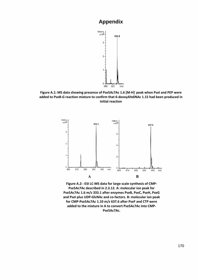
170
Appendix
Figure A.1: MS data showing presence of Pse5Ac7Ac 1.6 [M-H]- peak when PseI and PEP were added to PseB-G reaction mixture to confirm that 6-deoxyAltdiNAc 1.15 had been produced in
initial reaction
A B
Figure A.2: -ESI LC-MS data for large scale synthesis of CMP-Pse5Ac7Ac described in 2.3.12. A: molecular ion peak for
Pse5Ac7Ac 1.6 m/z 333.1 after enzymes PseB, PseC, PseH, PseG and PseI plus UDP-GlcNAc and co-factors. B: molecular ion peak
for CMP-Pse5Ac7Ac 1.10 m/z 637.6 after PseF and CTP were added to the mixture in A to convert Pse5Ac7Ac into CMP-
Pse5Ac7Ac.
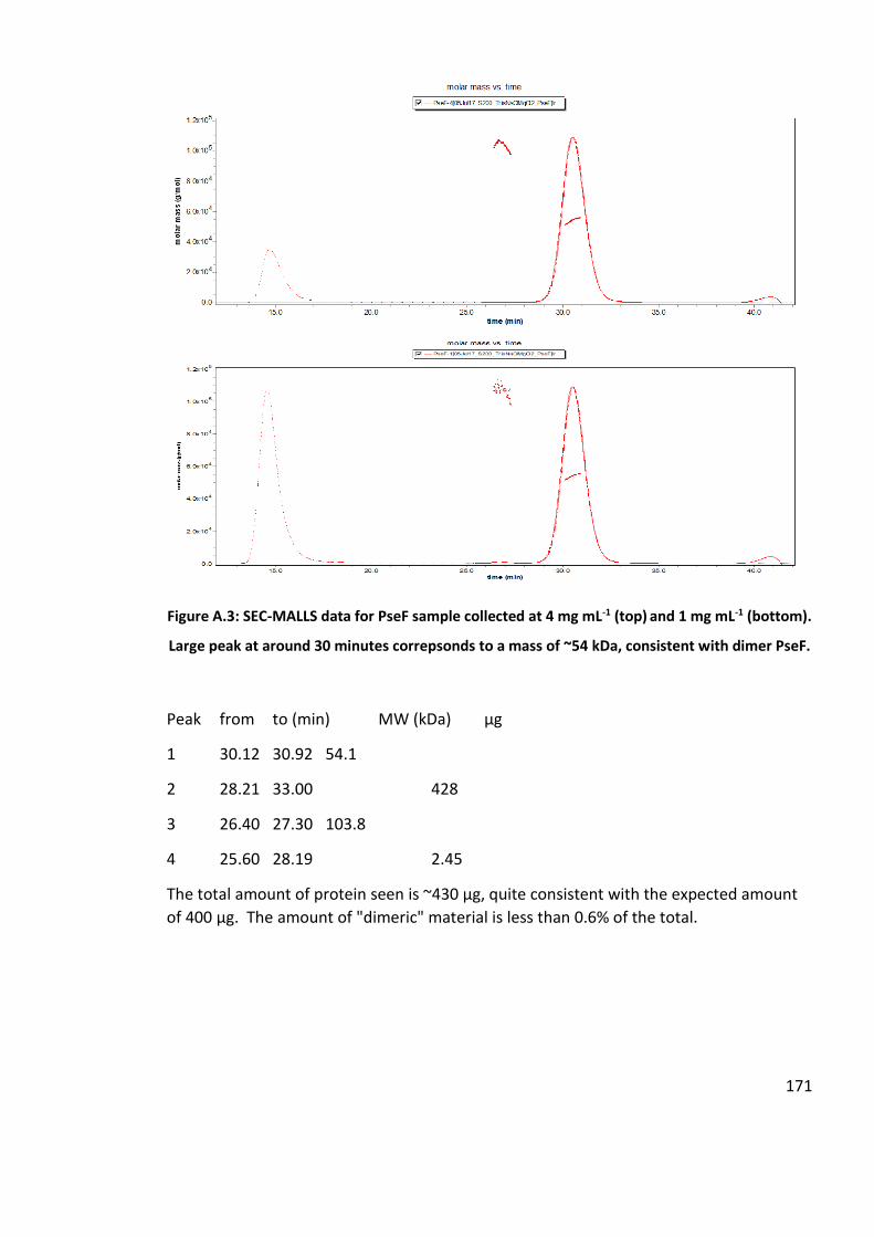
171
Figure A.3: SEC-MALLS data for PseF sample collected at 4 mg mL-1 (top) and 1 mg mL-1 (bottom).
Large peak at around 30 minutes correpsonds to a mass of ~54 kDa, consistent with dimer PseF.
Peak from to (min) MW (kDa) µg
1 30.12 30.92 54.1
2 28.21 33.00 428
3 26.40 27.30 103.8
4 25.60 28.19 2.45
The total amount of protein seen is ~430 µg, quite consistent with the expected amount
of 400 µg. The amount of "dimeric" material is less than 0.6% of the total.

172
Table A.1: 24 crystal well plate conditions for PseF crystallisation trials. Wells contained 10 mL hanging drops contained 1 µL 10 mg mL-1 PseF plus 1 µL well liquor.
1 2 3 4 5 6
A 60 mM Bis Tris
propane, 50
mM citric acid,
pH 4.5, 12%
PEG 3350
(v/w), 0.3 %
(v/v) C8E5
60 mM Bis Tris
propane, 50
mM citric acid,
pH 4.5, 14%
PEG 3350
(v/w), 0.3 %
(v/v) C8E5
60 mM Bis Tris
propane, 50
mM citric acid,
pH 4.5, 16%
PEG 3350
(v/w), 0.3 %
(v/v) C8E5
60 mM Bis Tris
propane, 50
mM citric acid,
pH 4.5, 18%
PEG 3350
(v/w), 0.3 %
(v/v) C8E5
60 mM Bis Tris
propane, 50
mM citric acid,
pH 4.5, 20%
PEG 3350
(v/w), 0.3 %
(v/v) C8E5
/
B 60 mM Bis Tris
propane, 50
mM citric acid,
pH 5.0, 12%
PEG 3350
(v/w), 0.3 %
(v/v) C8E5
60 mM Bis Tris
propane, 50
mM citric acid,
pH 5.0, 14%
PEG 3350
(v/w), 0.3 %
(v/v) C8E5
60 mM Bis Tris
propane, 50
mM citric acid,
pH 5.0, 16%
PEG 3350
(v/w), 0.3 %
(v/v) C8E5
60 mM Bis Tris
propane, 50
mM citric acid,
pH 5.0, 18%
PEG 3350
(v/w), 0.3 %
(v/v) C8E5
60 mM Bis Tris
propane, 50
mM citric acid,
pH 5.0, 20%
PEG 3350
(v/w), 0.3 %
(v/v) C8E5
/
C 60 mM Bis Tris
propane, 50
mM citric acid,
pH 5.5, 12%
PEG 3350
(v/w), 0.3 %
(v/v) C8E5
60 mM Bis Tris
propane, 50
mM citric acid,
pH 5.5, 14%
PEG 3350
(v/w), 0.3 %
(v/v) C8E5
60 mM Bis Tris
propane, 50
mM citric acid,
pH 5.5, 16%
PEG 3350
(v/w), 0.3 %
(v/v) C8E5
60 mM Bis Tris
propane, 50
mM citric acid,
pH 5.5, 18%
PEG 3350
(v/w), 0.3 %
(v/v) C8E5
60 mM Bis Tris
propane, 50
mM citric acid,
pH 5.5, 20%
PEG 3350
(v/w), 0.3 %
(v/v) C8E5
/
D 60 mM Bis Tris
propane, 50
mM citric acid,
pH 6.0, 12%
PEG 3350
(v/w), 0.3 %
(v/v) C8E5
60 mM Bis Tris
propane, 50
mM citric acid,
pH 6.0, 14%
PEG 3350
(v/w), 0.3 %
(v/v) C8E5
60 mM Bis Tris
propane, 50
mM citric acid,
pH 6.0, 16%
PEG 3350
(v/w), 0.3 %
(v/v) C8E5
60 mM Bis Tris
propane, 50
mM citric acid,
pH 6.0, 18%
PEG 3350
(v/w), 0.3 %
(v/v) C8E5
60 mM Bis Tris
propane, 50
mM citric acid,
pH 5.5, 20%
PEG 3350
(v/w), 0.3 %
(v/v) C8E5
/
1 2 3 4 5 6
A 50 mM Bis Tris
propane, 60
mM citric acid,
pH 7.0, 8% PEG
3350 (v/w)
50 mM Bis Tris
propane, 60
mM citric acid,
pH 7.0, 10% PEG
3350 (v/w)
50 mM Bis Tris
propane, 60
mM citric acid,
pH 7.0, 12% PEG
3350 (v/w)
50 mM Bis Tris
propane, 60
mM citric acid,
pH 7.0, 14% PEG
3350 (v/w)
50 mM Bis Tris
propane, 60
mM citric acid,
pH 7.0, 16% PEG
3350 (v/w)
/
B 50 mM Bis Tris
propane, 60
mM citric acid,
pH 7.0, 8% PEG
3350 (v/w), 0.3
% (v/v) C8E5
50 mM Bis Tris
propane, 60
mM citric acid,
pH 7.0, 10% PEG
3350 (v/w), 0.3
% (v/v) C8E5
50 mM Bis Tris
propane, 60
mM citric acid,
pH 7.0, 12% PEG
3350 (v/w), 0.3
% (v/v) C8E5
50 mM Bis Tris
propane, 60
mM citric acid,
pH 7.0, 14% PEG
3350 (v/w), 0.3
% (v/v) C8E5
50 mM Bis Tris
propane, 60
mM citric acid,
pH 7.0, 16% PEG
3350 (v/w), 0.3
% (v/v) C8E5
/
C 50 mM Bis Tris
propane, 60
mM citric acid,
pH 7.7, 8% PEG
3350 (v/w)
50 mM Bis Tris
propane, 60
mM citric acid,
pH 7.7, 10% PEG
3350 (v/w)
50 mM Bis Tris
propane, 60
mM citric acid,
pH 7.7, 12% PEG
3350 (v/w)
50 mM Bis Tris
propane, 60
mM citric acid,
pH 7.7, 14% PEG
3350 (v/w)
50 mM Bis Tris
propane, 60
mM citric acid,
pH 7.7, 16% PEG
3350 (v/w)
/
D 50 mM Bis Tris
propane, 60
mM citric acid,
50 mM Bis Tris
propane, 60
mM citric acid,
50 mM Bis Tris
propane, 60
mM citric acid,
50 mM Bis Tris
propane, 60
mM citric acid,
50 mM Bis Tris
propane, 60
mM citric acid,
/

173
pH 7.7, 8% PEG
3350 (v/w), 0.3
% (v/v) C8E5
pH 7.7, 10% PEG
3350 (v/w), 0.3
% (v/v) C8E5
pH 7.7, 12% PEG
3350 (v/w), 0.3
% (v/v) C8E5
pH 7.7, 14% PEG
3350 (v/w), 0.3
% (v/v) C8E5
pH 7.7, 16% PEG
3350 (v/w), 0.3
% (v/v) C8E5
Figure A.4: Extracted ion count peak intensities from LC-MS chromatograms of known concentrations of CMP-Pse5Ac. Data used in combination with EIC peak intensities of
Pse5Ac7Ac to calculate conversions to products for SiaT reactions
Figure A.5: Extracted ion count peak intensities from LC-MS chromatograms of known concentrations of CMP-Pse5Ac. Data used in combination with EIC peak intensities of
Pse5Ac7Ac to calculate conversions to products for SiaT reactions.
y = 1E+07x - 125691
0
5000000
10000000
15000000
20000000
25000000
0 0.5 1 1.5 2 2.5
EIC
pea
k in
tesi
ty f
or
m/z
63
8.1
CMP-Pse5Ac7Ac concentration / mM
EIC peak intensities (LC-MS) for known concentrations of CMP-Pse5Ac7Ac
y = 6E+06x + 875054
0
2000000
4000000
6000000
8000000
10000000
12000000
14000000
0 0.5 1 1.5 2 2.5
EIC
pea
k in
ten
sity
fo
r m
/z 3
32
.8
Pse5Ac7Ac concentration / mM
EIC peak intensities (LC-MS) for known concentrations of Pse5Ac7Ac

174
Figure A.6: SDS-PAGE of expression trials for Gtr16 (MW: 59.5 kDa) in E. coli BL21 (DE3) cultured at 30 °C. A: Cells lysed in Bugbuster mix, soluble fractions. B: Cells lysed in Bugbuster mix,
insoluble fractions. Lanes loaded as follows: 1-PAGE-RULER MW marker 2- OD600: 0.4, 0.1 mM IPTG, 4 h; 3- OD600: 0.4, 0.1 mM IPTG, 24 h; 4- OD600: 0.4, 1 mM IPTG, 4 h; 5- OD600: 0.4, 1 mM IPTG,2 4h; 6- OD600: 0.6, 0.1 mM IPTG, 4 h; 7- OD600: 0.6, 0.1 mM IPTG, 24 h; 8- OD600: 0.6, 0.1
mM IPTG, 4 h Uninduced sample; 9- : 0.6, 1 mM IPTG, 24 h 10-Uninduced sample.

175
Figure A.7: SDS-PAGE of expression trials for Lst (MW: 40.3 kDa) in E. coli BL21 (DE3) cultured at 30 °C. A: Cells lysed in Bugbuster mix, soluble fractions. B: Cells lysed in Bugbuster mix,
insoluble fractions. Lanes loaded as follows: 1-PAGE-RULER MW marker 2- OD600: 0.4, 0.1 mM IPTG, 4 h; 3- OD600: 0.4, 0.1 mM IPTG, 24 h; 4- OD600: 0.4, 1 mM IPTG, 4 h; 5- OD600: 0.4, 1 mM IPTG,2 4h; 6- OD600: 0.6, 0.1 mM IPTG, 4 h; 7- OD600: 0.6, 0.1 mM IPTG, 24 h; 8- OD600: 0.6, 0.1
mM IPTG, 4 h Uninduced sample; 9- : 0.6, 1 mM IPTG, 24 h 10-Uninduced sample.

176
Figure A.8: LC-MS data for control reaction for activity screen of Im9-KpsS1 with CMP-Pse5Ac7Ac and CMP-Neu5Ac as potential donors with pNP-β-D-Glc as acceptor. A: Molecular ion
peak CMP-Neu5Ac (m/z 613.1). B: Molecular ion peak α-2,6-Pse5Ac7Ac-pNP-β-D-Glc (m/z 616.2).
Figure A.9: LC-MS data for large scale synthesis of α-2,6-Pse5Ac7Ac-pNP-β-D-Glc (m/z 616.2). Remaining CMP-Pse5Ac7Ac shown in A, α-2,6-Pse5Ac7Ac-pNP-β-D-Glc molecular ion peak
shown in B.

177
Plasmid information, gene sequences and recombinant protein sequences
Genes for putative pseudaminyltransferases, KpsS1, Gtr16, WfdL and Orf14 were inserted
into pET15b vectors between BamHI and NdeI restriction sites. Lst gene was in pET15b
vector (RE sites? Truncated). AcPseF plasmid was kindly gifted by Jonathon Shaw.
PseB
MGSSHHHHHHSSGLVPRGSHMFNGKNILITGGTGSFGKTYTKVLLENYKPNKIIIYSRDELKQFEMSSI
FNSNCMRYFIGDVRDKERLSVAMRDVDFVIHAAAMKHVPVAEYNPMECIKTNIHGAQNVIDACFEN
GVKKCIALSTDKACNPVNLYGATKLASDKLFVAANNIAGNKQTRFSVTRYGNVVGSRGSVVPFFKKLIA
QGSKELPITDTRMTRFWISLEDGVKFVLSNFERMHGGEIFIPKIPSMKITNLAHALAPNLSHKIIGIRAGE
KLHEIMISSDDSHLTYEFENYYAISPSIKLVDQESDFSINALGEKGQKVKDGFSYSSDNNPQWASEKELL
DIINHTEGF
PseC
MGSSHHHHHHSSGLVPRGSHMLTYSHQNIDQSDIDTLTKALKDEILTGGKKVNEFEEALCEYMGVKH
ACVLNSATSALHLAYTALGVQEKIVLTTPLTFAATANAALMAGAKVEFIDIKNDGNIDEKKLEARLLKES
ENIGAISVVDFAGNSVEMDEISNLTKKYNIPLIDDASHALGALYKSEKVGKKADLSIFSFHPVKPITTFEG
GAVVSDNEELIDKIKLLRSHGIVKKRLWDSDMVELGYNYRLSDVACALGINQLKKLDHNLEKREEIANF
YDKEFEKNPYFSTIKIKDYKKSSRHLYPILLFPEFYCQKEELFESLLHAGIGVQVHYKPTYEFSFYKKLLGEIK
LQNADNFYKAELSIPCHQEMNLKDAKFVKDTLFSILEKVKKGYCG
PseH
ATGCTGATTAAACTGAAAAACTTCGCGGAACTGAATAGCCAGGAAATTAAACTGATCTTCAAATG
GCGTAACCACCCGGACATTAGCCAATTCATGAAGACCAAACACATCGACTTCGAGGAACACCTGC
GTTTTATTCGTAACCTGCACCAGGATAGCAACAAGAAATACTTCCTGGTGTTTCAGGACGAGCAA
ATCATTGGTGTGATCGATTTCGTTAACATTACCACCAAAAGCTGCGAATTTGGCCTGTATGCGATC
CCGGACCTGAAGGGTGTGGGCCAAGTTCTGATGAACGAGATCAAGAAATACGCGTTCGAAATTC
TGAAGGTGGACACCCTGAAAGCGTATGTTTTTAAGGATAACCACAAGGCGCTGAAACTGTACCA
GCAAAACCACTTTACCATTTATGATGAGGACAAGGACTTTTATTATGTGTGCCTGAAACAGAGCC
ACTGCAAGGCGCTGCCGAGCTAA
MGSSHHHHHHSSGLVPRGSHMIKLKNFTELNSQEIELIFKWRNHPDINQFMKTKYIDFEEHLRFLKKL
HQDSSKKYFLVFQDEQIIGVIDFVNITTKSCEFGLYAKPNLKGVGQILMNEIIKYAFENLKVNTLKAYVFK
DNRKALKLYQQNHFTIYDEDKDFYHICLKQSDCKALPS
PseG
ATGAAGGTGCTGTTCCGTAGCGACAGCAGCAGCCAGATCGGTTTTGGCCACATTAAGCGTGACCT
GGTGCTGGCGAAACAATACAGCGATGTTAGCTTTGCGTGCCTGCCGCTGGAGGGTAGCCTGATC
GACGAAATTCCGTACCCGGTTTATGAACTGAGCAGCGAGAGCATCTACGAACTGATCAACCTGAT
TAAAGAGGAGAAATTCGAGCTGCTGATCATTGATCACTATGGTATCAGCGTGGACGATGAGAAG
CTGATTAAACTGGAAACCGGCGTTAAGATCCTGAGCTTTGACGATGAAATTAAACCGCACCACTG
CGACATCCTGCTGAACGTGAACGCGTACGCGAAGGCGAGCGATTATGAGGGTCTGGTGCCGTTC

178
AAATGCGAAGTTCGTTGCGGCTTTAGCTACGCGCTGATCCGTGAGGAATTCTATCAGGAAGCGAA
GGAAAACCGTGAGAAGAAATACGACTTCTTTATTTGCATGGGTGGCACCGATATCAAAAACCTGA
GCCTGCAGATTGCGAGCGAGCTGCCGAAGACCAAAATCATTAGCATCGCGACCAGCAGCAGCAA
CCCGAACCTGAAGAAACTGCAAAAGTTCGCGAAACTGCACAACAACATCCGTCTGTTTATTGATC
ACGAGAACATTGCGAAGCTGATGAACGAAAGCAACAAACTGATCATTAGCGCGAGCAGCCTGGT
GAACGAGGCGCTGCTGCTGAAGGCGAACTTTAAAGCGATCTGCTACGTTAAGAACCAAGAAAGC
ACCGCGACCTGGCTGGCGAAGAAAGGCTATGAGGTTGAATACAAATATTAA
MGSSHHHHHHSSGLVPRGSHMLDKILYFKTLIRADSGSKIGHGHVRRDLILAKNFKDVSFACIDLPGSL
TGEIPCPVFTLKSADINELVNLIKEHKFELLIIDHYGISAADEKLIKEQTNVKILCFDDNYKEHFCDYLLNVN
IYAQPQKYVNLVPANCELVFSPLVRSEFYDEAKIKREKKFDCFIALGGTDALNLTAKIASNLLAKNKKVA
AITTSANANLANLQNLADSESNFSLFINSNEVARLMNESEILVISASSLVNEALVLGAKFKAVRVADNQ
NEMAQWLAANGREIYEADEICLNL
PseI
ATGCATATGCAAATTGGTAACTTTAACACCGACAAGAAGGTTTTTATCATTGCGGAACTGAGCGC
GAATCATGCGGGTAGCCTGGAGATGGCGCTGAAGAGCATCAAAGCGGCGAAGAAAGCGGGTGC
GGACGCGATCAAGATTCAGACCTACACCCCGGATAGCCTGACCCTGAACAGCGACAAAGAGGAC
TTCATCATTAAAGGTGGCCTGTGGGACAAGCGTAAACTGTACGAACTGTATGAGAGCGCGAAAA
CCCCGTATGAATGGCACAGCCAGATCTTCGAAACCGCGCAAAACGAGGGTATTCTGTGCTTCAGC
AGCCCGTTTGCGAAGGAAGACGTGGAGTTCCTGAAACGTTTTGATCCGATCGCGTACAAGATTGC
GAGCTTCGAAGCGAACGATGAGAACTTTGTGCGTCTGATTGCGAAAGAGAAGAAACCGACCATC
GTTAGCACCGGCATTGCGACCGAGGAAGAGCTGTTCAAGATCTGCGAAATTTTTAAGGAAGAGA
AAAACCCGGACCTGGTGTTCCTGAAGTGCACCAGCACCTATCCGACCGCGATCGAGGATATGAAC
CTGAAAGGTATTGTTAGCCTGAAGGAAAAATTTAACGTTGAGGTGGGTCTGAGCGACCACAGCTT
CGGCTTTCTGGCGCCGGTGATGGCGGTTGCGCTGGGTGCGCGTGTTATCGAAAAGCACTTCATGC
TGGACAAAAGCATTGAAAGCGAGGATAGCAAGTTTAGCCTGGACTTCGATGAATTTAAAGCGAT
GGTGGATGCGGTTCGTCAAGCGGAGAGCGCGCTGGGTGATGGCAAGCTGGACCTGGATGAAAA
GGTGCTGAAAAACCGTGTTTTCGCGCGTAGCCTGTACGCGAGCAAAGATATCAAGAAAGGCGAG
ATGTTTAGCGAAGAGAACGTGAAGAGCGTTCGTCCGAGCTTCGGTCTGCACCCGAAATTTTATCA
AGAACTGCTGGGCAAGAAGGCGAGCAAGGACATCAAGTTCGGTGACGCGCTGAAGCAAGGCGA
TTTCCAATAA
MGSSHHHHHHSSGLVPRGSHMQIGNFNTDKKVFIIAELSANHAGSLEMALKSIKAAKKAGADAIKIQ
TYTPDSLTLNSDKEDFIIKGGLWDKRKLYELYESAKTPYEWHSQIFETAQNEGILCFSSPFAKEDVEFLKR
FDPIAYKIASFEANDENFVRLIAKEKKPTIVSTGIATEEELFKICEIFKEEKNPDLVFLKCTSTYPTAIEDMNL
KGIVSLKEKFNVEVGLSDHSFGFLAPVMAVALGARVIEKHFMLDKSIESEDSKFSLDFDEFKAMVDAVR
QAESALGDGKLDLDEKVLKNRVFARSLYASKDIKKGEMFSEENVKSVRPSFGLHPKFYQELLGKKASKD
IKFGDALKQGDFQ
CjPseF- Codon optimised
pET-15b NdeI-BamHI

179
CATATGCGTGCGATTGCGATTGTTCTGGCGCGTAGCAGCAGCAAACGTATCAAGAATAAGAATATCATTGACTTTTTCAACAAGCCGATGCTGGCGTACCCGATCGAGGTGGCGCTGAACAGCAAGCTGTTCGAAAAAGTTTTTATTAGCAGCGACAGCATGGAGTACGTGAACCTGGCGAAGAACTATGGTGCGAGCTTCCTGAACCTGCGTCCGAAAATCCTGGCGGACGATCGTGCGACCACCCTGGAAGTTATGGCGTACCACATGGAGGAACTGGAGCTGAAGGACGAAGATATTGCGTGCTGCCTGTATGGTGCGAGCGCGCTGCTGCAGGAGAAGCACCTGAAAAACGCGTTTGAAACCCTGAACAAAAACCAAAACACCGATTACGTTTTCACCTGCAGCCCGTTTAGCGCGAGCCCGTATCGTAGCTTCAGCCTGGAGAACGGTGTGCAGATGGCGTTTAAGGAACACAGCAACACCCGTACCCAAGACCTGAAAACCCTGTACCACGATGCGGGTCTGCTGTATATGGGCAAGGCGCAGGCGTTCAAAGAGATGCGTCCGATCTTTAGCCAAAACAGCATTGCGCTGGAGCTGAGCCCGCTGGAAGTGCAGGACATCGCGCACTTCCGTCGTTTCCGTATTAGCCAGGCGCAGATTCAGCCGTTTGAGAAGCGTATGCCGGTGTAA (Codon optimsed for E. coli) CjPseF- Non-Codon optimised
CATATGAGAGCGATCGCTATTGTTTTAGCCAGAAGTTCCAGTAAAAGGATCAAGAATAAAAATATTATTGATTTTTTCAATAAACCCATGCTCGCTTACCCTATTGAGGTGGCGCTAAATTCCAAGCTCTTTGAAAAGGTGTTTATCTCTAGCGATAGCATGGAGTATGTTAATCTGGCTAAAAATTATGGGGCGAGTTTTTTGAATTTGCGCCCTAAAATTTTAGCGGACGACAGAGCCACGACTTTAGAGGTGATGGCCTATCACATGGAAGAATTAGAATTAAAAGATGAAGATATTGCGTGTTGTTTGTATGGCGCTTCAGCGCTTTTACAAGAAAAGCATTTAAAAAACGCTTTTGAAACTTTAAACAAAAACCAAAATACGGATTATGTTTTCACATGCTCTCCATTTAGCGCTTCGCCCTATCGTTCTTTTAGTCTTGAAAACGGCGTTCAAATGGCTTTTAAAGAGCATTCAAACACGCGCACGCAAGATTTAAAAACGCTCTATCATGACGCGGGGTTGCTTTATATGGGGAAGGCTCAAGCCTTTAAAGAAATGCGGCCTATTTTTAGTCAAAATTCTATCGCTTTAGAATTATCGCCCTTAGAAGTCCAAGATATTGCACACTTTAGAAGATTTAGAATTAGCCAAGCTCAAATACAGCCGTTTGAAAAACGCATGCCAGTATAA (Wild type sequence- non-codon optimised)
MGSSHHHHHHSSGLVPRGSHMRAIAIVLARSSSKRIKNKNIIDFFNKPMLAYPIEVALNSKLFEKVFISS
DSMEYVNLAKNYGASFLNLRPKILADDRATTLEVMAYHMEELELKDEDIACCLYGASALLQEKHLKNA
FETLNKNQNTDYVFTCSPFSASPYRSFSLENGVQMAFKEHSNTRTQDLKTLYHDAGLLYMGKAQAFK
EMRPIFSQNSIALELSPLEVQDIAHFRRFRISQAQIQPFEKRMPV*
AcPseF
ATGAATATTGCCATCATCCCTGCCCGTGGTGGCAGTAAGCGTATTCCTAGGAAAAATATCAAACC
ATTTCATAGCAAGCCCATGATCGCATGGTCCATCTTAGCTGCTAAGAAGGCTGGTTGTTTTGAACG
TATAATTGTTTCAACCGATGATGCTGAAATTGCTGCTGTTGCACTCGAATATGGTGCTGAAGTGCC
ATTTACTCGCCCGGCAGAGATTGCTAACGATTATGCCACTACTGGTGAGGTGATAAGCCATGCCA
TTAATTGGTTGATAAATCAGCAAGGGCAAGTGCCGGAAAACGTATGCTGCCTCTATGCAACAGCA
CCTTTTGTTGAGCCTGATGATTTATGCCAGGGATTAGAATTGTTAACGTTCAACAAGGAATGCCAA
TTTGTTTTCAGTGCTACTCGCTTTTCGTTTCCGATTCAACGCGCTATCAAGCTTGATGAGTCAGGTT
GGGTGAGTATGTTTCATCCCGAGTATCAACTAACTCGTTCCCAAGATCTGGAGGAAGCCTACCAT
GATGCGGGGCAATTTTATTGGGGAAAGGCTAATGCTTGGCTTAATAAATTACCTATATTTGCCGT

180
GCATACACAGGTAGTTCTATTACCCAGCCACAGGGTGCAAGATATTGATACTCAGGATGATTGGC
TGCGTGCTGAGAAGCTATTTACGCTAAGGTAA
MGSSHHHHHHSSGLVPRGSHMNIAIIPARGGSKRIPRKNIKPFHSKPMIAWSILAAKKAGCFERIIVST
DDAEIAAVALEYGAEVPFTRPAEIANDYATTGEVISHAINWLINQQGQVPENVCCLYATAPFVEPDDL
CQGLELLTFNKECQFVFSATRFSFPIQRAIKLDESGWVSMFHPEYQLTRSQDLEEAYHDAGQFYWGK
ANAWLNKLPIFAVHTQVVLLPSHRVQDIDTQDDWLRAEKLFTLR*
Gtr16
Vector: pET-15b
Restriction Enzymes: NdeI, BamHI
Sequence of Insert:
ATGTACGTTTATGGTGTGAAACTGGTGGTGTCTCAGCGTAACCTGCTGATCTGCTTCACCCCGCTG
CAAATCCTGATTGCGAGCAAGATCCTGGTTGAGAAAGACTTTGATACCCTGCTGATTAGCTACGT
GGACAACGATAAGTACCGTTTCTACTTCGATAAGATCAGCGCGATTAGCCGTAAGAGCTGGTTCT
TTAAAATCAACAGCACCAACAAGTTTAGCCGTATGATGGACATGATCAAGCTGAAGAAAATCATC
CGTGAATTCGATCCGCACTACAACATCGTTTATTTTGCGAGCCTGGACAACGCGTTCCTGCACCTG
GTGGTTAGCAACATCAGCTTTAACAGCATTGAGACCTTCGACGATGGTAGCGCGAACATTAACAA
GGATAGCACCTATTTCAAGGGCGAACGTAAAAGCAGCTTTCAGCTGCTGTTTAGCGCGCTGCTGG
GTATCAAGTTTAACAAAAGCATCATTCTGGACAAGATCTACAAACACTATAGCATTTTCGAGGGCT
ACAGCAACATTGTTCCGAACGTGGAATACATCAAGATCTTCGAGAGCGAAAACCTGGTTCCGCCG
AACAAGGTGATCAAAATTTTCCTGGGTCAGCCGTTTGAGGAAATGGGCTTCATCGATAAAGAGG
AGCTGTACCTGTTTCTGCGTAAAATCGGTATTGACTACTATTTCCCGCACCCGCGTGAGAAGCACG
ACAAGGATTTCTACTTCGAAATCGTGCAAAGCAAACTGATCTTCGAGGAATTTATTCTGGAGCTGC
TGACCGATGGTAACCTGATTGAAGTTTACACCCTGCTGAGCACCGCGGGCCTGAACGTGAGCATG
CTGGACTATGTGACCGTTAAGGTGATCCGTAGCAAAGATCTGTACATGCGTTATAGCAGCCTGTA
CAAGGTTTTCCAAGACATGAAAGTGGAGTTCATCGACTTTGATGGCATTAGCCTGTAA
MGSSHHHHHHSSGLVPRGSHMYVYGVKLVVSQRNLLICFTPLQILIASKILVEKDFDTLLISYVDNDKYR
FYFDKISAISRKSWFFKINSTNKFSRMMDMIKLKKIIREFDPHYNIVYFASLDNAFLHLVVSNISFNSIETF
DDGSANINKDSTYFKGERKSSFQLLFSALLGIKFNKSIILDKIYKHYSIFEGYSNIVPNVEYIKIFESENLVPP
NKVIKIFLGQPFEEMGFIDKEELYLFLRKIGIDYYFPHPREKHDKDFYFEIVQSKLIFEEFILELLTDGNLIEV
YTLLSTAGLNVSMLDYVTVKVIRSKDLYMRYSSLYKVFQDMKVEFIDFDGISL
KpsS1
Vector: pET-15b
Restriction Enzymes: NdeI, BamHI
Sequence of Insert:
ATGAACTTTCTGATCCTGATTAACAGCGCGCCGAACTACAAGTATTTCTTTTATGAACTGGCGAAA
GAAATCGAGAGCCGTGGCCACAACATTTATTTCGCGATCGATAGCCACCGTAGCAAGTACCTGGA

181
ACCGCTGCCGGAGCTGGACAACAACCAGAACAGCTTCTTTTTCGATAGCTACCTGGAAAAGAACT
TTGACAAAAACCTGAGCGTTAGCCACAACAACAACCAAGAGTATTGGGGTGATTACTTCTATAGC
GACTACGATCGTTTTCTGACCCACGATTTCAACCTGAACAAGGACAAAAACTACTGGCTGAACGT
GAAAGTTAGCCTGGACAGCTTTTTCGAAGACATCATTAAGGATAAACAGATCGACTTTGTGCTGT
ATGAGAACATTAGCAACAGCTTCGCGTACGCGGCGTATCTGCAATGCACCAAACTGGGCAAGAA
ATACATCGGTCTGATGGGCAGCCGTCTGCCGAACCACTTTGAAATTCAGAACAGCATCGTTGAGG
AAGAGCTGAAGAAACTGGAGATCCTGGCGCAGAAGCCGATTACCCAAGATGAAATGGAGTGGTT
CGAAAACTATAAGAAAAGCATCGTGGATATTCAGCCGGACTACATGAAACAAAACGGTCTGGAC
AACGTGGCGATCAGCCGTATTGTTAAGCTGAACAAATTTCTGAAGGCGCTGCGTCTGCTGACCAT
CGGCTTCAAGTACAAGCACTACTACGATTACCAGTTCGGTAACCCGTTCATGGTGCCGATCAAGG
CGATTCGTGTTAACATTAAACGTTATCTGAACACCAAGAAAAGCCAAAAGTTTTACATCAACAACG
ACGAACTGGAGATTTGCAGCAGCAAAGAACGTTTTTACATCTATCCGATTCACTTCCATCCGGAGA
GCAGCACCAGCGTTCTGGCGCCGGAATACACCAACGAGTATAGCAACATCATTAACATCGCGAAC
AACCTGCCGTTCGGCACCTACCTGTATGTGAAAGATCACAAGAGCGCGAAAGGTGTTCAGAGCTA
CGAGTTTTATAAGAAAGTGAGCAGCCTGCCGAACGTGCGTCTGGTTAACTTCGACGTGAACATTA
AGCGTCTGATCCTGAAAAGCCTGGGCGTGATTACCGTTAACAGCACCGCGGGTTACGAAGCGCT
GCTGCTGGGCAAGCCGGTGTACCTGCTGGGTCGTGTGTTCTACGAGAACTTCAACAACGTTTACA
ACCTGAAAAGCTTCCGTGACATCCGTGATATTCGTGACATCCTGGATTTTCAATTCCTGGATGTGA
AGAAAGACTTTATCGCGTACAAGAAATACGTGTATAAAGGCGTTATCTTCATTGATTATGGTAACC
GTGTTAACGACAAGAAACGTTACTTTAGCGAACTGGTGGATAGCATTTTCCTGAACATCAACACC
GACATTGAGTAA
MGSSHHHHHHSSGLVPRGSHMNFLILINSAPNYKYFFYELAKEIESRGHNIYFAIDSHRSKYLEPLPELD
NNQNSFFFDSYLEKNFDKNLSVSHNNNQEYWGDYFYSDYDRFLTHDFNLNKDKNYWLNVKVSLDSF
FEDIIKDKQIDFVLYENISNSFAYAAYLQCTKLGKKYIGLMGSRLPNHFEIQNSIVEEELKKLEILAQKPITQ
DEMEWFENYKKSIVDIQPDYMKQNGLDNVAISRIVKLNKFLKALRLLTIGFKYKHYYDYQFGNPFMVP
IKAIRVNIKRYLNTKKSQKFYINNDELEICSSKERFYIYPIHFHPESSTSVLAPEYTNEYSNIINIANNLPFGT
YLYVKDHKSAKGVQSYEFYKKVSSLPNVRLVNFDVNIKRLILKSLGVITVNSTAGYEALLLGKPVYLLGRV
FYENFNNVYNLKSFRDIRDIRDILDFQFLDVKKDFIAYKKYVYKGVIFIDYGNRVNDKKRYFSELVDSIFLN
INTDIE
Lst
Vector: pET-28a
Restriction Enzymes: NdeI, BamHI
Sequence of Insert:
ATGAATTTAGTTGTTTGTAATACACCATTTCAAGCTTTGCAGATTATCAATTTGGTTGCCAAAGGA
ATAATAACAAACTTCGATTTTTTTTATTTTTGTAAAAATAAAACTGCACAAGTAGAATATTACTTTG
AGCAAGTTAAGAAGCACGCTATTTCATCACAACTCTATATTAGTGACAAGCGTTATCCTTATCATA
TATTAGACATCAGGCGTCTATTCAAAGGTAGGTATTACCAATCTGTCTACAGTGCCTCTGTTGATA
GTGTATTCACACACAGTATTTTATCGTTTATAAAATTTGACAATTTTTACTCTTTTGATGATGGGTC
TGCTAATTTAAATAGAAACTCTACATATTACATTGAGCAGAGAGGTTATTTTAAACGGATATTGTT

182
CAGATTGACAGGTTGTAAATATGACTTGAGAAAAACGAAATCAATAATTCGTTCTCATTATACCGT
ATATAAGAATAAAAAAAATATAGTATCCAATACAATATATATTGATTTTGAGTTTCCTGCTCAATG
TTCTGATGGCCGAAAGGAAACTAACGACAAAAACTCAGTCGCCAATGTATTGTTAGGTACAGTTT
ATGATGAGGTGTTTTCGGATAATGAGGTAATAATTAAAAAATTGTCATGCTTTTTTGCCGATAAAG
ATTTCTATTATATCCCACACCCAAGAGATACACATAACTTCTTTATAAATGGAATGCGTATCGATG
GGCCTGAGATTGCTGAAAGCAAAATTCTATCTTTGTTAAAAAGATATGAAACAATAAATTTATATG
GTTTTGGAAGTTCAGTTCAAATAAATATGAGCTCACATCGTGGAGTTATCAATTATATATTTGATA
TAGGAAGTCATGAATATGAATATGACCATGATTATGATTATGGTTATGAAATACTGAAGATATAG
Recombinant Protein Sequence:
MGSSHHHHHHSSGLVPRGSHMNLVVCNTPFQALQIINLVAKGIITNFDFFYFCKNKTAQVEYYFEQV
KKHAISSQLYISDKRYPYHILDIRRLFKGRYYQSVYSASVDSVFTHSILSFIKFDNFYSFDDGSANLNRNST
YYIEQRGYFKRILFRLTGCKYDLRKTKSIIRSHYTVYKNKKNIVSNTIYIDFEFPAQCSDGRKETNDKNSVA
NVLLGTVYDEVFSDNEVIIKKLSCFFADKDFYYIPHPRDTHNFFINGMRIDGPEIAESKILSLLKRYETINLY
GFGSSVQINMSSHRGVINYIFDIGSHEYEYDHDYDYGYEILKI
Orf14
Vector: pET-15b
Restriction Enzymes: NdeI, BamHI
Sequence of Insert:
ATGAACACCTACTTCGACATCCCGCACCGTCTGGTGGGTAAAGCGCTGTATGAGAGCTACTATGA
TCACTTTGGTCAGATGGACATCCTGAGCGATGGCAGCCTGTACCTGATTTATCGTCGTGCGACCG
AGCACGTTGGTGGCAGCGACGGTCGTGTGGTTTTCAGCAAACTGGAAGGTGGCATCTGGAGCGC
GCCGACCATTGTGGCGCAGGCGGGTGGCCAAGACTTTCGTGATGTTGCGGGTGGCACCATGCCG
AGCGGTCGTATTGTGGCGGCGAGCACCGTTTACGAGACCGGTGAAGTTAAGGTGTATGTTAGCG
ACGATAGCGGCGTGACCTGGGTTCACAAATTCACCCTGGCGCGTGGTGGCGCGGATTACAACTTC
GCGCACGGCAAGAGCTTTCAAGTGGGCGCGCGTTACGTTATCCCGCTGTATGCGGCGACCGGTG
TGAACTACGAGCTGAAATGGCTGGAAAGCAGCGATGGTGGCGAGACCTGGGGTGAAGGCAGCA
CCATTTATAGCGGCAACACCCCGTACAACGAGACCAGCTATCTGCCGGTGGGTGATGGCGTTATC
CTGGCGGTGGCGCGTGTTGGTAGCGGTGCGGGTGGCGCGCTGCGTCAATTCATTAGCCTGGACG
ATGGTGGCACCTGGACCGACCAGGGTAACGTGACCGCGCAAAACGGCGACAGCACCGATATCCT
GGTTGCGCCGAGCCTGAGCTACATTTATAGCGAAGGTGGCACCCCGCACGTGGTTCTGCTGTACA
CCAACCGTACCACCCACTTTTGCTACTATCGTACCATCCTGCTGGCGAAAGCGGTTGCGGGTAGCA
GCGGTTGGACCGAGCGTGTTCCGGTTTACAGCGCGCCGGCGGCGAGCGGTTATACCAGCCAGGT
GGTTCTGGGTGGCCGTCGTATTCTGGGTAACCTGTTCCGTGAAACCAGCAGCACCACCAGCGGCG
CGTACCAATTTGAGGTGTATCTGGGTGGCGTTCCGGACTTCGAAAGCGATTGGTTTAGCGTGAGC
AGCAACAGCCTGTACACCCTGAGCCATGGTCTGCAGCGTAGCCCGCGTCGTGTGGTTGTGGAGTT
CGCGCGTAGCAGCAGCCCGAGCACCTGGAACATCGTTATGCCGAGCTATTTTAACGACGGTGGCC
ACAAAGGTAGCGGCGCGCAAGTGGAAGTTGGTAGCCTGAACATTCGTCTGGGTACCGGTGCGGC
GGTGTGGGGTACCGGCTACTTCGGTGGCATTGATAACAGCGCGACCACCCGTTTTGCGACCGGCT
ACTATCGTGTTCGTGCGTGGATTTAA
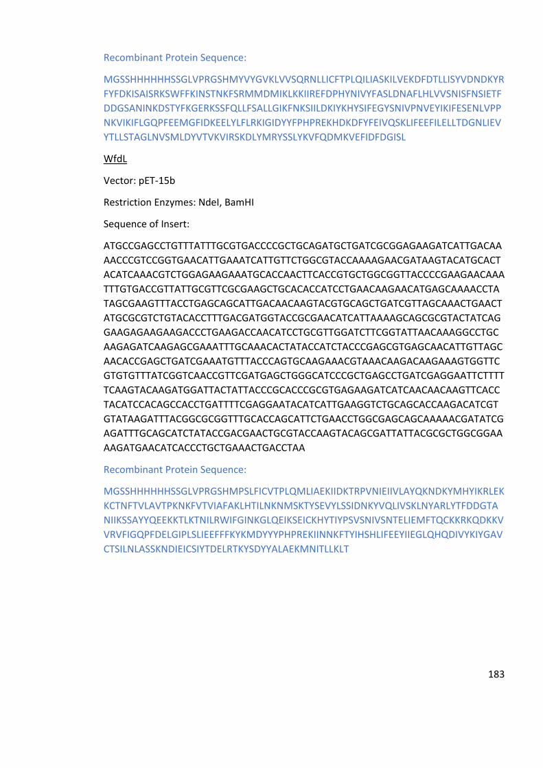
183
Recombinant Protein Sequence:
MGSSHHHHHHSSGLVPRGSHMYVYGVKLVVSQRNLLICFTPLQILIASKILVEKDFDTLLISYVDNDKYR
FYFDKISAISRKSWFFKINSTNKFSRMMDMIKLKKIIREFDPHYNIVYFASLDNAFLHLVVSNISFNSIETF
DDGSANINKDSTYFKGERKSSFQLLFSALLGIKFNKSIILDKIYKHYSIFEGYSNIVPNVEYIKIFESENLVPP
NKVIKIFLGQPFEEMGFIDKEELYLFLRKIGIDYYFPHPREKHDKDFYFEIVQSKLIFEEFILELLTDGNLIEV
YTLLSTAGLNVSMLDYVTVKVIRSKDLYMRYSSLYKVFQDMKVEFIDFDGISL
WfdL
Vector: pET-15b
Restriction Enzymes: NdeI, BamHI
Sequence of Insert:
ATGCCGAGCCTGTTTATTTGCGTGACCCCGCTGCAGATGCTGATCGCGGAGAAGATCATTGACAA
AACCCGTCCGGTGAACATTGAAATCATTGTTCTGGCGTACCAAAAGAACGATAAGTACATGCACT
ACATCAAACGTCTGGAGAAGAAATGCACCAACTTCACCGTGCTGGCGGTTACCCCGAAGAACAAA
TTTGTGACCGTTATTGCGTTCGCGAAGCTGCACACCATCCTGAACAAGAACATGAGCAAAACCTA
TAGCGAAGTTTACCTGAGCAGCATTGACAACAAGTACGTGCAGCTGATCGTTAGCAAACTGAACT
ATGCGCGTCTGTACACCTTTGACGATGGTACCGCGAACATCATTAAAAGCAGCGCGTACTATCAG
GAAGAGAAGAAGACCCTGAAGACCAACATCCTGCGTTGGATCTTCGGTATTAACAAAGGCCTGC
AAGAGATCAAGAGCGAAATTTGCAAACACTATACCATCTACCCGAGCGTGAGCAACATTGTTAGC
AACACCGAGCTGATCGAAATGTTTACCCAGTGCAAGAAACGTAAACAAGACAAGAAAGTGGTTC
GTGTGTTTATCGGTCAACCGTTCGATGAGCTGGGCATCCCGCTGAGCCTGATCGAGGAATTCTTTT
TCAAGTACAAGATGGATTACTATTACCCGCACCCGCGTGAGAAGATCATCAACAACAAGTTCACC
TACATCCACAGCCACCTGATTTTCGAGGAATACATCATTGAAGGTCTGCAGCACCAAGACATCGT
GTATAAGATTTACGGCGCGGTTTGCACCAGCATTCTGAACCTGGCGAGCAGCAAAAACGATATCG
AGATTTGCAGCATCTATACCGACGAACTGCGTACCAAGTACAGCGATTATTACGCGCTGGCGGAA
AAGATGAACATCACCCTGCTGAAACTGACCTAA
Recombinant Protein Sequence:
MGSSHHHHHHSSGLVPRGSHMPSLFICVTPLQMLIAEKIIDKTRPVNIEIIVLAYQKNDKYMHYIKRLEK
KCTNFTVLAVTPKNKFVTVIAFAKLHTILNKNMSKTYSEVYLSSIDNKYVQLIVSKLNYARLYTFDDGTA
NIIKSSAYYQEEKKTLKTNILRWIFGINKGLQEIKSEICKHYTIYPSVSNIVSNTELIEMFTQCKKRKQDKKV
VRVFIGQPFDELGIPLSLIEEFFFKYKMDYYYPHPREKIINNKFTYIHSHLIFEEYIIEGLQHQDIVYKIYGAV
CTSILNLASSKNDIEICSIYTDELRTKYSDYYALAEKMNITLLKLT

184
Figure A.10: Gst vector map

185
Figure A.11: MBP vector map

186
Figure A.12: Im9 vector map
Im9-KpsS1
Vector: pET-15b
Restriction Enzymes:
Sequence of Insert:
TCCAGGGACCAGCAATGAACTTTCTGATCCTGATTAACAGCGCGCCGAACTACAAGTATTTCTTTT
ATGAACTGGCGAAAGAAATCGAGAGCCGTGGCCACAACATTTATTTCGCGATCGATAGCCACCGT
AGCAAGTACCTGGAACCGCTGCCGGAGCTGGACAACAACCAGAACAGCTTCTTTTTCGATAGCTA
CCTGGAAAAGAACTTTGACAAAAACCTGAGCGTTAGCCACAACAACAACCAAGAGTATTGGGGT
GATTACTTCTATAGCGACTACGATCGTTTTCTGACCCACGATTTCAACCTGAACAAGGACAAAAAC

187
TACTGGCTGAACGTGAAAGTTAGCCTGGACAGCTTTTTCGAAGACATCATTAAGGATAAACAGAT
CGACTTTGTGCTGTATGAGAACATTAGCAACAGCTTCGCGTACGCGGCGTATCTGCAATGCACCA
AACTGGGCAAGAAATACATCGGTCTGATGGGCAGCCGTCTGCCGAACCACTTTGAAATTCAGAAC
AGCATCGTTGAGGAAGAGCTGAAGAAACTGGAGATCCTGGCGCAGAAGCCGATTACCCAAGATG
AAATGGAGTGGTTCGAAAACTATAAGAAAAGCATCGTGGATATTCAGCCGGACTACATGAAACA
AAACGGTCTGGACAACGTGGCGATCAGCCGTATTGTTAAGCTGAACAAATTTCTGAAGGCGCTGC
GTCTGCTGACCATCGGCTTCAAGTACAAGCACTACTACGATTACCAGTTCGGTAACCCGTTCATGG
TGCCGATCAAGGCGATTCGTGTTAACATTAAACGTTATCTGAACACCAAGAAAAGCCAAAAGTTT
TACATCAACAACGACGAACTGGAGATTTGCAGCAGCAAAGAACGTTTTTACATCTATCCGATTCAC
TTCCATCCGGAGAGCAGCACCAGCGTTCTGGCGCCGGAATACACCAACGAGTATAGCAACATCAT
TAACATCGCGAACAACCTGCCGTTCGGCACCTACCTGTATGTGAAAGATCACAAGAGCGCGAAAG
GTGTTCAGAGCTACGAGTTTTATAAGAAAGTGAGCAGCCTGCCGAACGTGCGTCTGGTTAACTTC
GACGTGAACATTAAGCGTCTGATCCTGAAAAGCCTGGGCGTGATTACCGTTAACAGCACCGCGG
GTTACGAAGCGCTGCTGCTGGGCAAGCCGGTGTACCTGCTGGGTCGTGTGTTCTACGAGAACTTC
AACAACGTTTACAACCTGAAAAGCTTCCGTGACATCCGTGATATTCGTGACATCCTGGATTTTCAA
TTCCTGGATGTGAAGAAAGACTTTATCGCGTACAAGAAATACGTGTATAAAGGCGTTATCTTCATT
GATTATGGTAACCGTGTTAACGACAAGAAACGTTACTTTAGCGAACTGGTGGATAGCATTTTCCT
GAACATCAACACCGACATTGAGTAACGCGCCTTCTCCTCA
Recombinant Protein Sequence:
MGSSHHHHHHSSMELKHSISDYTEAEFLQLVTTICNADTSSEEELVKLVTHFEEMTEHPSGSDLIYYPK
EGDDDSPSGIVNTVKQWRAANGKSGFKQGLEVLFQGPAMNFLILINSAPNYKYFFYELAKEIESRGHN
IYFAIDSHRSKYLEPLPELDNNQNSFFFDSYLEKNFDKNLSVSHNNNQEYWGDYFYSDYDRFLTHDFNL
NKDKNYWLNVKVSLDSFFEDIIKDKQIDFVLYENISNSFAYAAYLQCTKLGKKYIGLMGSRLPNHFEIQN
SIVEEELKKLEILAQKPITQDEMEWFENYKKSIVDIQPDYMKQNGLDNVAISRIVKLNKFLKALRLLTIGF
KYKHYYDYQFGNPFMVPIKAIRVNIKRYLNTKKSQKFYINNDELEICSSKERFYIYPIHFHPESSTSVLAPE
YTNEYSNIINIANNLPFGTYLYVKDHKSAKGVQSYEFYKKVSSLPNVRLVNFDVNIKRLILKSLGVITVNST
AGYEALLLGKPVYLLGRVFYENFNNVYNLKSFRDIRDIRDILDFQFLDVKKDFIAYKKYVYKGVIFIDYGN
RVNDKKRYFSELVDSIFLNINTDIE*

188
Figure A.13: Plasmid map for Im9-KpsS1
Gst-KpsS1
TCCAGGGACCAGCAATGAACTTTCTGATCCTGATTAACAGCGCGCCGAACTACAAGTATTTCTTTT
ATGAACTGGCGAAAGAAATCGAGAGCCGTGGCCACAACATTTATTTCGCGATCGATAGCCACCGT
AGCAAGTACCTGGAACCGCTGCCGGAGCTGGACAACAACCAGAACAGCTTCTTTTTCGATAGCTA
CCTGGAAAAGAACTTTGACAAAAACCTGAGCGTTAGCCACAACAACAACCAAGAGTATTGGGGT
GATTACTTCTATAGCGACTACGATCGTTTTCTGACCCACGATTTCAACCTGAACAAGGACAAAAAC
TACTGGCTGAACGTGAAAGTTAGCCTGGACAGCTTTTTCGAAGACATCATTAAGGATAAACAGAT
CGACTTTGTGCTGTATGAGAACATTAGCAACAGCTTCGCGTACGCGGCGTATCTGCAATGCACCA
AACTGGGCAAGAAATACATCGGTCTGATGGGCAGCCGTCTGCCGAACCACTTTGAAATTCAGAAC
AGCATCGTTGAGGAAGAGCTGAAGAAACTGGAGATCCTGGCGCAGAAGCCGATTACCCAAGATG
AAATGGAGTGGTTCGAAAACTATAAGAAAAGCATCGTGGATATTCAGCCGGACTACATGAAACA
AAACGGTCTGGACAACGTGGCGATCAGCCGTATTGTTAAGCTGAACAAATTTCTGAAGGCGCTGC

189
GTCTGCTGACCATCGGCTTCAAGTACAAGCACTACTACGATTACCAGTTCGGTAACCCGTTCATGG
TGCCGATCAAGGCGATTCGTGTTAACATTAAACGTTATCTGAACACCAAGAAAAGCCAAAAGTTT
TACATCAACAACGACGAACTGGAGATTTGCAGCAGCAAAGAACGTTTTTACATCTATCCGATTCAC
TTCCATCCGGAGAGCAGCACCAGCGTTCTGGCGCCGGAATACACCAACGAGTATAGCAACATCAT
TAACATCGCGAACAACCTGCCGTTCGGCACCTACCTGTATGTGAAAGATCACAAGAGCGCGAAAG
GTGTTCAGAGCTACGAGTTTTATAAGAAAGTGAGCAGCCTGCCGAACGTGCGTCTGGTTAACTTC
GACGTGAACATTAAGCGTCTGATCCTGAAAAGCCTGGGCGTGATTACCGTTAACAGCACCGCGG
GTTACGAAGCGCTGCTGCTGGGCAAGCCGGTGTACCTGCTGGGTCGTGTGTTCTACGAGAACTTC
AACAACGTTTACAACCTGAAAAGCTTCCGTGACATCCGTGATATTCGTGACATCCTGGATTTTCAA
TTCCTGGATGTGAAGAAAGACTTTATCGCGTACAAGAAATACGTGTATAAAGGCGTTATCTTCATT
GATTATGGTAACCGTGTTAACGACAAGAAACGTTACTTTAGCGAACTGGTGGATAGCATTTTCCT
GAACATCAACACCGACATTGAGTAACGCGCCTTCTCCTCA
MGSSHHHHHHSSMSPILGYWKIKGLVQPTRLLLEYLEEKYEEHLYERDEGDKWRNKKFELGLEFPNLP
YYIDGDVKLTQSMAIIRYIADKHNMLGGCPKERAEISMLEGAVLDIRYGVSRIAYSKDFETLKVDFLSKL
PEMLKMFEDRLCHKTYLNGDHVTHPDFMLYDALDVVLYMDPMCLDAFPKLVCFKKRIEAIPQIDKYL
KSSKYIAWPLQGWQATFGGGDHPPKGLEVLFQGPAMNFLILINSAPNYKYFFYELAKEIESRGHNIYFA
IDSHRSKYLEPLPELDNNQNSFFFDSYLEKNFDKNLSVSHNNNQEYWGDYFYSDYDRFLTHDFNLNKD
KNYWLNVKVSLDSFFEDIIKDKQIDFVLYENISNSFAYAAYLQCTKLGKKYIGLMGSRLPNHFEIQNSIVE
EELKKLEILAQKPITQDEMEWFENYKKSIVDIQPDYMKQNGLDNVAISRIVKLNKFLKALRLLTIGFKYK
HYYDYQFGNPFMVPIKAIRVNIKRYLNTKKSQKFYINNDELEICSSKERFYIYPIHFHPESSTSVLAPEYTN
EYSNIINIANNLPFGTYLYVKDHKSAKGVQSYEFYKKVSSLPNVRLVNFDVNIKRLILKSLGVITVNSTAG
YEALLLGKPVYLLGRVFYENFNNVYNLKSFRDIRDIRDILDFQFLDVKKDFIAYKKYVYKGVIFIDYGNRV
NDKKRYFSELVDSIFLNINTDIE

190
Figure A.14: Plasmid map for Gst-KpsS1

191
Figure A.14: Chromatogram for PseI Purification using Ni-affinity column

192
Figure A.15: Chromatogram for PseC Purification using Ni-affinity column

193
Figure A.16: AKTA Trace for PseG Purification using Ni-affinity column

194
Figure A.17: AKTA Trace for PseH Purification using Ni-affinity column

195
Figure A.18: Chromatogram for PseI Purification using Ni-affinity column

196
Figure A.19: AKTA Trace for CjPseF (codon optimised for E. coli) Purification using Ni-affinity column. SDS-PAGE in Figure 2.12 contains material from this purification attempt.

197
Figure A.20: AKTA Trace for CjPseF (non-codon optimised for E. coli) Purification using Ni-affinity column. SDS-PAGE in Figure 2.12 contains material from this purification attempt.

198
Figure A.21: AKTA Trace for AcPseF Purification using Ni-affinity column

199
Figure A.22: AKTA Trace for PseF Purification using SEC

200
Figure A.23: AKTA Trace for Im9-KpsS1 Purification using Ni-affinity column

201

202

203

204
Figure A.24: MALDI-TOF-MS Spectra for Initial Sialyltransferase Activity Screen with CMP-Pse5Ac7Ac and ITag-LacNAc, as described in 3.2.1 and 3.2.3
Table A.2: Secretion signal analysis for putative PseTs
Protein Signal peptide (Sec/SPI) likelihood
TAT signal peptide (Tat/SPI) likelihood
Lipoprotein signal peptide (Sec/SPII) likelihood
KpsS1 0.0098 0.0003 0.0008
Orf14 0.0571 0.0453 0.0183
Lst 0.0254 0.0012 0.003
Gtr16 0.0361 0.0007 0.0449
WfdL 0.1531 0.001 0.0476
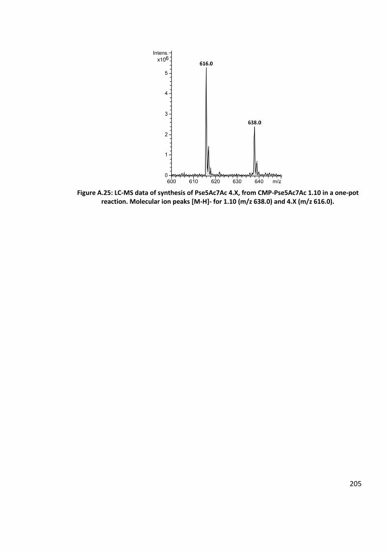
205
Figure A.25: LC-MS data of synthesis of Pse5Ac7Ac 4.X, from CMP-Pse5Ac7Ac 1.10 in a one-pot reaction. Molecular ion peaks [M-H]- for 1.10 (m/z 638.0) and 4.X (m/z 616.0).
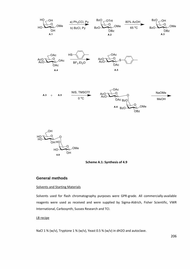
206
Scheme A.1: Synthesis of 4.9
General methods
Solvents and Starting Materials
Solvents used for flash chromatography purposes were GPR-grade. All commercially-available
reagents were used as received and were supplied by Sigma-Aldrich, Fisher Scientific, VWR
International, Carbosynth, Sussex Research and TCI.
LB recipe
NaCl 1 % (w/v), Tryptone 1 % (w/v), Yeast 0.5 % (w/v) in dH2O and autoclave.

207
LB agar recipe
NaCl 1 % (w/v), Tryptone 1 % (w/v), Yeast 0.5 % (w/v), Agar 1.5 % (w/v) in dH2O and autoclave.
Sodium dodecyl sulfate polyacrylamide gel electrophoresis (SDS PAGE);
5 x SDS reducing sample buffer recipe: SDS 10 % (w/v), Glycerol 20 % (w/v), Bromophenol blue
0.05 % (w/v), Tris-HCl 200 mM pH 6.8, β-mercaptoethanol 10 mM, in dH2O.
SDS resolving gel buffer: SDS 0.4 % (w/v), Tris-HCl 1.5 M pH 8.8, in dH2O.
SDS stacking gel buffer: SDS 0.4 % (w/v), Tris-HCl 0.5 M pH 6.8, in dH2O.
SDS running buffer 4X: Glycine 160 mM, Tris 0.1 M
Staining solution: 0.1 % Coomassie Brilliant Blue R-250, 50 % MeOH, 10 % glacial acetic acid
Destaining solution: 50 % MeOH, 10 % glacial acetic acid
Protein Purification
All protein purification, with the exception of PseF size-exclusion chromatography (SEC) was
performed on a AKTA Start, GE Healthcare Life Technologies. PseF SEC was performed on a AKTA
Prime, GE Healthcare Life Technologies.
Analytical TLC was performed on silica gel 60-F254 with detection by fluorescence and/or charring
following immersion in a solution of MeOH, 5% H2SO4.
Mass Spectrometry (MS)
Small-molecule HRMS data were obtained at room temperature on a Bruker Daltonics micrOTOF.
Liquid Chromatography Mass Spectrometry (LC-MS)
LC-MS data presented in 2.3.2, 2.2.3 (PseB inhibition only) and 2.3.6 was carried out using
negative ion mode electrospray ionisation (-ESI) on a Bruker HCTultra ETD II system (Bruker
Daltonics) MS in The University of York Centre of Excellence in Mass Spectrometry (CoEMS). A
Waters C18 column was fitted to a high performance Dionex UltiMate® 3000 LC system
(ThermoScientific) equipped with an UltiMate® 3000 Diode Array Detector. Chromeleon® 6.80
SR12 software (ThermoScientific) combined with esquireControl version 6.2, Build 62.24 software
(Bruker Daltonics), and Bruker compass Hystar 3.2-SR2, Hystar version 3.2, Build 44 software
(Bruker Daltonics). General procedure: Solvent A - Water, Formic Acid 0.1 % (v/v); Solvent B -

208
Acetonitrile, Formic Acid 0.1 % (v/v). A flow-rate of 300 μL min-1, followed pre-equilibrium of the
column in solvent A 95 % for 30 seconds followed by application of a linear gradient; solvent B 70
% to 30 % over 6 minutes. The column was washed in 95 % solvent B for 1 minute before solvent
A 95 % was applied for 1 minute to re-equilibrate the column.
All other LC-MS data was carried out using negative ion mode electrospray ionisation (-ESI) on a
Bruker HCTultra ETD II system (Bruker Daltonics) MS in The University of York Centre of Excellence
in Mass Spectrometry (CoEMS). A Waters C18 column was fitted to an ultra-performance Acquity
Class I LC system. General procedure: Solvent A - Water, Formic Acid 0.1 % (v/v); Solvent B -
Acetonitrile, Formic Acid 0.1 % (v/v). A flow-rate of 600 μL min-1, followed pre-equilibrium of the
column in solvent A 100 % for followed by application of a linear gradient; solvent B 0 % to 40 %
over 5 minutes. The column was washed in 95 % solvent B for 1 minute before solvent A 95 % was
applied for 4 minute to re-equilibrate the column.
All data was processed using Bruker Daltonik DataAnalysis 4.1 software.
Nuclear Magnetic Resonance Spectroscopy
For CMP-Pse5Ac7Ac 1.10 1H and 13C NMR spectra NMR data was collected on a Jeol ECS400 NMR
Spectrometer or the Bruker AVIIIHD500 FT-NMR Spectrometer made available at The University of
York Centre for Magnetic Resonance, using an internal deuterium lock at room temperature. Signals
were assigned using additional DEPT135, COSY, HSQC experiments. Chemical shifts are reported in
ppm according to the following references: D2O: δ: 4.80
For β-2,3-Pse5Ac7Ac-pNP-β-D-Gal 3.17 and α-2,6-Pse5Ac7Ac-pNP-β-D-Glc 1.12 1H and 13C NMR
spectra were recorded at 700 MHz and 126 MHz respectively on a Bruker Avance Neo 700 MHz
spectrometer operating at 658.78 MHz using an internal deuterium lock at room temperature.
Signals were assigned using additional DEPT135, COSY, HSQC experiments. Chemical shifts are
reported in ppm according to the following references: D2O: δ: 4.80
The following abbreviations were used to describe signal multiplicities or appearances: s, singlet; d,
doublet; t, triplet; dt, doublet of triplets; q, quartet; qd, quartet of doublets; quint, quintet; dq,
doublet of quintets; m, multiplet.
209
Abbreviations
β-ᴅ-Glcp-2,6-β-ᴅ-Galp-OMe β-ᴅ-glucopyranosyl-2,6-methyl-β-D-galactopyranoside
6-deoxy-AltdiNAc 2,4-diacetamido-2,4,6-trideoxy-L-altropyranose
6-deoxy-AltNAc4NAcN3 2-acetamido-4-azidoacetamido-2,4,6-trideoxy-β-L- altropyranose
6-F-Galp 6-deoxy-6-fluoro-ᴅ-Galactose
Abs Absorbance
Acetyl-CoA Acetyl-Coenzyme A
AcOH Acetic acid
Ada Adamantanyl
CAZy Carbohydrate-Active EnZymes
CDG Congenital Disorders of Glycosylation
CMP-Aci5Ac7Ac di-N-acetylacinetaminic acid
CMP-Kdo Cytidine-5’-monophosphate-2-keto-3-deoxy-manno-octonic acid
CMP-Neu5Ac Cytidine-5’-monophosphate N-acetylneuraminic acid
CMP-3F-Neu5Ac CMP-3-deoxy-3-fluoro-N-acetylneuraminic acid
CMP-Pse5Hb7Fm CMP-5-N-[(R)-3-hydroxybutanoyl]-7-N-formyl-3,5,7,9-tetradeoxy-˪-
glycero-˪-manno-2-nonulosonic acid
CMP-Pse5Ac7Ac Cytidine-5’-monophosphate-5,7-diacetamido-3,5,7,9-tetradeoxy-˪-
glycero-˪-manno-2-nonulosonic acid
CoA Coenzyme-A
CPS Capsular polysaccharide
CTP Cytidine-5’-triphosphate
C.V. Column volumes
D2O Deuterium oxide
DCM Dichloromethane
diNAcBac 2,4-diacetamido-2,4,6-trideoxy-D-glucose
DNA Deoxyribose nucleic acid
EIC Extracted ion chromatograph
210
-ESI Negative Ion-Mode Electrospray Ionisation
Equiv Equivalents
EPS extracellular polysaccharide
FucNAc N-acetyl fucosamine
Galp ᴅ-Galactopyranose (Galactose, Galp)
GalNAcp 2-Acetamido-2-deoxy-ᴅ-galactopyranose (GalNAc)
Glc ᴅ-Galactopyranose (Glucose, Glcp)
GH Glycosylhydrolase
GlcNAc N-acetylglucosamine
GRAVY the GRand AVerage of hydropathY
GST glutathione-S-transferase colicin
GT Glycosyltransferase
HOBt Hydroxybenzotriazole
HPLC High Performance Liquid Chromatography
Hrs Hours
HR-MS High Resolution-Mass Spectrometry
IC50 50% inhibitory concentration
Im9 E9 immunity protein
IPTG Isopropyl β-ᴅ-1-thiogalactopyranoside
kb Kilobases
kDa KiloDalton
Kdo 3-deoxy-d-manno-oct-2-ulosonic acid
KDN 2-keto-3-deoxy- ᴅ-glycero-ᴅ-galacto-2-nonulosonic acid
Lac β-D-galactopyranosyl-1,4-D-glucose (lactose)
LacNAc N-Acetyl-D-lactosamine
LacNAc-ITag imidazolium-tagged N-Acetyl-D-lactosamine
LB Lysogeny Broth
LBamp LB supplemented with 100 µg mL-1 ampicillin

211
LBkan LB supplemented with 100 µg mL-1 kanamycin
LC-MS Liquid Chromatography-Mass Spectrometry
Leg5Ac7Ac N, N’-diacetyl-legionaminic acid
˪-Glu ˪-Glutamate
LPS Lipopolysaccharide
LOS Lipooligosaccharide
Maf1 Motility associated factor-1
ManNAc N-acetyl-D-mannosamine
MBP maltose binding protein
MeOH Methanol
Me-β-ᴅ-Glcp methyl-β-ᴅ-galactopyranoside
Mins Minutes
µL Microlitre
µM Micromolar
mM Millimolar
MWCO molecular weight cut off
MS Mass spectrometry
MurNAc N-acetylmuramic acid
N10-fTHF N10-formyltetrahydrofolate
Neu5Ac 5-acetamido-3,5-ᴅ-glycero-ᴅ-galacto-2-nonulosonic acid, N-acetyl-neuraminic
acid
NMR Nuclear Magnetic Resonance
NulO Nonulosonic Acid
PCR Polymerase chain reaction
PEG Poly(ethylene glycol)
PEP Phosphoenolpyruvate
PdST Pasteurella dagmatis α-2,3-SiaT
PJT-ISH-224ST Photobacterium sp. JT-ISH-224 α-2,3-SiaT
PlST Photobacterium leiognathi α-2,6-SiaT

212
PmST Pasteurella multocida α-2,3-/α-2,6-SiaT
PLP Pyridoxal 5’-phosphate
pNP-β-ᴅ-Galp 4-nitrophenyl-β-D-galactopyranoside
pNP-β-ᴅ-Glcp 4-nitrophenyl-β-ᴅ-glucopyranoside
pMeP-β-ᴅ-Galp 4-methoxyphenyl β-ᴅ-galactopyranoside
pNP-β-ᴅ-Xylp 4-nitrophenyl-β-ᴅ-xylopyranoside
Pse Pseudaminic Acid (generic term)
Pse5Ac7Ac 5,7-diacetamido-3,5,7,9-tetradeoxy-˪-glycero-˪-manno-2-nonulosonic acid
Pse5Ac7Hb 5-N-acetyl- 7-(3-hydroxybutanoyl)- 3,5,7,9-tetradeoxy-˪-glycero-˪-manno-2-
nonulosonic acid
Pse5Ac7R 5-acetamido-7-R-3,5,7,9-tetradeoxy-˪-glycero-˪-manno-2-nonulosonic acid
Pse5Hb7Fm 5-N-[(R)-3-hydroxybutanoyl]-7-N-formyl-3,5,7,9-tetradeoxy-˪-glycero-˪-
manno-2-nonulosonic acid
R.p.m. Revolutions per minute
SDS-PAGE Sodium Dodecyl Sulphate Polyacrylamide Gel Electrophoresis
SEC Size-Exclusion Chromatography
SNAc N-acetyl-S-acetylcysteamine
SNFm N-acetyl-S-formylcysteamine
TLC Thin layer chromatography
Tris Tris(hydroxymethyl)aminomethane
UDP Uridine Diphosphate
UDP-6-deoxy-AltNAc4NAcCl UDP-2-acetamido-4-chloroacetamido-2,4,6-trideoxy-β-L-Alt
UDP-diNAcBac Uridine Diphosphate N, N’-diacetylbacillosamine
UDP-GlcNAc Uridine 5′-diphosphate-N-acetyl-D-glucosamine
UPLC Ultraperformance Liquid Chromatrography
UV Ultraviolet
Xyl xylose

213

214
References
1. Zunk M, Kiefel MJ. The occurrence and biological significance of the α-keto-sugars pseudaminic acid and legionaminic acid within pathogenic bacteria. RSC Adv. 2014;4(7):3413-3421. doi:10.1039/c3ra44924f
2. Tsvetkov YE, Shashkov AS, Knirel YA, Zaehringer U. Synthesis and NMR Spectroscopy of Nine Stereoisomeric 5,7-Diacetamido-3,5,7,9-tetradeoxynon-2-ulosonic Acids. Carbohydr Res. 2001;335(4):221-243. doi:10.1016/S0008-6215(01)00235-X
3. Lee YJ, Kubota A, Ishiwata A, Ito Y. Synthesis of pseudaminic acid, a unique nonulopyranoside derived from pathogenic bacteria through 6-deoxy-AltdiNAc. Tetrahedron Lett. 2011;52(3):418-421. doi:10.1016/j.tetlet.2010.11.078
4. Zunk M, Williams J, Carter J, Kiefel MJ. A new approach towards the synthesis of pseudaminic acid analogues. Org Biomol Chem. 2014;12(18):2918-2925. doi:10.1039/c3ob42491j
5. Williams JT, Corcilius L, Kiefel MJ, Payne RJ. Total Synthesis of Native 5 , 7-Diacetylpseudaminic Acid from N-Acetylneuraminic Acid. J Org Chem. 2016;81(6):2607–2611. doi:10.1021/acs.joc.5b02754
6. Liu H, Zhang Y, Wei R, Andolina G, Li X. Total Synthesis of Pseudomonas aeruginosa 1244 Pilin Glycan via de Novo Synthesis of Pseudaminic Acid. J Am Chem Soc. 2017;139(38):13420-13428. doi:10.1021/jacs.7b06055
7. Dhakal B, Crich D. Synthesis and Stereocontrolled Equatorially Selective Glycosylation Reactions of a Pseudaminic Acid Donor: Importance of the Side-Chain Conformation and Regioselective Reduction of Azide Protecting Groups. J Am Chem Soc. 2018;140(44):15008-15015. doi:10.1021/jacs.8b09654
8. Andolina G, Wei R, Liu H, et al. Metabolic Labeling of Pseudaminic Acid-Containing Glycans on Bacterial Surfaces. ACS Chem Biol. 2018;13(10):3030-3037. doi:10.1021/acschembio.8b00822
9. Wei R, Liu H, Tang AH, Payne RJ, Li X. A Solution to Chemical Pseudaminylation via a Bimodal Glycosyl Donor for Highly Stereocontrolled α- And β-Glycosylation. Org Lett. 2019;21(10):3584-3588. doi:10.1021/acs.orglett.9b00990
10. Biological Roles of Glycans - Essentials of Glycobiology - NCBI Bookshelf.
11. SCHMIDT RR. ChemInform Abstract: New Methods for the Synthesis of Glycosides and Oligosaccharides - Are there Alternatives to the Koenigs-Knorr-Method? Angew Chemie - Int Ed. 1986;17(27):212-235. doi:10.1002/chin.198627354
12. R. A. Laine. A Calculation of All Possible Oligosaccharide Isomers Both Branched And Linear Yields 1.05x10(12) Structures For A Reducing Hexasaccharide. Glycobiology. 1994;4(6):759-767.
13. Hurtley S, Service R, Szuromi P. Cinderella’s coach is ready. Science (80- ). 2001;291(5512):2337. doi:10.1126/science.291.5512.2337
14. Marth JD. A unified vision of the building blocks of life. Nat Cell Biol. 2008;10(9):1015-1016. doi:10.1038/ncb0908-1015
15. Varki A. Evolutionary forces shaping the Golgi glycosylation machinery: Why cell
215
surface glycans are universal to living cells. Cold Spring Harb Perspect Biol. 2011;3(6):1-14. doi:10.1101/cshperspect.a005462
16. Varki A. Biological roles of oligosaccharides : all of the theories are correct. Glycobiology. 1993;3(2):97-130. doi:10.1093/glycob/3.2.97
17. Varki A. Biological roles of glycans. Glycobiology. 2017;27(1):3-49. doi:10.1093/glycob/cww086
18. Szymanski CM, Ruijin Y, Ewing CP, Trust TJ, Guerry P. Evidence for a system of general protein glycosylation in Campylobacter jejuni. Mol Microbiol. 1999;32(5):1022-1030. doi:10.1046/j.1365-2958.1999.01415.x
19. Imperiali B. Bacterial carbohydrate diversity — a Brave New World. Curr Opin Chem Biol. 2019;53:1-8. doi:10.1016/j.cbpa.2019.04.026
20. Stephenson HN, Mills DC, Jones H, et al. Pseudaminic acid on Campylobacter jejuni flagella modulates dendritic cell IL-10 expression via Siglec-10 receptor: a novel flagellin-host interaction. J Infect Dis. 2014;210(9):1487-1498. doi:10.1093/infdis/jiu287
21. Lewis AL, Desa N, Hansen EE, et al. Innovations in host and microbial sialic acid biosynthesis revealed by phylogenomic prediction of nonulosonic acid structure. Proc Natl Acad Sci U S A. 2009;106(32):13552-13557. doi:10.1073/pnas.0902431106\r0902431106 [pii]
22. Lombard J. The multiple evolutionary origins of the eukaryotic N-glycosylation pathway. Biol Direct. 2016;11(1). doi:10.1186/s13062-016-0137-2
23. Sinnott ML. Catalytic Mechanisms of Enzymic Glycosyl Transfer. Chem Rev. 1990;90(7):1171-1202. doi:10.1021/cr00105a006
24. Gloster TM. Advances in understanding glycosyltransferases from a structural perspective. Curr Opin Struct Biol. 2014;28:131-141. doi:10.1016/j.sbi.2014.08.012
25. Ardèvol A, Rovira C. Reaction Mechanisms in Carbohydrate-Active Enzymes: Glycoside Hydrolases and Glycosyltransferases. Insights from ab Initio Quantum Mechanics/Molecular Mechanics Dynamic Simulations. J Am Chem Soc. 2015;137(24):7528-7547. doi:10.1021/jacs.5b01156
26. Lairson LL, Henrissat B, Davies GJ, Withers SG. Glycosyltransferases: structures, functions, and mechanisms. Annu Rev Biochem. 2008;77:521-555. doi:10.1146/annurev.biochem.76.061005.092322
27. Lombard V, Golaconda Ramulu H, Drula E, Coutinho PM, Henrissat B. The carbohydrate-active enzymes database (CAZy) in 2013. Nucleic Acids Res. 2013;42(D1):490-495. doi:10.1093/nar/gkt1178
28. Schmid J, Heider D, Wendel NJ, Sperl N, Sieber V. Bacterial glycosyltransferases: Challenges and Opportunities of a Highly Diverse Enzyme Class Toward Tailoring Natural Products. Front Microbiol. 2016;7(FEB):1-7. doi:10.3389/fmicb.2016.00182
29. Breton C, Šnajdrová L, Jeanneau C, Koča J, Imberty A. Structures and mechanisms of glycosyltransferases. Glycobiology. 2006;16(2):29-37. doi:10.1093/glycob/cwj016
30. Willis LM, Whitfield C. KpsC and KpsS are retaining 3-deoxy-D-manno-oct-2-ulosonic acid (Kdo) transferases involved in synthesis of bacterial capsules. Proc

216
Natl Acad Sci. 2013;110(51):20753-20758. doi:10.1073/pnas.1312637110
31. Ovchinnikova OG, Mallette E, Koizumi A, Lowary TL, Kimber MS, Whitfield C. Bacterial β-Kdo glycosyltransferases represent a new glycosyltransferase family (GT99). Proc Natl Acad Sci U S A. 2016;113(22):E3120-9. doi:10.1073/pnas.1603146113
32. Lin LYC, Rakic B, Chiu CPC, et al. Structure and mechanism of the lipooligosaccharide sialyltransferase from Neisseria meningitidis. J Biol Chem. 2011;286(43):37237-37248. doi:10.1074/jbc.M111.249920
33. Ovchinnikova OG, Doyle L, Huang BS, Kimber MS, Lowary TL, Whitfield C. Biochemical characterization of bifunctional 3-deoxy-β-D-manno-oct-2-ulosonic acid (β-Kdo) transferase KpsC from Escherichia coli involved in capsule biosynthesis. J Biol Chem. 2016;291(41):21519-21530. doi:10.1074/jbc.M116.751115
34. Brown C, Leijon F, Bulone V. Radiometric and spectrophotometric in vitro assays of glycosyltransferases involved in plant cell wall carbohydrate biosynthesis. Nat Protoc. 2012;7(9):1634-1650. doi:10.1038/nprot.2012.089
35. Glycan-Recognizing Probes as Tools - Essentials of Glycobiology - NCBI Bookshelf.
36. Schmölzer K, Ribitsch D, Czabany T, et al. Characterization of a multifunctional α-2,3-sialyltransferase from Pasteurella dagmatis. 2013;23(11):1293-1304. doi:10.1093/glycob/cwt066
37. Yu H, Takeuchi M, LeBarron J, et al. Notch-modifying xylosyltransferase structures support an SNi-like retaining mechanism. Nat Chem Biol. 2015;(September). doi:10.1038/nchembio.1927
38. Chen X, Fisher AJ. Crystal Structures of Pasteurella multocida Sialyltransferase Complexes with Acceptor and Donor Analogues Reveal Substrate Binding Sites and Catalytic. 2007:6288-6298. doi:10.1021/bi700346w
39. Takakura Y, Tsukamoto H, Yamamoto T. Molecular Cloning , Expression and Properties of an a / b -Galactoside a 2 , 3-Sialyltransferase from Vibrio sp . JT-FAJ-16. 2018;412(May):403-412. doi:10.1093/jb/mvm147
40. Tsukamoto H, Takakura Y, Mine T, Yamamoto T. Photobacterium sp . JT-ISH-224 Produces Two Sialyltransferases , α-/β-Galactoside a 2,3-Sialyltransferase and β-Galactoside. 2008;197:187-197. doi:10.1093/jb/mvm208
41. Gilbert M, Cunningham A, Watson DC, Martin A, Rlchards JC, Wakarchuk WW. Characterization of a recombinant Neisseria meningitidis α-2 ,3-sialyltransferase and its acceptor specificity. 1997;194:187-194. doi:10.1111/j.1432-1033.1997.t01-1-00187.x
42. Watson DC, Wakarchuk WW, Gervais C, et al. Preparation of legionaminic acid analogs of sialo-glycoconjugates by means of mammalian sialyltransferases. 2015:729-734. doi:10.1007/s10719-015-9624-4
43. Yu H, Chokhawala H, Karpel R, et al. A Multifunctional Pasteurella multocida Sialyltransferase : A Powerful Tool for the Synthesis of Sialoside Libraries. 2005;127:17618-17619. doi:10.1021/ja0561690
44. Koshland DE. STEREOCHEMISTRY AND THE MECHANISM OF ENZYMATIC

217
REACTIONS. 1953;(February):416-436. doi:10.1111/j.1469-185X.1953.tb01386.x
45. Ardèvol A, Iglesias-fernández J. The reaction mechanism of retaining glycosyltransferases. 2016. doi:10.1042/BST20150177
46. Persson K, Ly HD, Dieckelmann M, Wakarchuk WW, Withers SG, Strynadka NCJ. Crystal structure of the retaining galactosyltransferase LgtC from Neisseria meningitidis in complex with donor and acceptor sugar analogs. Nat Struct Biol. 2001;8(2):166-175. doi:10.1038/84168
47. Schuman B, Evans S V., Fyles TM. Geometric Attributes of Retaining Glycosyltransferase Enzymes Favor an Orthogonal Mechanism. PLoS One. 2013;8(8). doi:10.1371/journal.pone.0071077
48. Lee SS, Hong SY, Errey JC, Izumi A, Davies GJ, Davis BG. Mechanistic evidence for a front-side, SNi-type reaction in a retaining glycosyltransferase. Nat Chem Biol. 2011;7(9):631-638. doi:10.1038/nchembio.628
49. Withers SG, Aebersold R. Approaches to labeling and identification of active site residues in glycosidases. Protein Sci. 1995;4:361-372. doi:10.1002/pro.5560040302
50. Jamaluddin H, Tumbale P, Withers SG, Acharya KR, Brew K. Conformational Changes Induced by Binding UDP-2F-galactose to α-1,3 Galactosyltransferase- Implications for Catalysis. J Mol Biol. 2007;369:1270-2181. doi:10.1016/j.jmb.2007.04.012
51. Lee SS, Hong SY, Errey JC, Izumi A, Davies GJ, Davis BG. Mechanistic evidence for a front-side, SNi-type reaction in a retaining glycosyltransferase. Nat Chem Biol. 2011;7(9):631-638. doi:10.1038/nchembio.628
52. Lairson LL, Chiu CPC, Ly HD, et al. Intermediate trapping on a mutant retaining α-galactosyltransferase identifies an unexpected aspartate residue. J Biol Chem. 2004;279(27):28339-28344. doi:10.1074/jbc.M400451200
53. Mcarthur JB, Chen X. Glycosyltransferase engineering for carbohydrate synthesis. 2018;44(1):129-142. doi:10.1042/BST20150200.Glycosyltransferase
54. Schmolzer K, Czabany T, Luley-goedl C, et al. Complete switch from a-2,3- to a-2,6-regioselectivity in Pasteurella dagmatis b-D-galactoside sialyltransferase by active-site redesign. Chem Commun. 2015;51:3083-3086. doi:10.1039/C4CC09772F
55. Angata T, Varki A. Chemical diversity in the sialic acids and related α-keto acids: An evolutionary perspective. Chem Rev. 2002;102(2):439-469. doi:10.1021/cr000407m
56. Samuel BS, Hansen EE, Manchester JK, et al. Genomic and metabolic adaptations of Methanobrevibacter smithii to the human gut. Proc Natl Acad Sci U S A. 2007;104(25):10643-10648. doi:10.1073/pnas.0704189104
57. Li Y, Chen X. Sialic acid metabolism and sialyltransferases: natural functions and applications. 2012;94(4):887-905. doi:10.1007/s00253-012-4040-1.Sialic
58. Tanner ME. The enzymes of sialic acid biosynthesis. Bioorg Chem. 2005;33(3):216-228. doi:10.1016/j.bioorg.2005.01.005
59. Varki A. Uniquely human evolution of sialic acid genetics and biology. Proc Natl
218
Acad Sci. 2010;107(Supplement_2):8939-8946. doi:10.1073/pnas.0914634107
60. Vimr ER. Unified theory of bacterial sialometabolism: how and why bacteria metabolize host sialic acids. ISRN Microbiol. 2013;2013:816713. doi:10.1155/2013/816713
61. Schwarzkopf M, Knobeloch K-P, Rohde E, et al. Sialylation is essential for early development in mice. Proc Natl Acad Sci U S A. 2002;99(8):5267-5270. doi:10.1073/pnas.072066199
62. Traving C, Schauer R. Structure, function and metabolism of sialic acids. Cell Mol Life Sci. 1998;54(12):1330-1349. doi:10.1007/s000180050258
63. Vimr ER, Kalivoda KA, Deszo EL, Steenbergen SM. Diversity of microbial sialic acid metabolism. Microbiol Mol Biol Rev. 2004;68(1):132-153. doi:10.1128/MMBR.68.1.132
64. Varki A. Diversity in the sialic acids. Glycobiology. 1992;2(2):169. doi:10.1093/glycob/2.2.169
65. Severi E, Randle G, Kivlin P, et al. Sialic acid transport in Haemophilus influenzae is essential for lipopolysaccharide sialylation and serum resistance and is dependent on a novel tripartite ATP-independent periplasmic transporter. Mol Microbiol. 2005;58(4):1173-1185. doi:10.1111/j.1365-2958.2005.04901.x
66. Allen S, Zaleski A, Johnston JW, Gibson BW, Apicella MA. Novel sialic acid transporter of Haemophilus influenzae. Infect Immun. 2005;73(9):5291-5300. doi:10.1128/IAI.73.9.5291-5300.2005
67. Steenbergen SM, Lee YC, Vann WF, Vionnet J, Wright LF, Vimr ER. Separate pathways for O acetylation of polymeric and monomeric sialic acids and identification of sialyl O-acetyl esterase in Escherichia coli K1. J Bacteriol. 2006;188(17):6195-6206. doi:10.1128/JB.00466-06
68. Thomas GH. Sialic acid acquisition in bacteria-one substrate, many transporters. Biochem Soc Trans. 2016;44(3):760-765. doi:10.1042/bst20160056
69. Severi E, Hood DW, Thomas GH. Sialic acid utilization by bacterial pathogens. Microbiology. 2007;153(9):2817-2822. doi:10.1099/mic.0.2007/009480-0
70. Vimr E, Lichtensteiger C. To sialylate, or not to sialylate: that is the question. Trends Microbiol. 2002;10(6):254-257. http://www.ncbi.nlm.nih.gov/pubmed/12088651.
71. Ng MK, Ferreyra AJ, Higginbottom KS, et al. Microbiota-liberated host sugars facilitate post-antibiotic expansion of enteric pathogens. Nature. 2013;504(4):996-999.
72. Yu H, Chen X. Carbohydrate post-glycosylational modifications. Org Biomol Chem. 2007;5(6):865-872. doi:10.1039/b700034k
73. Harvey HA, Swords WE, Apicella MA. The mimicry of human glycolipids and glycosphingolipids by the lipooligosaccharides of pathogenic Neisseria and Haemophilus. J Autoimmun. 2001;16(3):257-262. doi:10.1006/jaut.2000.0477
74. Nizet V, Uchiyama S, Lewis AL, Varki A, Carlin AF, Chang Y-C. Molecular mimicry of host sialylated glycans allows a bacterial pathogen to engage neutrophil Siglec-9 and dampen the innate immune response. Blood. 2009;113(14):3333-3336.

219
doi:10.1182/blood-2008-11-187302
75. Fong JJ, Tsai C-M, Saha S, Nizet V, Varki A, Bui JD. Siglec-7 engagement by GBS β-protein suppresses pyroptotic cell death of natural killer cells. Proc Natl Acad Sci. 2018;115(41):10410-10415. doi:10.1073/pnas.1804108115
76. Schauer R. Sialic acids as regulators of molecular and cellular interactions. Curr Opin Struct Biol. 2009;19(5):507-514. doi:10.1016/j.sbi.2009.06.003
77. Vimr E, Lichtensteiger C, Steenbergen S. Sialic acid metabolism’s dual function in Haemophilus influenzae. Mol Microbiol. 2000;36(5):1113-1123. doi:10.1046/j.1365-2958.2000.01925.x
78. Kakuta Y, Okino N, Kajiwara H, et al. Crystal structure of Vibrionaceae Photobacterium sp . JT-ISH-224 α 2 , 6-sialyltransferase in a ternary complex with donor product CMP and acceptor substrate lactose : catalytic mechanism and substrate recognition. 2018;18(1):66-73. doi:10.1093/glycob/cwm119
79. Chiu CPC, Watts AG, Lairson LL, et al. Structural analysis of the sialyltransferase CstII from Campylobacter jejuni in complex with a substrate analog. Nat Struct Mol Biol. 2004;11(2):163-170. doi:10.1038/nsmb720
80. Yamamoto T, Hamada Y, Ichikawa M, et al. A β -galactoside α 2 , 6-sialyltransferase produced by a marine bacterium , Photobacterium leiognathi JT-SHIZ-145 , is active at pH 8. 2018;17(11):1167-1174. doi:10.1093/glycob/cwm086
81. Knirel, Y. A., Vinogradov, E. V., L’vov, V. L., Kocharova, N. A., Shashkov AS, Dmitriev, B. A., Kochetkov NK. Sialic acids of a new type from the lipopolysaccharides of Pseudomonasaeruginosa and Shigella boydii. Elsevier Sci Publ BV. 1984;133:6-8. doi:10.1016/0008-6215(84)85213-1
82. Kandiba L, Eichler J. Analysis of putative nonulosonic acid biosynthesis pathways in Archaea reveals a complex evolutionary history. FEMS Microbiol Lett. 2013;345(2):110-120. doi:10.1111/1574-6968.12193
83. Miller, Terry L; Wolin, M. J., Conway de Macario, Everly, Macario JL. Isolation of Methanobrevibacter smithii from Human Feces. Appl Environ Microbiol. 1982;43(1):227-232.
84. Castric P, Cassels FJ, Carlson RW. Structural characterization of the Pseudomonas aeruginosa 1244 pilin glycan. J Biol Chem. 2001;276(28):26479-26485. doi:10.1074/jbc.M102685200
85. Thibault P, Logan SM, Kelly JF, et al. Identification of the Carbohydrate Moieties and Glycosylation Motifs in Campylobacter jejuni Flagellin. J Biol Chem. 2001;276(37):34862-34870. doi:10.1074/jbc.M104529200
86. Kiss E, Kereszt a, Barta F, et al. The rkp-3 gene region of Sinorhizobium meliloti Rm41 contains strain-specific genes that determine K antigen structure. Mol Plant Microbe Interact. 2001;14(12):1395-1403. doi:10.1094/MPMI.2001.14.12.1395
87. DiGiandomenico A, Matewish MJ, Bisaillon A, Stehle JR, Lam JS, Castric P. Glycosylation substrate specificity of Pseudomonas aeruginosa 1244 pilin: glycan substrate specificity. Mol Microbiol. 2002;46(2):519-530. doi:10.1074/jbc.M510975200
88. Russo TA, Luke NR, Beanan JM, et al. The K1 capsular polysaccharide of Acinetobacter baumannii strain 307-0294 is a major virulence factor. Infect Immun.

220
2010;78(9):3993-4000. doi:10.1128/IAI.00366-10
89. Parker JL, Day-Williams MJ, Tomas JM, Stafford GP, Shaw JG. Identification of a putative glycosyltransferase responsible for the transfer of pseudaminic acid onto the polar flagellin of Aeromonas caviae Sch3N. Microbiologyopen. 2012;1(2):149-160. doi:10.1002/mbo3.19
90. Tomás JM. The Main Aeromonas Pathogenic Factors. ISRN Microbiol. 2012;2012:1-22. doi:10.5402/2012/256261
91. Schirm M, Soo EC, Aubry AJ, Austin J, Thibault P, Logan SM. Structural, genetic and functional characterization of the flagellin glycosylation process in. 2003;48(6). doi:10.1046/j.1365-2958.2003.03527.x
92. Guerry P, Alm RA, Power ME, Logan SM, Trust TJ. Role of two flagellin genes in Campylobacter motility. J Bacteriol. 1991;173(15):4757-4764. doi:10.1128/jb.173.15.4757-4764.1991
93. Thibault P, Logan SM, Kelly JF, et al. Identification of the Carbohydrate Moieties and Glycosylation Motifs in Campylobacter jejuni Flagellin. J Biol Chem. 2001;276(37):34862-34870. doi:10.1074/jbc.M104529200
94. Ewing CP, Andreishcheva E, Guerry P. Functional characterization of flagellin glycosylation in Campylobacter jejuni 81-176. J Bacteriol. 2009;191(22):7086-7093. doi:10.1128/JB.00378-09
95. Goon S, Kelly JF, Logan SM, Ewing CP, Guerry P. Pseudaminic acid, the major modification on Campylobacter flagellin, is synthesized via the Cj1293 gene. Mol Microbiol. 2003;50(2):659-671. doi:10.1046/j.1365-2958.2003.03725.x
96. Guerry P. Campylobacter flagella: not just for motility. Trends Microbiol. 2007;15(10):456-461. doi:10.1016/j.tim.2007.09.006
97. Ottemann KM, Lowenthal AC. Helicobacter pylori uses motility for initial colonization and to attain robust infection. Infect Immun. 2002;70(4):1984-1990. doi:10.1128/IAI.70.4.1984-1990.2002
98. Knirel YA, Sergei SD, Perepelov A V. Higher aldulosonic acids: components of bacterial glycans. Mendeleev Commun. 2011;21(3):173-182. doi:10.1016/j.mencom.2011.
99. Schoenhofen IC, McNally DJ, Brisson JR, Logan SM. Elucidation of the CMP-pseudaminic acid pathway in Helicobacter pylori: Synthesis from UDP-N-acetylglucosamine by a single enzymatic reaction. Glycobiology. 2006;16(9). doi:10.1093/glycob/cwl010
100. Margaret I, Crespo-Rivas JC, Acosta-Jurado S, et al. Sinorhizobium fredii HH103 rkp-3 genes are required for K-antigen polysaccharide biosynthesis, affect lipopolysaccharide structure and are essential for infection of legumes forming determinate nodules. Mol Plant-Microbe Interact. 2012;25(6):825-838. doi:10.1094/MPMI-10-11-0262
101. Datta De D, Roychoudhury S. To be or not to be: The host genetic factor and beyond in Helicobacter pylori mediated gastro-duodenal diseases. World J Gastroenterol. 2015;21(10):2883-2895. doi:10.3748/wjg.v21.i10.2883
102. Hofreuter D. Defining the metabolic requirements for the growth and colonization capacity of Campylobacter jejuni. Front Cell Infect Microbiol.

221
2014;4(September):137. doi:10.3389/fcimb.2014.00137
103. McNally DJ, Schoenhofen IC, Houliston RS, et al. CMP-Pseudaminic Acid is a Natural Potent Inhibitor of PseB, the First Enzyme of the Pseudaminic Acid Pathway in Campylobacter jejuni andHelicobacter pylori. ChemMedChem. 2008;3(1):55-59. doi:10.1002/cmdc.200700170
104. Tabei SMB, Hitchen PG, Day-Williams MJ, et al. An Aeromonas caviae Genomic island is required for both O-antigen lipopolysaccharide biosynthesis and flagellin glycosylation. J Bacteriol. 2009;191(8):2851-2863. doi:10.1128/JB.01406-08
105. Li Z, Hwang S, Ericson J, Bowler K, Bar-Peled M. Pen and Pal are nucleotide-sugar dehydratases that convert UDP-GlcNAc to UDP-6-deoxy-D-GlcNAc-5,6-ene and then to UDP-4-keto-6-deoxy-L-AltNAc for CMP-pseudaminic acid synthesis in Bacillus thuringiensis. J Biol Chem. 2015;290(2):691-704. doi:10.1074/jbc.M114.612747
106. Gil-serrano AM, Rodriguez-Carvajal MA, Tejero-Mateo P, et al. Structural determination of a 5-acetamido-3,5,7,9-tetradeoxy-7-(3-hydroxybutyramido)-L-glycero-L-manno-nonulosonic acid-containing homopolysaccharide isolated from Sinorhizobium fredii HH103. Biochem J. 1999;342:527-535.
107. Reimer JM, Harb I, Ovchinnikova OG, Jiang J, Whitfield C, Schmeing TM. Structural Insight into a Novel Formyltransferase and Evolution to a Nonribosomal Peptide Synthetase Tailoring Domain. ACS Chem Biol. 2018;13(11):3161-3172. doi:10.1021/acschembio.8b00739
108. McNally DJ, Hui JPM, Aubry AJ, et al. Functional characterization of the flagellar glycosylation locus in Campylobacter jejuni 81-176 using a focused metabolomics approach. J Biol Chem. 2006;281(27):18489-18498. doi:10.1074/jbc.M603777200
109. Rangarajan ES, Proteau A, Cui Q, et al. Structural and Functional Analysis of Campylobacter jejuni PseG. 2009;284(31):20989-21000. doi:10.1074/jbc.M109.012351
110. Ménard R, Schoenhofen IC, Tao L, et al. Small-molecule inhibitors of the pseudaminic acid biosynthetic pathway: targeting motility as a key bacterial virulence factor. Antimicrob Agents Chemother. 2014;58(12):7430-7440. doi:10.1128/AAC.03858-14
111. Shirai R, Ogura H. Improved syntheses of two 3-deoxyald-2-ulosonic acids (KDN, KDO) by condensation of oxalacetic acid with aldoses followed by Ni2+ catalyzed decarboxylation1). Tetrahedron Lett. 1989;30(17):2263-2264. doi:10.1016/S0040-4039(00)99665-5
112. Sharma M, Bernacki RJ, Paul B, Walter K. Fluorinated cabrohydrates as potential plasma membrane modifiers. Synthesis of 40 and 60fluro derivatives of 2-acetamido-2-deoxy-d-hexopyransoses. Carbohydr Res. 1990;198:205-221.
113. Zunk M, Williams J, Carter J, Kiefel MJ. A new approach towards the synthesis of pseudaminic acid analogues. Org Biomol Chem. 2014;12(18):2918-2925. doi:10.1039/c3ob42491j
114. Marra A, Sinaÿ P. Stereoselective synthesis of 2-thioglycosides of N-acetylneuraminic acid. Carbohydr Res. 1989;187(1):35-42. doi:10.1016/0008-6215(89)80054-0
115. Crich D, Li W. α-selective sialylations at -78°C in nitrile solvents with a 1-

222
adamantanyl thiosialoside. J Org Chem. 2007;72(20):7794-7797. doi:10.1021/jo7012912
116. Liu F, Aubry AJ, Schoenhofen IC, Logan SM, Tanner ME. The engineering of bacteria. Bearing azido-pseudaminic acid-modified flagella. ChemBioChem. 2009;10(8):1317-1320. doi:10.1002/cbic.200900018
117. Canals R, Ramirez S, Vilches S, et al. Polar flagellum biogenesis in Aeromonas hydrophila. J Bacteriol. 2006;188(2):542-555. doi:10.1128/JB.188.2.542-555.2006
118. Xu G, Ryan C, Kiefel MJ, Wilson JC, Taylor GL. Structural Studies on the Pseudomonas aeruginosa Sialidase-Like Enzyme PA2794 Suggest Substrate and Mechanistic Variations. J Mol Biol. 2009;386(3):828-840. doi:10.1016/j.jmb.2008.12.084
119. Soong G, Muir A, Gomez MI, et al. Bacterial neuraminidase facilitates mucosal infection by participating in biofilm production. J Clin Invest. 2006;116(8):2297-2305. doi:10.1172/JCI27920
120. Knirel YA, Vinogradov E V., Shashkov AS, et al. Somatic Antigens of Pseudomonas aeruginosa. 1986;651:129-138. doi:10.1111/j.1432-1033.1987.tb10913.x
121. Comer JE, Marshall MA, Blanch VJ, Deal CD, Castric P. Identification of the Pseudomonas aeruginosa 1244 pilin glycosylation site. Infect Immun. 2002;70(6):2837-2845. doi:10.1128/iai.70.6.2837-2845.2002
122. McNally DJ, Schoenhofen IC, Houliston RS, et al. CMP-pseudaminic acid is a natural potent inhibitor of PseB, the first enzyme of the pseudaminic acid pathway in Campylobacter jejuni and Helicobacter pylori. ChemMedChem. 2008;3:55-59. doi:10.1002/cmdc.200700170
123. Anonsen JH, Vik Å, Egge-Jacobsen W, Koomey M. An extended spectrum of target proteins and modification sites in the general O-linked protein glycosylation system in neisseria gonorrhoeae. J Proteome Res. 2012;11(12):5781-5793. doi:10.1021/pr300584x
124. Scott NE, Parker BL, Connolly AM, et al. Simultaneous Glycan-Peptide Characterization Using Hydrophilic Interaction Chromatography and Parallel Fragmentation by CID, Higher Energy Collisional Dissociation, and Electron Transfer Dissociation MS Applied to the N -Linked Glycoproteome of Campyloba. Mol Cell Proteomics. 2010;10(2):M000031-MCP201. doi:10.1074/mcp.m000031-mcp201
125. Shashkov AS, Liu B, Kenyon JJ, et al. Structures of the K35 and K15 capsular polysaccharides of Acinetobacter baumannii LUH5535 and LUH5554 containing amino and diamino uronic acids. Carbohydr Res. 2017;448:28-34. doi:10.1016/j.carres.2017.05.017
126. Heyes DJ, Levy C, Lafite P, et al. Structure-based mechanism of CMP-2-keto-3-deoxymanno-octulonic acid synthetase: Convergent evolution of a sugar-activating enzyme with DNA/RNA polymerases. J Biol Chem. 2009;284(51):35514-35523. doi:10.1074/jbc.M109.056630
127. Horsfall LE, Nelson A, Berry A. Identification and characterization of important residues in the catalytic mechanism of CMP-Neu5Ac synthetase from Neisseria meningitidis. FEBS J. 2010;277(13):2779-2790. doi:10.1111/j.1742-

223
4658.2010.07696.x
128. Knirel Y a, Rietschel ET, Marre R, Zähringer U. The structure of the O-specific chain of Legionella pneumophila serogroup 1 lipopolysaccharide. Eur J Biochem. 1994;221:239-245.
129. Song WS, Nam MS, Namgung B, Yoon S Il. Structural analysis of PseH, the Campylobacter jejuni N-acetyltransferase involved in bacterial O-linked glycosylation. Biochem Biophys Res Commun. 2015;458(4):843-848. doi:10.1016/j.bbrc.2015.02.041
130. Ud-Din AI, Liu YC, Roujeinikova A. Crystal structure of Helicobacter pylori pseudaminic acid biosynthesis N-acetyltransferase PseH: implications for substrate specificity and catalysis. PLoS One. 2015;10(3):e0115634.
131. Ishiyama N, Creuzenet C, Miller WL, et al. Structural studies of FlaA1 from Helicobacter pylori reveal the mechanism for inverting 4,6-dehydratase activity. J Biol Chem. 2006;281(34):24489-24495. doi:10.1074/jbc.M602393200
132. Umme S, Wahid H. Structural and functional characterization of the Helicobacter pylori cytidine 5 ′ -monophosphate- pseudaminic acid synthase PseF : molecular insight into substrate recognition and catalysis mechanism. 2017:79-88.
133. Kyte J, Doolittle RF, Diego S, Jolla L. Kyte1982.Pdf. 1982:105-132.
134. Wei Y, Thompson J, Floudas CA. CONCORD: A consensus method for protein secondary structure prediction via mixed integer linear optimization. Proc R Soc A Math Phys Eng Sci. 2012;468(2139):831-850. doi:10.1098/rspa.2011.0514
135. Yu NY, Wagner JR, Laird MR, et al. PSORTb 3.0: Improved protein subcellular localization prediction with refined localization subcategories and predictive capabilities for all prokaryotes. Bioinformatics. 2010;26(13):1608-1615. doi:10.1093/bioinformatics/btq249
136. Mosimann SC, Gilbert M, Dombroswki D, To R, Wakarchuk W, Strynadka NCJ. Structure of a Sialic Acid-activating Synthetase, CMP-acylneuraminate Synthetase in the Presence and Absence of CDP. J Biol Chem. 2001;276(11):8190-8196. doi:10.1074/jbc.M007235200
137. Mosimann SC, Gilbert M, Dombroswki D, To R, Wakarchuk W, Strynadka NCJ. Structure of a Sialic Acid-activating Synthetase , CMP- acylneuraminate Synthetase in the Presence and Absence of CDP *. 2001;276(11):8190-8196. doi:10.1074/jbc.M007235200
138. Ambrose MG, Freese SJ, Reinhold MS, Warner TG, Vann WF. 13C NMR investigation of the anomeric specificity of CMP-N-acetylneuraminic acid synthetase from Escherichia coli. Biochemistry. 1992;31(3):775-780. http://www.ncbi.nlm.nih.gov/pubmed/1731934.
139. Mu AK, Tiralongo J, Krapp S, et al. Structure and function of vertebrate CMP – sialic acid synthetases. Glycobiology. 2004;14(10):43-51. doi:10.1093/glycob/cwh113
140. Tsvetkov YE, Shashkov AS, Knirel YA, Za U. Synthesis and NMR spectroscopy of nine stereoisomeric. 2001;335:221-243.
141. Boons G, Demchenko A V. Recent Advances in O -Sialylation. Chem Rev. 2000;100:4539-4565. doi:10.1021/cr990313g

224
142. Boons G-J. Recent developments in chemical oligosaccharide synthesis. Contemp Org Synth. 2004;3(3):173. doi:10.1039/co9960300173
143. Mansuelle P, Yamakawa N, Qian X, Vincentelli R, Bourne Y. Glycosylate and move! The glycosyltransferase Maf is involved in bacterial flagella formation. 2018;20:228-240. doi:10.1111/1462-2920.13975
144. Kenyon JJ, Marzaioli AM, Hall RM, De Castro C. Structure of the K6 capsular polysaccharide from Acinetobacter baumannii isolate RBH4. Carbohydr Res. 2015;409:30-35. doi:10.1016/j.carres.2015.03.016
145. Yu H, Chokhawala H, Karpel R, et al. A Multifunctional Pasteurella multocida Sialyltransferase : A Powerful Tool for the Synthesis of Sialoside Libraries. 2005:17618-17619. doi:10.1021/ja0561690
146. Mine T, Katayama S, Kajiwara H, et al. An α 2,6-sialyltransferase cloned from Photobacterium leiognathi strain JT-SHIZ-119 shows both sialyltransferase and neuraminidase activity. Glycobiology. 2010;20(2):158-165. doi:10.1093/glycob/cwp157
147. Kakuta Y, Okino N, Kajiwara H, et al. Crystal structure of vibrionaceae Photobacterium sp. JT-ISH-224 α2,6-sialyltransferase in a ternary complex with donor product CMP and acceptor substrate lactose: Catalytic mechanism and substrate recognition. Glycobiology. 2008;18(1):66-73. doi:10.1093/glycob/cwm119
148. Galan MC, Tran AT, Bernard C. Ionic-liquid-based catch and release mass spectroscopy tags for enzyme monitoring. Chem Commun. 2010;46(47):8968-8970. doi:10.1039/c0cc04224b
149. Tsukamoto H, Takakura Y, Mine T, Yamamoto T. Photobacterium sp . JT-ISH-224 Produces Two Sialyltransferases , a - / b -Galactoside a 2 , 3-Sialyltransferase and b -Galactoside. 2008;197:187-197. doi:10.1093/jb/mvm208
150. Mine T, Katayama S, Kajiwara H, et al. An α 2 , 6-sialyltransferase cloned from Photobacterium leiognathi strain JT-SHIZ-119 shows both sialyltransferase and neuraminidase activity. 2018;20(2):158-165. doi:10.1093/glycob/cwp157
151. Schmo K, Czabany T, Luley-goedl C, et al. sialyltransferase by active-site redesign †. 2015;14:3083-3086. doi:10.1039/C4CC09772F
152. Sittel I, Galan MC. Chemo-enzymatic synthesis of imidazolium-tagged sialyllactosamine probes. Bioorganic Med Chem Lett. 2015;25(19):4329-4332. doi:10.1016/j.bmcl.2015.07.049
153. Xiao X, Liu S, Liu X, Jiang S. Application of ionic liquid in analytical chemistry. Fenxi Huaxue. 2005;33(4):569-574. doi:10.1016/j.trac.2004.09.005
154. Toukach P V. Bacterial carbohydrate structure database 3: Principles and realization. J Chem Inf Model. 2011;51(1):159-170. doi:10.1021/ci100150d
155. Tulskaya EM, Streshinskaya GM, Shashkov AS, et al. Novel teichulosonic acid from cell walls of some representatives of the genus Kribbella. Carbohydr Res. 2011;346(13):2045-2051. doi:10.1016/j.carres.2011.06.003
156. Lee I-M, Tu I-F, Yang F-L, et al. Structural basis for fragmenting the exopolysaccharide of Acinetobacter baumannii by bacteriophage ΦAB6 tailspike protein. Sci Rep. 2017;7(September 2016):42711. doi:10.1038/srep42711

225
157. Perepelov A V, Wang M, Guo X, et al. Structure and genetics of the O-antigen of Enterobacter cloacae G3054 containing di-N-acetylpseudaminic acid. Carbohydr Res. 2015;407:59-62.
158. Han R, Perepelov A V., Wang Y, et al. Structural and genetic characterization of the O-antigen of Enterobacter cloacae C5529 related to the O-antigen of E. cloacae G3054. Carbohydr Res. 2017;443-444:49-52. doi:10.1016/j.carres.2017.02.006
159. Knirel Y a, Bystrova O V, Kocharova N a, Zähringer U, Pier GB. Conserved and variable structural features in the lipopolysaccharide of Pseudomonas aeruginosa. J Endotoxin Res. 2006;12(6):324-336. doi:10.1179/096805106X118906
160. Kenyon JJ, Hall RM. Variation in the complex carbohydrate biosynthesis loci of Acinetobacter baumannii genomes. PLoS One. 2013;8(4):e62160. doi:10.1371/journal.pone.0062160
161. Arbatsky NP, Shneider MM, Shashkov AS, et al. Structure of the N-acetylpseudaminic acid-containing capsular polysaccharide of Acinetobacter baumannii NIPH67. Russ Chem Bull. 2016;65(2):588-591. doi:10.1007/s11172-016-1342-y
162. Benito-Alifonso D, Tremell S, Sadler JC, Berry M, Galan MC. Imidazolium-tagged glycan probes for non-covalent labeling of live cells. Chem Commun. 2016;52:4906-4909. doi:10.1039/C5CC10040B
163. Kim DU, Yoo JH, Ryu K, Cho HS. Crystallization and preliminary X-ray crystallographic analysis of the α-2,6-sialyltransferase PM0188 from Pasteurella multosida. Acta Crystallogr Sect F Struct Biol Cryst Commun. 2006;62(2):142-144. doi:10.1107/S1744309106000844
164. Kim D, Yoo J, Lee YJ, Kim KS, Cho H. Structural analysis of sialyltransferase PM0188 from Pasteurella multocida complexed with donor analogue and acceptor sugar.
165. Parker JL, Lowry RC, Couto N a S, Wright PC, Stafford GP, Shaw JG. Maf-dependent bacterial flagellin glycosylation occurs before chaperone binding and flagellar T3SS export. Mol Microbiol. 2014;92(2):258-272. doi:10.1111/mmi.12549
166. Sulzenbacher G, Roig-Zamboni V, Lebrun R, et al. Glycosylate and move! The glycosyltransferase Maf is involved in bacterial flagella formation. Environ Microbiol. 2018;20(1):228-240. doi:10.1111/1462-2920.13975
167. Kenyon JJ, Marzaioli AM, Hall RM, De Castro C. Structure of the K2 capsule associated with the KL2 gene cluster of Acinetobacter baumannii. Glycobiology. 2014;24(6):554-563. doi:10.1093/glycob/cwu024
168. Liu B, Knirel Y a, Feng L, et al. Structure and genetics of Shigella O antigens. FEMS Microbiol Rev. 2008;32(4):627-653. doi:10.1111/j.1574-6976.2008.00114.x
169. Canals R, Vilches S, Wilhelms M, Shaw JG, Merino S, Tomás JM. Non-structural flagella genes affecting both polar and lateral flagella-mediated motility in Aeromonas hydrophila. Microbiology. 2007;153(Pt 4):1165-1175. doi:10.1099/mic.0.2006/000687-0
170. Hyma KE, Lacher DW, Nelson AM, Bumbaugh AC, Janda JM, Strockbine NA. Evolutionary Genetics of a New Pathogenic Escherichia Species : Escherichia albertii and Related Shigella boydii Strains. 2005;187(2):619-628. doi:10.1128/JB.187.2.619-628.2005

226
171. Bourne Y, Henrissat B. Glycoside hydrolases and glycosyltransferases: Families and functional modules. Curr Opin Struct Biol. 2001;11(5):593-600. doi:10.1016/S0959-440X(00)00253-0
172. Kenyon JJ, Arbatsky NP, Sweeney EL, et al. Production of the K16 capsular polysaccharide by Acinetobacter baumannii ST25 isolate D4 involves a novel glycosyltransferase encoded in the KL16 gene cluster. Int J Biol Macromol. 2019;128:101-106. doi:10.1016/j.ijbiomac.2019.01.080
173. Kasimova AA, Shneider MM, Arbatsky NP, et al. Structure and Gene Cluster of the K93 Capsular Polysaccharide of Acinetobacter baumannii B11911 Containing 5 N Acetyl 7 N [( R ) 3 hydroxybutanoyl ] pseudaminic Acid. Biochem. 2017;82(4):483-489.
174. Kenyon JJ, Marzaioli AM, Hall RM, De Castro C. Structure of the K2 capsule associated with the KL2 gene cluster of Acinetobacter baumannii. Glycobiology. 2014;24(6):554-563. doi:10.1093/glycob/cwu024
175. Senchenkova SN, Shashkov AS, Shneider MM, et al. Structure of the capsular polysaccharide of Acinetobacter baumannii ACICU containing di-N-acetylpseudaminic acid. Carbohydr Res. 2014;391:89-92. doi:10.1016/j.carres.2014.04.002
176. Senchenkova SN, Popova A V, Shashkov AS, et al. Structure of a new pseudaminic acid-containing capsular polysaccharide of Acinetobacter baumannii LUH5550 having the KL42 capsule biosynthesis locus. Carbohydr Res. 2015;407:154-157. doi:10.1016/j.carres.2015.02.006
177. Kenyon JJ, Marzaioli AM, Hall RM, De Castro C. Structure of the K12 capsule containing 5,7-di-N-acetylacinetaminic acid from Acinetobacter baumannii isolate D36. Glycobiology. 2015;25(8):881-887. doi:10.1093/glycob/cwv028
178. Lee IM, Yang FL, Chen TL, et al. Pseudaminic Acid on Exopolysaccharide of Acinetobacter baumannii Plays a Critical Role in Phage-Assisted Preparation of Glycoconjugate Vaccine with High Antigenicity. J Am Chem Soc. 2018;140(28):8639-8643. doi:10.1021/jacs.8b04078
179. Pier GB. Pseudomonas aeruginosa lipopolysaccharide: A major virulence factor, initiator of inflammation and target for effective immunity. Int J Med Microbiol. 2007;297(5):277-295. doi:10.1016/j.ijmm.2007.03.012
180. Castric P, Cassels FJ, Carlson RW. Structural Characterization of the Pseudomonas aeruginosa 1244 Pilin Glycan. J Biol Chem. 2001;276(28):26479-26485. doi:10.1074/jbc.M102685200
181. Smedley JG, Jewell E, Roguskie J, et al. Influence of pilin glycosylation on Pseudomonas aeruginosa 1244 pilus function. Infect Immun. 2005;73(12):7922-7931. doi:10.1128/IAI.73.12.7922-7931.2005
182. Qutyan M, Neely AN, Cross AS, et al. Immunization with a Pseudomonas aeruginosa 1244 Pilin Provides O-Antigen-Specific Protection. Clin Vaccine Immunol. 2008;15(4):590-597. doi:10.1128/cvi.00476-07
183. Mecsas J, Green ER. Bacterial Secretion Systems – An overview. Microbiol Spectr. 2016;4(1):1-32. doi:10.1128/microbiolspec.VMBF-0012-2015.Bacterial
184. Coutinho PM, Deleury E, Davies GJ, Henrissat B. An evolving hierarchical family classification for glycosyltransferases. J Mol Biol. 2003;328(2):307-317.
227
doi:10.1016/S0022-2836(03)00307-3
185. Ni L, Chokhawala HA, Cao H, et al. Crystal structures of Pasteurella multocida sialyltransferase complexes with acceptor and donor analogues reveal substrate binding sites and catalytic mechanism. Biochemistry. 2007;46(21):6288-6298. doi:10.1021/bi700346w
186. Bennion BJ, Daggett V. The molecular basis for the chemical denaturation of proteins by urea. Proc Natl Acad Sci U S A. 2003;100(9):5142-5147. doi:10.1073/pnas.0930122100
187. Maxwell KL, Bona D, Liu C, Arrowsmith CH, Edwards AM. Refolding out of guanidine hydrochloride is an effective approach for high-throughput structural studies of small proteins. Prot Sci. 2003;12:2073-2080. doi:10.1110/ps.0393503.der
188. Yee-Guide Y, Edward N, Ruth A, E. Richard S. Removal of detergents from protein digests for mass spectrometry anal. Anal Biochem. 2008;382(2):135-137. doi:10.1016/j.cgh.2008.07.016.Cytokeratin
189. Costa S, Almeida A, Castro A, Domingues L. Fusion tags for protein solubility, purification, and immunogenicity in Escherichia coli: The novel Fh8 system. Front Microbiol. 2014;5(FEB):1-20. doi:10.3389/fmicb.2014.00063
190. Kenyon JJ, Kasimova AA, Shneider MM, et al. The KL24 gene cluster and a genomic island encoding a Wzy polymerase contribute genes needed for synthesis of the K24 capsular polysaccharide by the multiply antibiotic resistant Acinetobacter baumannii isolate RCH51. Microbiol (United Kingdom). 2017;163(3):355-363. doi:10.1099/mic.0.000430
191. Welner DH, Shin D, Tomaleri GP, et al. Plant cell wall glycosyltransferases: Highthroughput recombinant expression screening and general requirements for these challenging enzymes. PLoS One. 2017;12(6):1-20. doi:10.1371/journal.pone.0177591
192. Gilbert M, Karwaski M-F, Bernatchez S, et al. The Genetic Bases for the Variation in the Lipo-oligosaccharide of the Mucosal Pathogen, Campylobacter jejuni . J Biol Chem. 2002;277(1):327-337. doi:10.1074/jbc.m108452200
193. Spiwok V. CH/π interactions in carbohydrate recognition. Molecules. 2017;22(7):1-11. doi:10.3390/molecules22071038
194. Wiggins CAR, Munro S. Activity of the yeast MNN1-1,3-mannosyltransferase requires a motif conserved in many other families of glycosyltransferases. Proc Natl Acad Sci. 2002;95(14):7945-7950. doi:10.1073/pnas.95.14.7945