accelerator dosimetry: relationship and dna - pnas · application of the technology. ... isotopes...
TRANSCRIPT

Proc. Natl. Acad. Sci. USAVol. 87, pp. 5288-5292, July 1990Medical Sciences
Accelerator mass spectrometry in biomedical dosimetry:Relationship between low-level exposure and covalentbinding of heterocyclic amine carcinogens to DNA
("'C detection/2-amino-3,8-dimethylimidazo[4,5-flquinoxaline)
K. W. TURTELTAUB*t, J. S. FELTON*, B. L. GLEDHILL*, J. S. VOGELt, J. R. SOUTHONt, M. W. CAFFEE§,R. C. FINKEL§, D. E. NELSONt¶, 1. D. PROCTORt, AND J. C. DAVISt*Biomedical Sciences Division, tCenter for Accelerator Mass Spectrometry, and Nuclear Chemistry Division, Lawrence Livermore National Laboratory,University of California, 7000 East Avenue, Livermore, CA 94550; and TSimon Fraser University, Burnaby, British Columbia V5A 156, Canada
Communicated by H. H. Barschall, April 3, 1990 (received for review February 12, 1990)
ABSTRACT Accelerator mass spectrometry (AMS) is usedto determine the amount of carcinogen covalently bound tomouse liver DNA (DNA adduct) following very low-level ex-posure to a "'C-labeled carcinogen. AMS is a highly sensitivemethod for counting long-lived but rare cosmogenic isotopes.While AMS is a tool of importance in the earth sciences, it hasnot been applied in biomedical research. The ability ofAMS toassay rare isotope concentrations (10Be, 14C, 26Al, 41Ca, and129I) in microgram amounts suggests that extension to thebiomedical sciences is a natural and potentially powerfulapplication of the technology. In this study, the relationshipbetween exposure to low levels of 2-amino-3,8-dimethyl[2-14C~imidazo[4,5-f]quinoxaline and formation of DNA adductsis examined to establish the dynamic range of the technique andthe potential sensitivity for biological measurements, as well asto evaluate the relationship between DNA adducts and low-dosecarcinogen exposure. Instrument reproducibility in this studyis 2%; sensitivity is 1 adduct per 10"1 nucleotides. Formationof adducts is linearly dependent on dose down to an exposureof 500 ng per kg of body weight. With the present measure-ments, we demonstrate at least 1 order of magnitude Improve-ment over the best adduct detection sensitivity reported to dateand 3-5 orders of magnitude improvement over other methodsused for adduct measurement. An additional improvement of2 orders of magnitude in sensitivity is suggested by preliminaryexperiments to develop bacterial hosts depleted in radiocarbon.Expanded applications involving human subjects, includingclinical applications, are now expected because of the greatdetection sensitivity and small sample size requirements ofAMS.
Carcinogens covalently bound to any of the deoxynucleotidebases present in DNA (DNA adducts) have been proclaimedas markers ofcarcinogen exposure. The relationship betweenadduct formation and exposure, however, has been primarilyestablished at high carcinogen doses and not at lower, moreenvironmentally relevant, levels because of limitations inassay sensitivity. As a consequence, the significance of usingadducts as a measure of carcinogen exposure in the humanpopulation is unknown. Currently, the most sensitive tech-nique for adduct detection is the 32p postlabeling assay. The32p postlabeling assay has permitted measurement of 1 adductin 1010 nucleotides and has been used to detect carcinogen-DNA binding in occupationally exposed humans and smok-ers, but accurate quantitative measurement at levels <1adduct per 107-101 nucleotides is difficult because of vari-ability in adduct recovery (1-3). The ability of acceleratormass spectrometry (AMS) to measure concentrations of rare
isotopes in 20-Asg to 1-mg samples suggested to us that itsextension to the biomedical sciences was a natural andpotentially powerful application of the technology (4). Thegreat enhancement in 14C detection sensitivity available withAMS offers the distinct advantage of detecting extremelysmall amounts of covalently bound 14C-labeled carcinogensto DNA with known stoichiometry over a wide range ofcarcinogen binding.AMS was developed as a highly sensitive method for
counting long-lived but rare cosmogenic isotopes, typicallythose having half-lives between 103 and 2 x 10⁷ years (5).Isotopes with this range of half-lives are too long-lived todetect easily by conventional decay counting techniques butare too short-lived on geological time scales to be present inappreciable concentrations in the biosphere or lithosphere.Assay of these cosmogenic isotopes (10Be, 14C, 26AI, 36C1,41Ca, and 1291) by AMS has become a fundamental tool inarchaeology, oceanography, and the geosciences, but it hasnot been applied to problems of a biological or clinical nature(6, 7).
Historically, measurement of isotopically tagged materialshas been avoided by AMS laboratories due, at least in part,to concerns over facility contamination. Our initial measure-ments on biological materials have shown that contaminationof AMS instrumentation by samples prepared in biomedicallaboratories with a history of "'C usage is indeed a problem(8). In an effort to make this technology available to thebiomedical and environmental sciences communities, wedevised sample handling protocols to overcome such grosscontamination.
In this study, we used these protocols to determine therelationship between carcinogen dose and DNA adduct levelsin mice given very low levels of 2-amino-3,8-dimeth-ylimidazo[4,5-flquinoxaline (MeIQx; Fig. 1), a carcinogenfound in cooked meat (9). This study provides a report of thedynamic range, sensitivity, and general applicability ofAMStechnology to problems in biomedical and environmentaldosimetry, as well as presenting the relationship betweenDNA adducts and low-dose MeIQx exposure.
MATERIALS AND METHODSAnimals. Male C57BL/6 mice (23-27 g) were obtained
from Simonsen Laboratories, Gilroy, CA. Animals werehoused individually in disposable polystyrene cages withhardwood bedding on a 12-hr light/12-hr dark cycle. Animalswere fasted for 18 hr and then given either [2-14C]MeIQx (50
Abbreviations: AMS, accelerator mass spectrometry; MeIQx, 2-amino-3,8-dimethylimidazo[4,5-f]quinoxaline; TCDD, 2,3,7,8-tetrachlorodibenzo-p-dioxin.tTo whom reprint requests should be addressed.
5288
The publication costs of this article were defrayed in part by page chargepayment. This article must therefore be hereby marked "advertisement"in accordance with 18 U.S.C. §1734 solely to indicate this fact.
Proc. Natl. Acad. Sci. USA 87 (1990) 5289
NH2
N
H3C
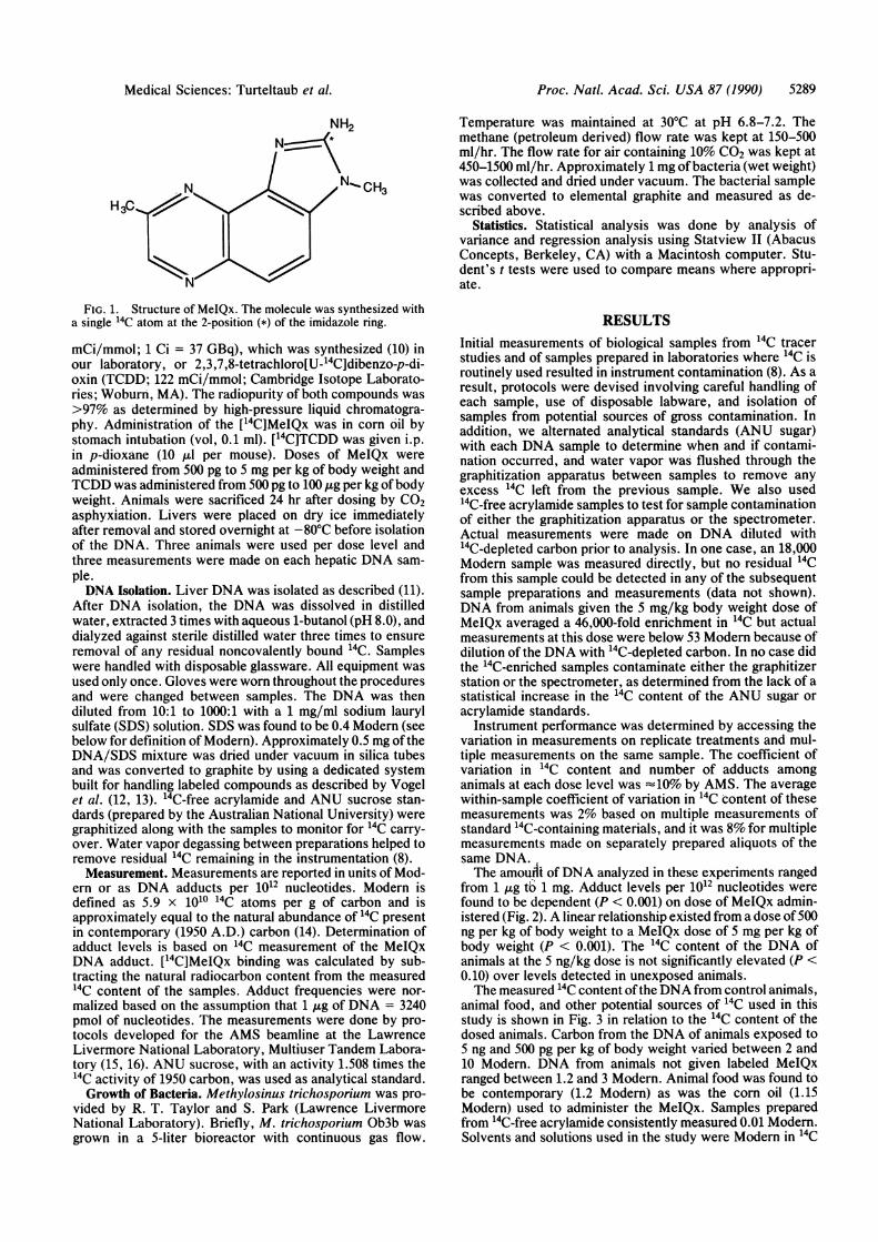
FIG. 1. Structure of MeIQx. The molecule was synthesized witha single 14C atom at the 2-position (*) of the imidazole ring.
mCi/mmol; 1 Ci = 37 GBq), which was synthesized (10) inour laboratory, or 2,3,7,8-tetrachloro[U-14C]dibenzo-p-di-oxin (TCDD; 122 mCi/mmol; Cambridge Isotope Laborato-ries; Woburn, MA). The radiopurity of both compounds was
>97% as determined by high-pressure liquid chromatogra-phy. Administration of the [14C]MeIQx was in corn oil bystomach intubation (vol, 0.1 ml). [14C]TCDD was given i.p.in p-dioxane (10 ,ul per mouse). Doses of MeIQx were
administered from 500 pg to 5 mg per kg of body weight andTCDD was administered from 500 pg to 100 ,ug per kg of bodyweight. Animals were sacrificed 24 hr after dosing by CO2asphyxiation. Livers were placed on dry ice immediatelyafter removal and stored overnight at -80°C before isolationof the DNA. Three animals were used per dose level andthree measurements were made on each hepatic DNA sam-
ple.DNA Isolation. Liver DNA was isolated as described (11).
After DNA isolation, the DNA was dissolved in distilledwater, extracted 3 times with aqueous 1-butanol (pH 8.0), anddialyzed against sterile distilled water three times to ensureremoval of any residual noncovalently bound 14C. Sampleswere handled with disposable glassware. All equipment was
used only once. Gloves were worn throughout the proceduresand were changed between samples. The DNA was thendiluted from 10:1 to 1000:1 with a 1 mg/ml sodium laurylsulfate (SDS) solution. SDS was found to be 0.4 Modern (seebelow for definition of Modern). Approximately 0.5 mg of theDNA/SDS mixture was dried under vacuum in silica tubesand was converted to graphite by using a dedicated systembuilt for handling labeled compounds as described by Vogelet al. (12, 13). 14C-free acrylamide and ANU sucrose stan-dards (prepared by the Australian National University) weregraphitized along with the samples to monitor for 14C carry-over. Water vapor degassing between preparations helped toremove residual 14C remaining in the instrumentation (8).Measurement. Measurements are reported in units of Mod-
ern or as DNA adducts per 1012 nucleotides. Modern isdefined as 5.9 x 10¹⁰ 14C atoms per g of carbon and isapproximately equal to the natural abundance of 14C presentin contemporary (1950 A.D.) carbon (14). Determination ofadduct levels is based on 14C measurement of the MeIQxDNA adduct. [14C]MeIQx binding was calculated by sub-tracting the natural radiocarbon content from the measured14C content of the samples. Adduct frequencies were nor-malized based on the assumption that 1 ,g of DNA = 3240pmol of nucleotides. The measurements were done by pro-tocols developed for the AMS beamline at the LawrenceLivermore National Laboratory, Multiuser Tandem Labora-tory (15, 16). ANU sucrose, with an activity 1.508 times the"4C activity of 1950 carbon, was used as analytical standard.Growth of Bacteria. Methylosinus trichosporium was pro-
vided by R. T. Taylor and S. Park (Lawrence LivermoreNational Laboratory). Briefly, M. trichosporium Ob3b was
grown in a 5-liter bioreactor with continuous gas flow.
Temperature was maintained at 30'C at pH 6.8-7.2. Themethane (petroleum derived) flow rate was kept at 150-500ml/hr. The flow rate for air containing 10% CO2 was kept at450-1500 ml/hr. Approximately 1 mg ofbacteria (wet weight)was collected and dried under vacuum. The bacterial samplewas converted to elemental graphite and measured as de-scribed above.
Statistics. Statistical analysis was done by analysis ofvariance and regression analysis using Statview II (AbacusConcepts, Berkeley, CA) with a Macintosh computer. Stu-dent's t tests were used to compare means where appropri-ate.
RESULTSInitial measurements of biological samples from "'C tracerstudies and of samples prepared in laboratories where 14C isroutinely used resulted in instrument contamination (8). As aresult, protocols were devised involving careful handling ofeach sample, use of disposable labware, and isolation ofsamples from potential sources of gross contamination. Inaddition, we alternated analytical standards (ANU sugar)with each DNA sample to determine when and if contami-nation occurred, and water vapor was flushed through thegraphitization apparatus between samples to remove anyexcess 14C left from the previous sample. We also used"4C-free acrylamide samples to test for sample contaminationof either the graphitization apparatus or the spectrometer.Actual measurements were made on DNA diluted with"'C-depleted carbon prior to analysis. In one case, an 18,000Modern sample was measured directly, but no residual 14Cfrom this sample could be detected in any of the subsequentsample preparations and measurements (data not shown).DNA from animals given the 5 mg/kg body weight dose ofMeIQx averaged a 46,000-fold enrichment in "4C but actualmeasurements at this dose were below 53 Modern because ofdilution of the DNA with 14C-depleted carbon. In no case didthe 14C-enriched samples contaminate either the graphitizerstation or the spectrometer, as determined from the lack of astatistical increase in the 14C content of the ANU sugar oracrylamide standards.
Instrument performance was determined by accessing thevariation in measurements on replicate treatments and mul-tiple measurements on the same sample. The coefficient ofvariation in 14C content and number of adducts amonganimals at each dose level was -10% by AMS. The averagewithin-sample coefficient of variation in 14C content of thesemeasurements was 2% based on multiple measurements ofstandard 14C-containing materials, and it was 8% for multiplemeasurements made on separately prepared aliquots of thesame DNA.The amouflt of DNA analyzed in these experiments ranged
from 1 ,ug to 1 mg. Adduct levels per 1012 nucleotides werefound to be dependent (P < 0.001) on dose of MeIQx admin-istered (Fig. 2). A linear relationship existed from a dose of500ng per kg of body weight to a MeIQx dose of 5 mg per kg ofbody weight (P < 0.001). The "'C content of the DNA ofanimals at the 5 ng/kg dose is not significantly elevated (P <0.10) over levels detected in unexposed animals.The measured 14C content ofthe DNA from control animals,
animal food, and other potential sources of 14C used in thisstudy is shown in Fig. 3 in relation to the 14C content of thedosed animals. Carbon from the DNA of animals exposed to5 ng and 500 pg per kg of body weight varied between 2 and10 Modern. DNA from animals not given labeled MeIQxranged between 1.2 and 3 Modern. Animal food was found tobe contemporary (1.2 Modern) as was the corn oil (1.15Modern) used to administer the MeIQx. Samples preparedfrom "'C-free acrylamide consistently measured 0.01 Modern.Solvents and solutions used in the study were Modern in 14C
Medical Sciences: Turteltaub et al.

5290 Medical Sciences: Turteltaub et al.
107
106
io5Immunoassay X104
Limit
0032P-postlabelin / 10 2
-10 °00Natural Abundance Limit 1 Z
10
AMS Sensitivity Limit[14C]-Depleted Biological ,0.2
Material 10
-10 -3
10-4 10-3 10-2 10-1 1 10 10 2 10 3 10 4105Animal Dose (gg [4C]MelQx/kg)
FIG. 2. Effect of MelQx exposure on DNA adduct levels foundin the hepatic DNA of mice as determined by AMS (K). Data (17, 18)from 32p postlabeling ofDNA isolated from mice given higher levelsof MeIQx (9) demonstrate the linearity of the dose-response aboveour limit of detection.
content, but radiocarbon levels in surface swipes ofwork areasshowed that some areas were contaminated with '4C fromprevious tracer experiments. These areas ranged between 18and 18,000 Modern (data not shown).The extreme sensitivity ofAMS for 14C suggests that very
small amounts of noncovalently bound 14C or 14C-boundmacromolecular contaminants in the DNA would be detect-able and thus could bias the adduct determinations. Toevaluate this possibility and to ensure that our proceduresadequately purify DNA, we measured DNA adduct forma-tion with TCDD, a carcinogen that has not been found tocovalently bind to DNA (19, 20). TCDD absorption is rapid
106I10 6
Dynamic Range of 14C-exposed animal 10DNA (present experiments) 102
corn oil-exposed 1 X0animal nimalfooda_>animal food corn oil vehicle1
10.1
102acrylamide 14C~epleted bacteria
(processing background) 10 3
FIG. 3. 14C content of control materials reported as fractionModem ([oo) in relation to the range in values obtained for the[14C]MeIQx-exposed mice (K). CO2 in present day air is -1.15Modem. Measurements of 14C content in methanotrophic bacteriagrown on petroleum-derived methane and CO2 (v) show the potentialsensitivity obtainable in selected biological systems.
10 6
lo 5
104
10 3
102
10
1
0
10.1
10 -2
0 0.5 5 50 500 105
ng TCDD/kg body weight
FIG. 4. 14C levels found in the DNA of [U-14C]TCDD-exposedmice. TCDD had 11.7 14Cs per molecule on average as opposed to 114C per molecule for MelQx. 14C levels were modem at all except thehighest TCDD doses, showing that animal handling and DNAextraction procedures were adequate in the removal of 14C non-covalently bound to DNA.
and -33% of the administered dose should reach the livertissue within the time frame of this study (21). These distri-bution kinetics are similar to that expected for MeIQx (22).The TCDD was uniformly labeled with 14C (11.7 14Cs permolecule on average) corresponding to 10 times more 14C ateach dose level than the MeIQx-exposed animals. HepaticDNA samples isolated from the TCDD-exposed mice (Fig. 4)were modem in 14C content (1.06-1.7 Modem) except at thehighest TCDD dose level (100 jig/kg; 14 Modem). Thehighest TCDD dose level is approximately the LD50 forTCDD in mice and the significance of the higher [14C]DNAvalues needs to be explored further. These data suggest thatno (or little) non-DNA-bound 14C remains after our DNApurifications since all DNA samples, except those from thehighest TCDD dose, were modem in "'C content. Thissuggestion is supported by the fact that 14C levels measuredin DNA from the MeIQx-exposed mice ranged from 100- to4000-fold greater than with the TCDD-exposed mice. Even atthe highest TCDD dose, >99.9% of the 14C distributed to theliver was removed through ourDNA purification protocol. Inaddition, this result shows that animal handling and DNAisolations can be carried out without radiocarbon contami-nation when appropriate procedures are used.The limiting factor for biological measurements in the
detection of 14C tagged molecules will be the natural abun-dance of 14C existing in the biosphere. Thus, we felt utiliza-tion of "4C-depleted hosts would be valuable for modelingdosimetry. Toward this end, M. trichosporium was grownwith petroleum-derived methane used as the sole carbonsource. Measurement of the 14C content of this organismverified that the 14C content can be easily depleted to an
equivalent of 0.01 Modern, which is 200 times less than our
lowest mouse sample (Fig. 3).
DISCUSSIONThe present detection limit for 14C-labeled DNA adducts byAMS is 1 adduct per 101" nucleotides. This corresponds to a
1 order of magnitude improvement over the very best sen-
sitivity offered to date by the 32P postlabeling assay. AMS
oM
OM
Proc. Natl. Acad. Sci. USA 87 (1990)

Proc. Natl. Acad. Sci. USA 87 (1990) 5291
provides a direct measure of the number of adducts presentwithout relying on enzymatic recognition of adducts and/orquantitative extraction recoveries (1, 2, 23). In addition,these measurements are 3-5 orders of magnitude better thanother techniques used for quantitative assay of DNA adductssuch as ultrasensitive radioimmunoassays (24, 25), surface-enhanced Raman spectroscopy (26), gas chromatography-mass spectrometry (27), laser-induced phosphorescence (28),fluorescence spectrometry (29), fluorescence line narrowingspectrometry (30), and synchronous scanning fluorescencespectrometry (31). Reproducibility of the measurements isvery high (within 10%) and is limited by animal to animalvariation.
Instrument precision in these measurements is 2% withfurther improvements likely. Thus, AMS is a uniquely sen-sitive and reproducible technique for adduct measurementand will easily complement existing methods.Our results show that the relationship between adduct
numbers and MeIQx exposure is linear to a MeIQx dosage of500 ng/kg based on the treatments used here. The 500 ng/kgdose level, corresponding to 1 adduct per 1011 nucleotides, isthe lowest dose we can statistically discriminate from thecontrols (P < 0.05). Linear extrapolations of adduct numbersfrom these exposures to adduct numbers reported previouslyby our laboratory and others (17, 18) from 32p postlabeling ofhepatic DNA from animals given much higher levels ofMeIQx (50 and 100 mg per kg of body weight) are similar,further demonstrating a linear dose-response (Fig. 2).Our inability to measure adducts in animals given 5 ng of
MeIQx per kg of body weight and less is seemingly due tocontamination of the DNA during isolation and does notrepresent a biological threshold. It is not the result of ourinability to detect modern or lower levels of 14C. Contami-nation most likely occurred during animal handling and/orDNA isolation. This is evident from comparison of theexpected amount of 14C in contemporary materials to theamounts actually found in the control animals (Fig. 3).Measurement of corn oil and animal food corresponded tocontemporary carbon (-1.2 Modern) but control animalswere 2-fold above contemporary in 14C content. Measure-ment of acrylamide and the 14C-depleted methanotrophicbacteria demonstrates the sensitivity of the carbon prepara-tion and measurement process and that we can measure up to100-fold below contemporary levels of 14C. Thus, the rela-tively high levels of 14C found in animals not given radioiso-tope are due to sample contamination and to the proximity ofthe total 14C content ofthe samples to the ambient 14C contentof the DNA itself. Extreme care must be taken to avoidexcess contamination above this natural limit. The workstation that measured a minimum of 18,000 Modern aptlydemonstrates the problems encountered in preparing samplesfor AMS measurement in laboratories with a history of 14Cuse. However, the data from the TCDD exposure study showthat, with proper handling, contamination problems andnonspecific binding of 14C can be eliminated.
Additional increases of 2- to 10-fold in the sensitivity ofDNA adduct detection by AMS will be possible throughcontamination reduction, but it will certainly be no betterthan that allowed by the natural abundance of 14C in biolog-ical molecules (modern carbon from the biosphere is 1/101214C). However, enhancement of sensitivity can be gained byusing 14C-depleted hosts. Growth of yeast and bacteria onpetroleum feedstocks has been reported (32, 33). We havegrown 14C-depleted M. trichosporium on petroleum-derivedmethane and verified that the 14C content can be easilydepleted to an equivalent of 0.01 Modern, demonstrating apotential 100-fold increase in sensitivity. Such model orga-nisms could be of use in studying the consequences of doseon the metabolism, kinetics, and effects of xenobiotic expo-
sures. Growth of other hosts on petroleum-based foodstuffsshould result in similarly low radiocarbon backgrounds.
Clinical applications and research with human subjects canbe envisioned with AMS radioisotope tracing. The detectionsensitivity and small sample size requirements ofAMS makeit ideal for measurements of small quantities of easily acces-sible human cells, in addition to the liver tissue demonstratedhere. Therapeutic parameters for individuals could be deter-mined by AMS through administration of small dosages of"C-labeled pharmaceuticals. Such custom tailoring of effec-tive therapeutic regimens would be particularly valuable forcancer chemotherapy as the extremely small human radiationdose from the drug should not be an issue. The estimatedeffective ["'C]MeIQx radiation dose equivalent in this study,based on a 24-hr biological half-life (22), corresponded to0.003 milliSieverts at the 500 ng/kg dose level. This exposureis -0.1% of the total annual adult exposure to ionizingradiation from known natural sources (34). Mutagen expo-sure in such protocols becomes a more significant issue thanthe radiation dose incurred.Use of AMS in the present measurements of low-level
DNA adducts provides no structural information on adducttype. Such information is better obtained with the postlabel-ing assay. However, molecular information on adduct typeshould be obtainable with AMS when it is used in conjunctionwith appropriate techniques to purify and separate adductsprior to measuring the 14C ratios. The need for use ofradiolabeled compounds with AMS is a limitation, but radi-ation exposure, because of the extreme sensitivity of thetechnique, is insignificant, particularly when used in thelaboratory and clinical setting where very low levels ofisotopically tagged compounds are being measured. AMSalso has the advantage of measuring these low 14C levels insmall samples. Utilization of rare stable isotopes seemspossible as well. Thus, AMS will be useful in any applicationin which sensitivity of detection is limiting.The present results with DNA adduct dosimetry demon-
strate the utility of AMS for quantitive measurement oflow-frequency biomolecular events following exposure tosmall concentrations of 14C-labeled xenobiotics. The tech-nique will be useful in clinical and laboratory environmentswhere sensitivity of detection is not possible by other assaysand in a wide number of applications beyond the adductdetection reported here. The technique has a dynamic rangecovering many orders of magnitude, is reproducible andsensitive, and 14C contamination is controllable. Further-more, requirements for 14C enrichment are 5-6 orders ofmagnitude below traditional decay-counting methods. Otherobvious candidate isotopes for low-level biomedical andenvironmental dosimetry applications are 3H and 41Ca. Thesepotential applications, coupled with the 14C measurementsreported here, show AMS technology to be an important toolfor the biomedical and environmental sciences community.
The authors thank D. J. Massoletti and E. E. Stilwell for assis-tance in sample preparation. This work was performed under theauspices of the U.S. Department of Energy by the LawrenceLivermore National Laboratory (W-7405-ENG-48) and was partiallysupported by the National Institutes of Environmental Health Sci-ences (222Y-01-ES-70158), the National Cancer Institute (CA40811),and the Natural Science and Engineering Research Council (Cana-da).
1. Reddy, M. V. & Randerath, K. (1986) Carcinogenesis 7, 1543-1551.
2. Gupta, R. C. (1985) Cancer Res. 45, 5656-5662.3. Gupta, R. C. & Early, K. (1988) Carcinogenesis 9, 1687-1693.4. Elmore, D. (1987) Biol. Trace Elem. Res. 12, 231-245.5. Brown, L. (1984) Annu. Rev. Earth Planet. Sci. 12, 39-45.6. Elmore, D. & Phillips, F. M. (1987) Science 236, 543-550.7. Kielson, J. & Waterhouse, C. (1978) Radiocarbon Dating,
Medical Sciences: Turteltaub et al.

5292 Medical Sciences: Turteltaub et al.
Proceedings 1st International Conference, ed. Grove, H. E.(Nuclear Structure Research Laboratory, University of Roch-ester, NY), pp. 391-395.
8. Vogel, J. S., Southon, J. R. & Nelson, D. E. Radiocarbon 32,81-83.
9. Felton, J. S., Knize, M. G., Shen, N. H., Andresen, B. D.,Bjeldanes, L. F. & Hatch, F. T. (1986) Environ. Health Per-spect. 67, 17-24.
10. Grivas, S. (1985) Acta Chem. Scand. Ser. B. 39, 213-217.11. Gupta, R.C. (1984) Proc. Natl. Acad. Sci. USA 81, 6943-6947.12. Vogel, J. S., Nelson, D. E. & Southon, J. R. (1989) Radiocar-
bon 31, 145-149.13. Vogel, J. S., Nelson, D. E. & Southon, J. R. (1987) Radiocar-
bon 29, 323-333.14. Stuiver, M. & Polach, H. A. (1977) Radiocarbon 19, 355-363.15. Davis, J. C. (1989) Nucl. Instrum. Methods B40/41, 705-708.16. Proctor, I. D. (1989) Nucl. Instrum. Methods B40/41, 727-730.17. Yamashita, K., Umemoto, A., Grivas, S., Kato, S. & Sug-
imura, T. (1988) Mutagenesis 3, 515-520.18. Turteltaub, K. W., Watkins, B. E., Vanderlaan, M. & Felton,
J. S. (1990) Carcinogenesis 11, 43-49.19. Randerath, K., Putman, K. L., Randerath, E., Mason, G.,
Kelley, M. & Safe, S. (1988) Carcinogenesis 9, 2285-2289.20. Poland, A. & Glover, D. (1979) Cancer Res. 39, 3341-3344.21. Abraham, K., Krowke, R. & Neubert, D. (1988) Arch. Toxicol.
62, 359-368.
22. Turesky, R. J., Aeschbacher, H. U., Malnoe, A. & Wurzner,H. P. (1988) Food Chem. Toxicol. 26, 105-110.
23. Dunn, B. P. & San, H. C. (1988) Carcinogenesis 9, 1055-1060.24. Hsu, I.-C., Poirier, M. C., Yuspa, S. H., Grunberger, D.,
Weinstein, I. B., Yolken, R. H. & Harris, C. C. (1981) CancerRes. 41, 1091-1095.
25. Harris, C. C., Vahakangas, K., Newman, M. J., Trivers,G. E., Shamsuddin, A., Sinopoli, N., Mann, D. L. & Wright,W. E. (1985) Proc. Nail. Acad. Sci. USA 82, 6672-6676.
26. Dinh, T.-V., Uziel, M. & Morrison, A. L. (1987) Appl. Spec-trosc. 41, 605-610.
27. Mohammed, G. B., Nazareth, A., Hayes, M. J., Giese, R. W.& Vouros, P. (1984) J. Chromatogr. 314, 211-217.
28. Dinh, T.-V. & Uziel, M. (1987) Anal. Chem. 59, 1093-1095.29. Rahn, R. O., Change, S. S., Holland, J. M. & Shugart, L. R.
(1982) Biochem. Biophys. Res. Commun. 109, 262-268.30. Sanders, M. J., Cooper, R. S. & Small, G. J. (1985) Anal.
Chem. 57, 1148-1152.31. Vahakangas, K., Trivers, G., Rowe, M. & Harris, C. C. (1985)
Environ. Health Perspect. 62, 101-104.32. Litchfield, J. H. (1977) Adv. Appl. Microbiol. 22, 266-304.33. Bassosi, R., Rossi, C., Trizzi, E. & Valension, G. (1983)
Angew. Chem. 95, 55-57.34. United Nations Scientific Committee on the Effects of Atomic
Radiation Report to the General Assembly (1988) Sources,Effects and Risks ofIonizing Radiation (United Nations, NewYork), Suppl. A/43/45, pp. 49-52.
Proc. Natl. Acad. Sci. USA 87 (1990)