academic program for master of science degree in medical physics
TRANSCRIPT

AAPM REPORT NO. 44
ACADEMIC PROGRAM FORMASTER OF SCIENCE DEGREE
IN MEDICAL PHYSICS
Published for theAmerican Association of Physicists in Medicine
by the American Institute of Physics
ACADEMIC PROGRAM FORMASTER OF SCIENCE DEGREE
IN MEDICAL PHYSICS
A REPORT OF THE EDUCATION AND TRAININGOF MEDICAL, PHYSICISTS COMMITTEE,
AMERICAN ASSOCIATION OFPHYSICISTS IN MEDICINE
Members
Paul M. DeLuca, Jr. (Chairman), University of Wisconsin-Madison, WisconsinF. H. Attix, University of Wisconsin-Madison, Wisconsin
Daniel A. Bassano, SUNY Health Science Center, Syracuse, New YorkJ. Larry Beach, Lexington Clinic, Kentucky
L. Stephen Graham, Veterans Administration Medical Center, UCLA, CaliforniaDavid Gur, University of Pittsburgh, Pennsylvania
Gerda B. Krefft, Methodist Hospital Lubbock, TexasRichard L. Maughan, Harper-Grace Hospitals, Michigan
Edwin C. McCullough, Mayo Clinic/Foundation, MinnesotaE. Russell Ritenour, University of Minnesota Medical School, Minnesota
Robert L. Siddon, Harvard Medical School, MarylandThomas G. Stir&comb, DePaul University, Illinois
David L. Vassy, Spartanburg Regional Medical Center, South CarolinaJoseph P. Windham, Henry Ford Hospital, Michigan
Lawrence T. Fitzgerald (Consultant), University of Florida, FloridaLawrence Lanzl (Consultant), Rush-Presbyterian-St Luke’s Medical Center, Illinois
October 1993
Published for theAmerican Association of Physicists in Medicine
by the American Institute of Physics
DISCLAIMER: This publication is based on sources and informationbelieved to be reliable, but the AAPM and the editors disclaim any warrantyor liability based on or relating to the contents of this publication.
The AAPM does not endorse any products, manufacturers or suppliers.Nothing in this publication should he interpreted as implying suchendorsement.
Further copies of this report may be obtained from:American Association of Physicists in Medicine
One Physics EllipseCollege Park, MD 20740-3846
International Standard Bock Number: 1-56396-287-XInternational Standard Serial Number: 0271-7344
©1993 by the American Associationof Physicists in Medicine
All rights reserved. No part of this publication may be reproduced, storedin a retrieval system, or transmitted in any form or by any means(electronic, mechanical, photocopying, recording, or otherwise) withoutthe prior written permission of the publisher.
Published by the American Institute of Physics, Inc.335 East 45th Street, New York, NY 10017
Printed in the United States of America

Contents
1 I N T R O D U C T I O N 1
2 T O P I C A L D I S C U S S I O N 3
2.1 Anatomy and Physiology . . . . . . . . . . . . . . . . . . . 3
2.2 Diagnostic Imaging . . . . . . . . . . . . . . . . . . . . . . . 3
2.3 Electronics . . . . . . . . . . . . . . . . . . . . . . . . . . . . 32.4 Health Physics-Radiation Protection . . . . . . . . . . . . . 42.5 Nuclear Medicine . . . . . . . . . . . . . . . . . . . . . . . . 52.6 Physics of Medicine and Biology . . . . . . . . . . . . . . . 52.7 Radiation Biology . . . . . . . . . . . . . . . . . . . . . . . 52.8 Radiological Physics and Dosimetry . . . . . . . . . . . . . 6
2.9 Radiation Therapy Physics . . . . . . . . . . . . . . . . . . 6
3 T O P I C A L O U T L I N E 7
3.1 Anatomy and Physiology . . . . . . . . . . . . . . . . . . . 73.2 Diagnostic Imaging . . . . . . . . . . . . . . . . . . . . . . . 9
3.3 Electronics . . . . . . . . . . . . . . . . . . . . . . . . . . . 113.4 Health Physics-Radiation Protection . . . . . . . . . . . . . 143.5 Nuclear Medicine . . . . . . . . . . . . . . . . . . . . . . . . 183.6 Physics of Medicine and Biology . . . . . . . . . . . . . . . 21
3.7 Radiation Biology . . . . . . . . . . . . . . . . . . . . . . . 243.8 Radiological Physics and Dosimetry . . . . . . . . . . . . . 263.9 Radiation Therapy Physics . . . . . . . . . . . . . . . . . . 30
4 B I B L I O G R A P H Y 3 6
4.1 Anatomy and Physiology . . . . . . . . . . . . . . . . . . . 364.2 Diagnostic Imaging . . . . . . . . . . . . . . . . . . . . . . 36
4.2.1 General References . . . . . . . . . . . . . . . . . . 364.2.2 Image Science . . . . . . . . . . . . . . . . . . . . . . 374.2.3 Imaging Systems . . . . . . . . . . . . . . . . . . . . 374.2.4 AAPM Monographs . . . . . . . . . . . . . . . . . . 38
4.3 Electronics . . . . . . . . . . . . . . . . . . . . . . . . . . . 384.4 Health Physics-Radiation Protection . . . . . . . . . . . . . 39
4.4.1 General . . . . . . . . . . . . . . . . . . . . . . . . . 39

4.4.2 Instrumentation . . . . . . . . . . . . . . . . . . . . 4 04.4.3 Shielding . . . . . . . . . . . . . . . . . . . . . . . . 4 14.4.4 Radiation Biology . . . . . . . . . . . . . . . . . . . 424.4.5 Miscellaneous References . . . . . . . . . . . . . . . 42
4.5 Nuclear Medicine . . . . . . . . . . . . . . . . . . . . . . . 424.6 Intermediate Physics for Medicine and Biology . . . . . . . 44
4.6.1 Additional References: . . . . . . . . . . . . . . . . . 444.7 Radiobiology . . . . . . . . . . . . . . . . . . . . . . . . . . 4 44.8 Radiation Therapy Physics . . . . . . . . . . . . . . . . . . 4 6
5 L A B O R A T O R Y T R A I N I N G 515.1 Laboratory for Diagnostic Imaging . . . . . . . . . . . . . . 515.2 Laboratory for Electronics . . . . . . . . . . . . . . . . . . . 535.3 Laboratory for Health Physics-Radiation Protection . . . . 545.4 Laboratory for Nuclear Medicine Instrumentation . . . . . . 5 7
5.5 Laboratory for Radiation Therapy Physics . . . . . . . . . . 5 8
6 G R A D U A T E P R O G R A M S I N M E D I C A L P H Y S I C S 60

1 I N T R O D U C T I O N
In the past two decades, Medical Physics has emerged from small splin-ter groups of applied physicists to a well-defined scientific discipline. Notunexpectedly, academic programs providing training in Medical Physicswere established to meet the monotonically increasing demand for medicalphysicists. As this discipline has matured, the educational requirementshave also become more clearly defined.
Existing academic programs contain a core of similarity yet reflect theindividual strengths and resources of personnel and facilities. As more pro-grams are created and existing programs seek AAPM accreditation, someguidelines for an academic program are needed. Such an endeavor is fraughtwith pitfalls, however. Hence this committee has collected a set of topicsthat provides the minimum level of training an M.S. graduate would beexpected to have. By no means is it suggested that the program outlinedhere is ideal or should be viewed as a satisfactory endpoint. Bather, theproposed program represents a solid foundation to build upon.
Beyond these caveats, we anticipate that some of this training mightbe provided in earlier academic experiences, e.g., a B.S. degree or an M.S.degree in a related field. Obviously, selective admission requirements couldimplement such criteria.
Training is organized into topical areas. Each area is outlined by topicsand subtopics. The format is similar to a course outline. This is notto imply that a topical area translates into a one or two semester courseformat. Rather the integral of all areas indicates the proposed training tobe accomplished by some convenient course structure. For some topics,training beyond tutorial contact is through laboratory sequences. Theseare separately indicated.
A bibliography of suggested resources is included. Again, selections aresegregated by topical area. Entries are often duplicated as appropriate.
A special question concerns “clinical” training. Ultimately a majorityof medical physicists practice their training in a clinical environment, mostnotably in radiation therapy. This situation may eventually lead to “cer-tification” or “licensure.” Without excessive elaboration, formal academictraining can never hope to provide nor is it necessarily the proper environ-ment for clinical training. Every attempt is made to provide a foundation
1

for a smooth transition to clinical applications in the suggested program.The final solution to this recurrent problem may reside with a “residency”program similar to the postgraduate training for clinical physicians.

2 TOPICAL DISCUSSION
2.1 Anatomy and Physiology
After completing this material, the student should be able to interpret com-mon medical terminology from knowledge of Greek and Latin root words.The student should be able to identify gross anatomical structures, definethe major organ systems and describe the physiological mechanisms for re-pair, maintenance, and growth. Anatomical structures and physiologicalfunction should be correlated with the imaging modalities used to viewthem.
2.2 Diagnostic Imaging
The topics listed in this section cover many (not all) of those that shouldbe included in a medical physics master’s degree training program. Thedesired level of learning to be attained in each topic area is not specified,in order to allow different programs the flexibility to emphasize selectedareas. However, the broad nature and ever-expanding scope of diagnosticimaging necessitate that most of these topics be covered to some extent asan integral part of the training program.
2.3 Electronics
The primary purpose of an electronics course in medical physics training istwo-fold.
1. Since medical physicists make use of a wide variety of electronic testand measurement equipment, the material should give the studentenough basic knowledge to understand the principles involved in ameasurement, the applicability of the instrumentation to that mea-surement and its limitations in regard to successfully completing themeasurement.
2. Much of the apparatus used by medical physicists is electronic orelectro-mechanical equipment of a high level of sophistication (e.g.
3

linac, CT scanners, x-ray units). The physicist should be able torecognize electronic malfunctions in the equipment and pinpoint thefault to a general area or functional unit within the system. Thecourse should aim to enable the student to recognize faults in simplecircuits and to understand the general philosophy behind fault findingin electronic circuits. It should give sufficient, basic knowledge to allowthe physicist. to converse intelligently with the electronic/electricalengineering staff who arc responsible for the repair and maintenanceof the equipment.
The prerequisites for the course should be elementary electricity andmagnetism as taught in a general Physics Course as offered by a Uni-versity Physics Department in the freshman year. An introductoryGeneral Physics course designed for physics and engineering studentswould be the ideal, although a course designed for students in biology,chemistry and medicine would suffice.
The course itself should be at the level of an undergraduate basic elec-tronics course as taught by a University Physics Department, againalthough a course designed for physics and engineering students wouldbe ideal, a. course for biologists, chemists and medical students wouldsuffice. The course should include a laboratory class which runs inparallel with the didactic lectures, and reinforces the material taughtin the lectures. An outline of the laboratory class is given. Simplecircuits which relate to the course content should be built and thentheir operating characteristics should be investigated using the rele-vant instrumentation. The time spent on the laboratory class shouldbe at least equal to that, spent in didatic lectures.
2.4 Health Physics-Radiation Protection
Radiation protection pervades the various subspecialties of medical physics.It provides the basic connection between microscopic interactions and cel-lular response. As such, a broad spectrum of topics are discussed. Specialattention is given detection apparatus and shielding analysis. An increas-ingly litiguous society is reflected in extensive presentation of the regulatoryenvironment. Complimenting tutorial instruction are a sequence of labora-
4

tory experiences focussing upon instrumentation, environmental sampling,bioassay, and several aspects of shielding. The emphasis in this topic isto provide a broad base supportive of the varied environments of medicalphysics.
2 .5 Nuc lear Medic ine
The purpose of this course of instruction is to provide a comprehensiveoverview of nuclear medicine physics. Previous experience in Nuclear Medi-cine is not required but students must have a solid knowledge of lower andupper division physics and appropriate facility with mathematics.
Since many of the instruments share a common underpinning with othersubspecialties and topics, careful integration is needed. An alternative ap-proach would be to segregate instrumentation essentials in a separate topic.
2.6 Physics of Medicine and Biology
Numerous applications of physics principles occur in medicine, biology, andphysiology that are not directly covered in the subspecialties. Examples arefluid flow dynamics encountered in the cardiovascular system, electrolyticsolutions and membrane-ion transport phenomena, and absorption and dis-solution of soluble gases. These topics provide the physical principles es-sential to physiology and anatomy and greatly expand the understandingof such topics by physicists.
2.7 Radiat ion Biology
Effects of ionizing radiation occur in all fields of medical physics. Lackof understanding of the biological consequences of ionizing radiation hasproduced a recent flood of disinformation. Only by education can thissituation be alleviated and eventually rectified. These topics should bepresented in a cohesive and consistent manner; not distributed among sev-eral related subjects such as radiation therapy physics, health physics, andnuclear medicine.
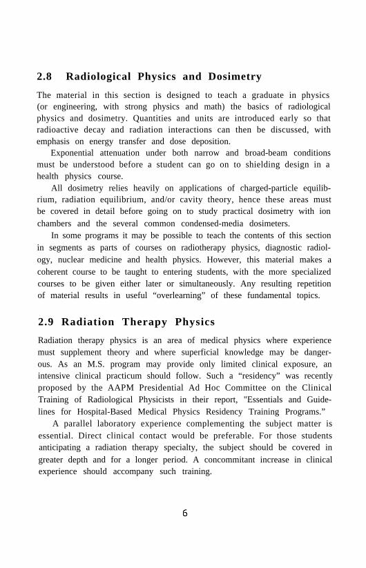
2.8 Radiological Physics and Dosimetry
The material in this section is designed to teach a graduate in physics(or engineering, with strong physics and math) the basics of radiologicalphysics and dosimetry. Quantities and units are introduced early so thatradioactive decay and radiation interactions can then be discussed, withemphasis on energy transfer and dose deposition.
Exponential attenuation under both narrow and broad-beam conditionsmust be understood before a student can go on to shielding design in ahealth physics course.
All dosimetry relies heavily on applications of charged-particle equilib-rium, radiation equilibrium, and/or cavity theory, hence these areas mustbe covered in detail before going on to study practical dosimetry with ionchambers and the several common condensed-media dosimeters.
In some programs it may be possible to teach the contents of this sectionin segments as parts of courses on radiotherapy physics, diagnostic radiol-ogy, nuclear medicine and health physics. However, this material makes acoherent course to be taught to entering students, with the more specializedcourses to be given either later or simultaneously. Any resulting repetitionof material results in useful “overlearning” of these fundamental topics.
2.9 Radiation Therapy Physics
Radiation therapy physics is an area of medical physics where experiencemust supplement theory and where superficial knowledge may be danger-ous. As an M.S. program may provide only limited clinical exposure, anintensive clinical practicum should follow. Such a “residency” was recentlyproposed by the AAPM Presidential Ad Hoc Committee on the ClinicalTraining of Radiological Physicists in their report, "Essentials and Guide-lines for Hospital-Based Medical Physics Residency Training Programs.”
A parallel laboratory experience complementing the subject matter isessential. Direct clinical contact would be preferable. For those studentsanticipating a radiation therapy specialty, the subject should be covered ingreater depth and for a longer period. A concommitant increase in clinicalexperience should accompany such training.

3 TOPICAL OUTLINE
3.1 Anatomy and Physiology
1. Anatomical Nomenclature
(a) Origin of anatomical names
(b) Prefixes and suffixes
(c) Anatomical position and body planes
2. Bones
(a) Classification
(b) Structure
(c) Development
3. Spinal Column
(a) Divisions of the spinal column
(b) Vertebral structure
(c) Radiography of the spinal column
4. The Thorax
(a) Bones of the thorax
(b) Organs in the thorax
(c) Radiography of the thoracic structures
5. The Abdomen
(a) Divisions and regions
(b) Organs in the abdomen
(c) Abdominal systems
6. Respiratory System
(a) Organs
(b) Function
(c) Radiography of system
7. Digestive System
(a) D ivisions
(b) Location, extension
(c) Function
(d) Radiography of system
8. Urinary System
(a) Organs
(b) Location
(c) Function
(d) Radiography of system
9. Reproductive System
(a) Organs
(b) Location
(c) Radiography of system
10. Circulatory System
(a) Major components
(b) Radiography of system
(c) Visit to Catheterization Laboratory
11. Pathology
(a) Identification of organs at autopsy
(b) Organ location
(c) Organ composition
(d) Correlation of radiographic findings
8
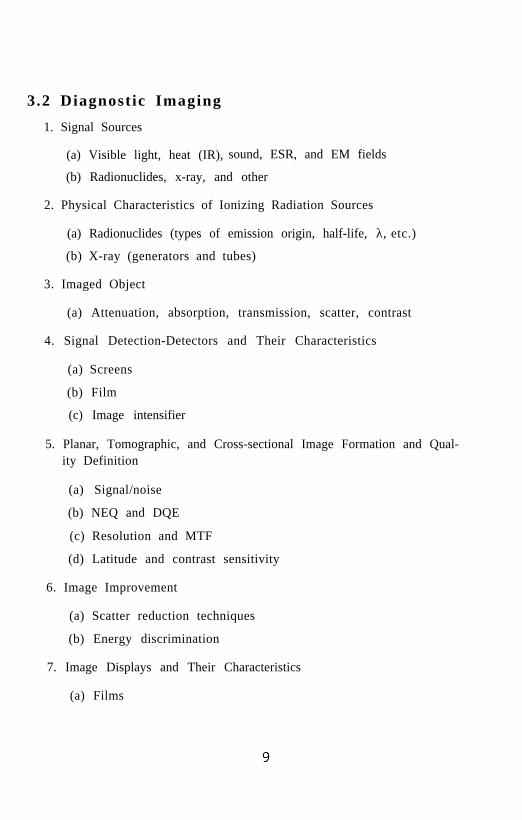
3.2 Diagnostic Imaging
1. Signal Sources
(a) Visible light, heat (IR), sound, ESR, and EM fields
(b) Radionuclides, x-ray, and other
2. Physical Characteristics of Ionizing Radiation Sources
(a) Radionuclides (types of emission origin, half-life, λ, etc.)
(b) X-ray (generators and tubes)
3. Imaged Object
(a) Attenuation, absorption, transmission, scatter, contrast
4. Signal Detection-Detectors and Their Characteristics
(a) Screens
(b) Film
(c) Image intensifier
5. Planar, Tomographic, and Cross-sectional Image Formation and Qual-ity Definition
(a) Signal/noise
(b) NEQ and DQE
(c) Resolution and MTF
(d) Latitude and contrast sensitivity
6. Image Improvement
(a) Scatter reduction techniques
(b) Energy discrimination
7. Image Displays and Their Characteristics
(a) Films

(b) Paper-printed images
(c) CRT, stereo displays, holographic
8. Image Enhancement-Post Processing
(a) Color display
(b) Subtraction
(c) AHE, USM, LPF, HPF
9. Special Requirements for Specific Procedures
(a) Mammography
(b) Bone density absorptiometry
(c) CT
(d) Angio-magnification
(e) Dental
10. Measurements of Image Quality
11. QA Procedures
12. Generation of Images from Nonionizing Radiation Sources
(a) Visible light
(b) Thermography
(c) Ultrasound
(d) NMR
(e) SQUID
13. Contemporary Issues
(a) Patient dose documentation
(b) PACS
(c) Measurements of diagnostic accuracy (ROC)
10

3 .3 E lec tron ics
1. Passive Components and Networks
(a) Review of DC circuits
(b) Review of AC circuits
(c) Network analysis
Thevenin’s Theorem, Norton’s Equivalent Circuit Theorem.Networks requiring Thevenin’s Theorem or Kirchhoff’s Lawsfor solution
2. Electronic Instrumentation
(a) Basic analog meters
Moving coil and moving iron meters, dynamometer. Mea-surement of RMS values, AC and DC measurements. Useof basic meters as an ammeter, voltmeter, ohmmeter orwattmeter.
(b) Measuring instruments
Analog multimeter, Wheatstone bridge, AC bridges, poten-tiometer, DVM, DMM and electrometer.
(c) Oscilloscope
Basic components, operation and use.
(d) Other instruments
Signal generator, frequency counter and attenuator.
(e) Related topics
Transmission lines and relays and other electro-mechanicaldevices.
3. Input Transducers
(a) Resistive input transducers
Photoresistor and thermistor
(b) Current input transducers
11

Ionization chamber
(c) Voltage input transducer
Thermocouple, photovoltaic cell and photodiode
4. Diodes and Some Applications
(a) Diodes
Vacuum and semiconductor diodes, physical model for semi-conductor diode, the p-n junction. Diode action; current-voltage characteristics.
(b) Rectification and filtering
Half-wave and full-wave rectification. Low pass and highpass filters.
(c) Zener diode
Current-voltage characteristic; use as a voltage regulator.
5. Transistors
(a) Transistor types
Bipolar junction, junction FET, MOSFET; basic structureand operation, current-voltage characteristics, load line.
(b) Applications of transistors
Amplifier circuits and switching circuits. Choice of transis-tor; interpretation of specification sheets.
6. Amplifier Circuits and Operational Amplifiers
(a) Fundamental amplifier properties
Input and output impedance, gain, frequency response, feed-back
(b) Amplifier types
Cascade, AC and DC, noninverting and inverting, differenceand differential amplifiers
12
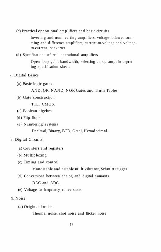
(c) Practical operational amplifiers and basic circuits
Inverting and noninverting amplifiers, voltage-follower sum-ming and difference amplifiers, current-to-voltage and voltage-to-current converter.
(d) Specifications of real operational amplifiers
Open loop gain, bandwidth, selecting an op amp; interpret-ing specification sheet.
7. Digital Basics
(a) Basic logic gates
AND, OR, NAND, NOR Gates and Truth Tables.
(b) Gate construction
TTL, CMOS.
(c) Boolean algebra
(d) Flip-flops
(e) Numbering systems
Decimal, Binary, BCD, Octal, Hexadecimal.
8. Digital Circuits
(a) Counters and registers
(b) Multiplexing
(c) Timing and control
Monostable and astable multivibrator, Schmitt trigger
(d) Conversions between analog and digital domains
DAC and ADC.
(e) Voltage to frequency conversions
9. Noise
(a) Origins of noise
Thermal noise, shot noise and flicker noise
13

(b) Reduction of noise
Shielding, ground loops, differential amplifier configurations,filtering and signal averaging techniques
3.4 Health Physics-Radiation Protection
1. Introduction and Historical Perspective
(a) Discovery and early application of ionizing radiation
(b) Observed radiation injury
(c) Suggested radiation protection practices
(d) Pre-regulatory initiatives
2. Interaction Physics as Applied to Radiation Protection
(a) Indirectly and directly ionizing radiation
(b) Bethe-Bloch formalism for coulomb scattering, shell effects, po-larization phenomena, nuclear processes, adiabatic scattering,track structure, target phenomena, radiative processes, Anderson-Ziegler parameterization, Janni tabulation, and effects due tomixtures and compounds.
(c) Electromagnetic interaction: photoelectric effect, Compton ef-fect, pair production, shower cascade phenomena.
(d) Neutron interactions: elastic and nonelastic processes
3. Operational Dosimetry
(a) Units
(b) Kerma and absorbed dose
(c) Dose equivalent
(d) Recommendations of the ICRU
(e) Recent changes in the neutron quality factor
4. Radiation Detection Instrumentation
14
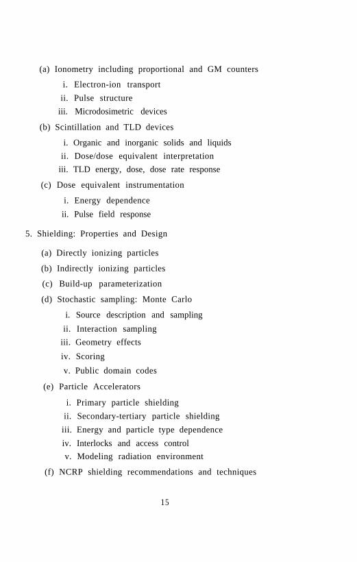
(a) Ionometry including proportional and GM counters
i. Electron-ion transportii. Pulse structure
iii. Microdosimetric devices
(b) Scintillation and TLD devices
i. Organic and inorganic solids and liquidsii. Dose/dose equivalent interpretation
iii. TLD energy, dose, dose rate response
(c) Dose equivalent instrumentation
i. Energy dependence
ii. Pulse field response
5. Shielding: Properties and Design
(a) Directly ionizing particles
(b) Indirectly ionizing particles
(c) Build-up parameterization
(d) Stochastic sampling: Monte Carlo
i. Source description and sampling
ii. Interaction samplingiii. Geometry effects
iv. Scoring
v. Public domain codes
(e) Particle Accelerators
i. Primary particle shieldingii. Secondary-tertiary particle shielding
iii. Energy and particle type dependenceiv. Interlocks and access controlv. Modeling radiation environment
(f) NCRP shielding recommendations and techniques
15

6. Statistics
(a) Statistical interpretation of instrument response
(b) Design of experiments
(c) Stochastic and nonstochastic error analysis
(d) Interpreting experimental results
7. Radiation Monitoring of Personnel
(a) Instrumentation and techniques
(b) Integral and active devices
(c) Dynamic range and response sensitivities
(d) Film, TLD, Lexan, and CR-39
(e) Pocket ion chambers and GM counters
8. Internal Exposure
(a) ICRP 26, ICRP 2A recommendations
(b) MIRD dosimetry
(c) Monitoring and radiation control
(d) Biological assay
(e) Dispersion in a working environment
(f) Allowed limit of intake and derived air (or water) concentrations
9. Environmental Dispersion
(a) Release of radionuclides to the environment
(b) Dosimetric consequences
(c) EPA and UNSNRC air and water dispersion models
10. Biological Effects
(a) Basic radiation biology
(b) Nonstochastic and stochastic responses
16
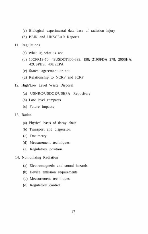
(c) Biological experimental data base of radiation injury
(d) BEIR and UNSCEAR Reports
11. Regulations
(a) What is; what is not
(b) 10CFR19-70; 49USDOT300-399, 198; 219SFDA 278; 290SHA;42USPHS; 40USEPA
(c) States: agreement or not
(d) Relationship to NCRP and ICRP
12. High/Low Level Waste Disposal
(a) USNRC/USDOE/USEPA Repository
(b) Low level compacts
(c) Future impacts
13. Radon
(a) Physical basis of decay chain
(b) Transport and dispersion
(c) Dosimetry
(d) Measurement techniques
(e) Regulatory position
14. Nonionizing Radiation
(a) Electromagnetic and sound hazards
(b) Device emission requirements
(c) Measurement techniques
(d) Regulatory control
17

3.5 Nuclear Medicine
1. Radioactive Materials
(a) Nuclear structure
(h) Decay schemes
(c) Production of radionuclides
i. Reactorsii. Accelerators
(d) Statistics of radioactive decay
2. Instrumentation
(a) Rectilinear scanners
i. Design and operationii. Quality control
iii. Clinical applications
(b) Scintillation cameras
i. P1anar systemsA. Design and operation (including computer interface)B. Hard copy output devices
C. Acceptance testingD. Quality controlE. Clinical applications (including transmission scanning)
ii. SPECT systemsA. Gantry control and computer interfacingB. Principles of reconstructionC. Acceptance testingD. Quality control
E. Clinical applicationsiii. PET systems
A. Design and operationB. Acceptance testing
18

C. Quality controlD. Clinical applications
(c) Alternative imaging systems
i. Pressurized proportional chambersii. Harvard brain imager
iii. Dedicated SPECT systems
(d) Scintillation counter systems (including thyroid uptake)
i. Electronic and mechanical componentsii. Acceptance testing and quality control
iii. Interfacing to computer systemsiv. Clinical applications
(e) Liquid scintillation counters
i. Principles of operation and design
ii. Quality controliii. Interfacing to computer systems
(f) Semiconductor systems
i. Spectroscopyii. Fluorescence scanning
(g) Well ionization chambers (dose calibrators)
i. Design and operationii. Sources of error
iii. Quality control
3. Radiopharmacy
(a) Generator systems
(b) Preparation of radiopharmaceuticals from kits
(c) Quality control of radiopharmaceuticals
(d) Radionuclides for specific clinical applications
4. Computers
19

(a) Hardware operation
(b) Software
(c) Peripheral connections and operation
(d) Acceptance testing and quality control
(e) Clinical applications
5. Internal Radiation Dosimetry
(a) Systems for calculating absorbed dose
i. Classical equations and methodologyii. Medical Internal Radiation Dose (MIRD) methodology
iii. Introduction to modeling and tracer kinetics
iv. Calculations for radionuclide therapy
(b) Typical patient absorbed doses for diagnostic and therapeuticprocedures
6. Radiation Safety
(a) Surveys of radiation areas
(b) Principles and mechanisms of protection for personnel and pa-tients
(c) Exposure rate levels associated with the preparation and admin-istration of radioactive materials for diagnostic and therapeuticuse
(d) Exposure rate levels from patients undergoing diagnostic andtherapeutic procedures
(e) Handling of radiation accident patients
(f) Implementation of the ALARA principle
7. Design of Nuclear Medicine Facilities
(a) Radioactive materials storage and laboratory areas
(b) Air exhaust design
20

(c) Patient procedures area
(d) Dark room
(e) Departmental layout (personnel and patient flow)
(f) General safety requirements
(g) Floor loading for equipment
(h) Electrical power and air conditioning requirements
8. Regulations
(a) Federal, state, and local regulations and requirements
(h) Regulatory inspections (state, NRC)
(c) License application, renewal
(d) Accreditation requirements (JCAHO)
(e) Accreditation site visits (JCAHO)
3.6 Physics of Medicine and Biology
1. Review of Statistics and Probability
(a) Parent populations and samples
(b) Binomial, Gaussian and Poisson distributions
2. Mechanics Applied to Body Systems
(a) Forces on the Achilles tendon and hip
(b) Mechanics of using a cane
3. Hydrostatics
(a) Viscous flow in a tube
(b) Transport in an infinite medium
(c) Flow, flux, and continuity
(d) Particle motion in a liquid-the Langevin form of Newton’s sec-ond law
2 1

(e) Newtonian fluids-viscosity-Stokes’ law
4. Diffusion
(a) Fick’s first law
(b) Diffusion related to viscosity
(c) Fick’s second law and applications
5. Transport Through Semipermeable Membranes
(a) Osmotic pressure
(b) Plasma exchange in capillaries
(c) Edema; osmotic diuresis; osmotic fragility of red blood cells
(d) Volume transport; solute transport; the artificial kidney
(e) External forces on solute molecules; ionic solutes and equilibriumelectric fields in membranes
(f) Ion movement in solution involving diffusion, solvent drag andelectric fields
(g) Nernst-Planck equation and the Goldman equation
6. Nerve Cell Structure
(a) Nonmyelinated and myelinated axons
(b) Introduction to the electrical nature of nerve impulse transmis-sion
(c) Review of electrostatics
7. Electrodynamics Emphasizing Relevance to Nerve Impulse Conduc-tion
(a) Current density
(b) Conductivity
(c) Kirchhoff’s laws
8. Charge Distribution in the Resting Nerve Cell
2 2

9. Leakage Currents Across the Axon Membrane in the Absence ofMyelination
10. Resistance of the Axon
11. Nerve Impulse and Transmission Across a Synapse
(a) Application of Kirchhoff’s laws to wave equation for nerve im-pulse transmission in nonmyelinated axons.
(b) Small voltage changes not involving changes in membrane con-ductance (electronus)
(c) Voltage clamp experiments
12. Hodgkin-Huxley Model for Membrane Current
(a) Voltage changes in the axially clamped axon following electricalstimulation as tests of the Hodgkin-Huxley model
(b) Solutions of the wave equation [propagation) for nonmyelinatedaxons employing the Hodgkin-Huxley model.
13. Nerve Impulse Propagation in Myelinated Axons; Myelin Sheath Con-ductance and Capacitance; Saltatory Conduction.
14. Electrocardiograms Taking the Body to be a Uniform Conductor
15. Kidney
(a) Structure of the nephron
(b) A physicist’s view of the glomerulus
16. Nonionic Filtration by the Glomerulus-Transport Through Pores ofParticles Having Diameters of the Order of the Pore Diameters.
17. Theory and Experiment of Verniory
18. Biomagnetism, Including Generation of Magnetic Fields by ElectricCurrents in the Body and Their Detection; Physical Principles of theDC SQUID.
23
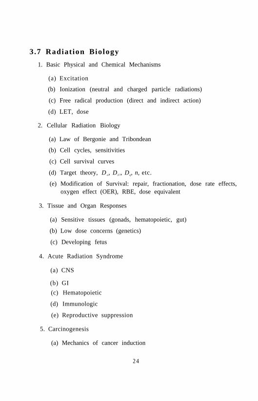
3.7 Radiat ion Biology
1. Basic Physical and Chemical Mechanisms
(a) Excitation
(b) Ionization (neutral and charged particle radiations)
(c) Free radical production (direct and indirect action)
(d) LET, dose
2. Cellular Radiation Biology
(a) Law of Bergonie and Tribondean
(b) Cell cycles, sensitivities
(c) Cell survival curves
(d) Target theory, D o, D3 7, Dq, n, etc.
(e) Modification of Survival: repair, fractionation, dose rate effects,oxygen effect (OER), RBE, dose equivalent
3. Tissue and Organ Responses
(a) Sensitive tissues (gonads, hematopoietic, gut)
(b) Low dose concerns (genetics)
(c) Developing fetus
4. Acute Radiation Syndrome
(a) CNS
(b) GI(c) Hematopoietic
(d) Immunologic
(e) Reproductive suppression
5. Carcinogenesis
(a) Mechanics of cancer induction
2 4
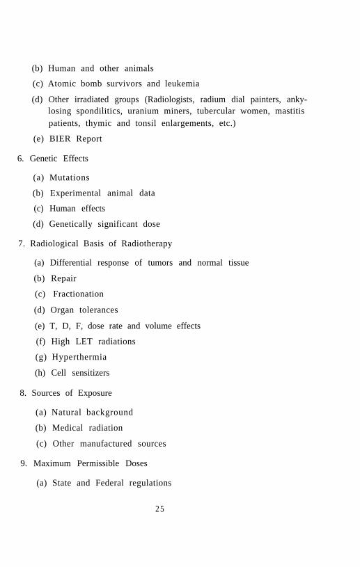
(b) Human and other animals
(c) Atomic bomb survivors and leukemia
(d) Other irradiated groups (Radiologists, radium dial painters, anky-losing spondilitics, uranium miners, tubercular women, mastitispatients, thymic and tonsil enlargements, etc.)
(e) BIER Report
6. Genetic Effects
(a) Mutations
(b) Experimental animal data
(c) Human effects
(d) Genetically significant dose
7. Radiological Basis of Radiotherapy
(a) Differential response of tumors and normal tissue
(b) Repair
(c) Fractionation
(d) Organ tolerances
(e) T, D, F, dose rate and volume effects
(f) High LET radiations
(g) Hyperthermia
(h) Cell sensitizers
8. Sources of Exposure
(a) Natural background
(b) Medical radiation
(c) Other manufactured sources
9. Maximum Permissible Doses
(a) State and Federal regulations
2 5

(b) Recommendations (NCRP, ICRU, etc.)
10. Risk Versus Benefit
(a) Screening
(b) Diagnostic radiation doses
3.8 Radiological Physics and Dosimetry
1. Ionizing Radiation
(a) Types and sources of ionizing radiation
(b) Description of ionizing radiation fields
2. Quantities for Describing the Interaction of Ionizing Radiation withMatter
(a) Energy imparted, energy transferred, and net energy transferred
(b) Kerma and collision-kerma
(c) Absorbed dose
(d) Exposure
(e) Dose equivalent and quality factor
3. Exponential Attenuation
(a) Simple exponential attenuation
(b) Exponential attenuation for plural modes of absorption
(c) Narrow- vs. broad-beam attenuation
(d) Spectral effects in attenuation
(e) Buildup factor
(f) Reciprocity theorem
4. Charged-Particle and Radiation Equilibria
(a) Radiation equilibrium
2 6

(b) Charged-particle equilibrium (CPE)
(c) Relationships between absorbed dose, collision kerma and expo-sure under CPE conditions
(d) Conditions that enable CPE, or cause its failure
(e) Transient CPE
5. Absorbed Dose in Radioactive Media
(a) Radioactive disintegration processes
(b) Energy available for absorbed dose
6. Radioactive Decay
(a) Total and partial decay constants
(b) Units of activity
(c) Mean life and half-life
(d) Parent-daughter relationships
(e) Activity equilibria
(f) Harvesting of daughter products
(g) Radioactivation by nuclear interactions
(h) Exposure-rate and air-kerma-rate constants
7. Photon Interactions in Matter
(a) Compton effect
(b) Photoelectric effect
(c) Pair production
(d) Rayleigh scattering
(e) Photonuclear interactions
(f) Coefficients for attenuation, energy transfer, and energy absorp-tion
8. Charged-Particle Interactions in Matter
2 7

(a) Types of Coulomb-force interactions
(b) Stopping power
(c) Range
(d) Calculation of absorbed dose
9. X-Ray Production and Quality
(a) Fluorescence
(b) Bremsstrahlung
(c) Beam quality
(d) Filtering
10. Cavity Theory
(a) Bragg-Gray theory and corollaries
(b) Spencer cavity theory
(c) Burlin cavity theory
(d) Stopping-power averaging
(e) Fano theorem
(f) Dose near interfaces
11. Dosimetry Fundamentals
(a) ICRU definitions of dosimetric qualities and units
(b) Interpretation of dosimeter measurements
(c) General characteristics of dosimeters
12. Ionization Chambers
(a) Free-air and free-electron chamber
(b) Cavity chambers
(c) Charge and current measurements
(d) Saturation and ionic recombination
28
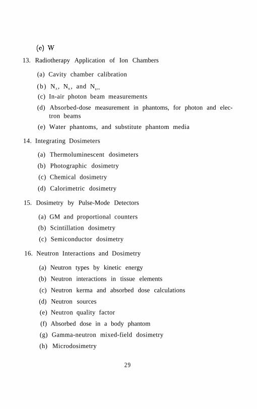
13. Radiotherapy Application of Ion Chambers
(a) Cavity chamber calibration
(b ) NX, NK, and Ng a s
(c) In-air photon beam measurements
(d) Absorbed-dose measurement in phantoms, for photon and elec-tron beams
(e) Water phantoms, and substitute phantom media
14. Integrating Dosimeters
(a) Thermoluminescent dosimeters
(b) Photographic dosimetry
(c) Chemical dosimetry
(d) Calorimetric dosimetry
15. Dosimetry by Pulse-Mode Detectors
(a) GM and proportional counters
(b) Scintillation dosimetry
(c) Semiconductor dosimetry
16. Neutron Interactions and Dosimetry
(a) Neutron types by kinetic energy
(b) Neutron interactions in tissue elements
(c) Neutron kerma and absorbed dose calculations
(d) Neutron sources
(e) Neutron quality factor
(f) Absorbed dose in a body phantom
(g) Gamma-neutron mixed-field dosimetry
(h) Microdosimetry
29

3.9 Radiation Therapy Physics
1. Overview of Clinical Radiation Oncology
(a) Cancer incidence/etiology
(b) Cancer classification/staging
i. Review lymphatic drainage
(c) Overview of treatment modalities
i. Surgeryii. Chemotherapy
iii. RadiotherapyA. Teletherapy
B. Brachytherapy
C. Neutron, proton and high Z therapy
iv. Hyperthermia
(d) Review of pertinent, radiobiology
i. Dose response curvesii. The 4 R’s
iii. The relationship of volume and time to radiation effects(TDF, Alpha/Beta Ratios)
iv. Side effects/complicationsv. Tolerance doses for normal tissues and tumors
(e) The role of a Clinical Medical Physicist
2. Phantom Systems
(a) Calibration phantoms
(b) Anthropomorphic phantoms
(c) Tissue-mimicking materials
(d) Properties of beam scanning systems
3. Radiation Machines
(a) Kilovoltage units
30
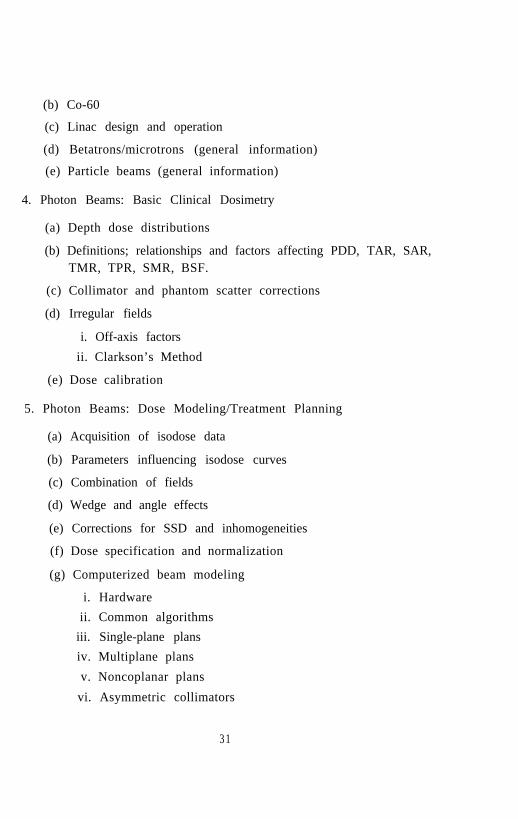
(b) Co-60
(c) Linac design and operation
(d) Betatrons/microtrons (general information)
(e) Particle beams (general information)
4. Photon Beams: Basic Clinical Dosimetry
(a) Depth dose distributions
(b) Definitions; relationships and factors affecting PDD, TAR, SAR,TMR, TPR, SMR, BSF.
(c) Collimator and phantom scatter corrections
(d) Irregular fields
i. Off-axis factors
ii. Clarkson’s Method
(e) Dose calibration
5. Photon Beams: Dose Modeling/Treatment Planning
(a) Acquisition of isodose data
(b) Parameters influencing isodose curves
(c) Combination of fields
(d) Wedge and angle effects
(e) Corrections for SSD and inhomogeneities
(f) Dose specification and normalization
(g) Computerized beam modeling
i. Hardwareii. Common algorithms
iii. Single-plane plansiv. Multiplane plans
v. Noncoplanar plans
vi. Asymmetric collimators
3 1

vii. Compensator designviii. 3-D planning (general information)
6. Photon Beams: Patient Application
(a) Patient data acquisition
i. Images (plain films, CT, US, NM, MRI)
ii. Contours
(b) Simulator techniques
i. Positioning/immobilizationii. Use of contrast, markers, etc.
iii. Image parameters/optimization
(c) Accessory devices/techniques
i. Block-cutting
ii. Compensators
iii. Portal/verification films
(d) Special considerations
i. Skin dose
ii. Adjacent fieldsiii. Tangential fields
iv. Integral dosev. Hemibody/whole body techniques
7. Electron Beams: Basic Clinical Dosimetry
(a) Basic characteristics
i. Electron interactions
ii. Detector characteristicsiii. Measurement techniques
(b) Beam characteristics
i. Energy determination
3 2

ii. Depth-dose (surface dose, x-ray contamination, isodose/D max
shift, etc.)iii. Profiles/isodose curves
iv, Output factors/virtual source position
8. Electron Beams: Dose Modeling and Treatment Planning
(a) Special effects
i. Air gap/obliquity effects
ii. Internal inhomogenity
(b) Computer algorithms
(c) Treatment planning
i. Energy selection/photon-electron mixingii. Use of bolus
iii. Field shaping
iv. Field abuttmentv. Conventional techniques
(d) Advanced techniques (general information)
i. Total skin irradiation
ii. Electron arc therapy
9. Brachytherapy
(a) Source characteristics and strength specifications
(b) Implant dosimetry systems
(c) Implantation/application techniques
(d) Dose computations/dose specifications
(e) Dose rate considerations
(f) Clinical examples: 1 3 7C s , 1 9 2Ir, 1 2 5I, 1 9 8A u
(g) Neutron sources
(h) Other radionuclides
3 3

10. Radiation Protection
(a) Regulatory requirements
(b) Structural shielding design
i. Cobalt and linac teletherapyii. Neutrons
iii. Brachytherapy (low-dose rate/high-dose rate)iv. X-ray Rooms (superficial, orthovoltage, simulators)
(c) Operational safety guidelines
i. Machine sourcesii. Brachytherapy
iii. Area and personnel monitoringiv. Radiation surveys (techniques and instruments)
11. Quality Assurance/Quality Control
(a) Error analysis of total treatment process
(b) Sources of QA/QC standards
(c) Organizing a QA program
i. Staff assignments
ii. Equipmentiii. Traceability/redundancy
(d) Treatments/dose delivery
i. Documentation requirements
ii. Portal/verification film techniques
iii. Record and verification systems
iv. Real time surface dosimetry (diodes)
(e) Machine acquisition (purchase, installation, acceptance)
(f) Specific guidelines
i. Machine sources
ii. Brachytherapy Sources and applicators
3 4

iii. Block-cutting/compensation systemsiv. Computers/dosimetry systems
12. Special Topics (Optional)
(a) Hyperthermia
(b) Intra-operative radiotherapy (photon and electron)
(c) Radiosurgery
(d) Hyperfractionation
(e) Conformal beam therapy
3 5

4 B I B L I O G R A P H Y
4.1 Anatomy and Physiology
l Clemente, Carmine D., 1987, Anatomy A Regional Atlas of the Hu-man Body, (Urban & Schwartzenberg, Baltimore).
l Dennerll, Jean T., 1988, Medical Terminology-A Programmed Text,(John Wiley & Sons, New York).
l Mallett, M., Anatomy and Physiology for Students of Medical Radi-ation Technology, (The Burnell Company/Publishers, Inc., Mankato,MN).
l Merrill, Vinita, 1982, Atlas of Roentgenographic Positionings andStandard Radiological Procedures (Mosby, St. Louis).
l Squire, Lucy F., 1964, Fundamentals of Roentgenology, (Harvard Uni-versity Press).
l Taylor, Elizabeth J., 1988, Dorland's Illustrated Medical Dictionary,2 7th Ed., (W. B. Saunders Co., Philadelphia).
l Vander, Arthur J., Shamon, James H. and Luciano, Dorothy S., 1985,Human Physiology, The Mechanisms of Body Function, (McGraw HillBook Company, New York).
l Weir. Jamie and Abrams, Peter, 1986, An Atlas of Radiological Anatomy,(Year Book Medical Publishers, Inc., Chicago)
l Wicke, Lothar, 1982, Atlas of Radiologic Anatomy, 3rd Ed., (Urban& Schwarzenberg, Baltimore).
4.2 Diagnostic Imaging
4 .2 .1 General References
l Curry, Thomas S., Dowdey, J.E., Murry, R.C., 1984, Christensen’sIntroduction to the Physics of Diagnostic Radiology (Lea and Febiger,Philadelphia, PA).
36

l Evans, Robly D., 1955, The Atomic Nucleus, (McGraw-Hill BookCompany, New York).
l Johns, H.E. and Cunningham, J.R., 1983, The Physics of Radiology,3rd Ed., (Charles C. Thomas, Springfield, IL).
l Knoll, Glenn F., 1989, Radiation Detection and Measurement, 2n d
Ed., (John Wiley and Sons, New York).
l Ter-Pogossian, Michel M., 1967, The Physical Aspects of DiagnosticRadiology, (Harper and Row, New York).
4 .2 .2 Image Sc ience
l Barrett, Harrison H., and William Swindell, 1981, Radiological Imag-ing, The Theory of Image Formation, Detection, and Processing,Vols. I & II, (Academic Press, New York).
l Brody, W.R., 1984, Digital Radiography, (Raven Press, New York).
l Dainty, J.C. and R. Shaw, 1974, Image Science, (Academic Press,New York).
l Haung, H.K., 1987, Elements of Digital Radiology, (Prentice-Hall,Englewood Cliffs, NJ).
l Herman, Gabor T., 1980, Image Reconstruction from Projections:The Fundamentals of Computerized Tomography, (Academic Press,New York).
l Swetis, John A. and Ronald M. Pickett, 1982, Evaluation of Diagnos-tic Systems: Methods from Signal Detection and Theory, (AcademicPress, New York).
4 .2 .3 Imaging Sys tems
l Gadian, David G., 1982, Nuclear Magnetic Resonance and Its Appli-cation to Living Systems, (Oxford University Press, New York).

l Gottschalk, Alexander, et al., 1988, Diagnostic Nuclear Medicine,Vol. I, (Williams and Wilkins, Baltimore).
l Macovski, Albert, 1983, Medical Imaging Systems, (Prentice-Hall, En-glewood Cliffs, N.J).
l Morris, Peger G., 1986, Nuclear Magnetic Resonance Imaging inMedicine, (Clarendon Press, Oxford).
l Nudelman, Sol and Dennis D. Patton, 1980, Imaging for Medicine,Vol. I, (Plenum Press, New York).
l Partain, C. Leon, et al., 1988, Magnetic Resonance Imaging, Vol. II,(W. B. Saunders Co., Philadelphia).
l Webb, Steve, 1988, The Physics of Medical Imaging, (Adam Hilger,Philadelphia).
4 .2 .4 AAPM Monographs
l Doi, Kunio, et al., 1984, Recent Developments in Digital Imaging,Monograph No. 12, (American Institute of Physics, New York).
l Fullerton, Gary D., et al., 1984, Monograph No. 11, Electronic Imag-ing in Medicine, (American Institute of Physics, New York).
l Haus, Arthur G., 1979, The Physics of Medical Imaging: RecordingSystem Measurements and Techniques, Monograph No. 3, (AmericanInstitute of Physics, New York).
l Thomas, Stephen R. and Robert L. Dixon, 1986, NMR in Medicine:The Instrumentation and Clinical Applications, Monograph No. 14,(American Institute of Physics, New York).
4 .3 E lec tron ics
l Barnaal, Dennis, 1982, Analog Electronics for Scientific Applications,1982, (Waveland Press, Inc., Prospect Heights, IL, ISBN 0-88133-422-
7).
3 8
l Barnaal, Dennis, Digital Electronics for Scientific Application, 1982,(Waveland Press, Inc., Prospect Heights, IL, ISBN 0-88133-421-9).
l Diefenderfer, A. James, 1979, Principles of Electronic Instrumenta-tion, (Saunders College Publishing, New York, ISBN 0-7216-3076-6).
l Holler, F. James, Avery, James P., Crouch, Stanley R. and Enke,Christie G., 1982, Experiments in Electronics, Instrumentation andMicrocomputers, (Benjamin/Cummings Publishing Company, Inc.,California, ISBN 0-8053-6918-X).
l Malmstadt, Howard V., Enke, Christie G. and Crouch, Stanley R.,1981, Electronics and Instrumentation for Scientists (Benjamin/Cum-mings Publishing Company, Inc., California, ISBN 0-8053-6917-1).
4 Health Physics-Radiation Protection
4.1 General
l Attix, F.H., Roesch, W.C., and Tochilin, E., eds., 1968, RadiationDosimetry, Vols. I-III, (Academic Press, New York).
l Attix, F.H., 1986, Introduction to Radiological Physics and RadiationDosimetry, (Wiley-Interscience, New York).
l Cember, H., 1983, Introduction to Health Physics, 2nd Ed., (PergamonPress, New York).
l Morgan, K.Z. and Turner, J.E., eds., 1973, Principles of RadiationProtection, (Krieger, Huntington, New York)
l Patterson and Thomas, 1973, Accelerator Health Physics, (AcademicPress, New York).
l Radiological Health, Handbook, 1970, (Bureau of Radiological Healthand the Training Institute, Washington, DC)
l Shapiro, J., 1981, Radiation Protection: A Glide for Scientists andPhysicists, 2nd Ed., (Harvard University Press, Cambridge, MA).
39

4 .4 .2 Ins trumentat ion
l Becker, Klaus, 1972, Solid State Dosimetry, (Chemical Rubber Co.).
l Cameron and Lein, Thermoluminescent Dosimetry, Health Physics14, 495 (1968).
l Cameron, J.R., and Suntharlingam, N., 1968, ThermoluminescentDosimetry, (University of Wisconsin-Madison).
l Hine, G.J., ed., 1967, Instrumentation in Nuclear Medicine, Vol. I,(Academic Press, New York).
l Hine, G.J. and Sorenson, J.A., eds., 1974, Instrumentation in NuclearMedicine, Vol. II, (Academic Press, New York).
l ICRU Report No. 20, 1971, Radiation Protection Instrumentationand Its Application, (International Commission on Radiation Unitsand Measurements, Washington, DC).
l ICRU Report No. 22, 1972, Measurement of Low-Level Radioactivity,(International Commission on Radiation Units and Measurements,Washington, DC).
l Knoll, Glenn F., 1989, Radiation Detection and Measurement, 2n d
Ed., (John Wiley and Sons, New York).
l Laboratory Manual A, Semiconductor Detectors and Associated Elec-tronics, (ORTEC, Inc.)
l Leo, William R., 1987, Techniques for Nuclear and Particle PhysicsExperiments, A How-To Approach, (Springer-Verlag, Berlin).
l NCRP Report. No. 50, 1977, Environmental Radiation Measurements,(National Council on Radiation Protection and Measurements, Wash-ington, DC).
l ORTEC Report AN 34, Experiments in Nuclear Science, (ORTEC,Inc., Oak Ridge, TN 615/482-4411).
40
l Price, W.J., 1964, Nuclear Radiation Detection, (McGraw Hill, NewYork).
4 .4 .3 Shie ld ing
l Berger, M.J., 1956, Proceedings of Shielding Symposium, USNRDL.
l Carter, L.L. and Cashwell, E.D., 1975, Particle Transport Simulationwith the Monte Carlo Method, Nat. Tech. Inf. Sev.: TID-26607.
l Chilton, A.B., Shultis, J.K. and Faw, R.E., 1984, Principles of Radi-ation Shielding, (Prentice-Hall, New Jersey).
l Jager, R.G., 1968-1975, Engineering Compendium on Radiation Shield-ing, (Springer-Verlag, Berlin).
l NCRP Report No. 38, 1971, Protection Against Neutron Radiation,(National Council on Radiation Protection and Measurements, Wash-ington, DC).
l NCRP Report No. 49, 1976, Structural Shielding Design and Evalua-tion for Medical Use of X-rays and Gamma Rays of Energies up to 10MeV, (National Council on Radiation Protection and Measurements,Washington, DC).
l NCRP Report No. 51, 1977, Radiation Protection Guidelines for 0.1-100 MeV Particle Accelerator Facilities, (National Council on Radi-ation Protection and Measurements, Washington, DC).
l NCRP Report No. 79, 1984, Neutron Contamination From MedicalElectron Accelerators, (National Council on Radiation Protection andMeasurements, Washington, DC).
l Schaeffer, N.M., ed., 1973, Reactor Shielding for Nuclear Engineers,NTIS: TID-25951.
l Wood, James, 1982, Computational Methods in Reactor Shielding,(Pergamon Press, Oxford).
41
4 .4 .4 Radiat ion Bio logy
l Casarett, A.P., 1963, Radiation Biology, (Prentice-Hall, New Jersey).
l Dalrymple, Gaulden, et al., 1973, Medical Radiation Biology, (W.B.Saunders Co., Philadelphia).
l Hall, Eric J., 1988, Radiobiology for the Radiologist, 3rd Ed., (Harperand Row, New York).
4.4.5 Miscellaneous References
l Bevington, P.R., 1969, Data Reduction and Error Analysis for thePhysical Sciences, (McGraw-Hill, New York).
l Evans, Robly D., 1955, The Atomic Nucleus, (McGraw-Hill BookCompany, New York).
l Kase, K.R. and Nelson, W.R., 1978, Concepts of Radiation Dosime-try, (Pergamon Press, New York).
l Lederer, C.M., Hollander, J.M., and Perlman, I., 1967, Table of Iso-topes, 6th Ed., (John Wiley and Sons, New York).
4.5 Nuclear Medicine
l AAPM Report No. 6, 1980, Scintillation Camera Acceptance Testingand Performance Evaluation, (American Institute of Physics, NewYork).
l AAPM Report No. 9, Computer-Aided Scintillation Camera Accep-tance Testing, 1982, (American Institute of Physics, New York).
l AAPM Report No. 22, 1987, Rotating Scintillation Camera SPECTAcceptance Testing and Quality Control, (American Institute of Physics,New York).
l Ell, P.J. and Holman, B.L., 1982, Computed Emmision Tomography,(Oxford Medical Publications, New York).
4 2

l English, R. J. and Brown, S. E., 1986, Single-Photon Emission Com-puted Tomography: A Primer, (The Society of Nuclear Medicine, Inc.,New York).
l Goris, M. L. and Briandet, P. A., 1983, A Clinical and MathematicalIntroduction to Computer Processing of Scintigraphy Images, (RavenPress, New York).
l Knoll, Glenn F., 1989, Radiation Detection and Measurement, 2n d
Ed., (John Wiley and Sons, New York).
l Lassen, N. A. and Perl, W., 1979, Tracer Kinetic Methods in MedicalPhysiology, (Raven Press, New York).
l Loevinger, R., Budinger, T.F., Watson, E.E., 1988, MIRD Primer forAbsorbed Dose Calculations, (The Society of Nuclear Medicine, NewYork).
l Phan, T. and Wasnich, R., 1981, Practical Nuclear Pharmacy, 2n d
Revised Ed., (Banyan Enterprises, Ltd, Honolulu, Hawaii).
l Rao, D. V., Chandra, R., and Graham, M. C., 1984, Physics of Nu-clear Medicine: Recent Advances, AAPM Medical Physics MonographNo. 10, (American Institute of Physics, New York).
l Shapiro, J., 1981, Radiation Protection: A Guide for Scientists andPhysicians, 2nd Ed., ( Harvard University Press, Cambridge, MA).
l Sodee, D. B. and Early, P. J., 1981, Mosby’s Manual of NuclearMedicine Procedures 3rd Ed., (The C. V. Mosby Company, St. Louis).
l Sorenson, J. A. and Phelps, M. E., 1987, Physics in Nuclear Medicine,2 nd Ed., (Grune & Stratton, Inc., Orlando, Florida).
l Sprawls, P., 1981, The Physics and Instrumentation of Nuclear Medicine,(University Park Press, Baltimore).
l Waggener, R. G., Kereiakes, J. G., and Shalek, R. J., 1984, Handbookof Medical Physics, Vol. II & III, (CRC Press, Inc., Boca Raton,Florida).
4 3

l Williams, L., 1987, Nuclear Medical Physics, Vols. I, II, & III, (CRCPress, Inc., Boca Raton, Florida.).
4.6 Intermediate Physics for Medicine and Biology
l Hobbie, Russell K., 1988, Intermediate Physics for Medicine and Bi-ology, (Wiley, New York).
4.6.1 Additional References:
l Guyton, A.C., 1976, Textbook of Medical Physiology, 5th Ed., (W. B.Saunders Co., Philadelphia).
l Ganong, W.F., 1975, Review of Medical Physiology, 7th Ed., (Lange,Los Altos, CA).
4.7 Radiobiology
l BEIR Report, 1980, The Effects on Populations of Exposure to LowLevels of Ionizing Radiation: 1980, (National Academy Press, Wash-ington, DC).
l Fullerton G.D., Kopp D.T., Waggener R.G., Webster E.W., 1980, Bi-ological Risks of Medical Irradiations, AAPM Medical Physics Mono-graph No. 5, (American Institute of Physics, New York).
l Hall, Eric J., 1988, Radiobiology for the Radiologist, 3rd Ed., (Harperand Row, New York).
l NCRP Report No. 43, 1975, Review of the Current State of Radia-tion Protection Philosophy, (National Council on Radiation Protec-tion and Measurements, Washington, DC).
l NCRP Report, No. 48, 1976, Radiation Protection for Medical andAllied Health Personnel, (National Council on Radiation Protectionand Measurements, Washington, DC).
4 4

l NCRP Report No. 49, 1976, Structural Shielding Design and Evalua-tion for Medical Use of X-rays and Gamma-Rays of Energies up to 10MeV, (National Council on Radiation Protection and Measurements,Washington, DC).
l NCRP Report No. 53, 1977, Review of NCRP Radiation Dose Limitfor Embryo and Fetus in Occupationally Exposed Women, (NationalCouncil on Radiation Protection and Measurements, Washington,DC).
l NCRP Report No. 54, 1977, Medical Radiation Exposure of Pregnantand Potentially Pregnant Women, (National Council on RadiationProtection and Measurements, Washington, DC).
l NCRP Report No. 64, 1980, Influence of Dose and Its Distribution inTime on Dose-Response Relationships for Low-LET Radiations, (Na-tional Council on Radiation Protection and Measurements, Washing-ton, DC).
l NCRP Report No. 66, 1980, Mammography, (National Council onRadiation Protection and Measurements, Washington, DC).
l NCRP Report No. 68, 1981, Radiation Protection in Pediatric Radi-ology, (National Council on Radiation Protection and Measurements,Washington, DC).
l NCRP Report No. 79, 1984, Neutron Contamination from MedicalElectron Accelerators, (National Council on Radiation Protection andMeasurements, Washington, DC).
l NCRP Report No. 80, 1985, Induction of Thyroid Cancer by IonizingRadiation, (National Council on Radiation Protection and Measure-ments, Washington, DC).
l NCRP Report No. 82, 1985, SI Units in Radiation Protection andMeasurements, (National Council on Radiation Protection and Mea-surements, Washington, DC).
45

l NCRP Report No. 84, 1985, General Concepts for the Dosimetry ofInternally Deposited Radionuclides, (National Council on RadiationProtection and Measurements, Washington, DC).
l NCRP Report No. 91, 1987, Recommendations on Limits for Expo-sure to Ionizing Radiation, (National Council on Radiation Protectionand Measurements, Washington, DC).
l NCRP Report No. 93, 1987, Ionizing Radiation Exposure of the Popu-lation of the United States, (National Council on Radiation Protectionand Measurements, Washington, DC).
l NCRP Report No. 94, 1987, Exposure to the Population in the UnitedStates and Canada from Natural Background Radiation, (NationalCouncil on Radiation Protection and Measurements, Washington,DC).
l Pizzarello D.J., 1982, Radiation Biology, (CRC Press, Boca Raton).
l Prasad K.N., 1974, Human Radiation Biology, (Harper, Hagerstown,MD).
l Rubin P. and Casarett G.W., 1968, Clinical Radiation Pathology,(W.B. Saunders Co., Philadelphia).
4.8 Radiation Therapy Physics
l AAPM Report No. 13, 1984, Physical Aspects of Quality Assurancein Radiation Therapy, (American Institute of Physics, New York).
l AAPM Report No. 17, 1986, The Physical Aspects of Total & HalfBody Photon Irradiation, (American Institute of Physics, New York).
l AAPM Report No. 19, 1987, Neutron Measurements Around High En-ergy X-Ray Radiotherapy Machines, (American Institute of Physics,New York).
l AAPM Report No. 21, 1987, Specification of Brachytherapy SourceStrength, (American Institute of Physics, New York).
4 6

l AAPM Report No. 23, 1988, Total Skin Electron Therapy: Technique& Dosimetry, (American Institute of Physics, New York).
l AAPM Task Group 21, “A Protocol for the Determination of Ab-sorbed Dose from High Energy Photon and Electron Beams,” MedicalPhysics 10(6), 741-771, 1983.
l Atlas of Radiation Dose Distributions, Volumes I, II, III, IV and V,various years, (International Atomic Energy Agency, Vienna).
l Attix, F.H., 1986, Introduction to Radiological Physics and RadiationDosimety, (Wiley-Interscience, New York).
l Berquest, T.H., Ehman, R.L., and May, G.R., 1987, Pocket Atlas ofMRI Body Anatomy, (Haven Press, New York).
l Curry, T.S., Dowdey, J.E., Murry, R.C., 1984, Christensen’s Intro-duction to the Physics of Diagnostic Radiology, (Lea and Febiger,Philadelphia).
l DeVita, V.T., Hellman, S., Rosenburg, S.A., 1985, Cancer: Principlesand Practice of Oncology, Volumes I and II, 2nd Ed., (J. B. Lippincott,Philadelphia).
l Dobbs, J. and Barrett, A., 1985, Practical Radiotherapy Planning,(Arnold, Baltimore).
l Epstein, L.C., 1987, Thinking Physics, (Insight Press, San Francisco).
l Gilbert, H.A. and Kagan, A.R., 1978, Modern Radiation Oncology:Classic Literature and Current Management, (Harper and Row, NewYork).
l Hall, Eric J., 1988, Radiobiology for the Radiologist, 3rd Ed., (Harperand Row, New York).
l Hendee, W.R., Chaney, E.L., and Rossi, R.P., 1977, Radiologic PhysicsEquipment and Quality Control, (Year Book Medical Publishers, Chicago).
47
l Horton, J.L., 1987, Handbook of Radiation Therapy Physics, (PrenticeHall, Engelwood Cliffs, NJ).
l Hyckcs, D., Hedrick, W.R., Starchman, D.E., 1985, Ultrnsound Physicsand Instrumentation, (Churchill Livingstone, New York).
l IAEA Report No. 277, 1987, Absorbed Dose Determination in Pho-ton and Electron Beams: An International Code of Practice, (Inter-national Atomic Energy Agency, Vienna).
l ICRU Report No. 23, 1973, Measurement of Absorbed Dose in a Phan-tom Irradiated by a Single Beam of X or Gamma Rays, (Interna-tional Commission on Radiation Units and Measurements, Washing-ton, DC).
l ICRU Report No. 24, 1976, Determination of Absorbed Dose in aPatient Irradiated by Beams of X or Gamma Rays in RadiotherapyProcedures, (International Commission on Radiation Units and Mea-surements, Washington, DC).
l ICRU Report No. 25, 1976, Conceptual Basis for the Determinationof Dose Equivalent, (International Commission on Radiation Unitsand Measurements, Washington, DC).
l ICRU Report No. 29, 1978, Dose Specification for Reporting ExternalBeam Therapy with Photons & Electrons, (International Commissionon Radiation Units and Measurements, Washington, DC).
l ICRU Report No. 33, 1980, Radiation Quantities & Units, (Interna-tional Commission on Radiation Units and Measurements, Washing-ton, DC).
l ICRU Report No. 35, 1984, Radiation Dosimetry: Electron Beamswith Energies Between 1 & 50 MeV, (International Commission onRadiation Units and Measurements, Washington, DC).
l ICRU Report No. 38, 1985, Dose & Volume Specifications for Report-ing Intracavitary Therapy in Gynecology, (International Commissionon Radiation Units and Measurements, Washington, DC).
4 8
l ICRU Report No. 42, 1987, Use of Computers in External Beam Ra-diotherapy Procedures with High-Energy Photons & Electrons, (Inter-national Commission on Radiation Units and Measurements, Wash-ington, DC).
l Johns, H.E. and Cunningham, J.R., 1983, The Physics of Radiology,3 rd Ed., (Charles C. Thomas, Springfield, IL).
l Karzmark, C.J. and Morton, R.J., 1981, A Primer on Theory and Op-eration of Linear Accelerators in Radiation, (RRH #FDA 82-8181).
l Kereiakes, J.G., Elson, H.R.., Born, C.G., 1987, Radiation OncologyPhysics-1986, AAPM Medical Physics Monograph No. 15, (AmericanInstitute of Physics, New York).
l Khan, F.M., 1984, The Physics of Radiation Therapy, (Williams &Wilkins, Baltimore).
l Knoll, Glenn F., 1989, Radiation Detection and Measurements, 2n d
Ed., (John Wiley and Sons, New York).
l Meredith, W.J. and Massey, J.B., 1977, Fundamental Physics of Ra-diology, (Year Book, Chicago).
l Mizer, S., Schiller, R.R., and Deye, J.A., 1986, Radiation TherapySimulation Workbook, (Pergamon Press, New York).
l Moss, W.T., Brand, W.N., and Battifora, H., 1973, Radiation Oncol-ogy: Rationale, Technique, Results (The C.V. Mosby Company, St.Louis).
l NCRP Report No. 49, 1976, Structural Shielding Design and Evalua-tion for Medical Use of X-rays and Gamma-Rays of Energies up to 10MeV, (National Council on Radiation Protection and Measurements,Washington, DC).
l NCRP Report No. 51, 1977, Radiation Protection Guidelines for 0.1-100 MeV Particle Accelerator Facilities, (National Council on Radi-ation Protection and Measurements, Washington, DC).
49

l NCRP Report No. 79, 1984, Neutron Contamination from MedicalElectron Accelerators, (National Council on Radiation Protection andMeasurements, Washington, DC).
l NCRP Report No. 91, 1987, Recommendations on Limits for Expo-sure to Ionizing Radiation, (National Council on Radiation Protectionand Measurements, Washington, DC).
l Orton, C.G., 1982, Progress in Medical Radiation Physics, (PlenumPress, New York).
l Orton, C.G., 1986, Radiation Dosimetry: Physical and Biological As-pect, (Plenum Press, New York).
l Rubin, P., 1978, Clinical Oncology for Medical Students and Physi-cians, (A.C.S., Publishers).
l Shapiro, J., 1981, Radiation Protection: A Guide for Scientists andPhysicians 2nd Ed., (Harvard University Press, Cambridge).
l Sorenson, J. A. and Phelps, M. E., 1987, Physics in Nuclear Medicine,2 nd Ed., (Grune & Stratton, Inc., Orlando, Florida).
l Tortora, G., 1986, Principles of Human Anatomy, (Harper and Row,New York).
l Turner, J.E., 1986, Atoms, Radiation & Radiation Protection, (Perga-mon, New York).
l Turner, J.E., Wright, H.A., and Hamm, R.N., “A Monte Carlo Primerfor Health Physicists,” Health Physics 48(6), 717-733, 1985.
l Weinstein, J.B., Lee, J.K.T., and Sagel, S.S., 1985, Pocket Atlas ofNormal CT Anatomy, (Raven Press, New York).
l Weir, J. and Abrams, P., 1986, An Atlas of Radiological Anatomy,(Year Book Medical Publishers, Inc., Chicago)
50

5 L A B O R A T O R Y T R A I N I N G
5.1 Laboratory for Diagnostic Imaging
1. X-ray Production and Machine Output
(a) Ionization chamber measurement
(b) Effects of kVp, mA, exposure time
(c) Effects of filtration
(d) Measurement of half value layer
2. Radiographic (Film) Contrast
(a) Densitometry, sensitometry
(b) Effects of kV, mA, exposure time
(c) H and D curves
(d) Processor
3. Film/Screen Systems
(a) Speed
(b) Resolution
(c) Noise
(d) Processors
4. Scatter Reduction
(a) Grids
(b) Air Gap
(c) Collimation
5. Roentgenographic and Fluoroscopic Quality Control
(a) Focal spot size
(b) Radiation field/light field
51

(c) Reproducibility, linearity
(d) Dose calculation
(e) Voltage measurement
(f) Tomography, cine, rapid film changers
(g) Fluoroscopy
(h) Mammography
(i) Dental
6. Image Storage and Display Systems
(a) Video systems
(b) Hardcopy cameras
(c) Optical disk
(d) Magnetic storage media
(e) Image processing
7. Nonionizing Imaging Techniques
(a) Thermography
(b) Visible light
(c) Biomagnetism
8. Evaluation of Imaging System Performance
(a) MTF
(b) ROC
(c) Figures of Merit
9. Ultrasound
(a) Imaging principles
(b) QC
(c) Measurement of intensity, power
5 2

10. Magnetic Resonance Imaging
(a) Imaging principles
(b) Receiver coil design
(c) Magnetic field mapping
(d) QC
11. Computed Tomography
(a) Imaging principles
(b) QC
5.2 Laboratory for Electronics
1. Electronic instrumentation
(a) Brief review on the practical use of measuring instruments.
(b) Instruction on the use of the oscilloscope.
(c) Instruction on the use of a signal generator and a function gen-erator.
2. Diodes
(a) Plot diode characteristics.
(b) Rectification and filtering. Build half-wave and full-wave recti-fier circuits. Low and high-band pass filters and investigate theiroperational characteristics.
(c) Zener diode. Plot diode characteristics and build a simple volt-age regulator.
3. Transistors
(a) Plot transistor current-voltage characteristics and load-line.
(b) Build simple amplifier and switching circuits.
4. Operational Amplifiers
53

(a) Investigate AC and DC response, gain limitations, frequencyresponse and the effects of input and output impedance in asimple voltage amplifier circuits.
(b) Practical operational amplifiers. Build circuits for noninvert-ing and inverting amplifiers, voltage-follower, current-to-voltageconverter, summing and difference amplifiers. Investigate re-sponse of these circuits in relation to specification data.
5. Digital Basics
(a) Basic logic gates. Investigate characteristics of AND, OR, NAND,NOR gates using TTL or CMOS.
(b) Use logic gates to build flip-flops and investigate their charac-teristics,
6. Digital Circuits
(a) Build simple circuits using TTL or CMOS counter and registerchip.
(b) Use multiplexer chip in a simple circuit.
(c) Build simple circuit using TTL or CMOS Schmitt trigger, a sta-ble and monostable multivibrator chips.
(d) Use ADC and DAC chips in circuit.
(e) Build simple circuit using a VFC.
7. Noise
(a) Reduction of noise. This topic should be dealt with as it arisesin the laboratory class.
5.3 Laboratory for Health Physics-Radiation Protec-t ion
1. Sample Analysis by Scintillation Detection
(a) Detector response vs. energy
54

(b) Statistical considerations
(c) USNRC leak test requirements
(d) Sample preparation
(e) Data analysis
(f) Result interpretation
2. Personnel Dosimeters: Photon-Electron
(a) Detector types and properties
(b) Gamma-ray energy response
(c) Dose response
(d) Stability and reproducibility
3. Personnel Dosimeters: Neutrons
(a) Detector types and properties
(b) Neutron energy response
(c) Dose response
(d) Dose-equivalent response
(e) Stability and reproducibility
4. Leakage Radiation Prom Linear Accelerators
(a) Anticipated radiation fields
(b) Detector types and calibrations
(c) AAPM recommendations
(d) Measurement and analysis
(e) Neutron leakage
5. Neutron Survey Instruments
(a) Dose equivalent response: Bonner Sphere
(b) Energy independent response: Long Counter
55

(c) Calibration: Pu-Be
(d) Effective center and neutron response
(e) Data analysis and interpretation
6. Tritium Air Concentrations-Biological Burden Determination
(a) Air dispersion and sample collection
(b) Biosample collection
(c) Liquid scintillation counting techniques
(d) Derived air concentrations
(e) Deduced body burdens
7. CT-Diagnostic Suite Shielding Calculation
(a) Special needs and characteristics of sources
(b) Use of existing building materials
(c) Suite layout and personnel flow
(d) Calculation and interpretation
(e) Presentation of results
8. Analysis of Iodine and Cesium in Milk
(a) Reactor produced air concentrations
(b) Sample preparation
(c) Measurement techniques
(d) Data analysis and interpretation
9. Particle Transport by Stochastic Sampling
(a) Generation of source histories
(b) Cross section preparation
(c) Geometry preparation
(d) Explicit transport of histories
(e) Scoring of results
5 6

5.4 Laboratory for Nuclear Medicine Instrumentation
1. Mo-Tc Radionuclide Generator
(a) Elution and assay
(b) Quality control
2. Radioisotope Calibrator
(a) Quality control: constancy, linearity, accuracy
(b) Wipe testing of radionuclide standards
3. Scintillation Detector Counting System
(a) Pulse output characteristics of each component
(b) Determination of optimum photomultiplier tube voltage
4. Gamma Ray Spectrometry (NaI System)
(a) Calibration of single channel and multichannel analyzer systems
(b) Measurement of linearity
(c) Quality control
(d) Dual isotope counting
5. Scintillation Camera (Anger Type)
(a) Quality control: Flood field uniformity and spatial resolution;Use of asymmetric windows for evaluating field uniformity andcrystal hydration
(b) Effect of pulse height analyzer window size on contrast and spa-tial resolution
(c) Measurement of resolving time
(d) Measurement of intrinsic, extrinsic, and extrinsic in scatter spa-tial resolution and calculation of modulation transfer functions
(e) Measurement of multiple window spatial registration errors
(f) Quantitation of flood field uniformity
5 7
6. Single Photon Emission Computed Tomography (SPECT)
(a.) Quality control: center-of-rotation calibration and high countfloods
(b) Comparison of planar and tomographic spatial resolution
(c) Measurement of field uniformity, RMS noise, accuracy of atten-uation correction, and contrast
7. Positron Emission Tomography (PET)
(a) Quality control
(b) Measurement of singles rate, RMS noise, and contrast
5.5 Laboratory for Radiation Therapy Physics
1. Overview of Clinical Radiation Oncology
(a) Attend multidisciplinary cancer conferences/tumor boards.
2. Absorbed Dose Determinations
(a) Calibrate a linac photon beam using C λ and TG-21 protocols
(b) Calibrate a cobalt-60 beam, both isocentrically and for SSD ge-ometry.
(c) Calibrate an electron beam, beginning with energy determina-tion, using both CE and TG-21 protocols.
(d) Perform 2 clinical TLD measurements, including requisite cali-brations.
(e) Use film dosimetry to measure electron depth doses and to mea-sure the flatness and symmetry of an electron beam.
3. Radiation Machines-None
4. Photon Beams: Basic Dose Descripters
(a) Perform direct PDD and TMR measurements. Calculate TMRsfrom the PDD data and compare to measurements.
58

(b) Calculate treatment times for every clinical case.
(c) Measure linac output factors.
(d) Calculate SARs (or SMRs) from TMR data.
(e) Calculate three cases of irregular fields, including one mantlefield, both manually and by computer.
(f) Calculate a rotational beam average TMR manually and by com-puter.
5. Photon Beams: Dose Modeling
6. Photon Beams: Patient Application
7. Electron Beam Therapy
(a) Participate in all clinical patient treatment activities, includingsimulation, block cutting, treatment planning, and treatmentdelivery. Participate in chart rounds and patient follow-up.
8. Brachytherapy
(a) In addition to clinical participation, perform cervix and planarimplant calculations by hand and by computer.
9. Radiation Protection
(a) Calculate required shielding for a linac installation without beam-stop.
10. Quality Assurance/Quality Control
(a) Carry out routine quality control tests on all radiation sources,block cutters, etc.
(b) Perform a complete annual quality control test on each beamtype (cobalt, linac photon, electron, superficial/orthovoltage,simulator).
5 9

6 GRADUATE PROGRAMS IN MEDICALPHYSICS
An updated listing of graduate programs in Medical Physics may be ob-tained from the AAPM headquarters office.
6 0
M.S . AND PH.D. PROGRAMS IN MEDICAL PHYSICS(* Indicates Institutions Offering Postdoctoral Programs)
(† Indicates Institutions Offering Clinical Residency Programs)(‡ Indicates Institutions Offering Bioengineering Programs)
A L A B A M AGary T. Barnes; Tel: (205) 934-5131University of Alabama at Birmingham *169 South 19th StreetBirmingham, AL 35233
CALIFORNIAGeoffrey Owen; Tel: (415) 642-4131University of California-Berkeley102 Donner LabBerkeley, CA 94720
Moses A. Greenfield; Tel: (310) 206-2967University of California-LADepartment of Radiological SciencesMedical Physics Division10833 LeConte AvenueLos Angeles, CA 90024-1721
Lynn J. Verhey; Tel: (415) 476-1208University of California-SFRadiation Oncology DepartmentPhysics Section L-75, Box 0226San Francisco, CA 94143-0226
COLORADOR. Edward Hendrick; Tel: (303) 270-7379University of Colorado HSCDepartment of Radiology4200 East Ninth AvenueContainer C-278Denver, CO 80262

FLORIDAFrank J. Bova; Tel: (904) 395-0316University of Florida †Department of Radiation OncologyP. O. Box 100385Gainesville, FL 32610-0385
Libby Brateman; Tel: (904) 392-1401University of FloridaDepartment of Nuclear Engineering Sciences202 Nuclear Science CenterGainesville, FL 32611
Richard L. Morin; Tel: (904) 223-2000Mayo Clinic †Clinical Medical Physics Residency ProgramRadiologic Physics4500 San Pablo RoadJacksonville, FL 32224(Prereq.: Medical Physics Ph.D. or Equivalent)
GEORGIASaid Abdel-Khalik; Tel: (404) 894-3721Georgia Institute of TechnologyNuclear Engineering & Health PhysicsAtlanta, GA 30322
ILLINOISKunio Doi; Tel: (312) 962-6779University of Chicago *Department of Radiology5841 S. Maryland AvenueChicago, IL 60637
John H. LeVan; Tel: (312) 578-3000 x406UHS/The Chicago Medical SchoolDivision of Medical PhysicsPhysiology & Biophysics3333 Green Bay RoadNorth Chicago, IL 60064
James C. H. Chu; Tel: (312) 942-5751Rush University *Rush-Presbyterian/St. Lukes Medical CenterDepartment of Medical Physics1653 W. Congress ParkwayChicago, IL 60612
Herman Cember; Tel: (312) 492-3351Northwestern UniversityDepartment of Civil EngineeringTechnological InstituteEvanston, IL 60201
I N D I A N AGeorge A. Sandison; Tel: (317) 274-1303Associate ChairmanIndiana University Medical CenterDept. of Radiation Oncology535 Barnhill DriveIndianapolis, IN 46220
John S. Kent; Tel: (317) 929-3172Methodist Hospital of Indiana †Radiation Therapy DepartmentP. O. Box 1367Indianapolis, IN 46206-1367

K E N T U C K YRalph Christensen; Tel: (606) 233-6350University of KentuckyDept. of Radiation MedicineRm. 130 Medical Center Annex 2Lexington, KY 40536-0080
MARYLANDThomas G. Mitchell; Tel: (301) 955-3350Johns Hopkins UniversitySchool of Hygiene & Public HealthDept. of Environmental Health Sciences615 N. Wolfe StreetBaltimore, MD 21205-2179
MASSACHUSETTSGoren Svensson; Tel: (617) 732-3596Harvard University Joint Centerfor Radiation Therapy *44 Binney StreetBoston, MA 02115
Nathan Alpert; Tel: (617) 726-8358Post-DoctoralMassachusetts General Hospital *Physics Research LaboratoryBoston, MA 02114
Gordon L. Brownell; Tel: (617) 726-3805Pre-DoctoralMassachusetts Institute of TechnologyWhitaker College77 Massachusetts AvenueCambridge, MA 02139

MICHIGANCohn G. Orton; Tel: (313) 745-2489Wayne State University †Harper HospitalRadiation Oncology Department3990 John R. StreetDetroit, MI 48201
Norman Tepley; Tel: (313) 377-3410Oakland Univers i tyDepartment of PhysicsRochester, MI 48063
MINNESOTAFaiz Khan; Tel: (612) 626-6445University of Minnesota Hospital †Therapeutic Radiology, Box 494Harvard St. at East River Rd.Minneapolis, MN 55455
E. Russell Ritenour; Tel: (612) 626-0131University of MinnesotaDept. of Radiology, Box 292420 Delaware Street, S.E.Minneapolis, MN 55455
Joel E. Gray; Tel: (507) 284-7374Mayo Clinic †Clinical Medical Physics Residency ProgramRadiologic Physics200 First Street, S.W.Rochester, MN 55905(Prereq.: Medical Physics Ph.D. or Equivalent)
Stephen J. Riederer; Tel: (507) 284-9770Mayo Clinic *Postdoctoral ProgramRadiologic Physics200 First Street, S.W.Rochester, MN 55905

Richard A. Robb; Tel: (507) 284-2997Mayo ClinicGraduate Program in Biophysics200 First Street, S.W.Rochester, MN 55905
M I S S O U R IRobert Brugger & Jay F. Kunze; Tel: (314) 882-7027University of Missouri-ColumbiaNuclear Engineering333 Electrical Engineering BuildingColumbia, MO 65211
James A. Purdy; Tel: (314) 362-2629Washington University School of MedicineMallinckrodt Inst. of Radiology, Physics SectionP.O. Box 8131, 510 S. Kingshighway Blvd.St. Louis, MO 63110
N E W Y O R KStephen Rudin; Tel: (716) 898-3500University of Buffalo (SUNY) †Department of Radiology & Biophysics462 Grider StreetBuffalo, NY 14215-3021
John S. Laughlin; Tel: (212) 639-8301Memorial Sloan-Kettering Cancer Center *Medical Physics1275 York AvenueNew York, NY 10021
NORTH CAROLINADinko Plenkovich; Tel: (919) 757-6320East Carolina UniversityMedical Physics Graduate ProgramDepartment of PhysicsGreenville, NC 27858
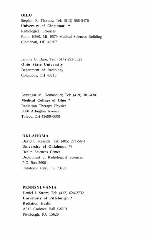
OHIOStephen R. Thomas; Tel: (513) 558-5476University of Cincinnati *Radiological SciencesRoom E560, ML 0579 Medical Sciences BuildingCincinnati, OH 45267
Jerome G. Dare; Tel: (614) 293-8323Ohio State UniversityDepartment of RadiologyColumbus, OH 43210
Ayyangar M. Komanduri; Tel: (419) 381-4301Medical College of Ohio *Radiation Therapy Physics3000 Arlington AvenueToledo, OH 43699-0008
OKLAHOMADavid E. Raeside; Tel: (405) 271-5641University of Oklahoma *†Health Sciences CenterDepartment of Radiological SciencesP.O. Box 26901Oklahoma City, OK 73190
PENNSYLVANIADaniel J. Strom; Tel: (412) 624-2732University of Pittsburgh *Radiation HealthA512 Crabtree Hall GSPHPittsburgh, PA 15626
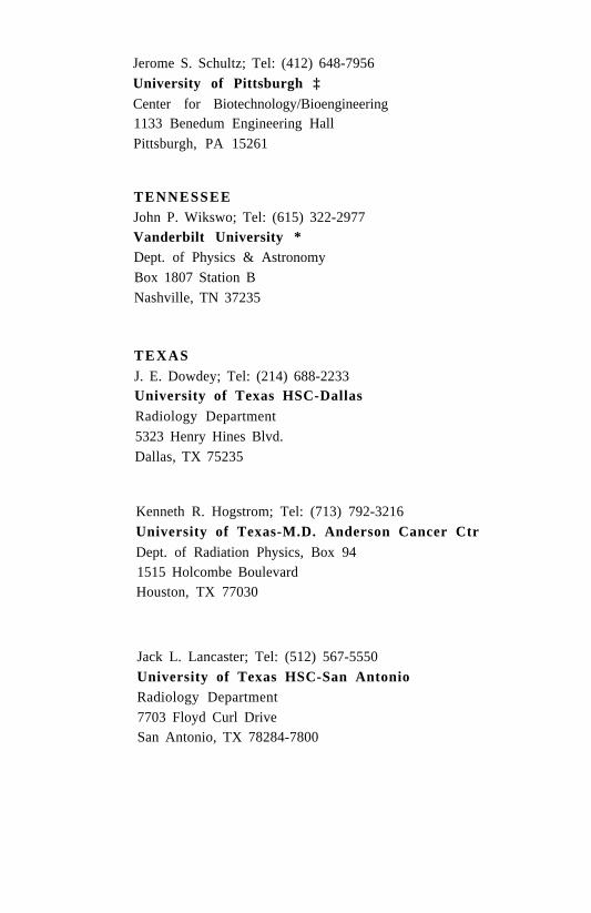
Jerome S. Schultz; Tel: (412) 648-7956University of Pittsburgh ‡Center for Biotechnology/Bioengineering1133 Benedum Engineering HallPittsburgh, PA 15261
TENNESSEEJohn P. Wikswo; Tel: (615) 322-2977Vanderbilt University *Dept. of Physics & AstronomyBox 1807 Station BNashville, TN 37235
TEXASJ. E. Dowdey; Tel: (214) 688-2233University of Texas HSC-DallasRadiology Department5323 Henry Hines Blvd.Dallas, TX 75235
Kenneth R. Hogstrom; Tel: (713) 792-3216University of Texas-M.D. Anderson Cancer CtrDept. of Radiation Physics, Box 941515 Holcombe BoulevardHouston, TX 77030
Jack L. Lancaster; Tel: (512) 567-5550University of Texas HSC-San AntonioRadiology Department7703 Floyd Curl DriveSan Antonio, TX 78284-7800

WISCONSINPaul M. DeLuca, Jr.; Tel: (608) 262-2173University of Wisconsin *†Department of Medical Physics1300 University Avenue, 1530 MSCMadison, WI 53706
C A N A D ABrendan McLean; Tel: (403) 492-8771Cross Cancer Institute *†Department of Medical Physics11560 University AvenueEdmonton, Alberta T6G 1Z2, Canada
Larry John Filipow; Tel: (403) 492-4094University of Alberta HospitalDept. of Radiation & Diag. ImagingEdmonton, AB T6G 2B7, Canada
Brian C. Wilson; Tel: (416) 387-9495McMaster UniversityOntario Cancer FoundationDepartment of Physics711 Concession StreetHamilton, ON L8V 1C3, Canada
Ervin B. Podgorsak; Tel: (514) 934-8052McGill University *Department of Medical PhysicsLivingston Hall, Room L5-1091650 Avenue Des CedresMontreal, PQ H3G 1A4, Canada
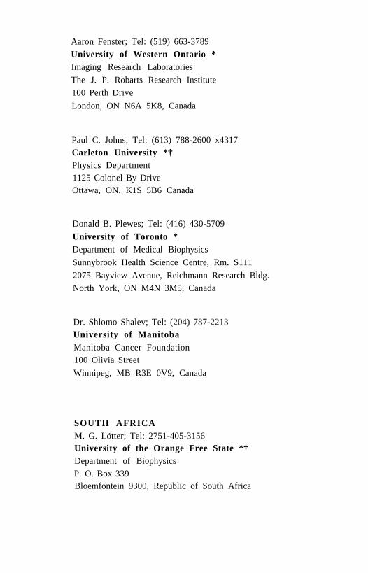
Aaron Fenster; Tel: (519) 663-3789University of Western Ontario *Imaging Research LaboratoriesThe J. P. Robarts Research Institute100 Perth DriveLondon, ON N6A 5K8, Canada
Paul C. Johns; Tel: (613) 788-2600 x4317Carleton University *†Physics Department1125 Colonel By DriveOttawa, ON, K1S 5B6 Canada
Donald B. Plewes; Tel: (416) 430-5709University of Toronto *Department of Medical BiophysicsSunnybrook Health Science Centre, Rm. S1112075 Bayview Avenue, Reichmann Research Bldg.North York, ON M4N 3M5, Canada
Dr. Shlomo Shalev; Tel: (204) 787-2213University of ManitobaManitoba Cancer Foundation100 Olivia StreetWinnipeg, MB R3E 0V9, Canada
SOUTH AFRICAM. G. Lötter; Tel: 2751-405-3156University of the Orange Free State *†Department of BiophysicsP. O. Box 339Bloemfontein 9300, Republic of South Africa
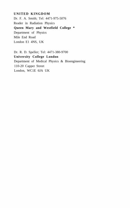
U N I T E D K I N G D O MDr. F. A. Smith; Tel: 4471-975-5076Reader in Radiation PhysicsQueen Mary and Westfield College *Department of PhysicsMile End RoadLondon E1 4NS, UK
Dr. R. D. Speller; Tel: 4471-380-9700University College LondonDepartment of Medical Physics & Bioengineering110-20 Capper StreetLondon, WC1E 6JA UK