acaaddemmiicc sccii eennccess international journal of ... · evaluation of macroelements (ca, na,...
TRANSCRIPT
Research Article
BOTANICAL STUDY, DNA FINGERPRINTING, NUTRITIONAL VALUES AND CERTAIN PROXIMATES OF ENTADA RHEEDII SPRENG
MONA M. OKBA1, FATHY M. SOLIMAN1, KADRIYA S. EL DEEB1 AND MIRIAM F. YOUSIF1, 2*
1Pharmacognosy Department, Faculty of Pharmacy, Cairo University, Kasr El-Ainy, 11562, Cairo, Egypt, 2Pharmacognosy Department, Faculty of Pharmaceutical Sciences and Pharmaceutical Industries, Future University, Al Tagamoa Al Khames, 11528, New Cairo, Egypt.
Email:[email protected]
Received: 26 May 2013, Revised and Accepted: 17 Jun 2013
ABSTRACT
Entada rheediiSpreng. (Family Fabaceae) seeds are used in Egypt in folk medicine.Macro- and micro-morphological characters of E. rheedii Spreng.seeds imported from India together with that of roots, stems and leaves cultivated in Egypt were presented with the aim of their identification in entire and powdered forms. Cultivation in Egypt gave a climbing plant instead of the huge fruiting trees bordering the Indian Ocean. Plant materials were fixed, freehand sectioned and stained with Safranin. Leaves are compound, bipinnate; their blades exhibit rubiaceous and few ranunculaceous stomata, non-glandular trichomes and dorsiventral mesophyll. The stem upper part is cylindrical with six ridges. The stem has relatively wide pith surrounded by open collateral vascular bundles. Study of Deoxyribonucleic acid (DNA) fingerprinting, total seed protein profiling, nutritional value and certain proximates was carried out in order to contribute to the identification of the plant material.A total of 53 different fragments have been recorded in DNA fingerprinting, produced mainly by (A-19) primer, showing 15 bands ranging from 1.337 Kbp to 0.356 Kbp,Eight bands were recorded in seed total protein banding profile of E. rheedii Spreng seed with molecular weights ranging from 52 to 9 KDa.High levels of Glutamic acid and Phenyl alanine amino acids were determined.Moisture, carbohydrates and ash percentages were 7.35, 16.47 and 2.83 respectively. Evaluation of macroelements (Ca, Na, K and P); and microelement (Fe) revealed that potassium (K) and phosphorous (P) occupied the highest positions (1264 and 1240 mg/100 g seeds respectively) among the macroelements, whereas the micro element Iron (Fe) level was 3.3 mg/100 g seeds.
Keywords: Entada; Macro- and micro-morphology; Nutritive-value; Amino acids; Macro and micro elements; Proximate analysis.
INTRODUCTION
E. rheedii Spreng. (= E. pursaetha DC, common name Match Box Bean [1]) family Fabaceae are widely grown in tropical and subtropical countries bordering the Indian Ocean [2]. Seeds of E. rheediiSpreng.are known in the Egyptian herbal market as anti-rheumatic, anti-inflammatory, dietary supplement and in weight gain preparations [3,4]. Therefore, it was necessary to identify this species together with a trial for its cultivation. Nothing was traced in the available literature concerning its botanical characters. It was, thus, necessary to study the macro- and micro-morphological features of the roots, stems, leaves and seeds. Further identification was achieved by a DNA and seed protein profiling, nutritional value and certain proximates.
MATERIAL AND METHODS
Plant material for botanical study
Samples of seeds imported from India were purchased from Egyptian market. Samples of cultivated roots, stems and leaves of E. rheediiSpreng.were obtained in July 2010 from plants grown in the Experimental Station of Medicinal Plants, Pharmacognosy Department, Faculty of Pharmacy, Cairo University, Giza. Voucher sample (herbarium no. 14.4.2013.1) was deposited at the Museum of the Pharmacognosy Department, Faculty of Pharmacy, Cairo University.Fresh samples of root, stem and leaves, as well as, samples preserved in ethanol 70 % containing 5 % glycerin were examined. Seeds and air-dried roots, stems and leaves were finely powdered and packed in dark-colored, tightly closed containers for examination of powdered organs.
Material for molecular investigation
Plant material for molecular investigations
Samples of fresh leaves were stored at -70°C, freeze-dried and ground to a fine powder prior to DNA isolation.
Buffers and agarose gel
Extraction buffer and agarose gel were prepared according to [5].
Primers: Five primers were used for randomly amplified polymorphic DNA (RAPD) analysis obtained from Operon Technologies Inc., Almeda,
California, USA with the following sequence: A-10: 5'-GTGATCGCAG-3'; A-17: 5'-GACCGCTTGT-3'; A-18: 5'-AGGTGACCGT-3'; A-19: 5'-CAAACGTCGG-3'; G-18: 5'-GGCTCATGTG-3'.
Molecular weight marker: 100 bp ladder, Promega Corporation, Madisson, USA.
Apparatus: DNA thermocycler (Hybaid PCR Express) used for amplification of DNA, agarose gel electrophoresis tool (Biorad Wide Mini Sub Cell) used for separation of RAPD fragments according to size and ultraviolet Polaroid camera used for visualization of RAPD fragments.
Material for seed protein profiling
A- Solution for gel preparation
1- Acrylamide/Bisacrylamide: 30% stock solution.
2- Stacking gel buffer: Tris-HCl solution (pH 6.6)
3- Separating gel: Tris-HCl solution (pH 8.8)
4- Running buffer: Tris base, glycine and sodium dodecyl sulphate in distilled water
5-Sample buffer: Tris borate solution (pH 8.2)
6- Staining solution: 0.1 g coomassie blue powder in a solution composed of 400 ml distilled water, 70 ml acetic acid, 200 ml ethanol and 60 ml trichloroacetic acid
7- Destaining solution: 15 ml ethanol, 50 ml glacial acetic acid and 300 ml distilled water
B- Protein molecular weight marker
A promega mixture (Heidelberg, Germany) containing nine proteins with the indicated molecular weights Mwt= 9, 13, 19, 26, 39, 52, 80, 125 and 175 KDa
Plant material for determination of certain pharmacopoeial constants and the macro and micro elements content
Seeds were coarsely powdered and packed in dark-colored, tightly closed containers for determination of certain pharmacopoeial constants[6] and macro and micro elements [7].
International Journal of Pharmacy and Pharmaceutical Sciences
ISSN- 0975-1491 Vol 5, Suppl 3, 2013
AAccaaddeemmiicc SScciieenncceess
Yousif et al. Int J Pharm Pharm Sci, Vol 5, Suppl 3, 311-329
312
Method for botanical study
Different organs were separately imbedded in paraffin and serial transverse sections, 10 -15 μm thick, were performed with a manual microtome and stained with Safranin and Fast Green [8]. Sections were mounted in synthetic balsam. Epidermis was obtained by scraping fresh material. Transverse sections, epidermises and powder of different organs were examined by light microscope. Dimensions of different elements were determined using Reichert Austria Mikrometer 2 mm langGeteilt in 200 Teile C. Reichert Wien.
Methods for molecular investigations
DNA extraction and quantification, amplification of RAPD markers and analysis of RAPD data were performed according to [5]
Methods for protein extraction
Seed protein was analyzed using continuous polyacrylamide gel electrophoresis in vertical slab apparatus in the presence of sodium dodecyl sulphate (SDS-PAGE). A 0.1 g of the mature seed was powdered and mixed with 1 ml sample buffer. The slurry was centrifuged at 6000 rpm for 10 min. The supernatant was used immediately for electrophoresis.
Gel preparation
The gel used for separation was prepared and poured between the glass plates immediately after adding ammonium persulphate. After polymerization of the gel (11 cm), the stacking gel (3 cm) was poured into the glass plates and then comb was inserted to form sample wells.
Gel electrophoresis [9]
1-Sample preparation
Protein extracts were diluted with sample buffer (1:3 v/v), then 500 µl of 10% SDS were added followed by 25 µl of mercatoethanol. They were then placed in a boiling water bath for 5 min and 5 µl of bromophenol was added as a tracking dye. Ten µl of the sample was loaded and 10 µl of marker protein mixture was used as standard.
2-Running conditions
Runs were carried out at a constant voltage of 200 volt. Usual runs took approximately 120 min.
3-Gel staining and destaining
The chromatograms were stained with excess of coomassie brilliant blue stain R-250 for about one hr. After gel staining, the gel was transferred to destaining solution to remove excess stain.
Methods for determination of certain pharmacopoeial constants
The moisture, ash and crude fiber were determined in the seeds according to the Egyptian Pharmacopoeia [6].
Determination of the macro and micro elements
The percentage of sodium (Na), phosphorous(P), potassium (K), calcium (Ca), magnesium (Mg) and iron (Fe) were determined in the seeds according to Cotennine[7].
Methods for amino acids analysis
The total protein was extracted from the seeds using 50mM Tris-HCl PH (7.5) and then acid hydrolysis of the protein was carried out [10]. One ml 6N HCI (HCI Suprapure® from Merck)was mixed with one mg protein in a hydrolysis tube. The solution was frozen in a mixture of dry ice/ethanol in a test tube. The tube was evacuated with a vacuum pump and sealed using gas-burner. The sealed tube was placed in an oven at 110 °C for 72 hours for hydrolysis, and then cooled down in an ice-bath. The solution was centrifuged to precipitate insoluble components. The supernatant was evaporated at 40°C in a rotary evaporator. The residue was then dissolved in a diluting buffer.
Apparatus and conditions Eppendrof – Germany. LC3000 Amino acid analyzer.
Conditions: flow rate: 0.2 ml/min, pressure of buffer from 0 to 50 bar, pressure of reagent from 0 to 150 bar, reaction temperature 123 °C
RESULTS AND DISCUSSION
Cultivation of E. rheedii Spreng.seeds in Egypt failed to give huge fruiting trees as in countries bordering the Indian Ocean (Fig.1)[11]. It gave a climbing plant (Fig.2) through four years of cultivation, which may be attributed to the difference in the environmental conditions. The seeds germinate only when their coats are mechanically abraded(Fig 3:B). It was found that this is the only method that induces water imbibition.
Macro- and micro morphologically of roots, stems, leaves, as well as, imported seeds of E. rheedii Spreng. were presented (Figs.2-13). Dimensions in microns of the different elements of the studied organs are recorded (Table 1). Numerical values [12] of the leaves are recorded (Table 2).
Table 1: Dimensions of different elements of the organs under investigation in microns of E. rheedii Spreng.
Parameter measured Size (min-max) µm
L W or D H
Root Cork 18,37,50 45,61,77 8,15,21 Vessels 32,54,73 Starch granules 4,7,9 Calcium oxalate prism 9,15,18 8,13,16 Stem Epidermis 25,50,82 7,12,15 8,10,13 Stomata 12,15,21 7,10,16 Non-glandular trichomes 186,236,280 10,15,31 Vessels 11,15,23 Calcium oxalate prism 9,16,25 8,10,16 Leaves Upper epidermis 26,35,47 15,20,26 7,9,15 Lower epidermis 15,19,26 31,45,68 7,13,15 Neural epidermis 66,81,108 25,31,37 Stomata 20,23,26 7,10,12 Palisade 21,26,31 12,16,21 Non-glandular trichomes 151,200,265 7,10,12 Vessels 12,18,25 Tracheids 61,72,82 5,7,10 Calcium oxalate prism 20,27,36 15,18,21

Yousif et al. Int J Pharm Pharm Sci, Vol 5, Suppl 3, 311-329
313
Seed Palisade like epidermis 11,13,17 169,185,192 Hypodermis 29,33,38 38,40,42 Sclereids 42,65,83 stellate aerenchyma 76,88,107 cotyledon parenchyma 18,20,22 47,56,66 Starch granules 4,7,10
Fig. 1: Macromorphology of E. rheedii Spreng. tree and pod [11]
A.Photograph of E. rheedii L. fruiting trees, B. Photographs of E. rheedii L. pods. g.p., giant pod; stk., stalk
Yousif et al. Int J Pharm Pharm Sci, Vol 5, Suppl 3, 311-329
314
Table 2: Numerical values of E. rheedii Spreng.leaves
Numerical values* E. rheedii L. leaves Stomatal index -Upper epidermis 10.31 -Lower epidermis 11.15 Stomatal number -Upper epidermis 100-120 -Lower epidermis 100-120 Palisade ratio 4.25--5 Vein islet number 9--13 Veinlet termination number 16.75--23.75
*average of ten determinations
Macro-morphology
The root: (Fig.2: A)
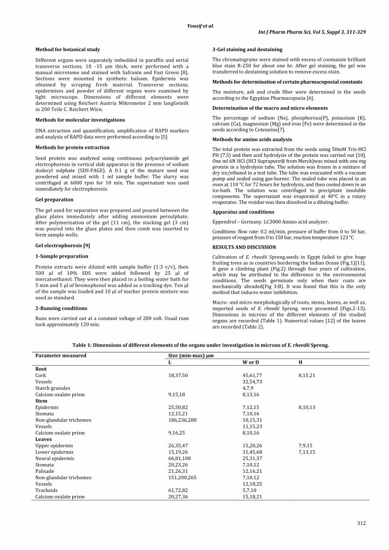
The plant consists of a distinct tap-root system. The main root attains up to 25-40 cm in length, and 0.3-0.7 cm in diameter, bearing tiny fibrous rootlets. It has dark brown color, with longitudinally wrinkled surface, and breaks with fibrous fracture. The root has faint odor and characteristic taste.
The stem: (Fig.2: B)
The main stem reaches up to 3 m height. The stem lower parts are green, more or less cylindrical, solid and flexible, about 0.5 cm in diameter. The stem upper parts are more or less cylindrical with six ridges and dark greyish-brown color. The surface of the stem is rough with numerous whitish spots. It is tough with a fibrous fracture. The stem is green with faint odor and a characteristic taste.
The leaf: (Fig. 2: C)
The leaves are compound, bipinnate. The main axis of the leaf terminates in a bifid tendril. The leaflets are 2 (rarely 3) pairs oppositely arranged on a nearly cylindrical rachis. The lamina is ovate to oval with entire margin, emarginate apex, with a very short petiolule (1-2mm L and about 0.8 mm D) and with asymmetric base. The upper surface is dark-green and the lower one is light-green; both surfaces are apparently glabrous. The midrib is slightly prominent on the lower side. The venation is pinnately reticulate; the veins leave the midrib at an angle of about 45°. The lamina measures 1.1-6.5 cm in length and 0.6-2.6 cm in width. The petiole is green in color 2.7-4.2 cm in length and 1 mm in diameter. The petiole shows a swollen pulvinus at its base, which measures 3mm in diameter. The leaflets have faint odor and a characteristic taste.
Fig. 2: Macromorphology of cultivated E. rheedii Spreng.
A. The root, B. The climbing plant, C.The leaf.c.p., climbing plant; l., leaflet; m.r., main root; p., petiole; pe.,petiolule; r.t, rootlet; ra., rachis; t., tendril.
Yousif et al. Int J Pharm Pharm Sci, Vol 5, Suppl 3, 311-329
315
The seed: (Fig.3)
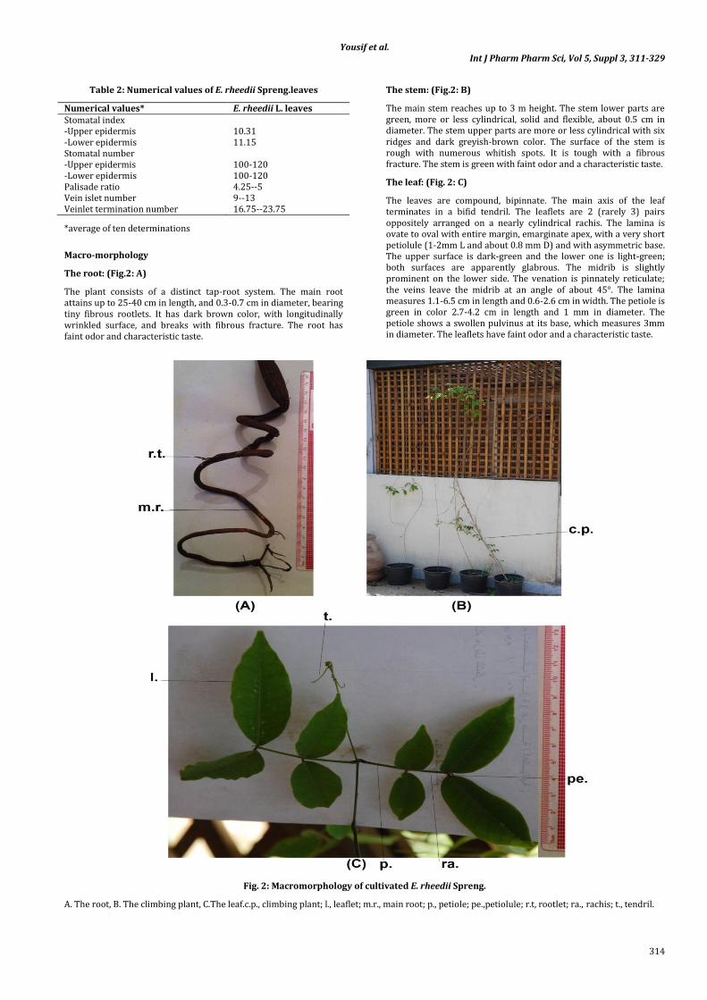
The seeds are round disc shaped or elliptically flat, smooth dark brown to black in color and glossy with fine striations. It measures 3-6.5 cm W, 3-7.8 cm L and 0.7-2.5 cm thickness. They have thick and hard seed coats. One hundred seeds weight 2.46 to 3.06 kg.
The hilum appears as a pale scar beside the micropyle in a small depression on the margin. The seed is derived from anatropous ovule.
The seed, as in most other leguminous plants [13], is essentially devoid of endosperm (exalbuminous) and consists of a seed coat that surrounds a large, well developed central embryo. The embryo completely fills the seed. It is yellowish- white, straight, with the radicle directed towards the micropyle. It consists of two concavo-convex cotyledons, the concave surfaces inclosing a rather large cavity (Fig 3:B) which explains the ability of the seed to float upon water [14]. The seeds have a slight characteristic odor when crushed and a characteristic taste.
Micro-morphology
The root: (Fig.4)
A transverse section (T.S.) in the root is almost circular in outline. It is formed of cork, a very narrow cortex, and a pericycle. The pericycle is parenchymatous and interrupted by groups of lignified fibers. The vascular bundles show well developed secondary thickening and occupy three forth the diameter of the root.
The cork is formed of 4-6 rows of brown radially arranged elongated thick-walled polygonal or tabular cells with suberized lignified walls.
The cortex is narrow composed of 4-7 rows of parenchymatous cells. They are nearly oval in outline with narrow intercellular spaces. Starch granules are occasionally present. They are rounded oval, mostly simple, with neither visible hila nor striations. Idioblasts with yellowish-orange content that give reddish-color with potassium hydroxide are scattered in the cortex. Prisms of calcium oxalate are scattered in the parenchymatous cells, especially, those surrounding the cortical fibers forming a crystal sheath. The fibers are long having acute tips, mostly narrow lumen some have moderate lumen, with straight walls.
The pericycle is formed of 4-6 rows of parenchymatous cells interrupted by small patches of lignified fibers. The cells are oval or rounded with narrow intercellular spaces and contain starch granules, calcium oxalate prisms similar to those in the cortex.
The fibers are long with acute tips with moderately thick straight lignified walls. They are accompanied by crystal sheath.
The vascular system is formed of a continuous ring of vascular tissue. It is traversed by uni- or biseriate medullary rays.
The phloem is formed of sieve tubes, companion cells, and phloem parenchyma containing occasional starch granules and prisms of calcium oxalate similar to those of the cortex.
The cambium is formed of 4-6 rows of cambiform cells. The xylem is formed of a continuous ring of lignified vessels, wood fibers and parenchyma. The vessels are mostly solitary or arranged in small groups. They are radially arranged with wide boardered pitted thickening,
Fig. 3: Macromorphology of E. rheedii Spreng. seed
Yousif et al. Int J Pharm Pharm Sci, Vol 5, Suppl 3, 311-329
316
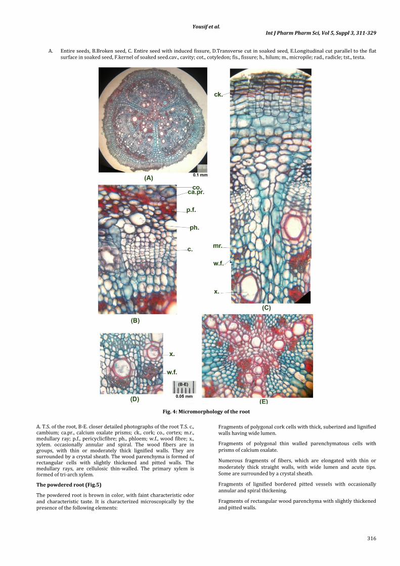
A. Entire seeds, B.Broken seed, C. Entire seed with induced fissure, D.Transverse cut in soaked seed, E.Longitudinal cut parallel to the flat surface in soaked seed, F.kernel of soaked seed.cav., cavity; cot., cotyledon; fis., fissure; h., hilum; m., micropile; rad., radicle; tst., testa.
Fig. 4: Micromorphology of the root
A. T.S. of the root, B-E. closer detailed photographs of the root T.S. c., cambium; ca.pr., calcium oxalate prisms; ck., cork; co., cortex; m.r., medullary ray; p.f., pericyclicfibre; ph., phloem; w.f., wood fibre; x., xylem. occasionally annular and spiral. The wood fibers are in groups, with thin or moderately thick lignified walls. They are surrounded by a crystal sheath. The wood parenchyma is formed of rectangular cells with slightly thickened and pitted walls. The medullary rays, are cellulosic thin-walled. The primary xylem is formed of tri-arch xylem.
The powdered root (Fig.5)
The powdered root is brown in color, with faint characteristic odor and characteristic taste. It is characterized microscopically by the presence of the following elements:
Fragments of polygonal cork cells with thick, suberized and lignified walls having wide lumen.
Fragments of polygonal thin walled parenchymatous cells with prisms of calcium oxalate.
Numerous fragments of fibers, which are elongated with thin or moderately thick straight walls, with wide lumen and acute tips. Some are surrounded by a crystal sheath.
Fragments of lignified bordered pitted vessels with occasionally annular and spiral thickening.
Fragments of rectangular wood parenchyma with slightly thickened and pitted walls.
Yousif et al. Int J Pharm Pharm Sci, Vol 5, Suppl 3, 311-329
317
Rounded and oval starch granules, mostly simple, with neither visible hila nor striations.
The stem: (Figs. 6&7)
Lower part (Figs. 6: A& 7:A&B)
A transverse section in the stem lower part is almost circular. It shows an epidermis, a narrow cortex followed by pericycle formed of continuous band of lignified fibers. The vascular tissue is composed of a continuous collateral ring surrounding a wide parenchymatous pith which occupies about 2/3 the diameter of the stem.
The epidermis consists of polygonal axially elongated cells with straight anticlinal walls covered with thin smooth cuticle. Stomata are mostly of rubiaceous type, few are ranunculaceous type. Trichomes are common, being non-glandular, unicellular and
multicellular (up to 5 cells), conical, with thin walls and wide lumen and covered with smooth cuticle. The trichomes are usually curved near the apex, occasionally straight
The cortex is formed of 6-9 rows of thin walled rounded or oval parenchyma. Scattered in the cortex are numerous cells with yellowish-brown content which acquires a dark-reddish brown color with potassium hydroxide. Starch granules are occasionally present, being rounded mostly simple rarely compound of two, with neither visible hila nor striations. Prisms of calcium oxalate are present. The innermost layer of the cortex is formed of cells containing prisms of calcium oxalate especially those abutting on the pericyclic fibers.
The pericycle is formed of a continuous band of lignified fibers. The fibers have pointed or rounded tips, wide lumen, some fibers have undulating walls. They are surrounded by a crystal sheath.
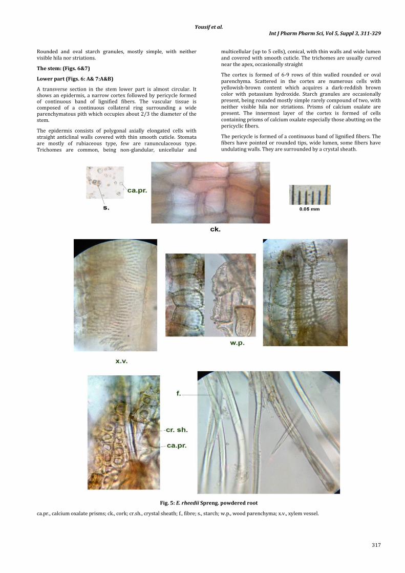
Fig. 5: E. rheedii Spreng. powdered root
ca.pr., calcium oxalate prisms; ck., cork; cr.sh., crystal sheath; f., fibre; s., starch; w.p., wood parenchyma; x.v., xylem vessel.

Yousif et al. Int J Pharm Pharm Sci, Vol 5, Suppl 3, 311-329
318
The vascular system is formed of a continuous ring of vascular tissue traversed, by uni- or biseriate medullary rays. The phloem is formed of sieve tubes, companion cells, phloem parenchyma and few bast fibers which are arranged in isolated groups. The fibers are accompanied with a crystal sheath. The phloem parenchyma contains occasional starch granules similar to those of the cortex. Medullary ray cells are thin-walled, slightly radially elongated and contain few starch granules. The cambium is formed of 5 - 10 rows of cambiform cells. The xylem is formed of lignified vessels, wood fibers and parenchyma. It is traversed by uniseriate lignified medullary rays. The vessels are radial1y arranged with pitted and spiral thickening. The wood fibers are abundant, with fairly thick walls. Tracheids are occasionally with pitted and reticulate thickening. The wood parenchyma is rectangular with pitted walls and free of content.
Upper part (Fig. 6: B&C)
A transverse section in the stem upper part is nearly circular with six wings.
The cork is formed of several rows (10-12) of radially arranged thick-walled, polygonal, brown, tabular cells having suberised and lignified walls.
The cortex is formed of 1-2 rows of parenchyma cells with scattered yellowish-brown content which acquires a dark-reddish brown color with potassium hydroxide. Starch granules are occasionally present, similar to that mentioned above. Prisms of calcium oxalate are present. The innermost layer of the cortex is formed of cells
containing prisms of calcium oxalate especially those abutting on the pericyclic fibers.The pericycle resembles that of the stem lower part.
The vascular system is similar to that of the stem lower part but it is discontinuous ring.
The pith is formed of rounded or oval parenchymatous cells with thin cellulosic or slightly lignified pitted walls. Starch granules, calcium oxalate prisms, as well as, idioblasts with yellowish-orange content giving reddish-color with potassium hydroxide are scattered in the pith.
Tendril: (Fig. 6: C&D)
A transverse section in the tendril is more or less rounded in outline with two ridges.
The epidermis consists of one row of cubical cells covered with smooth cuticle. The cortex is formed of 4-7 rows of thin walled rounded or oval parenchymatous cells with the same content as described in stem different parts. The innermost layer of the cortex forming crystal sheath.
The pericycle is formed of a continuous band of fibers (2-6 rows). The first rows of pericyclicfibres are with lignified walls and wide lumen.
The vascular system is nearly similar to that of stem upper part.
The pith is formed of rounded or oval parenchymatous cells with thin cellulosic walls. They have same content as in stem different parts.
Fig. 6: Micromorphology of E. rheedii Spreng. stem and tendril
A.T.S. in the stem lower part, B.T.S. in the stem upper part, C.T.S. in the tendril, D. closer detailed layers in tendril T.S. c., cambium; co., cortex; cr.sh., crystal sheath; ep., epidermis; m.r., medullary ray; p., pericycle; ph., phloem; pi., pith; x., xylem.
Yousif et al. Int J Pharm Pharm Sci, Vol 5, Suppl 3, 311-329
319
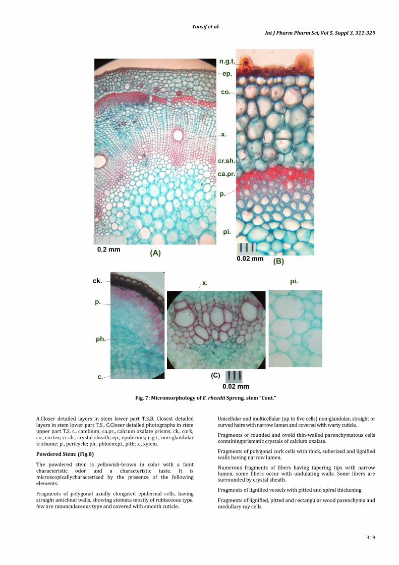
Fig. 7: Micromorphology of E. rheedii Spreng. stem “Cont.”
A.Closer detailed layers in stem lower part T.S,B. Closest detailed layers in stem lower part T.S., C.Closer detailed photographs in stem upper part T.S. c., cambium; ca.pr., calcium oxalate prisms; ck., cork; co., cortex; cr.sh., crystal sheath; ep., epidermis; n.g.t., non-glandular trichome; p., pericycle; ph., phloem;pi., pith; x., xylem.
Powdered Stem: (Fig.8)
The powdered stem is yellowish-brown in color with a faint characteristic odor and a characteristic taste. It is microscopicallycharacterized by the presence of the following elements:
Fragments of polygonal axially elongated epidermal cells, having straight anticlinal walls, showing stomata mostly of rubiaceous type, few are ranunculaceous type and covered with smooth cuticle.
Unicellular and multicellular (up to five cells) non-glandular, straight or curved hairs with narrow lumen and covered with warty cuticle.
Fragments of rounded and ovoid thin-walled parenchymatous cells containingprismatic crystals of calcium oxalate.
Fragments of polygonal cork cells with thick, suberized and lignified walls having narrow lumen.
Numerous fragments of fibers having tapering tips with narrow lumen, some fibers occur with undulating walls. Some fibers are surrounded by crystal sheath.
Fragments of lignified vessels with pitted and spiral thickening.
Fragments of lignified, pitted and rectangular wood parenchyma and medullary ray cells.
Yousif et al. Int J Pharm Pharm Sci, Vol 5, Suppl 3, 311-329
320
Fragments of wide parencymatous cells of the pith contain starch granules and calcium oxalate prisms.
Occasional yellowish-brown cells, which acquire reddish-brown color with potassium hydroxide.
Starch granules, mostly simple, rarely compound of two, with neither visible hila nor striations.
The leaf: (Figs. 9&10)
A transverse section in the leaflet (Fig. 9:A-C), shows an upper and a lower epidermises, a dorsiventral mesophyll. The midrib shows collateral crescent-shaped vascular bundle surrounded by continuous ring of pericyclic fibers.
The upper and lower epidermises (Fig.9:F&G) are formed of polygonal cells with slightly wavy anticlinal walls, more wavy in
lower epidermal cells, and covered with a smooth cuticle. The cells covering the midrib, upper and lower neural, (Fig.9:D&E) are axially elongated having straight anticlinal walls.
Trichomes are non-glandular, multi- or unicellular, conical with blunt or acute apices, covered with smooth cuticle. Fallen hairs show rounded cicatrices, each surrounded by 8 radiating cells. Trichomes of the lower epidermis are similar to those of the upper epidermis but rare.
Stomata are mostly of rubiaceous type, few are ranunculaceous type.
The mesophyll is dorsiventral, the palisade is not continuous over the midrib region, and formed of one row of columnar cells with straight walls. Spongy tissue is formed of 2-4 rows.
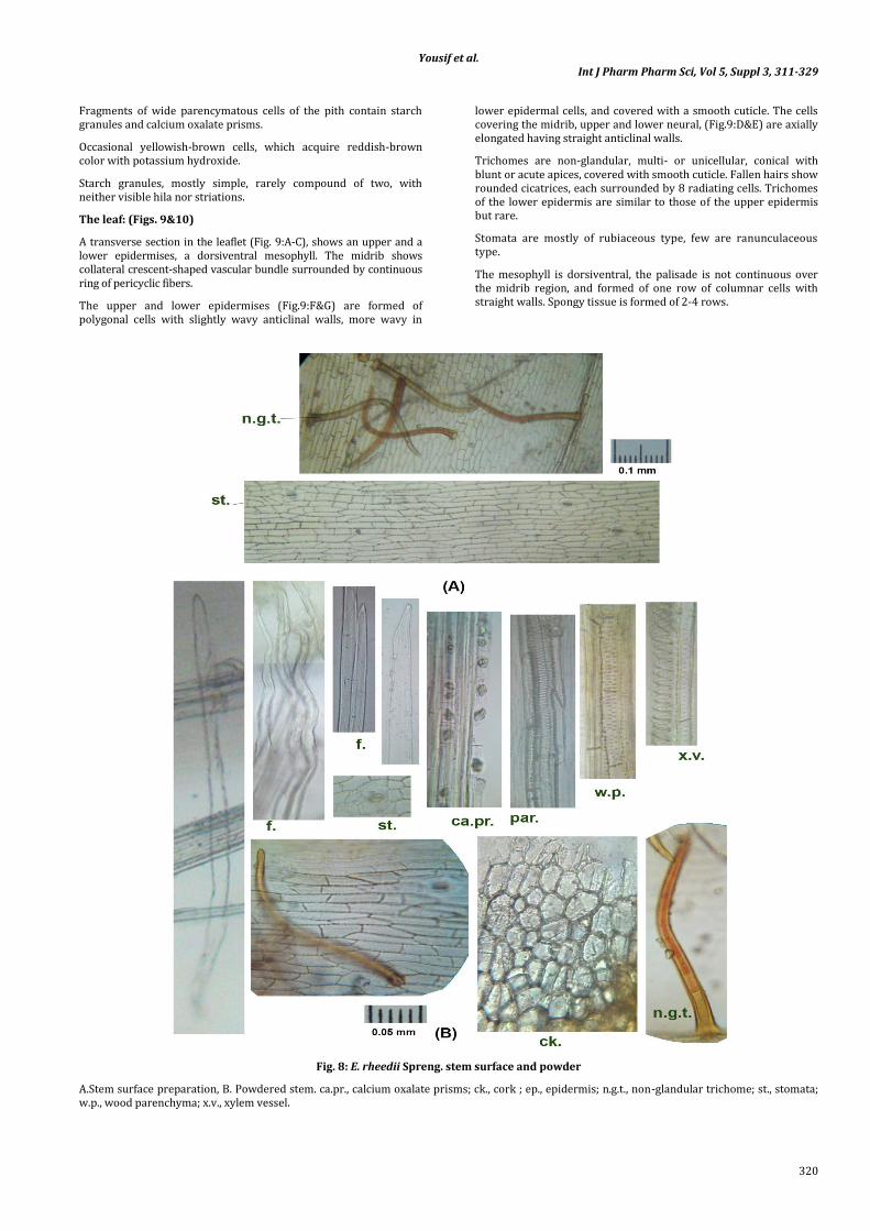
Fig. 8: E. rheedii Spreng. stem surface and powder
A.Stem surface preparation, B. Powdered stem. ca.pr., calcium oxalate prisms; ck., cork ; ep., epidermis; n.g.t., non-glandular trichome; st., stomata; w.p., wood parenchyma; x.v., xylem vessel.

Yousif et al. Int J Pharm Pharm Sci, Vol 5, Suppl 3, 311-329
321
Of thin walled parenchymatous cells with intercellular spaces. It is traversed by narrow vascular strands.
The cortical tissue of the midrib region consists of several rows of parenchyma abutting the upper and lower epidermises. The innermost layer of the cortical tissue (endodermis) is formed of slightly tangentially elongated cells containing prisms of calcium oxalate, forming crystal sheath.
The vascular system is enclosed by a continuous ring of pericyclic fibers, 1 - 8 fibers thick. The phloem is formed of sieve tubes, companion cells, phloem parenchyma and numerous patches of bast fibers. The xylem is formed of spiral, reticulate and pitted lignified xylem vessels and non-lignified wood parenchyma and wood fibers with uni- and biserriate medullary rays.
The Petiole (Fig.10: A-C)
A transverse section in the petiole is more or less rounded in outline with two ridges. It shows hairy epidermis, narrow parenchymatous cortex. The innermost layer of the cortical tissue (endodermis) is formed of slightly tangentially elongated cells containing prisms of calcium oxalate forming crystal sheath. The vascular system shows a narrow central ring of vascular tissue surrounding comparatively wide parenchymatous pith. Two small vasocentric vascular bundles are situated on the upper side of the petiole above the vascular ring and beneath the ridges. The main central ring, as well as, the two lateral bundles is surrounded by continuous bands of pericyclic fibers.
The Petiolule (Fig.10: D&E )
A transverse section in the petiolule is almost circular in outline. The epidermis is hairy on the upper side. The cortex is wide formed of
parenchymatous cell. The innermost layer of the cortical tissue (endodermis) is formed of slightly tangentially elongated cells containing calcium oxalate prisms forming crystal sheath. The vascular system is nearly similar to that of the midrib of the leaf.
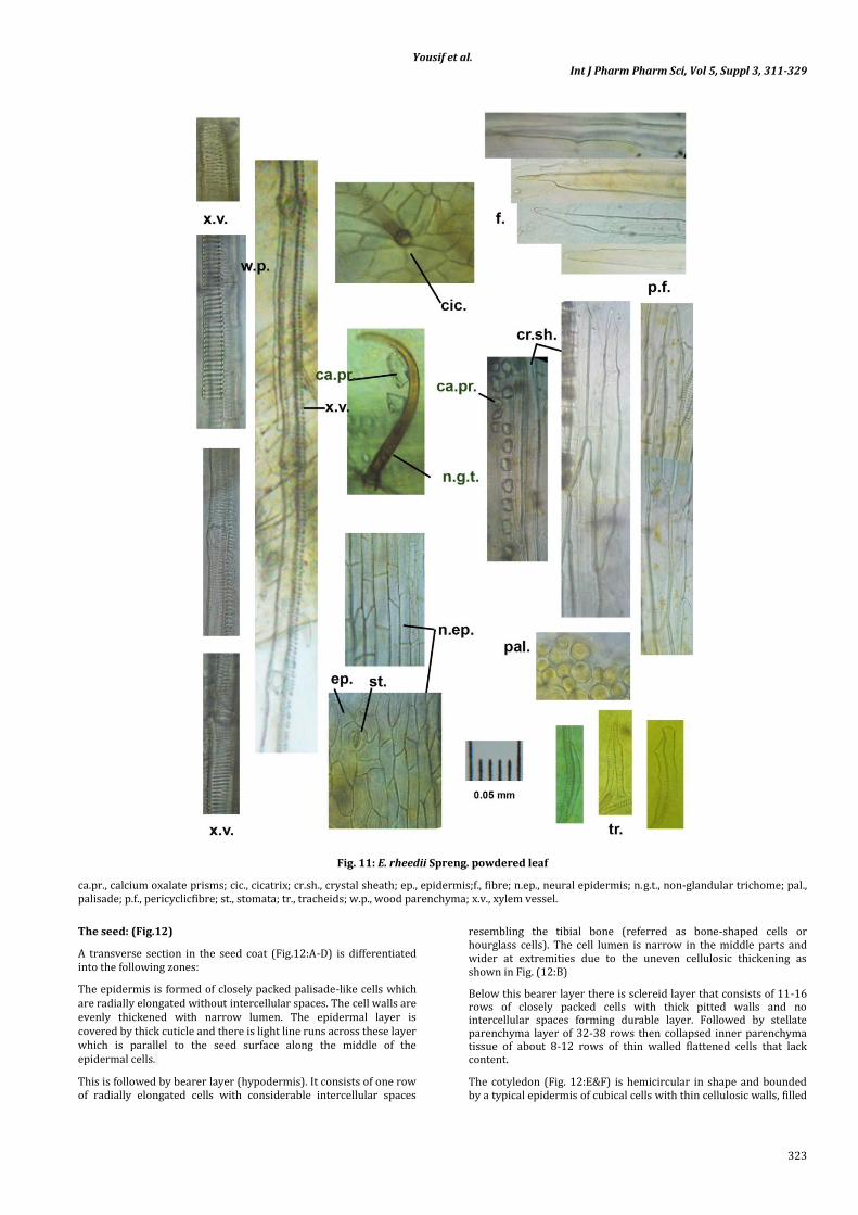
Powdered leaf (Fig.11)
The powdered leaf is light green in color with faint characteristic odor and characteristic taste. It is characterized microscopically by the following elements:
Fragments of the upper, lower and neural epidermises of polygonal cells, showing stomata mostly of rubiaceous type, few are ranunculaceous type and cicatrices of fallen trichomes.
Few non-glandular, unicellular and multicellular (up to five cells) trichomes covered with smooth cuticle mainly on the upper epidermis on the midrib and veins.
Fragments of palisade cells.
Fragments of broken narrow spiral and pitted lignified xylem vessels.
Fragments of pericyclic fibers accompanied by crystal sheath. The fibers are elongated with thin, lignified straight or undulating walls, wide lumina and blunt or pointed apices.
Numerous prisms of calcium oxalate.
Occasional fragments of tracheids or tracheidial fibers.
Fragments showing parenchymatous cells containing starch granules, mostly simple with neither visible hila nor striations
Fig. 9: Micromorphology of E. rheedii Spreng. leaflet
A. T.S. in the leaflet, B. Closer detailed photographs of T.S. in midrib, C.Closer detailed layers of T.S. in lamina, D. Upper surface of neural epidermis, E. Lower surface of neural epidermis, F. Upper surface of lamina, G.Lower surface of lamina. cic., cicatrix; cr.sh., crystal sheath; cu., cuticle; ep., epidermis; l.ep., lower epidermis; n.ep., neural epidermis; n.g.t., non-glandular trichome; pal., palisade; par., parenchyma; p., pericycle; ph., phloem; sp., spongy tissue; st., stomata; u.ep., upper epidermis; x., xylem

Yousif et al. Int J Pharm Pharm Sci, Vol 5, Suppl 3, 311-329
322
Fig. 10: Micromorphology of E. rheedii Spreng. petiole and petiolule
A.T.S. in the petiole, B. closer detailed layers in petiole T.S. corner, C. closer detailed layers in petiole T.S. side, D. T.S. in the petiolule, E. closer detailed layers in petiolule T.S. center .c., cambium; ca.pr., calcium oxalate prisms; co., cortex; cr.sh., crystal sheath; ep., epidermis; n.g.t., non-glandular trichome; par., parenchyma; p., pericycle; ph., phloem; x., xylem.
Yousif et al. Int J Pharm Pharm Sci, Vol 5, Suppl 3, 311-329
323
Fig. 11: E. rheedii Spreng. powdered leaf
ca.pr., calcium oxalate prisms; cic., cicatrix; cr.sh., crystal sheath; ep., epidermis;f., fibre; n.ep., neural epidermis; n.g.t., non-glandular trichome; pal., palisade; p.f., pericyclicfibre; st., stomata; tr., tracheids; w.p., wood parenchyma; x.v., xylem vessel.
The seed: (Fig.12)
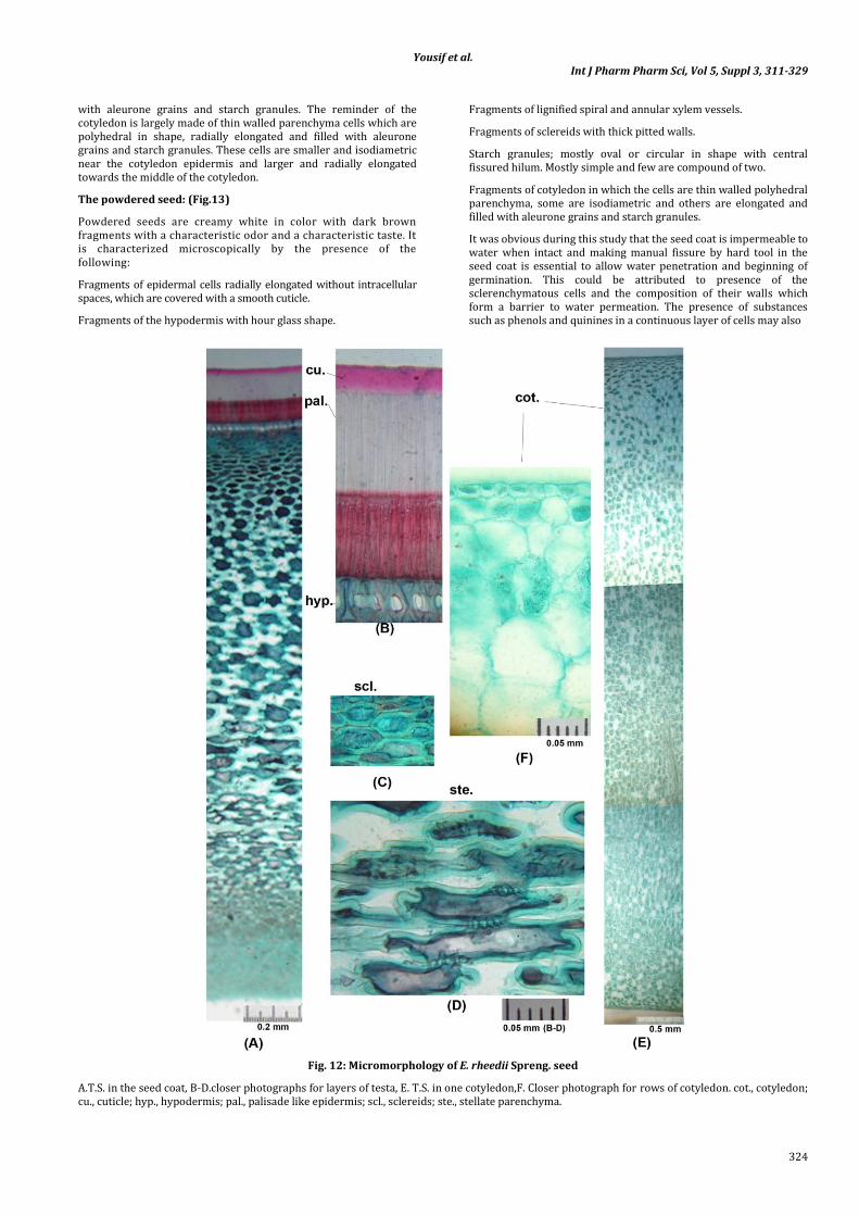
A transverse section in the seed coat (Fig.12:A-D) is differentiated into the following zones:
The epidermis is formed of closely packed palisade-like cells which are radially elongated without intercellular spaces. The cell walls are evenly thickened with narrow lumen. The epidermal layer is covered by thick cuticle and there is light line runs across these layer which is parallel to the seed surface along the middle of the epidermal cells.
This is followed by bearer layer (hypodermis). It consists of one row of radially elongated cells with considerable intercellular spaces
resembling the tibial bone (referred as bone-shaped cells or hourglass cells). The cell lumen is narrow in the middle parts and wider at extremities due to the uneven cellulosic thickening as shown in Fig. (12:B)
Below this bearer layer there is sclereid layer that consists of 11-16 rows of closely packed cells with thick pitted walls and no intercellular spaces forming durable layer. Followed by stellate parenchyma layer of 32-38 rows then collapsed inner parenchyma tissue of about 8-12 rows of thin walled flattened cells that lack content.
The cotyledon (Fig. 12:E&F) is hemicircular in shape and bounded by a typical epidermis of cubical cells with thin cellulosic walls, filled
Yousif et al. Int J Pharm Pharm Sci, Vol 5, Suppl 3, 311-329
324
with aleurone grains and starch granules. The reminder of the cotyledon is largely made of thin walled parenchyma cells which are polyhedral in shape, radially elongated and filled with aleurone grains and starch granules. These cells are smaller and isodiametric near the cotyledon epidermis and larger and radially elongated towards the middle of the cotyledon.
The powdered seed: (Fig.13)
Powdered seeds are creamy white in color with dark brown fragments with a characteristic odor and a characteristic taste. It is characterized microscopically by the presence of the following:
Fragments of epidermal cells radially elongated without intracellular spaces, which are covered with a smooth cuticle.
Fragments of the hypodermis with hour glass shape.
Fragments of lignified spiral and annular xylem vessels.
Fragments of sclereids with thick pitted walls.
Starch granules; mostly oval or circular in shape with central fissured hilum. Mostly simple and few are compound of two.
Fragments of cotyledon in which the cells are thin walled polyhedral parenchyma, some are isodiametric and others are elongated and filled with aleurone grains and starch granules.
It was obvious during this study that the seed coat is impermeable to water when intact and making manual fissure by hard tool in the seed coat is essential to allow water penetration and beginning of germination. This could be attributed to presence of the sclerenchymatous cells and the composition of their walls which form a barrier to water permeation. The presence of substances such as phenols and quinines in a continuous layer of cells may also
Fig. 12: Micromorphology of E. rheedii Spreng. seed
A.T.S. in the seed coat, B-D.closer photographs for layers of testa, E. T.S. in one cotyledon,F. Closer photograph for rows of cotyledon. cot., cotyledon; cu., cuticle; hyp., hypodermis; pal., palisade like epidermis; scl., sclereids; ste., stellate parenchyma.

Yousif et al. Int J Pharm Pharm Sci, Vol 5, Suppl 3, 311-329
325
Fig. 13: E. rheedii Spreng. powdered seed
A. palisade cells (side view), B. palisade cells (surface view), C. hypodermis (side view), D. hypodermis (surface view), E. starch granules, F. sclereids, G. cotyledon.cot., cotyledon; hyp., hypodermis; pal., palisade like epidermis; s., starch; scl., sclereids.
contribute to this barrier [15-18]. Isolation and identification of three phenolic compounds from the seed coat is under publication[19].
Analysis of RAPD data
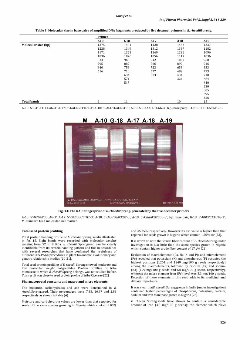
The banding profile produced by five decamer primers used in RAPD analysis of E. rheediiSpreng.is illustrated in Fig. (14).
The RAPD electrophoretic profile of the DNA sample amplified with the five decamer primers showed distinguishable bands and
generated 53 fragment pattern. The distribution of these bands is illustrated in Table (3).
The DNA was amplified using five decamer primers to reveal RAPD fragments. Each of the five primers successfully directed the amplification of a genom-specific fingerprint of DNA fragment; all amplifications were found to be prolific.
A total of 53 different fragments have been recorded, produced mainly by one of the five primers (A-19) showing 15 bands ranging from 1.337 Kbp to 0.356 Kbp, while primer A-10 produced only 8 bands.
Yousif et al. Int J Pharm Pharm Sci, Vol 5, Suppl 3, 311-329
326
Table 3: Molecular size in base pairs of amplified DNA fragments produced by five decamer primers in E. rheediiSpreng.
Primer A10 G18 A17 A18 A19 Molecular size (bp) 1375 1401 1428 1483 1337
1228 1349 1312 1337 1182 1171 1263 1149 1228 1096 1036 1076 1056 1117 1036 833 960 942 1007 960 795 882 866 890 916 640 758 723 658 833 616 710 577 482 773 634 373 434 710 571 324 664 515 640 530 505 395 356
Total bands 8 11 9 10 15
A-10: 5'-GTGATCGCAG-3'; A-17: 5'-GACCGCTTGT-3'; A-18: 5'-AGGTGACCGT-3'; A-19: 5'-CAAACGTCGG-3'; b.p., base pair; G-18: 5'-GGCTCATGTG-3'.
Fig. 14: The RAPD fingerprint of E. rheediiSpreng. generated by the five decamer primers
A-10: 5'-GTGATCGCAG-3'; A-17: 5'-GACCGCTTGT-3'; A-18: 5'-AGGTGACCGT-3'; A-19: 5'-CAAACGTCGG-3'; b.p., base pair; G-18: 5'-GGCTCATGTG-3'; M: standard DNA molecular size marker.
Total seed protein profiling
Total protein banding profile of E. rheedii Spreng seedis illustrated in fig. 15. Eight bands were recorded with molecular weights ranging from 52 to 9 KDa. E. rheedii Sprengseed can be clearly identifiable from its protein banding pattern and this in accordance with several researches that have confirmed the usefulness of different SDS-PAGE procedures in plant taxonomic, evolutionary and genetic relationship studies [20-21].
Total seed protein profiling of E. rheedii Spreng showed moderate and low molecular weight polypeptides. Protein profiling of tribe mimoseae to which E. rheedii Spreng belongs, was not studied before. This result was close to seed protein profile of tribe Cicereae [22].
Pharmacopoeial constants and macro and micro elements
The moisture, carbohydrates and ash were determined in E. rheediiSpreng.seed. Their percentages were 7.35, 16.47 and 2.83 respectively as shown in table (4).
Moisture and carbohydrate values are lower than that reported for seeds of the same species growing in Nigeria which contain 9.00%
and 45.35%, respectively. However its ash value is higher than that reported for seeds grown in Nigeria which contain 1.20% ash[23].
It is worth to note that crude fiber content of E. rheediiSpreng.under investigation is just little than the same species grown in Nigeria which contain higher crude fiber content of 17 g% [23].
Evaluation of macroelements (Ca, Na, K and P); and microelement (Fe) revealed that potassium (K) and phosphorous (P) occupied the highest positions (1264 and 1240 mg/100 g seeds respectively) among the macroelements; followed by calcium (Ca) and sodium (Na) (199 mg/100 g seeds and 68 mg/100 g seeds, respectively), whereas the micro element Iron (Fe) level was 3.3 mg/100 g seeds. Detection of these elements in this seed adds to its medicinal and dietary importance.
It was clear thatE. rheedii Spreng.grown in India (under investigation) contained higher percentages of phosphorous, potassium, calcium, sodium and iron than those grown in Nigeria [23].
E. rheedii Spreng.seeds have shown to contain a considerable amount of iron (3.3 mg/100 g seeds); the element which plays

Yousif et al. Int J Pharm Pharm Sci, Vol 5, Suppl 3, 311-329
327
essential role for the treatment of iron deficiency anemia; the most common type of anemia in Egypt [24]. Egypt’s 2005 Demographic Health Survey said that one quarter of adolescent males and one third of females in Egypt were anemic, according to the UN Children’s Fund (UNICEF). Anemia, according to the World Health Organization is a condition in which the number of red blood cells or their oxygen-carrying capacity is insufficient. It is mainly caused by iron deficiency.
Iron deficiency anemia affects a quarter of the world’s population and is widespread in most developing countries [25].Nutritional anemia occur when one or more of the nutrients; that are necessary
for red blood cells production are deficient; shortages of iron, vitamin B12 and folic acid are the most common causes of nutritional anemia [26].
Determination of macro and micro elements in this study had important relation to the potential use of E. rheedii Spreng.in medicine. Calcium, magnesium and phosphorus contribute to prevention of osteoporosis and are needed for tooth formation and cell growth. Sodium and potassium, together with calcium are crucial for muscle contractions. Besides, the two former elements help in maintenance of the normal heart rhythm and body water balance [27].
Fig. 15: Photograph of polyacrylamide gel illustrating electrophoretic band profiles of total protein extracted from E. rheedii Spreng seeds.
E, Entada rheedii; M, molecular weight marker
Table 4: Determination of some nutrients and macro and micro elements in E. rheedii Spreng. seeds
Nutrient g%* Elements mg%* Moisture 7.35 P 1240 Total Carbohydrates 16.47 Ca 199 Fiber 13.77 K 1264 Ash 2.83 Na 68 Fe 3.3
*Average of three determinations
Amino acid content
From Table (5) it could be concluded that the total percentage of the amino acids is 23.499 g/100 g seeds. The main essential amino acid was leucine (2.597 g/100 g seeds). This was followed by phenylalanine (2.116 g/100 g seeds) and lysine (1.776 g/100 g seeds). The lowest percentage of the essential amino acids was therionine (0.782 g/100 g seeds) followed by methionine, isoleucine, valine and tyrosine (1.004 9/100 g seeds, 1.033 g/100 g seeds, 1.145 g/100 g seeds and 1.655 g/100 g seeds, respectively).
Phenyl alanine amino acid is effective in painful arthritis [28].Its relatively high level in E. rheedii Spreng. seed may explain its use in folk medicine in treatment of arthritis and other rheumatoid diseases [3,4].
Glutamic acid (3.737 g/100 g seeds) was the main non-essential amino acid followed by aspartic acid and alanine (1.831 g/100 g
seeds and 1.695 g/100 g seeds respectively). The lowest percentage of the nonessential amino acids was both histidine and glycine (0.649 g/100 g seeds and 0.704 g/100 g seeds); followed by proline, arginine and serine (0.726 g/100 g seeds, 0.827 g/100 g seeds and 1.222 g/100 g seeds respectively).
Glutamic acid is important in the metabolism of sugars and fats and is used in treatment of ulcers [28]. The recent use of different Entada seed species as antiulcerogenic drugs [29] may be explained by the detected relatively high level of Glutamic acid.
It could be also concluded from table (5) that 100g of E. rheedii Spreng.seeds contained the recommended daily dietary allowance of the essential amino-acids according to [30]. It is worth mentioning that 100 g of E. rheedii Spreng.seeds afford the daily dietary requirements of the essential amino acids for infant, child and adult like wise.

Yousif et al. Int J Pharm Pharm Sci, Vol 5, Suppl 3, 311-329
328
Table 5: Percentage of amino acids in E. rheedii Spreng seeds
Amino Acid g/100gm Daily dietary allowance of the amino acids (g /Kg of body weight) [30] Infant Child (10 t0 12 years) Adult
Essential amino acids isoleucine 1.033 0.083 0.028 0.012 leucine 2.597 0.135 0.042 0.016 lysine 1.776 0.099 0.044 0.012 methionine 1.004 0.049* 0.022* 0.010* phenyl alanine 2.116 0.141** 0.022** 0.016** threonine 0.782 0.068 0.028 0.008 tyrosine 1.655 0.021 0.004 0.003 valine 1.145 0.092 0.025 0.014
Non-essential amino acids arginine 0.827 Proline 0.726 serine 1.222 glutamic acid 3.737 glycine 0.704 alanine 1.695 aspartic 1.831 histidine 0.649
Ammonium ion 1.507 Total essential amino acids 12.108 Total non-essential aminoacids 11.391 Total determined amino acids 23.499 Total determined amino acids and ammonia 25.006
*Methionine+cystine
**Phenyl alanine+tyrosine
CONCLUSION
The following macro- and micro morphological characteristics were established after the analysis of the studied plant material. The presence of the below described elements is considered to be useful for the botanical identification of the E. rheedii Spreng together with the numerical values of the leaf and the dimensions of the different organs.
Macromorphological characteristics
Compound, bipinnate leaf, terminates in a bifid tendril. The leaflets are 2 (rarely 3) pairs oppositely arranged. The lamina is ovate to oval with entire margin, emarginate apex, with a very short petiolule and with asymmetric base. Venation is pinnate reticulate. The stem lower parts are more or less cylindrical, while the upper parts are more or less cylindrical with six ridges.
Micromorphological characteristics
Leaflet with dorsiventral mesophyll. Stomata mostly of rubiaceous type, few are ranunculaceous in leaflets and stem. Unicellular and multicellular (up to five cells) non-glandular, straight or curved trichomes with narrow lumen and covered with warty cuticle in leaflets and stem. Sclereids with thick pitted walls in seed.Crystal sheath with the specified dimensions in different organs.
Botanical study presented in the current study appears to be an excellent method for identification. The botanical study is easy to conduct, rapid, sensitive, adaptable to simple laboratory conditions and inexpensive.
The analysis of RAPD data can select the use of primers A-19 for selective discrimination.
The SDS-PAGE procedure using total protein samples is not suitable to detect the seed storage protein polymorphism within varieties or within populations of legumes. This is because all varieties or cultivars related to same species showed identical number of bands with similar mobility [22]. So our SDS-PAGE results gave fingerprint profile for the studied species, without being able to define the accession of E. rheedii Spreng. Specific types of protein extracts (glutellins, albumins and isozymes) will be required to differentiate between accessions of E. rheedii Spreng, not the total protein extract [22].
Nutritional value and certain proximates determined can be used to confirm the identity of the plant.
ACKNOWLEDGEMENT
The Authors would like to thank Dr. Mohammed El-Gibali Senior botanist for his help in plant identification. The authors are grateful to Dr. SaharAbd El Tawab, Professor of Botany and Genetics, Faculty of Girls, Ain Shams University for revising the interpretation of the molecular investigations.
RERERENCES
1. http://keys.trin.org.au/key-server/data/0e0f0504-0103-430d-8004-60d07080d04/media/Html/taxon/Entada_rheedii.htm[ Accessed on May 3, 2013]
2. Langran X, Dezhao C, Xiangyun Z, Puhua H, Zhi W, Dianxianget. al.: Fabaceae (Leguminosae). 2009. p.p. 51-52 [Through http://hua.huh.harvard.edu/china/mss/volume10/FOC_10_Fabaceae_all.pdf] Flora of China on line volumes www.efloras.org
3. Burkill MH.The Useful Plants of West Tropical Africa, Families J-L, 1995; Vol. 3. Royal Botanic Garden Kew, pp: 229-230. [Through Tibiri A, Rakotonandrasana O, Nacoulma GO and Banzouzi JT: Radical scavenging activity, phenolic content and cytotoxicity of bark and leaves extracts of EntadaafricanaGuill. and Perr. (Mimosaceae).J. Biol. Sci. 2007; 7 (6): 959-963].
4. Diallo D, Paulsen BS, Liljeback TH and Michaelsen TE: Polysaccharides from the roots of EntadaafricanaGuill. etPerr., Mimosaceae, with complement fixing activity. J.Ethn. Pharmacol. 2001; 74(2): 159-171.
5. Doyle JJ and Doyle JL: A rapid DNA isolation procedure for small quantities of fresh leaf tissue. Phytochem.Bull. 1987, 19: 11-15.
6. Egyptian Pharmacopoeia: 4thed, central Administration of Pharmaceutical Affairs (CAPA), Ministry of Health and Population, Cairo, Egypt, 2005.
7. Cotennie, A.: Soil and plant testing as a basis of fertilizer recommendation. FAO Soils Bull.1980; 38 (2): 94-96.
8. Ruzin S: Plant microtechnique and microscopy. Ed. Oxford University Press New York 1999, p. 1-125.
9. Stegmann H, Burgermeister W, Shah AA, Francksen HE and Krogerrecklenfort E: Gel electrophoresis and isoelectric focusing, (PANTA- PHOR and MONO-PHOR Manual). Institut
Yousif et al. Int J Pharm Pharm Sci, Vol 5, Suppl 3, 311-329
329
fur Biochemie, BiologischeBundesanstlt, Messeweg 11, D-3300 Braunschweig West Germany. 1988
10. Spackman DH, Stein WH and Moore S: Automatic recording apparatus for use in chromatography of amino acid. Annal.Chem. 1958; 30:1190-1206.
11. (11)https://www.google.com.eg/search?q=entada+against+virus+viral+activity&hl=en&source=lnms&tbm=isch&sa=X&ei=h8NIUZ6BEcOXtQbN0oCABw&ved=0CAcQ_AUoAQ&biw=1366&bih=667#hl=en&tbm=isch&sa=1&q=entada+&oq=entada+&gs_l=img.3..0l5j0i24l5.39182.40860.0.42020.28.11.0.0.0.5.224.1116.6j4j1.11.0...0.0...1c.1.7.img.DE0zMhPBDDo&bav=on.2,or.r_qf.&bvm=bv.44011176,d.Yms&fp=5a638c3b016ad8b7&biw=1366&bih=667[ Accessed on May 3, 2013]
12. Wallis TE: Practical pharmacognosy. 6th Ed., J and A Churchill LTD, London; 1953.p.139-140.
13. (13)Rendle, A.B.: The classification of the flowering plants.vol II, Cambridge, 1959, p. 348-365
14. (14) http://www.beachbeans.com/descriptions.html 15. (15) Corner EJ: The Leguminosae seeds. Phytomorph.1951;
11:17-50. 16. (16) Spurny: The imbibitions process in seed ecology,
proceedings of the 19th Easter school in Agricultural Sciences. University of Nottingham, Butter Worths, London; 1973.p. 367-389..
17. Werker E and Fahn A: Seed Anatomy of Pancratium Species from Three Different Habitats. Bot. Gaz. 1975; 13:639-643.
18. Werker E, Marbache I, and Mayer AM: Relation between the Anatomy of the Testa, Water Permeability and the Presence of Phenolics in the Genus Pisum. Ann. Bot. 1979; 43:765-771
19. Mona MO, FathyMS ,Kadriya SE and Miriam FY: A quantitative study ofoptimal Entadasaponins and phenolics extraction methods and their spermistatic activity. 2013; Under publication
20. Ladizinsky G and Hymowitz T: Seed protein electrophoresis in taxonomic and evolutionary studies. Theor.And Appl. Genet., 1979; 51: 145-151.
21. Virinhos F and Murry DR: The seed Proteins of Chickpea: Comparative studies of Cicerarietinum, C. reticulatum and C. echinospermum. PI. Syst. Evol.1983;142: 11-22.
22. Valizadeh M: Seed storage protein of grain legumes grown in Iran, using SDS-PAGE. J. Agric. Sci. Tecnol. 2001; 3: 287-292
23. Oderinde RA and Ajayi IA: Composition of Entada pursaetha seed and seed oil grown in Nigeria. Rivist.Ital. delleSostanze-Grasse. 1998; 75(10):457-459.
24. Kamal M, Youssef E, Mohamed MA and Mehani MM: Incidence of iron deficiency anemia among girls of nursing faculty, Assuit University as affected by their nutritional status. Edited by the 5th International Conference and Exhibition, International Center for Research and Consultation (Food Quality 2003) Part II 899-918
25. Siddiqui MS and Siddiqui MK: Public health significance of iron deficiency anemia. Pak. Arm. For. Med. J.2008; 3(9).
26. Dudek SG: Nutrition handbook for nursing practice. Lippincott-Raven, Philadelphia; 1997.
27. Stillwell E:Vitamins and minerals. A comprehensive guide to understanding your daily diet and nutrition.PCR Publishing Ltd., London; 2002.
28. James FB and Phyllis AB: Prescription for nutritional healing. Avery Publishing Group, Garden City Park, New York, 2nd Ed., 1997, p. 38-41
29. Ramakrishna D, Pavan KK, Mukkanti K and Abedulla K.K.: Antiulcer Activity of The Seeds of EntadaPhaseoloides. Pharmacologyonline2008; 3:93-99
30. Al-Zohairy AD:Human nutrition.Mosul University, Iraq, 2nd Ed., 1992. p. 116-118.