abstracts menton 2016 word
TRANSCRIPT

1
6th International Workshop
on PET in Lymphoma
Palais de l’Europe. Menton, France
September 20 -21, 2016
Poster session

2
A1. Interim PET after 2 versus 4 cycles of immunotherapy in a Phase 2 randomized trial in diffuse large B-cell lymphoma (DLBCL) patients Authors Mónica Coronado (1), Marc Simó (2), Pilar Sarandeses (3), Montserrat Cortés (4), Ana Cristina Hernández (3), Amanda Rotger (5), Eva González-Barça (6), Carlos García-Grande (3), Dolores Caballero (7), Xavier Setoain (8) Author's affiliation (1) Hospital Universitario La Paz, (2) Hospital Universitari Vall d´Hebron, (3) Hospital Universitario 12 de Octubre, (4) IDI PET-TAC Hospital de Bellvitge, (5) Hospital General Universitario Gregorio Marañón, (6) Hospital Duran I Reynals, (7) Hospital Universitario de Salamanca, Hospital, (8) Clínic Barcelona 8. On behalf of the Spanish Group of Lymphoma (Grupo Español de Linfomas y Trasplante de Médula Ósea, GELTAMO). Text Background. Interim PET/CT (iPET) is a promising tool for tailoring risk-adapted therapeutic strategy in DLBLC patients. However, the optimal time to realize iPET is not well defined. iPET after 2 and 4 cycles was compared in a phase II randomized clinical trial comparing standard immunotherapy and a modified RCHOP regimen in young patients with unfavourable IPI score DLBCL (NCT01848132). Methods. A blinded, prospective, centralized review in real time of PET/CT images was realized by the GELTAMO PET network. For each patient, images of basal (PET0), iPET after 2 cycles (PET2), iPET after 4 cycles (PET4) and final PET after completion of chemotherapy (PET 6) were centrally reviewed. PET2 and PET4 were interpreted visually based on Deauville criteria (considering scores 4 and 5 as positive), and semiquantitavely (positive PET2 when ∆SUVmax≤66% and positive PET4 when ∆SUVmax≤70%). Semiquantitative analysis defined final positive or negative interim PET result. A positive PET4 result determined dropped out from trial. PET2 and PET4 final result were compared in order to see PET2 ability to predict PET4 result. Concordance between central review and on-site evaluation of PET2 and PET4 was also analyzed. Results. Ninety-nine patients underwent PET0, PET2 and PET4. In 90/99 (91%) patients PET2 and PET4 result were concordant (63 negative and 27 positive). PET2 result was predictive of PET4 (p<0.001). Nine patients (9%) had discordant PET2 and PET4 result; 8 of them being PET2 (+) and PET4 (-); 1 patient PET2 (-) and PET4 (+). PET6 was revised in 8 discordant PET2 - PET4 cases. PET6 was negative in 7 patients with PET2(+) and PET4 (-). PET6 was positive in 1 patient with PET2 (+) and PET4 (-). One patient with PET2 (-) and PET4 (+) did no undergo PET6.Concordance between central review and on-site evaluation: we found 63% concordant and 37% discordant PET2 studies; we found 93% concordant and 7% discordant PET4 results (most of them PET positive by local evaluation and negative in the central review). Concordance between on-site and central review was poor for PET2 and good for PET4 (Kappa= 0,34, p<0.001 and Kappa= 0,8, p<0.001, respectively). Conclusions. PET2 is predictive of PET4 in DLBCL patients treated with standard/modified R-CHOP regimen. In discordant PET2 and PET4 cases, PET4 is preferable to define final metabolic response. Concordance between on-site and central evaluation is better in PET4 than in PET2. Email [email protected]

3
A2. Interim 18F-FDG PET/CT in aggressive lymphoma: assessment of interobserver agreement and impact of baseline PET or CT scan and disease localization Authors Burggraaff CN (1), Cornelisse AC (1), Zijlstra JM (1), Hoekstra OS (2), De Vet HCW (3), Lugtenburg PJ (4), Celik F (5), Huijbregts JE (6), Arens AIJ (7), De Keizer B (6) Author's affiliation On behalf of the HOVON imaging working group. (1) Department of Hematology, VU university medical center, Amsterdam, The Netherlands, (2) Department of Radiology and Nuclear Medicine, VU university medical center, Amsterdam, The Netherlands, (3) Department of Epidemiology and Biostatistics, VU university medical center, Amsterdam, The Netherlands, (4) Department of Hematology, Erasmus MC Cancer Institute, Rotterdam, The Netherlands, (5) Department of Radiology and Nuclear Medicine, Deventer Ziekenhuis, Deventer, The Netherlands, (6) Department of Radiology and Nuclear Medicine, university medical center Utrecht, Utrecht, The Netherlands, (7) Department of Radiology and Nuclear Medicine, Radboud university medical center, Nijmegen, The Netherlands Text Aim: To assess the interobserver agreement of interim 18F-FDG PET/CT (iPET) using the Deauville 5-point scale (DS) in patients with aggressive lymphoma as a function of the baseline imaging modality (18F-FDG PET/CT or CT only) and of the nodal- and extra nodal localizations with residual 18F-FDG uptake. Methods: iPET scans were obtained from the HOVON84 study, an international multicenter randomized controlled trial, conducted from 2007-2012. Patients received R-CHOP immuno-chemotherapy and were randomized to receive rituximab intensification in the first 4 cycles or not. iPET scans were made after 4 cycles and were scored according to the DS by 2 reviewers from a pool of 10 experienced nuclear medicine physicians. DS results were dichotomized (DS 1-3: ‘negative’, DS 4-5: ‘positive’). Besides Cohen’s kappa we calculated the agreement separately for the positive and the negative scores, expressed as positive agreement (PA) and negative agreement (NA). Definition of PA: given one reviewer scores positive, the probability that another reviewer scores positive as well (de Vet et al. BMJ 2013) Results: 488 iPET scans were reviewed centrally. Kappa for iPET interobserver agreement was 0.65, NA was 92.1% and PA was 72.5%. Kappa and PA of the 369 iPET cases with a baseline 18F-FDG PET/CT available were 0.68 and 75.6% versus 0.52 and 60.9% in the 119 iPET cases with only a baseline CT scan for reference (p=0.10 and 0.16, respectively). Kappa for the first 100 reviewed scans was 0.38, while it was 0.72 for the last 100 scans, corresponding NA were 86.6 and 94.4% and PA were 51.2 and 76.9% respectively. NA for nodal localizations ranged from 98.3 (spleen and mesenterial) to 100%. Highest PA was observed for iliac left nodes (75%) and lowest PA for hilar left nodes (33.3%). NA in extra nodal sites ranged from 97.8 (liver) to 100%. Highest PA was observed for lung (80%) and lowest PA for skeletal localizations (30%). Conclusion: Availability of a baseline 18F-FDG PET/CT results in a better interobserver agreement of iPET, although not statistically significant. Despite reasonable kappas, the relatively low PA scores indicate that observer agreement needs to be improved before iPET can be used in treatment escalation trials in aggressive lymphoma. Although central reviewers were all experienced, a learning curve between first and last 100 reviewed scans was observed. These data indicate the importance of further training and research on iPET interpretation. Email [email protected]

4
A3 Semi-quantitative FDG-PET Assessment of Interim and End of Treatment Response in the CALGB/Alliance Phase III Study of R-CHOP vs Dose-Adjusted EPOCH-R in Untreated DLBCL: A Blinded, Arm-Pooled, Initial Analysis of Correlation with Outcomes. Authors Schoder, H1, Hall, N2, Knopp, MV2, Zhang, J2, Pitcher, B3, Jung, S-H3, Kelloff, G4, Dehdashti, F5, Kurdziel, K4, Lamonica, D6, Esposito, G7, Manzone, T8, Higley, H 9, Schwartz, L10, Bartlett, N5, Cheson, B7 Leonard, J11 and Wilson, W4. Author's affiliation 1Memorial Sloan Kettering Cancer Center, 2Ohio State University Imaging Core Laboratory, 3Duke University School of Medicine, 4National Cancer Institute, 5Washington University School of Medicine, 6Roswell Park Cancer Institute, 7Georgetown University Hospital, 8Christiana Care Health System, 9CCS Associates, 10Columbia University School of Medicine, 11Cornell University School of Medicine. Text CALGB 50303 is a phase III randomized comparison of R-CHOP, the de facto standard for DLBCL, to dose adjusted EPOCH-R. Considerable evidence supports the association of post-therapy FDG-PET results with outcome in lymphoma patients. Specifically, FDG uptake has been a significant early predictor of residual or recurrent disease and disease progression, as well as PFS and overall survival (OS). Although FDG-PET/CT imaging is increasingly being used in oncology clinical trials, there are as yet no standardized criteria for PET imaging or established procedures detailing transmission, storage, quality assurance, and analysis of PET images. Consequently, an imaging substudy CALGB 580603, was incorporated into the parent phase III trial to collect standardized images in a multicenter setting and assess the value of semi-quantitative methods in predicting response to treatment in this study cohort. Between 2007 and 2013, 536 subjects were randomized to the two treatment arms. Of these, 171 underwent standardized baseline FDG-PET scans at 29 different institutions, with 161 undergoing per protocol cycle 2 PET scans, and 151 a PET scan at cycle 6 end of treatment. PET images were read by two central reviewers with responses scored using both Deauville (5-PS) and, IWG+PET criteria, as well as assessed for ∆SUV% change at both time points. Data are still blinded as to treatment arm, per the requirement of the CALGB DSMB, until all imaged subjects have reached 3 years of follow-up and sufficient events have taken place to inform the primary endpoint. However, an initial arm-pooled analysis was performed on the imaged subjects who have at least reached at least 2 years of follow-up. With a median time to follow-up of 3.9 years (maximum 6.9 years), Kaplan-Meier plots of PET-negative (Deauville 1-3) vs PET-positive (Deauville 4-5) subjects showed significant prolongation of EFS (p=0.0322) and marginal significance of OS (p=0.0689) for PET- subjects when imaging was performed at the end of cycle 2 treatment. Cycle 6 correlation with progression and survival had p-values of 0.0544 and 0.0316, respectively between PET end of treatment responders and non-responders. A comparison between the performance of Deauville criteria and IWG+PET and ∆SUV% in predicting response will also be presented. Study supported by NCI grants: U10CA031946, U10CA180821 and HHSN261200800001E a public, private partnership project funded by the Foundation for the National Institutes of Health. Email [email protected]

5
A4. A quantitative approach to the interpretation of interim and final FDG-PET/CT studies in Diffuse Large B Cell Lymphoma. Authors Amanda Rotger 1, Montserrat Cortés 2, Xavier Setoain 3, Marc Simó 4, Ana Cristina Hernández 5, Pilar Sarandeses 5, Eva González-Balça 6, Dolores Caballero 7, Carlos García 5, Mónica Coronado 8 Author's affiliation 1 Hospital General Universitario Gregorio Marañón, 2 IDI PET-TAC Hospital de Bellvitge,3 Hospital Clínic Barcelona,4 Hospital Universitari Vall d´Hebron, 5Hospital Universitario 12 de Octubre, 6 Hospital Duran iReynals, 7Hospital Universitario de Salamanca, 8 Hospital Universitario La Paz. Text Background: The Deauville (DV) scale, currently considered the standard in evaluating treatment response with PET CT in FDG-avid lymphomas is a 5-point scoring scale based on visual interpretation of residual FDG uptake referred to mediastinal blood pool (MBP) and liver uptake. Inter-rater discrepancy in visual assessment is a challenge. ∆SUVmax has also been proposed as a predictor of response in interim PET studies in DLBCL patients. Preliminary analysis of our data from a centralized PET CT review panel in a randomized trial with DLBC patients yielded moderate-medium concordance between readers using DV, and very good concordance with ∆SUVmax. Objectives: To use a quantified continuous scale based on the ratio of target lesion maximum uptake / reference region medium uptake and correlate it with DV scores and ∆SUVmax. Patients and Methods: DLBCL patients included in a phase 2 randomized trial underwent 4 PET-CT studies, PET 0 (baseline), PET 2 (after 2 cycles), PET4 (after 4 cycles), and PET6 (final). Blinded central reviewing of the images was performed in real time by at least 3 of a total of 7 nuclear medicine experts. DV scores were given in PET2, 4, and 6, and target lesion ∆SUVmax was recorded for PET2 and 4. Studies were considered positive with DV4-5 scores and ∆SUVmax 66% and 70% for PET2 and 4 respectively. ∆SUVmax was determinant of final result in PET2-4. Subsequently 2 ratios were obtained for PET2, 4 and 6: target lesion/MBP (rTMBP) and target lesion/liver (rTL). We compared the ratios with de DV scores and the ∆SUVmax. Results: 122 patients were included, yielding a total of 430 PET CT studies and 2220 revisions. Each DV category translated to significantly different values in both ratios. Median rTMBP was1,07, 1,25, 1,87, 2,92, 10,11 for DV1-5 respectively and median rTL was 0,71, 0,90, 1,30, 2,12, 7,07 for DV1-5 respectively. The area under the curve was slightly higher for rTL (0,978) than rTMBP (0,965). When analyzing DV negative vs DV positive cases, using a rTL cutoff of 0,7 and 2 we obtained a sensibility of 90,9% and 78,2% and a specificity of 92,5 and 99,4% respectively. Both ratios proved strong significant inverse correlation with ∆SUVmax. Conclusions: Target lesion /reference region uptake ratios prove to be significantly correlated with each score in the visual scale and with ∆SUVmax providing an additional easily measurable tool for image interpretation. Follow up data is needed to evaluate a possible prognostic role. Email [email protected]

6
A5. Evaluating early interim fluorine-18 fluorodeoxyglucose positron emission tomography /computed tomography with Peking criteria for predicting the outcome in diffuse large B-cell lymphoma Authors Xuejuan Wang 1, Yang Fan 1, Yuewei Zhang 1, Zhi Yang 1, Nina Zhou 1, Chen Liu 1, Zhitao Ying 2, Jun Zhu 2 Author's affiliation 1Key Laboratory of Carcinogenesis and Translational Research (Ministry of Education), Department of Nuclear Medicine, Peking University Cancer Hospital & Institute, Beijing 100142, China 2Key Laboratory of Carcinogenesis and Translational Research (Ministry of Education), Department of Lymphoma, Peking University Cancer Hospital & Institute, Beijing 100142, China Text Purpose: To investigate whether a new interpretation scale, the Peking criteria, could be a superior method for evaluating early interim PET compared with the Deauville five-point scale (5-PS) and reduction rate of the maximum standardized uptake value (∆SUVmax) criteria. Methods: A total of 119 patients with DLBCL underwent 18F-FDG PET/CT at baseline (PET0) and after two chemotherapy cycles (PET2). PET2 were evaluated with the Peking, 5-PS, and ∆SUVmax criteria. The optimal threshold of the Peking criteria was calculated via reproducibility and prognostic analyses using the liver SUVmax as reference. Using the three criteria, prognostic factors were compared by the survival analysis. Uniand multivariate analyses of outcomes were performed using clinical variables and PET2. Results: The optimal threshold for the Peking criteria is 1.6 fold of the liver SUVmax. The Cohen’s k values for reproducibility of the Peking criteria were above 0.90 and were superior to the 5-PS or ∆SUVmax interpretation. Using the Peking criteria, the 3-year progression-free survival (PFS) and overall survival (OS) were 75.1% and 78.6%, respectively, for patients with a positive residue compared with 15.8% and 36.9%, respectively, for patients with a negative residue (P < 0.001). The Peking criteria demonstrated a slight superior prognostic value compared with the other two criteria. Uni- and multivariate analyses revealed that the Peking criteria was anindependent predictor for PFS (P = 0.000) and OS (P = 0.003). Conclusion: Together, these data indicate that early interim 18F-FDG PET/CT effectively predicts the outcome in patients with DLBCL using the Peking criteria. Email [email protected]
TABLE 1. Outcome prediction of 119 patients with DLBCL based on interim PET2 using different
assessment parameters
Assessment parameter Sensitivity Specificity PPV NPV Accuracy
PFS
Peking criteria (SUVmax-liver × 1.5) 56.7% 85.3% 69.4% 77.1% 74.8%
Peking criteria (SUVmax-liver × 1.6) 56.8% 88.0% 73.5% 77.6% 76.5%
Peking criteria (SUVmax-liver × 1.7) 54.5% 88.0% 72.7% 77.6% 75.6%
5-PS criteria (threshold: 4) 61.4% 80.0% 64.3% 77.9% 73.1%
66% ∆SUVmax scale 47.7% 90.7% 75.0% 74.7% 74.8%
OS
Peking criteria (SUVmax-liver ×1.5) 55.6% 77.2% 41.7% 85.5% 72.3%
Peking criteria (SUVmax-liver ×1.6) 55.6% 79.3% 44.1% 85.9% 73.9%
Peking criteria (SUVmax-liver ×1.7) 51.9% 78.3% 41.2% 84.7% 72.3%
5-PS criteria (threshold 4) 59.3% 71.7% 38.1% 85.7% 68.9%
66% ∆SUVmax scale 44.4% 82.6% 42.9% 83.5% 73.9%
PPV: positive predictive value, NPV: negative predictive value

7
A6. Can Peking Criteria accurately interpreting interim and end-of-treatment 18F-FDG PET/CT for the
prognosis of patients with diffuse large B cell lymphoma?
Authors Xuejuan Wang, Yuewei Zhang 1, Zhi Yang 1, Nina Zhou 1, Chen Liu 1, Zhitao Ying 2, Jun Zhu 2, Author's affiliation 1Key Laboratory of Carcinogenesis and Translational Research (Ministry of Education), Department of Nuclear Medicine, Peking University Cancer Hospital & Institute, Beijing 100142, China 2Key Laboratory of Carcinogenesis and Translational Research (Ministry of Education), Department of Lymphoma, Peking University Cancer Hospital & Institute, Beijing 100142, China Text Objective Our previous study demonstrated Peking Criteria, a new interpreting method of early interim 18F-FDG PET/CT using liver SUVmax as references, were the better criteria than five-point criteria and %∆SUVmax criteria in patients with diffuse large B cell lymphoma (DLBCL). In this study, our aim was to further investigate whether Peking criteria could be superior to the other two methods in analyzing interim and end-of-treatment PET. Method One hundred twenty six patients with DLBCL were recruited in the study and underwent baseline PET/CT scans. Eighty-eight patients carried out PET/CT after 4 cycles of chemotherapy (PET-4), 91 ones performed endof- treatment PET/CT (PET-end), and 53 ones were with both interim and posttherapy PET/CT scan. Peking criteria were adopted to analyze the interim and end-of-treatment PET/CT scans, comparing to five-point criteria and %∆SUVmax criteria. The optimal threshold of Peking Criteria was decided via interobserver agreements and prognostic accuracies. Residue SUVmax higher than the optimal threshold or new 18F-FDG avid lesions indicated the positive lesion in interim or end-of-treatment PET. Prognostic values of PET/CT interpreting with three criteria were compared via the accuracy of survival analysis. Survival curves were obtained using Kaplan-Meier estimates compared using the log-rank test. Uni- and multivariate analyses of outcomes were performed using clinical variables and PET2 scans. Results The median follow-up was 19 months for 88 patients with PET-4 and 24 months for 91 patients with PETend. Interobserver agreements were almost perfect (κ value: from 0.824 to 1) when the threshold was set above 1.4 times of SUVmax-liver. In both PET-4 and PET-end, the better specificity, positive predictive value (PPV), and good negative predictive value (NPV) were achieved for progress free survival (PFS) and overall survival (OS) using Peking Criteria, comparing to 5-PS or ∆SUVmax interpretation. The 3-year PFS and OS were 6.67% and 28.57% for PET-4 with positive lesion and 77.07% and 88.13% for negative PET-4, respectively. Also, all of patients with positive lesion in PET-end suffered the progression of disease, while 3-year PFS of negative PET-end was 86.81%. Three-year OS was 44.44% for positive PET-end while 93.03% for the negative. Univariate analysis suggested stage, level of LDH, IPI, and bulky disease were adverse factors for PFS and OS. Cox regression multivariate analysis showed positive residue interpre Email [email protected]

8
A7. Interim FDG-PET/CT in Hodgkin Lymphoma: the prognostic role of the ratio between target lesion and liver SUVmax (rPET) Authors Salvatore Annunziata1, Annarosa Cuccaro2, Maria Lucia Calcagni1, Stefan Hohaus2, Alessandro Giordano1, Vittoria Rufini1 Author's affiliation 1Institute of Nuclear Medicine, Università Cattolica del Sacro Cuore, Rome, ITALY. 2Institute of Hematology, Università Cattolica del Sacro Cuore, Rome, ITALY. Text Objective. To evaluate the prognostic role of the ratio between target lesion and liver SUVmax (rPET) in patients with Hodgkin Lymphoma (HL) undergoing interim FDG-PET/CT and to compare rPET with 5-point Deauville Score (5p-DS). Methods. Sixty-eight patients with HL undergoing interim FDG-PET/CT after first courses of chemotherapy were evaluated. The receiver operating characteristic (ROC) approach was applied to identify the optimal cut-point of rPET with respect to progression free survival (PFS). The prognostic significance of rPET was compared with 5p-DS (score 4 and 5 considered as positive). Positive predictive value (PPV) and negative predictive value (NPV) were calculated using the presence of adverse event as gold standard. Results. The ROC analysis for rPET as predictor of progression showed an optimal rPET cut-point of 1.14. Both 5p-DS and rPET were strong outcome predictors (p< 0.001). Patients with negative 5p-DS and patients with rPET<1.14 had a similar two-years PFS (86% and 87%, respectively). Patients with a positive 5p-DS had a two-years PFS of 27%, while patients with rPET> 1.14 had a two-years PFS of 15%. 5p-DS and rPET cutoff of 1.14 showed a PPV of 58% versus 70%, and a NPV of 85% versus 86%, respectively. Conclusions. rPET could be considered an accurate prognostic factor in patients with HL undergoing interim FDGPET/CT. Furthermore, larger prospective studies are needed to confirm these data. Email [email protected]

9
A8 The Prognostic Value of Residual Anatomical Disease in De-Novo Diffuse Large B-Cell Lymphoma (DLBCL) Patients With FDG-PET–Based Complete Response After First-Line Rituximab-CHOP Therapy Authors Novo Mattia, Kovalchuk Sofia, Puccini Benedetta, Mannelli Lara, Benelli Gemma, Berti Valentina 1, Bosi Alberto and Rigacci Luigi Author's affiliation Hematology Department and 1 Nuclear Medicine, AOU Careggi Firenze Text Objective: this preliminary study aimed to determinate the efficacy of 18F-fluoro-2-deoxy-D- glucose positron emission tomography on asses the end-of-treatment remission in de-novo diffuse large B-cell lymphoma patients treated with first-line R-CHOP immunochemoterapy and the prognostic value of residual anatomical disease detected by computed tomography (CT) imaging in those who had FDG-PET–based complete response. Methods: this retrospective study included patients with de-novo DLBCL diagnosed between January 2009 and December 2012, treated with R-CHOP regimen and evaluated with total body-CT and FDG-PET scans at baseline and end-of-treatment. Response assessment was based on Cheson Criteria 2007. Results: eighty-four de-novo DLBCL patients were evaluable for clinical characteristics and 62 of 84 had complete radiological assessment with CT and FDG-PET scan. Clinical characteristics of the 62 cases were: median age 61 years, 28 (45%) male, 37 (59%) stage III-IV and, according to International Prognostic Index (IPI), 29 (47%) had high-intermediate or high risk. 46 of 62 patients (74%) resulted with a negative post-treatment FDG-PET scan (complete response) and 6 of those presented a residual anatomical disease at the CT evaluation. All 16 patients with positive final FDG-PET progressed and started a salvage therapy. With a 51 months-median follow up (9-79 month) 5 of 46 patients relapsed (11%) so FDG-PET end-of-treatment assessment resulted on negative predictive value (NPV) of 89%; 2 of 5 relapsed patients had a post-treatment CT-based anatomical residue disease; the first of those two patients had an anatomical residue characterized by an abdominal mass of 5x2 cm (vs 7x4 cm at diagnosis) and the other hadn’t a real residue disease but a posttreatment persistent splenomegaly of 21 cm. The other 4 of 6 patients with CT- anatomical residue remained in a persistent complete response; one of those was characterized by a post-treatment retroperitoneal mass of 4.5x3 cm (vs 12x5 cm at diagnosis) but without any enhancement at CT-intravenous contrast; other patient presented an abdominal residue of 7x3.5cm (vs initial bulky of 17x13 cm) and this mass disappeared at subsequent follow up CT-scans; the third had a partial reduction of an initial 6x3 cm mass and the last of those presented a paraaortic residue no better described. Conclusions: our data showed, with a limit of a small sample size and a defect of non-revised CT and FDG-PET images, that Cheson Email [email protected]

10
A9 IS FDG-PET REALLY USEFUL IN THE RESPONSE EVALUATION OF PATIENTS WITH PRIMARY BONE LYMPHOMAS? Authors Rigacci Luigi, Kovalchuk Sofia, Puccini Benedetta, Benelli Gemma, Mannelli Lara, Novo Mattia, Berti Valentina1, Bosi Alberto Author's affiliation Hematology Department and 1 Nuclear Medicine, AOU Careggi Firenze Text Introduction: primary bone lymphoma (PBL) is a rare type of malignant lymphoma. Few data have been reported regarding the utility of FDG-PET/CT (PET)in this lymphoma in particular in the response evaluation. The aim of this retrospective study was to evaluate the utility of PET in the end of therapy evaluation of patients with PBL. Methods: A total of 35 PBL were diagnosed in our Institution between 1999-2014. Twenty-three patients were evaluated at the end of therapy for response with PET. These are the patients evaluated in this study. Patients were studied at the end of therapy and during follow-up with CT and NMR also. Results: A staging PET was performed in 20/23 patients and was positive in all bone lesions. Multiple lesions were observed in 13 patients (57%) and in 10 patients the bone lesion was only one. According to Ann Arbor staging system 8 patients were stage I, 2 were stage II and 13 were stage IV. Two patients presented systemic symptoms at diagnosis. According to IPI 15 patients had 0-1; 4 had IPI 2 and 4 had IPI 3. Twelve patients were treated with immuno-chemotherapy alone and 11 patients with combined therapy (immuno-chemo plus radiotherapy). All patients evaluated had a diagnosis of diffuse large B cell lymphoma. Patients were evaluated at the end of therapy, either chemotherapy alone or combined therapy, all obtained a response and, with the new Cheson criteria using PET, 11 patients obtained a complete remission (48%) and 12 a partial remission (52%) due to the PET positivity persistent in at least one bone lesion. All patients were followed with computed tomography (CT) or NMR during follow-up and in 6 patients a new PET was performed. In 4/6 cases the PET shifted to a negative result and in 2 remained positive. With a median follow-up period of observation of 64 months (range 10-117 months) none of the 23 patients relapsed or showed progressive disease and all patients are alive without disease. Conclusions: The result of this study suggests that PET was too sensitive technique to evaluate response to therapy in bone lesions. We can speculate that the positivity could be associated with an activation of osteoblasts or regeneration of bone tissue. Prospective and larger studies are needed to clearly define the role of this functional test in the management of PBL. Email [email protected]

11
A10.Is additional contrast-enhanced CT of any benefit to end-of-therapy PET/CT evaluation in follicular lymphoma? Authors Gaetano Paone, Raditchkova-Sarnelli Mariana, Anastasios Stathis, Luca Giovanella, Emanuele Zucca, Luca Ceriani Author's affiliation Oncology Institute of Southern Switzerland, Bellinzona, Switzerland Text BACKGROUND: Follicular lymphoma (FL) is generally characterized by a moderate metabolic activity (FDGavidity) in nodal and extra-nodal sites and by the frequent detection of residual disease in post-treatment PET scans. Since median age at diagnosis is over 60 years, in many FL patients several conditions that affect the elderly often occur complicating and limiting the use of conventional imaging for lymphoma staging and response assessment. The choice of the imaging technique should take into account clinical needs (preservation of renal function, reduction of diagnostic radiation exposure) as well as the necessity to constrain health care costs. OBJECTIVE: The aim of the study was to investigate if there is any advantage in the use of contrast-enhanced CT (ceCT) vs. unenhanced low-dose CT (ldCT) in routine protocols for end-of-therapy PET/CT evaluation of patients with FL. METHODS: Thirty FL patients who underwent end-of-therapy PET/CT protocol with ldCT and ceCT were analyzed retrospectively. Two different observers evaluated PET/ldCT and PET/ceCT in a blinded manner. Number and sites of nodal or extra-nodal disease were compared, using PET/ceCT as gold standard in order to evaluate if the type of CT could result in changes of the DS and therapeutic strategy. RESULTS: In 26 of 30 patients (87%; 95% confidence interval, 73%-98%), PET/ldCT showed the same number and sites of lesions highlighted by PET/ceCT. The inter-observer concordance and overall concordance between imaging procedures were excellent with a very high Cohen’s kappa (respectively 0.82 and 0.83). 97% of lesions (103/107) were found by PET/ldCT and in 4 of 30 patients (13%) PET/ceCT provided additional nodal lesions in the mesenteric and iliac regions (3 mesenteric nodes and 1 iliac node; <1%). In these 4 patients, DS and consequently the therapeutic strategy were not changed after additional ceCT findings. PET/ldCT accuracy, sensibility, specificity, positive predictive value and negative predictive value and NPV were respectively 87%, 83%, 100%,100% and 60%. CONCLUSION: Our results indicate that the clinical impact of PET/ceCT in assessing end-therapy evaluation in FL is limited. The PET/ldCT could be suggested as primary imaging modality of choice, thus limiting the acquisition of PET/ceCT images only for doubtful cases of residual disease in mesenteric area. This diagnostic approach would be less expensive, minimize diagnostic radiation exposure, and preserve renal function. Email [email protected]

12
A11. Prognostic value of 18-FDG-PET/CT in Plasmablastic lymphomas Authors Yassine Al Tabaa1, Emmanuelle Tchernonog2, Anne Laure Gazez2, Guillaume Cartron2 Author's affiliation 1 Department of Nuclear Medicine, Montpellier University, France; 2 Department of Hematology, Montpellier University, France Text Introduction: Plasmablastic lymphoma (PBL) is a rare disease commonly associated with immunosuppression, especially HIV infection. It has been recognized as a rare and agressive variant of diffuse large B cell lymphomas in the WHO classification. Most clinical data available derived from pooled cases reports and small series. Evaluation of the metabolic response by 18-Fluorodeoxyglucose positron emission tomography/computed tomography (18-FDGPET/CT) results have been only reported in isolated cases reports. Methods: Patients diagnosed as PBL identified by clinicians members of the LYSA were selected for this retrospective analysis. 18-FDG-PET/CT scans was performed at baseline and after treatment. Post-therapy PET scans were analysed using the 5-point-scale visual analysis in accordance with Lugano criteria. Patients were classified in complete metabolic response (CMR) or no CMR including partial metabolic response (PMR), stable disease (SD), and progression disease (PD). Post-therapy PET/CT results were assessed for the ability to predict progression-free survival (PFS) and overall survival (OS). Results: Thirty-nine PBL patients were treated and evaluated by 18-FDG-PET/CT. 18F-FDG avidity was found in all patients at baseline. More than two-third of patients (74%) achieved CMR, and 26% were no CMR including 13% PMR, 5% SD, and 8% PD. The median follow-up was 49 months (range 0-156 months). Complete metabolic response after immunochemotherapy predicted higher 5-year PFS (p < 0.001) and OS (p < 0.03). Conclusion: This study is the first attempt to test the use of PET-CT scanning in restaging an aggressive lymphoma at the completion of chemotherapy using the Lugano criteria. PBL subtype showed high 18F-FDG avidity. Moreover, post-therapy PET/CT results can predict unfavorable outcomes following treatment in patients with PBL. Email [email protected]

13
A12. PET-CT response after first line treatment is a prognostic factor for progression and overall survival in Peripheral T-cell Lymphoma (PTCL): results from a multicenter retrospective study in Spain R.Córdoba1, MC.Martínez-Losada2, E.Domingo3, J.López4, A.Martínez5, C.Carpio6, A.Bendaña7, AJ. González8, E.Conde9, J.Gómez10, B.Navarro11, G.Rodríguez12, M.Grande13, D.Caballero14 1Fundación Jiménez Díaz (Madrid), 2H.U.Reina Sofía (Córdoba), 3ICO Bellvitge (Barcelona), 4H.U.Ramón y Cajal (Madrid), 5H.Clinic (Barcelona), 6H.U. Val d’Hebron (Barcelona), 7H.C.U.Santiago (Santiago de Compostela), 8H.U.C. Asturias (Oviedo), 9H.U.Marqués de Valdecilla (Santander), 10H.U.La Fé (Valencia), 11H. Puerta de Hierro (Madrid), 12H.Universitario Virgen del Rocío (Sevilla) 13Departamento Médico, Takeda Farmacéutica España S.A (Madrid), 14H.C.U. Salamanca (Salamanca). Introduction The poor outcome of PTCL is mainly associated with primary disease and relapse after treatment response. A more accurate assessment of tumor viability after first line treatment is essential for predicting treatment failure in PTCL. PET-CT analysis has been adopted as the preferred assessment method in clinical trials. The impact in day clinical practice should be stated. Material&Methods Between 01 January 2008 and 31 December 2013, data from 173 patients with newly diagnosis of PTCL according the WHO classification were collected from 13 centers in Spain. Fifty-seven patients (32,94%) were evaluated at the time of diagnosis with PETCT, and most of them, 50 patients (28,90%) were also evaluated with end-of-treatment (EOT) PET-CT. Results After first line treatment, 29/50 patients (58%) achieved complete metabolic response (CMR). 41,37% of them had disease progression vs 80,95% with non-CMR at EOT PET-CT assessment (p=0,0037). With a median follow-up of 28,1 months, median Progression Free Survival (PFS) in the subgroup who achieved CMR was 65,7 months vs 5,3 months in the non-CMR group (p<0,0001), with a HR 0,17 (IC 95%, 0,07-0,41). Regarding Overall Survival (OS), median OS for the CMR group was 72,9 months vs 9,9 months in the non-CMR group (p<0,0001), with a HR 0,23 (IC 95%, 0,094-0,56). In the univariate analysis, 3 predictive factors were identified for CMR: absence of B symptoms at the time of diagnosis (p=0,0355), B2microglobuline levels (p=0,025) and elevation of serum LDH levels (p=0,0424). Neither age, histologic subtype, stage, extranodal involvement, ECOG, SUVMax, haemoglobin, leucocytes, lymphocytes, monocytes, platelets, CRP nor albumin, showed differences between both groups. Conclusion PET-CT is not routinely used for PTCL lymphoma patient’s response assessment in Spain outside clinical trials in the period of this study. The patients with an EOT PETCT in CMR seemed to have a better prognosis. The new predictive factors may help in the design of intensified chemotherapy in the bad prognosis group. Further studies are needed to confirm the best chemotherapy regimen for PTCL patients.

14
A13. PROGNOSTIC VALUE OF 18F-FDG PET-TC IN PATIENTS WITH LYMPHOMA WITH UNDERWENT BONE MARROW TRANSPLANTATION. PRELIMINARY RESULTS. Authors Luis G Díaz1, Berta Pérez1, Lucía López-Corral2, Carlos Montes3, Ricardo Ruano1, Paloma García-Talavera1, Dolores Caballero2, Pilar Tamayo1 Author's affiliation 1-Nuclear Medicine Department. Hospital Universitario de Salamanca. Spain. 2-Hematology Department.Hospital Universitario de Salamanca. Spain. 3-Radiological Protection Department. Hospital Universitario de Salamanca. Spain. Text INTRODUCTION-AIM Bone Marrow transplantation (BMT) has become a potentially curative therapeutic option in patients with Lymphoma and Mieloma. In autollogous transplantation (Auto-BMT), pre-tranplantation 18F-FDG PET has a high predictive value, as a positive PET is associated with a high percentage of BMT failure. In Allogeneic BMT (Allo-BMT), pre-BMT PET seems to have no predictive value, because of graft against Lymphoma therapeutic effect. However, there are evidences that suggest that post-BMT PET can detect relapse of disease earlier than CT, which would allow an early management of inmunosupression and cell infusion. On this base, our objective has been to study the prognostic role of 18F-FDG PET in treatment response in those patients with Hodgkin and non Hodgkin Lymphoma (HL and NHL) who underwent BMT, making a difference between Auto and Allo-BMT. METHOD Prospective study of 24 patients (12 patients with 7 Auto-BMT and 5 Allo-BMT; 12 NHL patients with 8 Auto-BMT and 4 Allo-BMT). In each patient, 3 PET-CT were performed: Inmediately before BMT to asess patient situation (PET0), 100 days after BMT (PET1) and a year post-BMT (PET2) to evaluate early and late treatment response, respectively. RESULTS 5 of 15 Auto-BMT (7 HL and 8 NHL) were PET0 positive, and so were in PET1 and PET2 (predictive value of failure: 100%). 100% of 10 patients with negative PET0 stayed in complete metabolic response in PET1. However, 50% of them were PET2 positive (50% HL and 50% NHL). From 9 Allo-BMT patients (5 HL, 4 NHL), 80% of HL and 100 % of NHL stayed negative in PET2. CONCLUSIONS According to our preliminary results, a positive pre-BMT PET-CT in patients who underwent Auto-TPH are a sign of poor therapeutic response, in HL as NHL patients. However, a positive PET in Allo-BMT patients does not predict response to BMT. Email [email protected]

15
A14. Predictive value of FDG PET/CT in adults with T lymphoblastic lymphoma Authors Stéphanie Becker1; Thomas Vermeulin2; Stéphane Leprêtre3 Author's affiliation 1. Nuclear medicine department, Centre H. Becquerel and Rouen University, QuantIF (Litis EA 418), Rouen,France. Text Backgroud: T lymphoblastic lymphoma (LL) is a rare and aggressive form on non-Hodgkin lymphoma (NHL) that most often affects young males. Although FDG-PET is a well-known powerful tool to predict outcome in aggressive NHL, it have not been studied for lymphoblastic lymphoma. Methods : We retrospectively evaluated FDG-PET scans in 36 patients included in a prospective single-arm phase II study (the GRAAL-LYSA LL03 study). They were treated with an adapted pediatric-like acute lymphoblastic leukemia protocol. PET was done at baseline (PET0) and after induction (PETi), and treatment was not modified according to the PETi result. SUVmax and total metabolic tumor volume (TMTV0) were measured at baseline.TMTV0 was comptuted by summing the volumes of all lymphomatous lesions with a semiautomatic method using a 41% SUVmax treshold. SUVmax reduction between PET0 and PETi (deltaSUVmax TEP0-i), Deauville score and Lugano classification were also computed. Based on ROC analysis, patients with a baseline SUVmax > 8.7 and TMTV0 > 380 cm3 were considered good responders. Pronostic value was assessed by Kaplan-Meier estimates of event-free survival (EFS) and overall survival (OS). Results: With a median follow-up of 27.8 months, 3-year event free survival was 64.9% (95% CI [46.4 to 78.4]) and 3-year overall survival was 72.1% (95% CI [51.4 to 85.2]). Median baseline SUVmax was 11.5 and median TMTV0 was 463 cm3. Baseline SUVmax (< 8.7 vs ≥ 8.7) was predictive of 3-year EFS: 31.6% vs 80.4% (p=0.01) and OS: 35% vs 83.7% (p=0.03) (figure 1). With a 380 cm3 cut off, patients with low TMTV0 had a 3 y-EFS of 44% compared with 77% for patient with a high TMTV (p=0.07). There was no significant prognostic value of deltaSUVmax, Deauville score and Lugano classification measured on the interim PET. Conclusion: Baseline SUVmax appears as an independent predictor of lymphoblastic lymphoma outcome. Interestingly low baseline SUVmax and low TMTV0 were associated with a worse prognostic. Interim PET factors (deltaSUVmax, Deauville and Lugano scores) were not relevant for predicting the outcome in this study. These result warrant further validation as a prognostic marker in this rare lymphoma. Email [email protected]
Figure 1: Kaplan Meier estimates of 3-year event free survival (EFS) and 3-year overall survival (OS) according to baseline SUVmax;
p=0.03 p=0.01

16
A15.Prognosis value of FDG-PET Parameters at Diagnosis and after Induction in Patients with Mantle Cell Lymphoma, Interim Results from the LyMa-PET Project, a LYSA study. C. Bailly1, T. Carlier1, M. Meignan2, C. Gallazzini-Crépin3, A. Beriollo-Riedinger4, A. Devillers5, F. Kraeber-Bodéré1, S. Le Gouill 6, C. Bodet-Milin1 1. Nuclear Medicine department, University Hospital, Nantes, France 2. Nuclear Medicine department, University Hospital, Creteil, France 3. Nuclear Medicine department, University Hospital, Grenoble, France 4. Nuclear Medicine department, Cancer Center, Dijon, France 5. Nuclear Medicine department, Cancer Center, Rennes, France 6. Haematology department, University Hospital, Nantes, France OBJECTIVES: Positron emission tomography using 18F-fluoro-2-deoxy-glucose (FDG-PET) has emerged as an important predictor of clinical outcome in lymphomas. Yet, its role and its prognostic value in mantle cell lymphoma (MCL) is less well defined as its utility for assessing disease burden and response to therapy remains unclear. The objective of the study was to analyse whether quantitative indices derived from FDG-PET at diagnosis and after induction can provide prognostic value for untreated MCL patients. Our work is an ancillary study of the prospective phase III Lyma trial (NCT00921414). METHODS: FDG-PET of 94 MCL patients have been independently and centrally reviewed by 2 lymphoma expert nuclear physicians. Quantitative metrics including SUVmax, SUVmean, SUVpeak, total lesion glycolysis (TLG) were extracted from the area with the highest uptake, at diagnosis and before ASCT (iPET). Textural features (TF) were extracted from the area with the highest uptake, at diagnosis. Whole-body functional volume (MTVwb) and wholebody TLG (TLGwb) were derived considering all detected lesions at diagnosis. Visual analysis with Deauville scale was also performed at iPET. The best cut-off values were determined for each metric using X-tile® analysis. Prognostic value was assessed using univariate analysis by Kaplan-Meier estimates of progression-free survival (PFS). RESULTS: The studied population did not differ from the entire Lyma cohort (n=299). At diagnosis, univariate analysis showed a prognostic value on PFS of SUVmax (p<0.001), SUVmean (p<0.001), SUVpeak (p<0.001), TLG (p=0.03) and some TF. The prognostic value of SUVmax was reinforced when combined with MIPI. Indeed, patients can be separated in 3 prognostic groups including a group of patients with a very good outcome (low SUVmax plus MIPI inter/low). MTVwb and TLGwb were not associated with PFS. Results of iTEP showed that SUVmax, SUVpeak, SUVmean but also ∆SUVmax, ∆SUVpeak, ∆SUVmean were predictive of PFS. iPET’s analysis according to Deauville scale (positivity cut-off: Deauville score ≥4) was not associated with PFS. CONCLUSION: The LyMa-PET project is the largest study addressing the question of FDG-PET in a homogeneously treated population of MCL. Results show a strong prognostic value on PFS of quantitative parameters such as SUVmax determined on FDG-PET at diagnosis and after induction in MCL untreated patients. TF may provide reliable and complimentary information to SUV-based metrics at diagnosis.

17
A16. Baseline tumor metabolic volume and EBV influence outcomes in newly diagnosed diffuse large B-cell lymphoma patients Authors Yassine Al Tabaa 1, Jeremy Delage 2, Anne Laure Gagez 2, Guillaume Cartron 2 Author's affiliation 1 Department of Nuclear Medicine, Montpellier Hospital, France 2 Department of Hematology, Montpellier Hospital, France Text Introduction: Epstein-Barr Virus (EBV) is associated with neoplasm including diffuse large B-cell lymphoma (DLBCL). The total metabolic tumor volume at baseline (T-MTV0) has been proposed as a prognostic factor at staging in this lymphoma subtype. We tested the association between circulating cell-free EBV DNA (cf-EBV DNA) in plasma and metabolic tumor burden in DLBCL. Methods: Forty-seven patients with untreated DLBCL were retrospectively included. cf-EBV DNA load was assessed using in-house BamW1 region quantitative PCR. T-MTV0 was measured at baseline using a 41%-SUVmax-threshold. Patients characteristics, T-MTV0, Cf-EBV DNA, progression free survival (PFS), and overall survival (OS) were compared. Results: The median follow up was 22.2 months (0.8 – 43.6 months). Seven patients (14,9%) were positive for EBV. T-MTV0 ranged from 8 to 830 cm3 (median 171 cm3). A T-MTV0 value of 206 cm3 and the presence EBV were predictive of PFS and OS (P< .001 and P< .001, respectively). Moreover, multivariate analysis identified T-MTV0 (p=0.0342; HR 18.72; 95%CI 1.244-281.8) and EBV positive status (p=0.018; HR, 17.89; 95%CI, 1.638-195.3) as pejorative prognostics factors in overall survival. Discussion: The association of high metabolic volume and EBV positive status influence outcomes in newly diagnosed DLBCL patients, offering some research directions in development of new treatment strategy. Email [email protected]
18
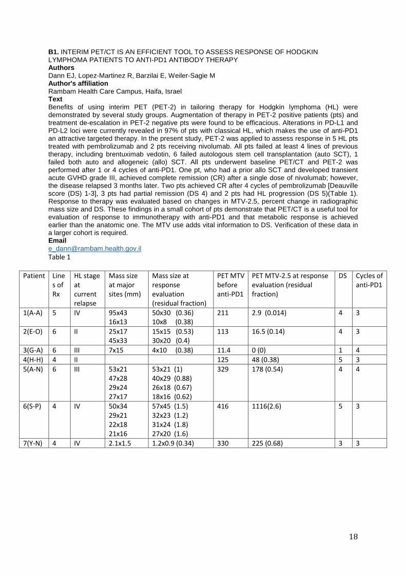
B1. INTERIM PET/CT IS AN EFFICIENT TOOL TO ASSESS RESPONSE OF HODGKIN LYMPHOMA PATIENTS TO ANTI-PD1 ANTIBODY THERAPY Authors Dann EJ, Lopez-Martinez R, Barzilai E, Weiler-Sagie M Author's affiliation Rambam Health Care Campus, Haifa, Israel Text Benefits of using interim PET (PET-2) in tailoring therapy for Hodgkin lymphoma (HL) were demonstrated by several study groups. Augmentation of therapy in PET-2 positive patients (pts) and treatment de-escalation in PET-2 negative pts were found to be efficacious. Alterations in PD-L1 and PD-L2 loci were currently revealed in 97% of pts with classical HL, which makes the use of anti-PD1 an attractive targeted therapy. In the present study, PET-2 was applied to assess response in 5 HL pts treated with pembrolizumab and 2 pts receiving nivolumab. All pts failed at least 4 lines of previous therapy, including brentuximab vedotin, 6 failed autologous stem cell transplantation (auto SCT), 1 failed both auto and allogeneic (allo) SCT. All pts underwent baseline PET/CT and PET-2 was performed after 1 or 4 cycles of anti-PD1. One pt, who had a prior allo SCT and developed transient acute GVHD grade III, achieved complete remission (CR) after a single dose of nivolumab; however, the disease relapsed 3 months later. Two pts achieved CR after 4 cycles of pembrolizumab [Deauville score (DS) 1-3], 3 pts had partial remission (DS 4) and 2 pts had HL progression (DS 5)(Table 1). Response to therapy was evaluated based on changes in MTV-2.5, percent change in radiographic mass size and DS. These findings in a small cohort of pts demonstrate that PET/CT is a useful tool for evaluation of response to immunotherapy with anti-PD1 and that metabolic response is achieved earlier than the anatomic one. The MTV use adds vital information to DS. Verification of these data in a larger cohort is required. Email [email protected] Table 1
Patient Line
s of
Rx
HL stage
at
current
relapse
Mass size
at major
sites (mm)
Mass size at
response
evaluation
(residual fraction)
PET MTV
before
anti-PD1
PET MTV-2.5 at response
evaluation (residual
fraction)
DS Cycles of
anti-PD1
1(A-A) 5 IV 95x43
16x13
50x30 (0.36)
10x8 (0.38)
211 2.9 (0.014) 4 3
2(E-O) 6 II 25x17
45x33
15x15 (0.53)
30x20 (0.4)
113
16.5 (0.14) 4 3
3(G-A) 6 III 7x15 4x10 (0.38) 11.4 0 (0) 1 4
4(H-H) 4 II 125 48 (0.38) 5 3
5(A-N) 6 III 53x21
47x28
29x24
27x17
53x21 (1)
40x29 (0.88)
26x18 (0.67)
18x16 (0.62)
329 178 (0.54) 4 4
6(S-P) 4 IV 50x34
29x21
22x18
21x16
57x45 (1.5)
32x23 (1.2)
31x24 (1.8)
27x20 (1.6)
416 1116(2.6) 5 3
7(Y-N) 4 IV 2.1x1.5 1.2x0.9 (0.34) 330 225 (0.68) 3 3

19
B2..EARLY FDG PET/CT RESPONSE ASSESSMENT IN PEDIATRIC NON HODGKIN LYMPHOMA: A REPORT FROM THE FRENCH PET LYMPHOMA STUDY Authors V Edeline1, M Texier2, F Montravers3, J Lumbroso4, I Borget2, H Brisse4, S Canale5, H. Ducou Le Pointe6, A Lambilliotte7, H. Pacquement8, N Garnier9, J Landmann-Parker10, G. Plat11,T Leblanc12, V Minard-Colin13, L Brugières13, on behalf of the French Society of Pediatric Oncology (SFCE) Author's affiliation 1Department of Imaging, Institut Curie, Hôpital René Huguenin, Paris 2Department of Biostatistics and Epidemiology, Gustave Roussy, Villejuif 3Department of Nuclear Medecine, Hôpital Tenon, APHP, Paris 4Departement of Imaging, Gustave Roussy, Villejuif 5Department of Imaging, Institut Curie, Paris 6Department of Radiology, Hôpital Trousseau, APHP, Paris 7Pediatric Hematology Department, Hôpital Jeanne de Flandres, CHRU, Lille 8Department of Pediatric Oncology, Institut Curie, Paris 9Pediatric Hemato-oncology unit, Leon Berard Center, Lyon 10Department of Paediatric Haemato-Oncology, Hôpital Trousseau AP-HP, Paris 11Department of Pediatric Hemato-oncology, Hôpital Purpan, Toulouse 12Department of Paediatric Haematology, Hôpital Robert Debré, AP-HP, Paris 13Department of Children and Adolescent Oncologgy, Gustave Roussy, Villejuif France Text BACKGROUND: The prognostic value of early metabolic response assessed with FDG PET/CT after 2 courses of chemotherapy remains an area of clinical research in aggressive NHL adults whereas there is still limited information on the feasibility and the prognostic significance of early metabolic response in pediatric NHL. PATIENTS AND METHODS: We performed a national multicentric prospective trial including a total of 230 patients (3-21y) treated according to current French protocols for the main NHL paediatric subtypes. With a median follow-up of 29.2 months, 3- year EFS and OS of this cohort are 85.3% and 95.4% respectively. The main objective of the study was to investigate the value of PET/CT at the time of remission assessment. PET/CT at initial staging and interim PET/CT during chemotherapy were recommended but not mandatory for inclusion. No therapeutic decision was based on PET/CT only. We report here the results of early PET/CT performed 7 to 45 days after the beginning of treatment according to NHL subtype. Early PET/CT response was assessed using the Deauville 5 points scale. Exploratory analyses were performed using ∆SUVmax. PET/CT at diagnosis was not required to evaluate early response based on Deauville score whereas ∆SUVmax was evaluable only in patients with available PET/CET at initial staging. RESULTS: Among 218 evaluable pts included between 2011 and 2015, 127(58%) had an early PET/CT. Central review of PET/CT is on-going and has been already performed for 89(71%) pts. Deauville score is available for only 69 patients. Deauville score could not be used in twenty cases due to technical issues (frequent brown fat tissue uptake in pediatric patients). Of the 69 fully interpretable pts, 31 (45%) had a Deauville score 1-3 (complete metabolic response). ∆SUVmax was >66% in 39 (72%) of the 54 evaluable patients. The metabolic response rate according to the histological subtypes is indicated in the table below.The prognostic impact of early PET/CT response will be evaluated once central review is completed for all patients. CONCLUSION: Interim PET/CT is feasible in a large proportion of paediatric NHL. The first results of this trial indicate that a low proportion (45%) of patients achieve complete metabolic response based on Deauville score 1-3 with this early evaluation of response (after C1 in most lymphoma subtypes). Email [email protected]

20
Lymphoma subtype
Timing of evaluatio
n
Nb of patient
s
Metabolic response
Deauville score missing
Deauville score
1-3
Deauville Score
4
Deauville
Score 5
∆SUVmax
missing
∆SUVmax > 66
Burkitt Lymphoma
After C1 28 10 10/18 4/18 4/18 15 11/13
Diffuse large B-cell lymphoma
After C 1 13 1 4/12 4/12 4/12 3 7/10
Primary Mediastinal B-cell Lymphoma
After C2 13 2 5/11 4/11 2/11 4 8/9
Lymphoblastic Lymphoma
After steroid
prephase
20 3 7/17 4/17 6/17 7 (35%) 5 /13
Anaplastic Large Cell Lymphoma
After C1 15 4 5/11 5/11 1/11 6 (40%) 8 /9
Total 89 20 (22%) 31/69 (45%)
21/69 (30.4%)
17/69 (24.6%)
35/89 (39%)
39/54 (72%)

21
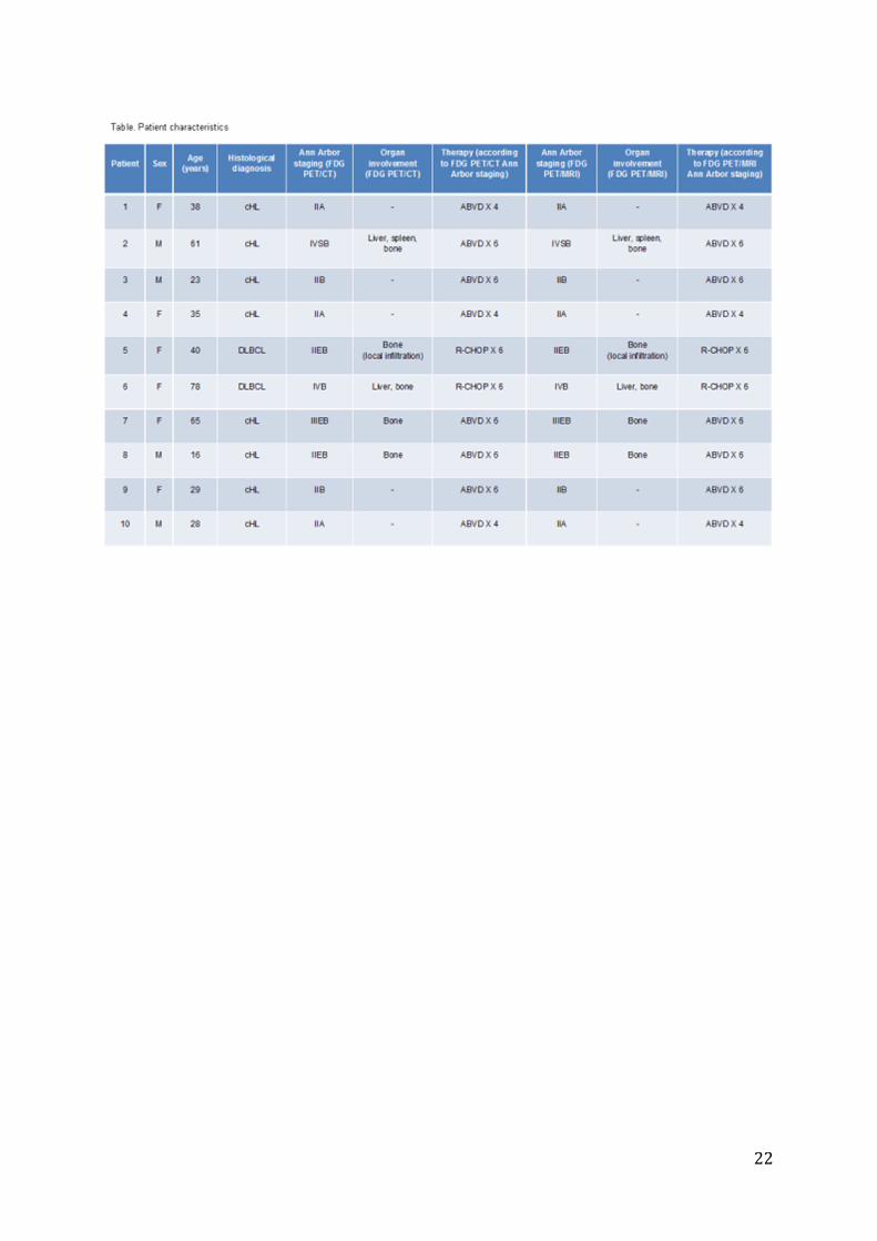
C1. FDG PET/whole-body MRI, including diffusion-weighted imaging, for staging patients with classical Hodgkin lymphoma and diffuse large B-cell lymphoma: comparison with FDG PET/contrast-enhanced CT in a prospective study Authors R. Della Pepa1, M. Picardi1, E. Nicolai2, A. Soricelli3, C. Cerchione1, N. Pugliese1, F. Pane1, M. Meignan4, M. Salvatore5 Author's affiliation 1 Department of Clinical Medicine and Surgery, Hematology, Federico II University, via Sergio Pansini, 5, Naples, 80131, Italy; 2 Department of Nuclear Medicine, SDN IRCCS, Via Gianturco 113, Napoli, Na 80143, Italy; 3Department of Radiology, University of Naples Parthenope -SDN IRCCS, Via F. Acton 38, Napoli, Na 80143, Italy; 4 Centre Universitaire Hospitalier Henri Mondor, Lymphoma Study Association Imaging, Créteil, France; 5 Department of Radiology and of Nuclear Medicine, SDN IRCCS, Via Gianturco 113, Napoli, Na 80143, Italy; Text Hodgkin lymphoma (HL) and non-Hodgkin lymphoma (NHL) require accurate staging for planning the most appropriate therapy. CT, together with FDG PET, are the crucial imaging tools in this setting. Important disadvantages of diagnostic CT are the exposure of ionizing radiation and contrast-induced acute kidney injury. Because the survival rates of patients with HL and NHL have considerably increased over the past years, the goal of current therapies is to maximize cure rates while minimizing toxicity, including the risk of second neoplasms. In line with this paradigm, prevention of exposure to CT-related ionizing radiation and i.v. contrast medium is important. Unenhanced whole-body magnetic resonance imaging (MRI) is feasible and may be a good radiation free alternative to CT for staging lymphoma. The aim of this study was to compare staging obtained with fused FDG PET/MRI with staging obtained with FDG PET/CT for patients with newly diagnosed lymphoma. At pretreatment staging, all patients seen for classical HL and diffuse large B-cell lymphoma (DLBCL) underwent same-day FDG PET/contrast-enhanced CT and FDG PET/whole-body MRI (Biograph mMR imager; Siemens Healthcare, Erlangen, Germany) with diffusion-weighted imaging (DWI). Lymph node and extra-nodal involvements were evaluated site by site using qualitative and quantitative image analysis. FDG PET, diagnostic CT and MRI scans were interpreted by consensus by radiologists and nuclear medicine physicians. Overall, 12 consecutive patients with newly diagnosed lymphoma were scheduled to receive imaging tools for staging. Of them, 2/12 (16.6%) have failed to carry out MRI examination because of claustrophobia. Characteristics of 10 evaluable patients are detailed in the Table. The agreement between FDG PET/MRI and PET/CT for all nodal and extra-nodal regions was 100%, with a low inter-observer variability (Pearson's r=0.958; P <0.01). Ann Arbor stages according to FDG PET/MRI were concordant with those of FDG PET/CT in 100% (10/10) of patients. However, the average dose of ionizing radiation and of i.v. nonionic contrast medium (diagnostic CT) received by each patient was 19.9 mSv (range, 13.9-25.8) and 140 ml (range, 120-150), respectively. The results of this study suggest that fused FDG PET/MRI equals FDG PET/CT for staging patients with newly diagnosed cHL and DLBCL. Whole-body MRI with DWI can be a good alternative to diagnostic CT if radiation exposure and i.v. contrast medium should be avoided. Email [email protected]
22

23
C2. Iron deposits within untreated lymphoma lesions detected on Diffusion-Weighted (DW) and T2-Weighted Gradient Echo (GRE) MR imaging Authors Cottereau AS1, Mule S2, Lin Chieh3, Belhadj K4, Itti E1, Tacher V2, Pigneur F2, Copie C5, Le Bras F4, Haioun C4, Luciani A2, Rahmouni A2. Author's affiliation 1 Nuclear Medicine Department, AP-HP, Groupe Henri Mondor Albert Chenevier, CHU Henri Mondor, 51 Avenue du Marechal de Lattre de Tassigny, 94010 Creteil, France. 2 Medical Imaging Department, AP-HP, Groupe Henri Mondor Albert Chenevier, CHU Henri Mondor, 51 Avenue du Marechal de Lattre de Tassigny, 94010 Creteil, France. 3 Nuclear Medicine Department and Molecular Imaging Cen¬ter, Chang Gung Memorial Hospital, Gueishan, Taiwan. 4 Lymphoproliferative Unit, AP-HP, Groupe Henri Mondor Albert Chenevier, CHU Henri Mondor, 51 Avenue du Marechal de Lattre de Tassigny, 94010 Creteil, France. 5 Pathology department, AP-HP, Groupe Henri Mondor Albert Chenevier, CHU Henri Mondor, 51 Avenue du Marechal de Lattre de Tassigny, 94010 Creteil, France. Text Purpose: To report and analyze the distribution of iron deposits within involved lymphoma lesions and to compare with PET/CT and inflammatory biological findings. Material and Methods: 43 untreated patients (12 with a diffuse large B cell lymphoma (DLBCL); 20 with a Hodgkin lymphoma (HL) and 11 with a Follicular lymphoma (FL)) were enrolled in an ongoing whole-body DW MR imaging prospective trial, Adamantius (NCT02300402). When focal low signal intensity on DW images was found, a T2 GRE sequence was systematically performed. Focal iron deposits were reported, visually scaled as moderate or marked and compared to inflammatory associated biological parameters (Mann-Whitney test) and baseline PET/CT quantitative parameters. Results: 13 patients had focal iron deposits, mostly observed in HL (8/20) and in DLBCL (4/12), mainly Ann Arbor stage 4 (n=10). Iron deposits were detected in spleen (n=9), liver (n=3) and nodal (n=9) lesions corresponding to focal intense FDG uptakes with mean SUVmax respectively of 8.7, 6.7 and 16.2. Seven patients had marked iron deposits, mostly localized in the spleen. Patients with iron deposits had a significant higher CRP, alpha1-globulin, alpha2-globulin levels and microcytic anemia than the others patients (p=0.025, p=0.0068, p=0.02 respectively). Conclusion: Focal iron deposits, a new imaging biomarker, are frequently observed in lymphoma lesions on DW imaging, mostly in stage 4 patients and are associated to inflammatory syndrome. Email [email protected]

24
D1. Combination of mean standard uptake value of whole body metabolic tumor volume and increased bone marrow uptake in baseline FDG-PET/CT improves progression-free survival prognostication in Hodgkin lymphoma patients Authors Michal Weiler-Sagie1, Einat Savin-Shalom5, Tanya Mashiach, John Kennedy1,6 and Eldad J Dann2,3,5 Author's affiliation 1Nuclear Medicine Department, 2Blood Bank and Apheresis Unit, 3Department of Hematology and Bone Marrow Transplantation 4Quality Assurance Unit, all at Rambam Health Care Campus, Haifa, Israel, 5Bruce Rappaport Faculty of Medicine, Technion, Israel Institute of Technology, Haifa Israel, 6Faculty of Biomedical Engineering, Technion, Israel Institute of Technology, Haifa Israel Text Background: Utilization of baseline FDG-PET/CT (bPET) is currently limited to assigning stage of disease and has not had further impact on the risk prognostication that guides initial treatment decisions in Hodgkin lymphoma (HL). The study objectives were to assess the prognostic value of multiple bPET-derived parameters. Patients and Methods: The study population in this retrospective study consisted of 107 HL patients from the H2 lymphoma study, initially treated with 2 cycles of adriamycin, bleomycin, vinblastine and dacarbazine (ABVD) according to predefined prognostic risk factors for early disease (ED) and international prognostic score (IPS) 0-2 for advanced disease (AD). Treatment was escalated after 2 cycles if PET-2 was positive. bPET studies were re-read and the following parameters were recorded: stage, presence of diffuse bone marrow uptake (BMU), whole body metabolic tumor volume (WBMTV), maximal and mean standard uptake values (SUVmaxWBMTV, SUVmeanWBMTV), standard deviation (SDWBMTV) and coefficient of variance (COVWBMTV). Results: At a median follow up of 4.2 years, 19/107 (18%) relapsed, 10/65 with ED and 9/42 with AD. There was no significant difference in PFS in patients with stage III-IV HL (76%) compared to stage I-II HL (86%). Median SUVmeanWBMTV was 4.6 g/mL, Youden’s index-derived cut-off point was 3.84 g/mL. There were 21 patients with SUVmeanWBMTV<3.8, with PFS of 56% compared to 91% in 86 patients with SUVmeanWBMTV ≥3.8 (p<0.001). In univariate analysis p value of <0.1 was found with BMU, WBMTV, SUVmaxWBMTV, SUVmeanWBMTV, SDWBMTV and COVWBMTV. In multivariate analysis, SUVmeanWBMTV (p=0.001, aHR=6.1) and BMU (p=0.008, aHR=3.9) were significant predictors of PFS. A model combining SUVmeanWBMTV and BMU separated the population into 3 risk groups. Forty-four patients with low BMU and SUVmeanWBMTV ≥3.8, 4 of whom relapsed, with PFS of 96%. Fifty-five patients with either high BMU or SUVmeanWBMTV<3.8, 8 of whom relapsed, with PFS of 85%. Eight patients with both high BMU and SUVmeanWBMTV<3.8, 7 of whom relapsed, with PFS of 13%. There was a significant difference between the low risk and the high-risk groups (p<0.001, aHR 18.5). Conclusions: In this cohort of HL patients initially treated with ABVD, combination of SUVmeanWBMTV < 3.8 and diffusely increased BMU in bPET identified a high risk group in which 7 of 8 patients eventually relapsed. Parameters derived from bPET, other than stage, may improve pronostication of HL patients. Email [email protected]

25
Prognostic model combining BMU and SUVmeanWBMTV
n Events PFS
(%)
p value aHR 95% CI
(upper-
lower)
BMUlow, SUVmeanWBMTV>3.8 44 4 96 1.0
BMUlow, SUVmeanWBMTV<3.8
Or
BMUhigh, SUVmeanWBMTV>3.8
55 8 85 0.362 1.7 (0.5-5.8)
BMUhigh, SUVmeanWBMTV<3.8 8 7 13 <0.001 18.5 (5.2-65.9)
Total 107 19 82
BMU – bone marrow uptake
SUV – standard uptake value
WBMTV – whole body metabolic tumor volume
PFS – progression free survival
aHR – adjusted hazard ratio
CI – confidence interval

26
D2. Descriptive results of FDG PET/CT in evaluation of nodular lymphocyte predominant Hodgkin lymphoma (NLPHL) in children: 5 years’ experience Authors F. Montravers1, H. Ducou le Pointe2, V. Huynh2, N. Jehanno3, S. Abbou4, T. Leblanc5, H. Pacquement6, Y.Reguerre7, S. Helfre8, TJ Molina9, S Boudjemaa10, J. Landman-Parker11 on behalf of the children lymphoma multidisciplinary team in the Ile-de-France region in France Author's affiliation 1 Service de médecine nucléaire, APHP and UPMC, Hôpital Tenon, Paris, France 2 Service de radiologie, APHP and UPMC, Hôpital ArmandTrousseau, Paris, France 3 Service de médecine nucléaire, Institut Curie, Paris, France 4 Département de Cancérologie de l'Enfant et l'Adolescent, Institut Gustave Roussy, Villejuif, France 5 Service d’hématologie pédiatrique, APHP and Université Paris Diderot, Hôpital Robert Debré, Paris, France 6 Service d’oncologie pédiatrique, Institut Curie, Paris, France 7 Service d'hémato-oncologie pédiatrique, Hôpital Felix Guyon, Saint Denis, La Réunion, France 8 Service de radiothérapie, Institut Curie, Paris, France 9 Service d’anatomie et cytologie pathologiques, APHP and Université Paris-Descartes, Hôpital Necker, Paris, France 10 Service d’anatomie et cytologie pathologiques, APHP and UPMC, Hôpital Armand Trousseau, Paris, France 11 Service d’hématologie pédiatrique, APHP and UPMC, Hôpital Armand Trousseau, Paris, France Text NLPHL is a very rare disease accounting for about 10 new cases a year in France in children. The disease differs clearly from classical Hodgkin lymphoma (cHL) in its histopathological characteristics, clinical presentation and management. The role of FDG PET/CT in this rare disease is not as well established as in cHL. It has especially been reported a less intense FDG uptake than in cHL at diagnosis, leading to a risk of underestimation of the disease and difficulty to evaluate the response to therapy. Methods Our aim was to describe the FDG PET characteristics of all the NLPHL children patients presented during our regional children lymphoma multidisciplinary team meetings (to which are also referred difficult cases from the whole country) from February 2011 to June 2016. Results During these 5 years, 496 consecutive patients with lymphoma (NHL and HL) were presented among them 23 (18 boys and 5 girls, median age = 12 years, range: 5-18) with newly diagnosed NLPHL. FDG PET was negative in 7 children imaged after complete resection of the involved node. These children were not treated (wait-and-see approach). The other 16 patients received a non-intensive chemotherapy (CVP courses x 3). FDG PET was clearly positive in all of them except one for whom the intensity of uptake was too low (SUVmax =2.5) to allow a correct evaluation of the disease. SUVmax was available in 12 on the 15 clearly positive FDG PET. The intensity of uptake was high with a median SUVmax = 10.6 (range: 5.5 – 20). Thirteen had a limited disease (stage I or II) but 3 patients had an advanced disease (stage III: n=2 and stage IV: n=1 with bone marrow involvement). To our knowledge, overall survival is 100%. Seven patients were not in complete remission at the end of treatment or relapsed. Four patients had a poor response to CVP. Three of them were explored by PET with SUVmax respectively = 14, 8 and 5. Three children relapsed. One patient relapsed 6 months after the initial complete resection of the involved node (SUVmax = 10). Two patients relapsed respectively 6 months (SUVmax = 10) and 10 months after the initial diagnosis (SUVmax = 11). Conclusion: These results in children with NLPHL show that FDG PET was effective both at diagnosis to correctly stage the disease with evidence of high FDG uptake in all children except one and in the relapse setting. In this pediatric series, FDG PET performance does not seem to differ significantly from that reported in cHL. Email [email protected]

27
D3. Prognosis value of baseline total metabolic tumor volume (TMTV) in advanced Hodgkin lymphoma (HL): Ancillary study of AHL2011 LYSA trial Authors S. Kanoun1,2,3, A. Berriolo-Riedinger2, I. Tal4, V. Edeline5, A. Cottereau6, P. Brice7,R.Bouabdallah8, G. Salles9, A. Stamatoullas10, J. Dupuis6, M. Andre11, N. Mounier12, C. Ferme13, M. Meignan6, R. Casasnovas14; Author's affiliation 1LE2I, UMR CNRS 6306, Dijon, FRANCE, 2Centre Georges Francois Leclerc, Dijon, FRANCE, 3CHU Dijon, Dijon, FRANCE, 4Beth Israel Deaconess Medical Center, Boston, MA, UNITED STATES, 5Institut Curie, Paris, FRANCE, 6Hopital H. Mondor, Creteil, FRANCE, 7AP-HP at Saint-Louis Hospital, Paris Diderot- Sorbonne University, Paris, FRANCE, 8Cancer Center Institut Paoli-Calmettes, Marseille, FRANCE, 9University De Lyon, Hospices Civils De Lyon, Lyon, FRANCE, 10Centre Henri Becquerel, Rouen, FRANCE, 11Centre Hospitalier Universitaire Mont-Godinne, Dinant, BELGIUM, 12CHU l'Archet, Nice, FRANCE, 13Institut Gustave Roussy, Villejuif, FRANCE, 14Hôpital Le Bocage, Dijon, FRANCE. Text Aim: The TMTV assessed on the baseline FDG-PET is a novel approach of tumor burden measurement. It has been reported to influence HL outcome in a retrospective series (Kanoun, EJNM 2014). We designed a study evaluating the TMTV prognosis value in patients (pts) prospectively enrolled in a phase III randomized trial testing a treatment strategy driven by PET, compared to a standard treatment not monitored by PET. Methods: Eligible pts had to be enrolled in the AHL2011 trial (NCT01358747) and to have a baseline PET (PET0) available for central review and TMTV calculation. Pts were 16-60 y, with a previously untreated advanced HL (Ann-Arbor stage III, IV or high risk IIB) and were randomly assigned to a treatment strategy driven by PET after 2 escalated BEACOPP (BEA) cycles (PET2), delivering 4 cycles of ABVD for PET2- pts and 4 cycles of BEA for PET2+ pts or a standard treatment not monitored by PET and delivering 6 cycles of BEA. PET2 were centrally reviewed and interpreted according to Deauville criteria. TMTV was computed on PET0 by summing the metabolic volumes of the individual lesions using the 41% SUVmax thresholding method already described in lymphoma. Results: 392 pts with a median age of 30 y were included: 64% were male, 89% had stage III/IV, and 59% an IPS≥3. Median TMTV was 200 ml (23 - 2149). Using a X-tile method a 350 ml cut off value was identified from a training set (n = 262) and confirmed in a validation set (n = 130) of pts obtained from the whole series. With a 16 months median follow up, 2y-PFS was 81% vs 93% in pts with high and low TMTV respectively in the whole population (p =0.0015; HR = 3). PET2 positivity was also related to a lower 2y-PFS compared to PET2- pts (76% vs 92% ;p<0.0001). Then 3 groups could be identified: pts with either [high TMTV and PET2+ (n = 23; 6%)], or [high TMTV and PET2-, or low TMTV and PET2+ (n = 103; 27%)], or [low TMTV and PET2- (n = 261; 67%)] had a 61%, 88%, 94% 2y-PFS respectively (p<0.0001). Conclusions: TMTV predicts the outcome of young advanced HL pts independently of the early metabolic response to treatment. The combination of TMTV and PET2 allows identifying 3 subsets of HL pts with significantly different outcome that may help clinician to better tailor therapy. Email [email protected]

28
D4. Prognostic Value Of Different F‐18 FDG PET/CT Quantitative Analytical Methodologies In Pediatric Hodgkin’s Lymphoma Authors Serry O M 1, Kandeel A A 2, El‐Sayed M A 2, Hussein A E 3, Omar W S 4 Author's affiliation 1Nuclear Medicine Department in Children’s Cancer Hospital Foundation in Egypt, 2Nuclear Medicine Department, Faculty of Medicine, Cairo University, Egypt, 3Nuclear Medicine Department, Faculty of Medicine, Sohag University, Egypt and 4Nuclear Medicine Department, National Cancer Institute, Cairo University, Egypt Text Introduction and aim of work: Assessment of the individualized SUVs, PET-derived total metabolic tumor volume (TMTV) and the product of both parameters, termed total lesion glycolysis (TLG) in both initial and interim PET if it carries a better PPV in early assessment of response to therapy in pediatric Hodgkin’s lymphoma (PHL) patients. Patients and Methods: Retrospective analysis of PET/CT results was performed on 60 patients (42 males and 18 females; mean age 8.7±4.2 years). To assess the prognostic value of initial and interim 18F-FDG PET/CT, different semi-quantitative parameters such as SUVmax, SUVmean, Total lesion glycolysis (TLG) and TMTV of all lesions using SUVmax & mean including SUV2.5 and 40% of SUVmax as cut-off values were calculated. Follow up for 24 months from initial treatment with calculation of Disease Specific Survival (DSS). According to the recommendations of Deauville criteria interim PET (PET2) results were identified into three groups; PET2-negative (PET2-ve), PET2-positive (PET2+ve), and PET2-minimal residual uptake (PET2-MRU), the cut-off between PET2+ve and PET2-MRU was 3-4 in the 5-point scale. Results: Out of the 60 interim-PET scans, 50 scans were considered as PET2-ve (83.3%), 5 scans as PET2+ve (8.3%) and 5 scans as PET2-MRU (8.3%). The risk of the disease and the visual scoring assessment were significantly correlated with patient's outcome (whether Negative or Residual/Relapse) (p <0.0001). Different results were obtained; the most important were TLGmax2.5 (cut-off 2.5), TLGmean2.5 (cut-off 2) and TMTV2.5 (cut-off 0.75 ccm) in interim PET showed the highest sensitivity, specificity, PPV and NPV (58.5%, 97.9%, 87.5% and 90.3% respectively for the 3 parameters). Conclusion: TLGmax2.5, TLGmean2.5 and TMTV2.5 are the most relevant parameters for predicting the outcome in patients with PHL, and can add a significant prognostic insight to interim PET response assessment. This may guide clinicians in their choice of therapeutic strategy. Email [email protected]

29
D5. VALUE OF FDG PET/CT IN THE INITIAL STAGING OF PEDIATRIC NON HODGKIN LYMPHOMA. A REPORT FROM THE FRENCH PET LYMPHOMA STUDY Authors F Montravers1, V Edeline2, M Texier3, J Lumbroso4, I Borget3, H Brisse5, S Canale4, H. Ducou Le Pointe6, A Lambilliotte7, H. Pacquement8, N Garnier9, J Landmann-Parker10, G. Plat11, T Leblanc12, L Brugières13, V. Minard-Colin13 on behalf of the French Society of Pediatric Oncology (SFCE) Author's affiliation 1Department of Nuclear Medecine, Hôpital Tenon, APHP, Paris 2Department of Imaging, Institut Curie, Hôpital René Huguenin, Paris 3Department of Biostatistics and Epidemiology, Gustave Roussy, Villejuif 4Departement of Imaging, Gustave Roussy, Villejuif 5Department of Imaging, Institut Curie, Paris 6Department of Radiology, Hôpital Trousseau, APHP, Paris 7Pediatric Hematology Department, Hôpital Jeanne de Flandres, CHRU, Lille 8Department of Pediatric Oncology, Institut Curie, Paris 9Pediatric Hemato-oncology unit, Leon Berard Center, Lyon 10Department of Paediatric Haemato-Oncology, Hôpital Trousseau AP-HP, Paris 11Department of Pediatric Hemato-oncology, Hôpital Purpan, Toulouse 12Department of Paediatric Haematology, Hôpital Robert Debré, APHP, Paris 13Department of Children and Adolescent Oncologgy, Gustave Roussy, Villejuif France Text BACKGROUND: FDG PET/CT is recommended for initial staging of aggressive adult Non Hodgkin Lymphoma (NHL), but there is limited data on the feasibility and the diagnostic performance of PET/CT for initial staging of paediatric NHL. PATIENTS AND METHODS: We performed a prospective multicentric study including 230 French patients (pts) (3-21y) treated according to current SFCE protocols for NHL. The main objective of the study was to investigate the value of PET/CT for remission assessment. Initial staging included clinical examination, cervico-thoraco-abdominal CTscan and/or MRI, examination of bone marrow (BM) and CSF and bone scan for patients with symptomatic bone lesions. Staging was based on St Jude’s classification. We report here the results of PET/CT performed at initial staging which was recommended but not mandatory for inclusion. RESULTS: 153/218(70%) evaluable pts included between 2011 and 2015 had a PET/CT for initial staging: 55/83(66%) Burkitt lymphoma (BL), 25/28(80%) diffuse large B-cell lymphoma (DLBCL), 16/21(80%) primary mediastinal Bcell lymphoma (PMBL), 34/57(60%) lymphoblastic lymphoma (LL) and 23/29(79%) anaplastic large cell lymphoma (ALCL). In all pts, except 7 with completely resected lymphoma, at least one lesion exhibited significant FDG uptake at PET/CT. In 60 pts, initial PET/CT detected at least one spot of hyperfixation in a region which had not been classified as abnormal on conventional imaging (mostly in bone and lymp-nodes). Only 11/32 pts with cyto/histologically proven BM involvement had a bone or BM FDG uptake on PET/CT. Finally, taking into account that pts with multiple bone lesions without BM involvement (n=18) are not classified as stage IV in St Jude’s classification, PET/CT findings resulted in a change in staging in only 3/218(1.4%) pts. SUVmax values are available for 84 pts for whom central review of the initial TEP is available so far. Median SUVmax is 13.3(4.4-41) for BL, 14.2(3.1-34) for DLBCL, 17.1(13.5–28.7) for PMBCL, 7.2(3.3–12.4) for LL and 15.0(4.2-37) for ALCL. CONCLUSION: PET/CT proved feasible at diagnosis in a large proportion of paediatric NHL. A positive uptake with high SUVmax was found in all pts except those with primary complete resection. The results of PET/CT rarely led to a treatment modification as compared to staging with conventional imaging alone. Nevertheless, initial PET at diagnosis is crucial for having a more accurate TEP interpretation during or at the end of treatment Email [email protected]

30
D6. Detection of Bone marrow involvement (BMI) by baseline FDG-PET/CT in Patients with High-Tumor Burden Follicular Lymphoma (FL): A pooled analysis of three prospective studies from the LYSA and the FIL.
Berivan Emsen1, Anne Ségolène Cottereau1, Annibale Versari2, Loïc Chartier3, Jehan Dupuis4, Vittoria Tarantino5, Rene-Olivier Casasnovas6, Antonella Franceschetto5, Hervé Tilly7, Corinne Haioun4 ,Massimo Federico5, Gilles Salles8, Emmanuel Itti1, Michel Meignan1 Judith Trotman9, Stefano Luminari5.
1 Nuclear Medicine Department, Hôpital Henri Mondor, University Paris-Est Créteil, France,2 Nuclear Medicine Department, Santa Maria Nuova Hospital, IRCCS, Reggio Emilia, Italy,3 Department of Biostatistics (LYSARC), Centre Hospitalier Lyon Sud, Pierre Bénite, France,4 Unité d’Hémopathies Lymphoïdes, Hôpital Henri Mondor, Université Paris-Est Créteil, France,5 Department of Diagnostic, Clinical, and Public Health Medicine, University of Modena and Reggio Emilia, Modena, Italy, 6 Hématologie Clinique, Hôpital le Bocage, CHU-Dijon, Dijon, France ,7 Département d’Hématologie, UMR918, Centre Henri Becquerel, Université de Rouen, Rouen, France, 8 Service d’Hématologie, Hospices Civils de Lyon 1, Université Claude Bernard Lyon 1, Pierre Bénite, France, 9Haematology Department, Concord Hospital, University of Sydney, Sydney, NSW, Australia
Introduction Although FDG-PET/CT has replaced Bone marrow biopsy (BMB) for the detection of BMI in Hodgkin lymphoma and in the majority of Diffuse Large B, its role in FL is controversial. Luminari et al have reported a lower sensibility of FDG-PET/CT to detect BMI using visual analysis compared to BMB. Perry et al have recently suggested that quantitative evaluation might improve its sensitivity. No large prospective study is currently available to evaluate its prognostic usefulness. The aim of this study was to investigate the prognostic role of the detection of bone marrow involvement on baseline FDG-PET/CT, using visual and quantitative analyses, in patients with high tumor burden FL from 3 prospective studies (PET-FL and PRIMA from the LYSA, FOLL05 from the FIL).
Patients and Methods A total of 168 baselines PET-CT scans with high tumor burden FL receiving R-chemotherapy (PRIMA, PET-FL and FOLL05 studies) were retrospectively reviewed by a central analysis. The median follow up of the population was 64 months; 5 y PFS was 55%, 5y OS was 92%. Visually, bone marrow involvement on PET (BMIPET V) was determined on the presence of a focal increased uptake. Three subgroups were individualized: one single site, equal or less than 6 sites and more than 6). Maximum standardized uptake value (SUVmax) were systematically measured at 8 different sites (spine T5, L4, L5, right and left iliac wing, right and left femur and sternum). In patients with focally increased uptake, the latter was also measured at that specific location. A background ratio (TBR) was calculated as the ratio between SUVmax in a bone region and SUVmax of liver. For quantitative approach, a patient with at least one TBR ratio>1 was classified BMI (BMIPETQ). BM biopsy results were available for 159 patients.
Results Focal lesions were present on PET in 62 patients: 17 had a single lesion, 11 equal or less than 6 lesions and 34 more than 6. 87 patients had a positive BMB among which only 31 had also focal lesions on PET. On the other hand, 31 patients had BMIPET V with negative BMB: in these cases PET detected BMI missed by BMB. A positive BMB was associated with a lower PFS (5yPFS of 69.4% vs 46.8%, p=0.0218, HR=1.8), but not with OS (p=0.19). Detection of focal lesions on PET did not reach statistical significance to predict outcome (p=0.12 for PFS and p=0.61 for OS), whatever the number of lesions observed. However, combined with BMB, PET improves risk patient stratification, identifying three groups with significantly different outcome: patients with negative BMIPET V and BMB, patients with a positive PET BMIPET V or positive BMB and patients with both positive BMIPET V and BMB had a 5yPFS of 72,6%, 53% and 38.5% (p=0.028, HR=2.4). Median TBR of spine T5, L4, L5, right and left iliac wing, right and left femur and sternum were respectively 0,95 (+/- 0,57 SD), 1,08 (+/- 0,54 SD), 1,22 (+/- 0,95 SD), 1,08 (+/- 0,68 SD), 0,94 (+/- 1,01 SD), 0,55 (+/- 0,91 SD), 0,57 (+/- 0,86 SD), 0,87 (+/- 1,05 SD). However, quantitative approach did not improve outcome prediction with similar prognostic value than visual analysis.
Conclusion PET-CT cannot replace BMB in FL. However, the risk of progression increases in the presence of a focal lesion in patients with negative BMB.
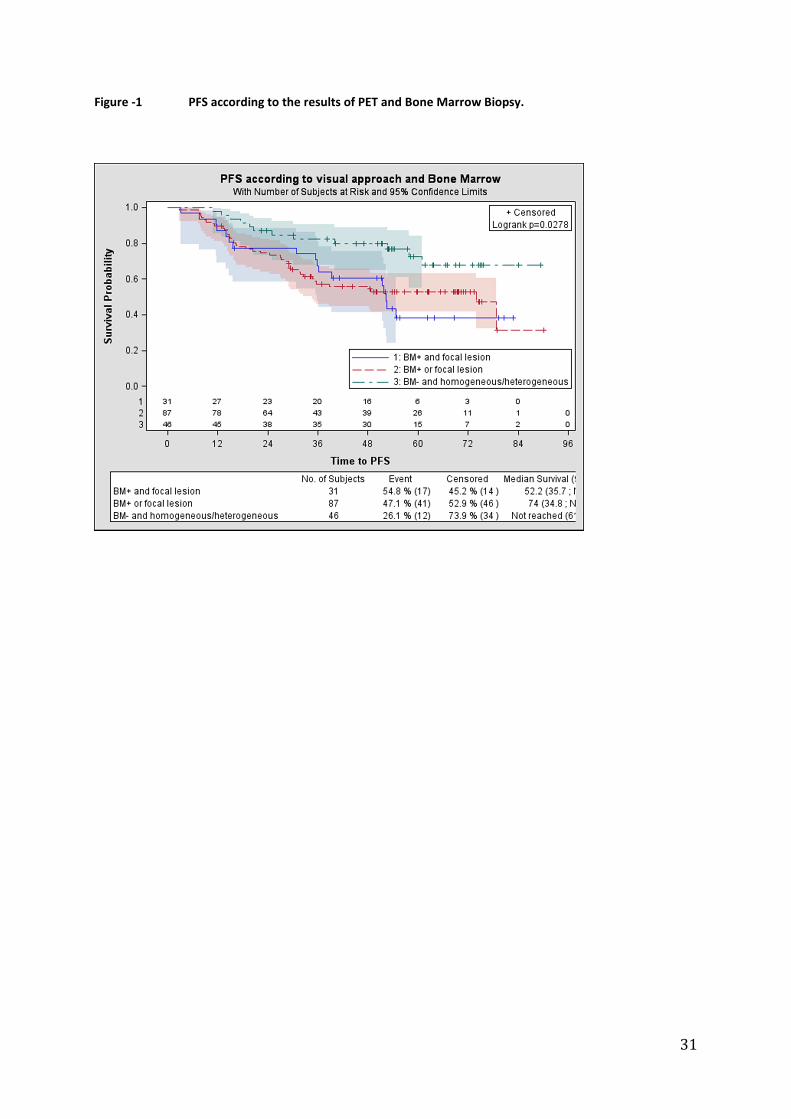
31
Figure -1 PFS according to the results of PET and Bone Marrow Biopsy.

32
E1. Rituximab exposure is influenced by baseline metabolic tumor volume and affects outcome of DLBCL patients: A LYSA study Authors Mira Tout (1), Olivier Casasnovas (2), Michel Meignan (3), Thierry Lamy (4), Franck Morschhauser (5), Gilles Salles (6), Emmanuel Gyan (7), Corinne Haioun (8), Mélanie Mercier (9), Pierre Feugier (10), Sami Boussetta (11), Gilles Paintaud (1,12), David Ternant (1,12), Guillaume Cartron (13) Author's affiliation (1) Université François-Rabelais de Tours, CNRS, GICC UMR 7292, Tours, France; (2) Department of Clinical Hematology, CHU Dijon, INSERM LNC UMR866, Dijon, France; (3) Department of Nuclear Medicine, Henri Mondor University Hospital, Créteil, France; (4) Department of Clinical Hematology, CHU Rennes, U917, Rennes 1 University, Rennes, France; (5) Department of Clinical Hematology , CHU Lille, Unité GRITA, Université de Lille 2, Lille, France; (6) Hospices Civils de Lyon, Université Claude Bernard Lyon-1, Department of Hematology, France; (7) Department of Hematology and cell therapy, CIC INSERM U1415, CHU Tours, Tours, France; (8) Lymphoid malignancies unit, Henri Mondor University Hospital, APHP and UPEC, Créteil, France; (9) Department of Hematology, CHU Angers, Angers, France; (10) Department of Hematology, CHU Nancy, Nancy, France; (11) Lymphoma Academic Research Organisation, CHU Lyon-Sud, Lyon, France; (12) CHU Tours, Laboratory of Pharmacology-Toxicology, Tours, France; (13) Department of Hematology, CHU Montpellier, CNRS UMR 5235, University of Montpellier, Montpellier, France Text Introduction: Rituximab (MabThera® and Rituxan®) in association with anthracycline-based chemotherapy (CHOP or ACVBP) has dramatically improved outcome of patients with diffuse large B-cell lymphoma (DLBCL). However, some patients failed to respond or relapsed early after treatment with poor prognosis. A murine model suggested that tumor burden could affect rituximab exposure and efficacy. The aim of this study was to evaluate the influence of the baseline total metabolic tumor volume (TMTV0) assessed by PET and rituximab pharmacokinetics (PK) on outcome of DLBCL patients receiving immunochemotherapy. Patients and Methods: Data on baseline total metabolic tumor volume (TMTV0) measured using 18F-FDG-PET/CT and rituximab pharmacokinetics were available in 108 DLBCL patients who received four infusions of rituximab (375 mg/m2) associated with anthracycline-based chemotherapy administered every 14 days (CHOP-14 or ACVBP-14) in two prospective multicenter studies. Patients with localized stage (n = 19) were included in GOELAMS 0203 trial (NCI number: NCT00841945), whereas those in advanced stage (n = 89) were included in GELA 073B trial (NCT00498043). The association of TMTV0 and rituximab exposure (AUC) with metabolic response (based on PET evaluation after C4 according to IHP criteria) as well as PFS and OS was assessed using logistic regression and Cox models, respectively. Cutoff values for patients’ outcome were determined using ROC curve analysis. Results: Baseline median TMTV0 was 313.5 cm3 (range 0.8–4339 cm3). Complete metabolic response after four cycles was 50.5%. Rituximab exposure decreased as TMTV0 increased (R2 = 0.41; p < 0.0001). Patients with rituximab exposure in cycle 1 (AUC1) above the 9600 mg.h/L cutoff value (n = 48; 44%) had significantly better response (odds ratio [OR] = 1.65; p = 0.0006), 4-y PFS (87% vs 62%; hazard ratio [HR] = 0.33, p = 0.025), and 4-y OS (96% vs 72%; HR = 0.066, p < 0.001). Rituximab dose to administer according to individual TMTV0, resulting in the optimal AUC1 was then calculated, and the standard dose of 375 mg/m2 would be suitable for patients with a TMTV0 of 216.3 cm3. Conclusion: Baseline metabolic tumor volume significantly influences rituximab exposure which predicts response and outcome of DLBCL patients treated by immunochemotherapy. Our results support evaluating dose individualization according to MTV0 in prospective studies. Email [email protected]

33
E2. Baseline Total Metabolic Tumor Volume measured with fixed or different adaptive thresholding methods equally predicts outcome in Peripheral T cell lymphoma. Authors A-S. Cottereau1, S. Hapdey2,3, L.Chartier4, O. Casasnovas5, E. Itti1, H. Tilly6, P. Vera2,3, M.Meignan1, S.Becker2,3. Author's affiliation 1 Nuclear Medicine Department, Hôpital Henri Mondor, University Paris-Est Créteil, France 2 Nuclear Medicine Department, Henri Becquerel Cancer Center and Rouen University Hospital, Rouen, France. 3 QuantIF–LITIS (EA[Equipe d’Accueil] 4108), Faculty of Medicine, University of Rouen, Rouen, France. 4 Department of Biostatistics (LYSARC), Centre Hospitalier Lyon Sud, Pierre Bénite, France. 5 Hematology Department, Hopital Le Bocage - CHU Dijon, France. 6 Hematology Department, UMR918, Henri Becquerel Cancer Center and Rouen University Hospital, Rouen, France. Text Objective: To compare the prognostic value of baseline Total Metabolic Tumor Volume (TMTV) measured on FDG-PET/CT with different adaptive thresholding methods to TMTV measured with a fixed 41% SUVmax threshold, in a large series of Peripheral T cell lymphoma (PTCL), as a model of diffuse disease. Methods: 106 patients with PTCL from 5 LYSA centers, staged with a PET/CT were enrolled. In this series TMTV computed with the 41% SUVmax threshold was a strong predictor of outcome. On a dedicated workstation, we measured the TMTV with four adaptive thresholding methods based on characteristic image parameters: Daisne (Da) modified based on signal/background ratio, Nestle (Ns) on tumor and background intensities, Fit including a 3D geometric model based on spatial resolution (Fit) and Black (Bl) based on mean SUVmax. The TMTV values obtained with each adaptive method were compared to the TMTV values obtained with 41% SUVmax method. Their respective prognostic impacts on outcome prediction were compared using ROC analysis and Kaplan Meier survival curves. Results: The median value of TMTV41%, TMTVDa, TMTVNs, TMTVFit, TMTVBl were respectively 231 cm3 (CI 95% 145-361), 175 cm3 (129-311), 198 cm3 (137-343), 175 cm3 (134-313), 333 cm3 (260-652). The correlation between TMTV values from the 41% SUVmax method and TMTVDa, TMTVFit, TMTVNs and TMTVBl was excellent with a Pearson coefficient of 0.99, 0.99, 0.98 and 0.94 and a Spearman coefficient of 0.99, 0.99, 0.97 and 0.96 respectively. There was no significant difference within the methods between the ROC curves (p>0.4) for Progression Free Survival (PFS), (except for Black) and for Overall Survival (OS). Survival curves with the ROC optimal cut off for each method separated the same groups of low risk (volume≤cut-off) from high risk patients (volume>cut-off) with similar 2y-PFS (range 66-72% vs 26-29%; HR 3.7-3.9) and 2y-OS (79-83% vs 50-53%, HR 2.8-3.2). Conclusion: Although each method produced some TMTV measurement differences, the prognostic value of TMTV remained quite similar whatever the methods, which supported its use as a strong prognosticator in lymphoma. For implementation of TMTV in clinical trials one single method easily applicable in a multicentric PET review is mandatory. Email [email protected]

34
F1. 18F-fluorocholine versus 18F-fluorodeoxyglucose for PET/CT imaging in patients with suspected relapsing or progressive multiple myeloma: A pilot study Authors Thibaut Cassou-Mounat1,2,3, Sona Balogova1,4, Françoise Montravers1,3,Valérie Nataf 1,5, Marie Calzada1,2, Virginie Huchet1, Khaldoun Kerrou1, Jean-Yves Devaux2,3, Mohamad Mohty3,6, Jean-Noël Talbot1,3, Laurent Garderet3,6. Author's affiliation 1- Department of Nuclear Medicine, Hôpital Tenon, AP-HP, Paris, France Text Hybrid positron emission tomography/computed tomography (PET/CT) has now become available, as well as whole-body low dose multidetector row computed tomography (MDCT) or magnetic resonance imaging (MRI). The radioactive glucose analogue 18F-fluorodeoxyglucose (FDG) is the most widely used tracer but has a relatively low sensitivity in detecting multiple myeloma (MM). We compared FDG with a more recent metabolic tracer, 18F-fluorocholine (FCH), for the detection of MM lesions at time of disease relapse or progression. Methods: We analysed the results of FDG and FCH imaging in 21 MM patients undergoing PET/CT for suspected relapsing or progressive MM. For each patient and each tracer, an on-site reader and a masked reader independently determined the number of intraosseous and extraosseous foci of tracer and the intensity of uptake as measured by their SUVmax and the corresponding target/non-target ratio (T/NT). Results: In the skeleton of 21 patients, no foci were found for two cases, uncountable foci were observed in 4 patients, including some mismatched FCH/FDG foci. In the 15 patients with countable bone foci, the on-site reader detected 72 FDG foci vs. 127 FCH foci (+76%), whereas the masked reader detected 69 FDG foci vs. 121 FCH foci (+75%), both differences being significant. Interobserver agreement on the total number of bone foci was very high, with a kappa coefficient of 0.81 for FDG and 0.89 for FCH. Measurement of uptake in the matched foci that took up both tracers revealed a significantly higher median SUVmax and T/NT for FCH vs. FDG. Almost all unmatched foci were FCH-positive FDG-negative (57/59=97% on-site and 56/60=93% on masked reading); they were more frequently observed than matched foci in the head and neck region. Conclusion: These findings suggest that, PET/CT performed for suspected relapsing or progressive MM would reveal more lesions when using FCH rather than FDG. Email [email protected]

35
Figure 1.
Patient #19: FDG PET MIP (A), FDG PET/CT axial slices (sacrum, pelvis and Th10) (B), CT (C), FCH
PET/CT axial slices (sacrum, pelvis and Th10) (D) and FCH PET MIP (E).
Patient with innumerable bone foci on both FDG and FCH PET/CTs. The majority of foci are matched,
taking-up both tracers (white full arrow). However, some foci appear more intense with FDG (black
full arrow) and other with FCH (black dotted arrow). Furthermore, some lesions visible on CT take-up
neither FDG nor FCH (white dashed arrow), probably as a consequence of the previous treatment.
Figure 2.
Maximum intensity pixel (MIP) visualization of PET in two different patients
FCH shows much more bone foci than FDG in patient #2 (A). FCH and FDG show the same bone foci in
patient #3 (B).
FDG PET/CT FCH PET/CT
A
FDG PET/CT FCH PET/CT
B
A B C D E

36
F2. HOVON Multiple Myeloma CT scanning, reformatting and interpretation protocol Authors De Keizer B(1), Quarles van Ufford HME(2), Hoekstra OSH(3), Zijlstra JM(4) Author's affiliation 1.Department of Radiology and Nuclear Medicine, university medical center Utrecht, Utrecht, The Netherlands 2.Department of Radiology and Nuclear Medicine, MCH-Bronovo, the Hague, The Netherlands 3.Department of Radiology and Nuclear Medicine, VU university medical center, Amsterdam, The Netherlands 4.Department of Hematology, VU university medical center, Amsterdam, The Netherlands. On behalf of the HOVON (Haemato Oncology Foundation for Adults in the Netherlands) imaging working group Text Introduction CT imaging is recommended by international guidelines for the evaluation of multiple myeloma localisations. Because of upcoming new multicentre studies there is need for an uniform scan protocol. However there are no guidelines on acquisition and interpretation of CT. Methods Using available literature and consulting multiple clinical physicists and radiologists a consensus imaging protocol was made, taking into account patient positioning, scanning, reconstructing and reformatting. Also recommendations for interpretation of CT scans were made. Results Patient should be positioned supine with arms ventral of the abdomen to decrease beam hardening artefacts. A low dose scanning protocol is recommended, preferably, if available with iterative reconstruction. Scans must be reconstructed with a sharp / bone kernel with slice thickness ≤1,5 mm. For interpretation CT scans must be reformatted with a slice thickness of 3 mm (slice interval 3 mm or less) in axial, coronal and sagittal planes with a pixel size < 1mm. For image interpretation positive myeloma lesions are: 1) Clear lytic lesions >5mm (smaller lesions can be discarded), 2) Soft tissue lesions with destruction of cortical bone, 3) Extra-osseous soft tissue lesions adjacent to bone or extending outside of cortical bone. Vertebral compression fractures should be reported, these are not myeloma localization per se, but might have clinical consequences. Conclusion A consensus CT scanning, reformatting and interpretation protocol was made allowing uniform scan quality and interpretation for multicenter studies. Email [email protected]

37
F3. FDG-PET/CT AT RELAPSE PREDICTS SURVIVAL IN MULTIPLE MYELOMA Authors B Jamet(1), C Bailly(1), L Planche(3,) T Carlier(1,2), T Eugene(1), C Touzeau(4), C Ansquer(1), P Moreau(4), F Kraeber-Bodéré(1,2), C Bodet-Milin(1,2) Author's affiliation (1) Nuclear Medicine Unit, University Hospital, Nantes, France (2)INSERM U892, CRCNA, Nantes, France (3)Statistics Unit, University Hospital, Nantes, France (4)Hematology Unit, University Hospital, Nantes, France Text AIM: This retrospective study evaluated the benefit of FDG-PET/CT to predict the survival of MM patients, at suspected or biochemical proved relapse. MATERIALS AND METHODS: 45 patients with a history of MM have been included, all initially treated as recommended by the IMWG’s guidelines (median time from diagnosis to suspected or biochemical proved relapse: 42 months (range, 5 to 169)). All patients were examined with FDGPET/CT. Visual focal lesions (FLs), bone marrow involvement (BMI), extra-medullary disease (EMD)) and quantitative PET parameters (SUVmax, metabolic tumor volume (MTV) and total lesion glycolysis (TLG) of hottest lesions, whole-body MTV (MTVT)) were evaluated. Kaplan-Meier method, Logrank tests, univariate and multivariate Cox analyses were used to analyze whether PET and usual international prognostic parameters (International staging system score (ISS), cytogenetic abnormalities (FISH analysis of del(13q), t(4;14), del(17p)) could predict free-progression survival (PFS) and overall survival (OS). RESULTS: The median age at diagnosis was 61 years (range 33, to 80) and median follow-up after PET/CT was 18 months (range, 1 to 64) for living and 19 months (range, 7 to 28) for dead patients. 34/45 patients had a positive FDG-PET/CT, including 6/11 (54,5%) without abnormality of electrophoresis or free light chain level (FLC). The first multivariate analysis performed in the entire cohort showed that positive FDG-PET/CT affected PFS, (P=0.02) whereas ISS and cytogenetic abnormalities did not. Univariate analysis realised in positive FDG-PET/CT patients showed that presence of ≥2 EMD lesions was correlated with shorter OS (P<0.001) and presence of ≥17FLs (P=0.003), ≥4FLs in the appendicular skeleton (P=0.0057), SUVmax (P=0.04) and TLG≤24.22 (P=0.01) predicted shorter PFS. MTVT, ISS, cytogenetic abnormalities were not predictive for PFS or OS. Multivariate analysis performed in this subgroup of positive FDG-PET/CT patients highlighted that only presence of ≥17FLs (P=0.03) was an independent variable adversely affecting PFS whereas presence of ≥2 EMD had a limit prognostic value for OS (P=0.06). CONCLUSION: Our study showed interest of FDG-PET/CT in MM patients with suspected relapse, especially in patients with normal electrophoresis and FLC. Positive FDG-PET/CT predicted PFS and number of FLs, SUVmax, TLG, and presence of EMD lesions showed prognostic value.

38
F4. Whole-body combined [18F]NaF and [18F]FDG PET/CT versus MRI for the detection of myeloma lesions Authors Nadia Withofs, François Cousin, Tino Tancredi, Paolo Simoni, Bernard De Prijck, Kaoutar Hafraoui, Christophe Bonnet, Victoria Alvarez-Miezentseva, Roland Hustinx, Yves Beguin, Jo Caers Author's affiliation CHU of Liege, Belgium Text The detection rate of whole-body combined [18F]NaF/[18F]FDG PET/CT versus MRI, low dose CT (ldCT) alone and X-ray (XR) was assessed in patients with newly diagnosed MM. METHODS: Patients were prospectively included; WB combined [18F]NaF/[18F]FDG PET/CT, XR and MRI were performed with a median delay of 6 days between scans. PET/CT scans were acquired 1 hour after injection of 134 ± 13 MBq [18F]NaF and 249 ± 18 MBq [18F]FDG. The MR images were acquired in T1-weighted and diffusion-weighted sequences. The FLs were listed and classified according to their location. The McNemar’s test was used to compare the detection rate of each technique and the Kruskal-Wallis test was used to estimate a relationship between the detection rate and the size of FLs measured with ldCT. RESULTS: Twelve myeloma patients (median age 64y) with stage 1 (n = 4), 2 (n = 5) or 3 (n = 3) were included in the analyses. The pattern of bone marrow involvement was focal (n = 7) or combined diffuse and focal (n = 5); no extramedullary disease was detected. The total number of FLs detected was 281. The detection rate between techniques was significantly different (p < 0.05): XR (89; 32%) < PET (158; 56%) < MRI (183, 65%) < LdCT alone (219; 78%) < PET/CT (277; 99%). Out of 158 FLs detected with PET, 125 (79%) were also detected with MRI. Out of 183 MM lesions detected with MRI, 125 (68%) were detected with PET; PET positivity was significantly associated with lesion size (p = 0.002). Out of 145 FLs ≥ 5 mm measured on ldCT images, MRI (n = 87; 60%) and PET (n = 96; 66%) detection rates were similar (p = 0.17) and significantly associated with lesion size only for MRI (p = 0.014). The detection rate of PET was higher than MRI only for rib lesions (p = 0.0005). At the patient’s level, MM diagnosis was based on biological data in 7/12 patients and required imaging in 5/12. PET/CT and ldCT alone correctly identified bone involvement in all 5 patients. Bone involvement was overlooked by MRI in 3/5 patients due to a diffuse pattern (n = 2) or the presence of a single FL (n = 1) that are not myeloma defining events. In 1/5 patient, FLs in pelvis, spine and scapula were overlooked by XR. CONCLUSION: The MM lesions detection rate of combined [18F]NaF/[18F]FDG PET/CT was higher than MRI, ldCT alone and XR, respectively. The detection rate of PET was higher than MRI only for rib lesions. Out of FLs detected with MRI, PET positivity was significantly associated with lesion size. Email [email protected]

39
G1. Association between textural and morphological tumor indices on baseline PET-CT and early metabolic response on interim PET-CT in malignant lymphomas with bulky mass Authors Yassine Al Tabaa1 • Fayçal Ben Bouallègue1 Guillaume Cartron2 • Fabien Vauchot1 • Denis Mariano-Goulart1 Author's affiliation 1 Department of Nuclear Medicine, Montpellier University, France Text Introduction: We investigated how statistical, textural and morphological tumor indices evaluated on baseline PET-CT were predictive of early metabolic response on interim PET-CT in a cohort of Hodgkin and non-Hodgkin patients presenting with a bulky mass. Methods: The retrospective study included 57 patients referred for initial PET-CT examination. An in-house dedicated software allowed to delineate the bulky tumor contours using a fixed 30% threshold of SUV max, then to compute tumor statistical parameters (SUV max, mean, peak, standard deviation, skewness and kurtosis, metabolic tumor volume, total lesion glycolysis, area under the curve of the cumulative histogram), textural parameters (Moran’s and Geary’s indices, energy, entropy, contrast, and correlation derived from the gray-level co-occurrence matrix, area under the curve of the power spectral density, auto-correlation distance, and granularity), and shape parameters (surface, asphericity, convexity, surface extension, 2D and 3D fractal dimensions). Metabolic response was assessed on interim PET-CT using Deauville 5-point scale and patients were ranked according to the Lugano classification as complete or partial metabolic responders. Results: Textural indices significantly associated with complete metabolic response were high contrast (p=0.01), high power spectral density AUC (p=0.004), and low granularity (p=0.02). Shape indices significantly associated with complete metabolic response were high surface extension (p=0.01), low 2D fractal dimension (p=0.002), and low 3D fractal dimension (p<0.001). Conclusion: Appropriately chosen textural and shape parameters evaluated on bulky tumours are independent predictors of early metabolic response. Email [email protected]

40
G2. Interobserver Reproducibility of Semi-Automated Assessment of Metabolic Tumour Volume (MTV) in Diffuse Large B-cell Lymphoma (DLBCL) patients Authors Burggraaff CN (1), Kaßner I (2), Rahman F (3), Pieplenbosch S (1), Müller S (2), Barrington SF (3), De Vet HCW(4), Hoekstra OS (5), Zijlstra JM (1), Boellaard R (5,6) Author's affiliation (1) Department of Hematology, VU university medical center, Amsterdam, The Netherlands (2)Department of Nuclear Medicine, University Hospital Essen, University of Duisburg-Essen, Germany (3) PET Imaging Centre at St Thomas’Hospital, Division of Imaging Sciences and Biomedical Engineering, King’s College London, United Kingdom (4) Department of Epidemiology and Biostatistics, VU university medical center, Amsterdam, The Netherlands (5) Department of Radiology and Nuclear Medicine, VU university medical center, Amsterdam, The Netherlands (6) Department of Radiology and Nuclear Medicine, University medical center Groningen, The Netherlands Text Introduction: Preliminary data suggest that baseline MTV has prognostic value in lymphoma. Most clinical lymphoma studies use an absolute SUV threshold or a percentage of SUVmax (e.g. SUV=2.5 or 41% SUVmax) for measuring MTV. For future use in practice and research a robust, reproducible and easy method is necessary. Therefore, the aim of this pilot project is to determine the interobserver reproducibility of two semi-automated methods for measuring MTV in patients with DLBCL. Material and methods: 10 baseline PET/CT scans from DLBCL patients with wide variation in number, size and (extra) nodal lymphoma involvement were selected from the international PETRA consortium database. 3 observers analysed the scans in a home-developed tool with two semi-automated methods. Method A: User defined selection of individual lesions followed by automated segmentation (4 thresholds: 41% SUVmax, A50% SUVpeak with local background correction and absolute SUV thresholds of 2.5 and 4.0). Method B: Automated preselection of FDG-avid structures (defined by SUVmax 4.0 and lower volume threshold of 3cc), followed by manually removing unwanted non tumour clusters. Manual adaptation of the generated volumes of interest (VOI’s) was not allowed in this stage. Interobserver reproducibility was expressed as coefficient of variation (CoV) and intraclass correlation coefficients (ICC). Results: Method A: Mean CoV for semi-automated MTV’s are respectively 53.9%, 40.7%, 23.4% and 20.0% for the 41% SUVmax, A50% SUVpeak, SUVmax2.5 and SUVmax 4.0 thresholds. ICC are 0.70, 0.28, 0.84 and 0.82, respectively.Method B is based on SUV 4.0 threshold and shows good correlation with SUV 4.0 threshold of method A with a Pearson correlation of 0.96 after removing 4 outliers (see figure 1). Outliers were caused by one patient with a high number of lesions, in which SUV 4.0 threshold failed and one patient with a large abdominal lesion that was interpreted as non-lymphoma by one of the observers. Conclusion: Lowest CoV in method A was observed for an absolute SUVmax threshold of 4.0. This method is highly correlated with method B after excluding outliers (Pearson’s R of 0.96). The outliers indicate that visual judgement of semiautomated MTV is necessary. This project will be extended to assess the influence of manual adaptation of the generated VOI’s on interobserver reproducibility and ease of use. Email [email protected]

41

42
G3. A new 3D full body lymphoma automated segmentation for PET images using machine learning multi-modality tumour characteristics Authors Eloïse Grossiord1, Hugues Talbot1, Laurent Najman1 and Michel Meignan2 Author's affiliation 1 Université Paris-Est, ESIEE, Laboratoire d’Informatique Gaspard-Monge, CNRS UMR 8049, 2 Département de Médecine Nucléaire, Hôpital Henri-Mondor, Créteil, France Text Purpose PET image segmentation is essential in lymphoma imaging for detecting lesions and quantifying their metabolic activity. Due to the spatial and spectral properties of PET images, most methods rely on intensity-based strategies, and usually require user interaction. This process can be laborious for lymphomas where lesions are numerous and located in multiple sites of the body. Recent methods also integrate anatomical priors to improve the segmentation process. In this work, we use hierarchical approaches involving multi-modality descriptors for automated PET lesion segmentation of the whole body. Materials and method We propose to combine a machine-learning process for an intelligent threshold over high-level knowledge. First, a tree-based representation of each PET image is produced to model the different PET regions, based on intensity and spatial information. In this representation, each region is characterized with a set of 33 features extracted from both PET and CT modalities (intensity, shape and textural features). Then, a random forest classifier is applied on those features to predict, for each region, tumour membership probability. PET segmentation using an interactively fixed 41% threshold of the maximum uptake in the lesion is considered as ground-truth. Finally, our segmentation consists of keeping the regions with the highest tumour membership probabilities. This work includes 35 multi-centric PET/CT images of patients treated for lymphoma. We computed sensitivity and specificity to evaluate the overlap between ground-truth and detected regions. Results ROC curve and AUC analysis were performed to evaluate the lymphoma detection performances. Figure 1 shows the 35 ROC curves obtained by varying the RF-produced probability, with the mean ROC curve. Our segmentation method proved good performances with a mean AUC of 0.61 ± 0.3. At the optimal cut-off, mean sensitivity and specificity were 0.70 ± 0.24 and 0.99 ± 0.01, respectively. Hence, most of the tumours in the ground truth were efficiently detected automatically with good volume correspondence. Nevertheless, some physiological hyperfixations can be detected in the process, such as the heart. Conclusion We have shown how hierarchical approaches involving machine-learning and multi-modality descriptors can automatically and efficiently segment lymphoma lesions in 3D over the whole body. Our results suggest that we detect 70% of all lesions, but some hyperfixating organs may remain Email [email protected]

43

44
G4. Beth Israel Plugin for FIJI: A free and open source software tool for PET/CT processing in research Authors S. Kanoun1,2,3, I. Tal4, J. Vrigneaud2, A. Cochet1,2,3; Author's affiliation 1LE2I, UMR CNRS 6306, Dijon, FRANCE, 2Centre Georges Francois Leclerc, Dijon, FRANCE, 3CHU Dijon, Dijon, FRANCE, 4Beth Israel Deaconess Medical Center, Boston, MA, UNITED STATES. Text Aim: To develop a new collaborative free and open source software tool, provide image processing capabilities and facilitate data collection on PET/CT. Presentation: The software is developed by Beth Israel Deaconess Medical Center (Boston) in collaboration with other institutions. Based on FIJI (ImageJ distribution) and distributed with the General Public License (GPL), the software can be used with any operating system (Windows, Mac OSX, GNU/Linux). The project is open for any collaboration to provide a free software support for processing and data collection on PET/CT images. Software quantifications has been validated using a PET/CT phantom and with a clinical dataset and has shown identical results than commercial workstations (Kanoun et al. Plos One 2015). Functionalities: - DICOM compatibility: parsing and reading. - PET/CT display: PET and CT display, image fusion, LUTs availability, MPR and MIP reconstruction, image annotation, secondary capture. - Quantification: SUV (max, mean, peak, SD), SUL, MTV (absolute or relative threshold), TLG. Regular or irregular ROI drawing. - Results export in CSV files (global results and for each ROI), ROI coordinate saving. - Possibility to develop dedicated tool for specific research topics: data organization in local or remote database, advanced image processing, multi reader collaborative studies.... - Other: MRI registration, additional image processing using ImageJ/Fiji functions, automatic updates... Conclusion: Beth Israel Plugin for FIJI provides free image processing and data collection services with personalized features capabilities. This free software solves usual software cost and availability issues. Based on a collaborative project, each researcher is welcome to suggest new image processing topics and participate in building an open source multipurpose software tool for scientific applications. Email [email protected]

45
G5. 89Zr-labeled-rituximab PET as an imaging biomarker to assess CD20 targeting: a pilot study in patients with relapsed/refractory diffuse large B cell lymphoma Authors Yvonne W.S. Jauw (1), Marc C. Huisman (2), Daphne de Jong (3), Danielle J. Vugts (2), Sonja Zweegman (1), Guus A.M.S. van Dongen (2), Otto S. Hoekstra (2), Josée M. Zijlstra (1) Author's affiliation (1) Department of Hematology, VU University Medical Center, Amsterdam, The Netherlands (2) Department of Radiology & Nuclear Medicine, VU University Medical Center, Amsterdam, The Netherlands (3) Department of Pathology, VU University Medical Center, Amsterdam, The Netherlands Text Patients with relapsed/refractory diffuse large B cell lymphoma (rrDLBCL) are treated with rituximab, an anti-CD20 monoclonal antibody (mAb). However, clinical benefit of repeated rituximab treatment might be limited by insufficient tumor targeting. Molecular imaging with 89Zirconium (89Zr)-labeled rituximab PET provides a potential imaging biomarker to assess CD20 targeting. The aim of this pilot study was to explore the relation between tumor uptake of 89Zr-rituximab and CD20 expression in biopsies of patients with DLBCL after first-line rituximab-containing treatment. Methods: 6 patients with rrDLBCL, scheduled for rituximab-containing treatment, were included. Biopsies were obtained to confirm DLBCL and to assess CD20 expression using immunohistochemistry (IHC). 74 MBq 89Zrrituximab (10 mg) was administered within 2 hours after the therapeutic dose of rituximab. Immuno-PET scans on day 0, 3 and 6 post injection (D0, D3 and D6 respectively) were visually assessed for tumor uptake, definedas focal uptake exceeding local background. Tumor volumes of interest were manually delineated. Peak activity concentrations were derived to calculate standardized uptake values (SUVpeak). Spearman’s rank correlation coefficient was used to assess the relation between tumor uptake of 89Zr-rituximab and ranking of CD20 expression in biopsies. Results: Tumor uptake of 89Zr-rituximab and CD20 expression were concordant in 5 patients: for 1 patient, both were negative, for the other 4 patients visible tumor uptake was concordant with CD20-positive biopsies. Intense tumor uptake of 89Zr-rituximab on PET (SUVpeak=12.8) corresponded with uniformly positive CD20 expression on IHC with in 1 patient. Moderate tumor uptake of 89Zr-rituximab (range SUVpeak=3.2-5.4) corresponded with positive CD20 expression on IHC in 3 patients. In 1 patient tumor uptake of 89Zr-rituximab was observed (SUVpeak=3.8), while the biopsy was CD20-negative. Overall, a positive correlation was observed between tumor uptake of 89Zr-rituximab and CD20 expression in the biopsied tumor lesions (rs=0.83, p=0.04, n=6). Conclusion: In this study tumor uptake of 89Zr-rituximab was correlated with CD20 expression in biopsies. These results strongly support the use of 89Zr-rituximab-PET as an imaging biomarker to assess CD20 targeting. Further studies are required to investigate whether 89Zr-rituximab-PET can be used to predict which patients with rrDLBCL will benefit from repeated rituximab treatment. Email [email protected]