abstract€¦ · web viewuk biobank has received funding from the uk medical research council,...
TRANSCRIPT

PROTOCOL AND QUALITY ASSURANCE FOR CAROTID IMAGING IN 100,000
PARTICIPANTS OF UK BIOBANK: DEVELOPMENT AND ASSESSMENT
Sean Coffey1,2, Adam J Lewandowski1, Sarah Hudson3, Steve Garratt3, Rudy Meijer4, Steven
Lynum5, Ram Bedi6, James Paterson7, Mohammad Yaqub8, J. Alison Noble8, Stefan
Neubauer1, Steffen E Petersen9, Naomi Allen3,10, Cathie Sudlow3,11, Rory Collins3,10, Paul M.
Matthews12, Paul Leeson1
Affiliations:
1 Division of Cardiovascular Medicine, Radcliffe Department of Medicine, University of
Oxford, Oxford, United Kingdom.
2 Department of Medicine, Dunedin School of Medicine, University of Otago, Dunedin, New
Zealand.
3 UK Biobank, Cheadle, Stockport, United Kingdom.
4 Julius Center for Health Sciences and Primary Care, University Medical Centre Utrecht,
The Netherlands.
5 Panasonic Healthcare Corporation of North America, New Jersey USA
6 Department of Bioengineering, University of Washington, Seattle WA USA
7 Intelligent Ultrasound Ltd, Abingdon, United Kingdom.
8 Institute of Biomedical Engineering, Department of Engineering Science, University of
Oxford, Oxford, United Kingdom.
1
1
2
3
4
5
6
7
8
9
10
11
12
13
14
15
16
17
18
19
20
21
22
23

9 William Harvey Research Institute, NIHR Cardiovascular Biomedical Research Unit at
Barts, Queen Mary University of London, London, United Kingdom.
10 Clinical Trial Service Unit & Epidemiological Studies Unit, Nuffield Department of
Population Health, University of Oxford, Oxford, United Kingdom.
11 Centre for Clinical Brain Sciences, University of Edinburgh, Edinburgh, United Kingdom.
12 Division of Brain Sciences, Imperial College, London, United Kingdom
Funding
UK Biobank has received funding from the UK Medical Research Council, Wellcome Trust,
Department of Health, British Heart Foundation, Diabetes UK, Northwest Regional
Development Agency, Scottish Government, and Welsh Assembly Government. SC was
supported by the UK Engineering and Physical Sciences Research Council (EPSRC) (Impact
acceleration award EP/K503769/1) and Heart Research Australia. The QA tool was
developed as part of the InnovateUK and EPSRC funded AQABUS project (EP/L505316/1).
RC is supported by a BHF personal Chair. PL is supported by the British Heart Foundation
FS/11/65/28865.
Address for correspondence
Professor Paul Leeson, Oxford Cardiovascular Clinical Research Facility, Division of
Cardiovascular Medicine, Radcliffe Department of Medicine, University of Oxford, John
Radcliffe Hospital, Oxford. OX39DU. e-mail: [email protected]. Tel:
+44(0)1865572846, Fax:+44(0)1865572840
2
1
2
3
4
5
6
7
8
9
10
11
12
13
14
15
16
17
18
19
20
21
22
23

Word count, including references: 3938
3
1
2

Abstract
Background - Ultrasound imaging is able to quantify carotid arterial wall structure for
assessment of cerebral and cardiovascular disease risks. We describe a protocol and quality
assurance process to enable carotid imaging at large scale that has been developed for the UK
Biobank Imaging Enhancement Study of 100,000 individuals.
Design - An imaging protocol was developed to allow measurement of carotid intima-media
thickness (CIMT) from the far wall of both common carotid arteries. Six quality assurance
criteria were defined and a web-based interface (Intelligent Ultrasound) was developed to
facilitate rapid assessment of images against each criterion.
Results and Conclusions – Excellent inter- and intra-observer agreements were obtained for
image quality evaluations on a test dataset from 100 individuals. The image quality criteria
then were applied in the UK Biobank Imaging Enhancement Study. Data from 2560
participants was evaluated. Feedback of results to the imaging team led to improvement in
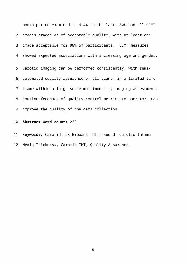
quality assurance (QA), with QA failures falling from 16.2% in the first two-month period
examined to 6.4% in the last. 80% had all CIMT images graded as of acceptable quality, with
at least one image acceptable for 98% of participants. CIMT measures showed expected
associations with increasing age and gender.
Carotid imaging can be performed consistently, with semi-automated quality assurance of all
scans, in a limited time frame within a large scale multimodality imaging assessment.
Routine feedback of quality control metrics to operators can improve the quality of the data
collection.
Abstract word count: 239
4
12
3
4
5
6
7
8
9
10
11
12
13
14
15
16
17
18
19
20
21
22
23

Keywords: Carotid, UK Biobank, Ultrasound, Carotid Intima Media Thickness, Carotid
IMT, Quality Assurance
5
1
2
INTRODUCTION
UK Biobank is a prospective study of 500,000 people aged between 40 and 69 years at
recruitment between 2006 and 2010.1 An initial feasibility assessment (pilot phase) was
initiated in 2014, to evaluate the feasibility of conducting a multi-modal imaging study in
100,000 participants over 6 years, including MRI scans of the brain, heart and body, a dual-
energy X-ray absorptiometry (DEXA) scan and a carotid ultrasound.2 The large scale and the
richness of the phenotyping in UK Biobank allows researchers to examine aspects of
cardiovascular epidemiology that previously would not have been possible.
Imaging of the carotid vessels can provide information on the thickness of the carotid vessels,
expressed as the carotid intima media thickness (CIMT), and the presence of carotid plaque.
Information concerning both has been associated with incident cardiovascular events.3–5 Use
of ultrasound to image the carotids has the advantage of being relatively low-cost, fast, free
of ionizing radiation, and requiring relatively little training to use,6 leading to its successful
use in a number of large population-based cohort studies.4,5,7,8 Good reproducibility is seen
even among relatively inexperienced operators under routine clinical conditions.6 However,
imaging on the scale of UK Biobank is unprecedented and requires adaptation of usual
approaches that allow both efficient data acquisition and exceptional attention to quality
assurance, to avoid bias and minimise measurement variation over time. Previous
publications have described the UK Biobank in general,1 and the imaging substudies
involving cardiac MRI,2,9 liver MRI,10 and brain MRI.11 In this article, we describe the carotid
ultrasound scanning protocol used within the UK Biobank Imaging Enhancement study and
the development of a quality assurance process. We also report an evaluation of this process
based on carotid ultrasound scans from the first 2,560 participants.
6
1
2
3
4
5
6
7
8
9
10
11
12
13
14
15
16
17
18
19
20
21
22
23
24

METHODS
Protocol Development
A protocol was developed based on reported guidelines for carotid image acquisition12,13 that
took account of the practical aspects of space for equipment, time available and the limited
experience of the technicians. In addition, the requirement for potential future analysis of raw
ultrasound data was considered in acquisition of images. The protocol was devised to operate
alongside the other planned assessments for the Imaging Enhancement of UK Biobank
(cardiovascular, brain and abdominal magnetic resonance imaging, DEXA, as well as a
repeat of the study’s baseline measures, which incorporate a comprehensive computer-based
questionnaire with interview, physical measures, and blood and urine sampling).
The flowchart for the protocol developed is shown in Figure 1. An abbreviated version of the
UK Biobank Standard Operating Protocol is available from
http://biobank.ctsu.ox.ac.uk/crystal/refer.cgi?id=511. In the pilot phase, imaging was
performed solely at the UK Biobank imaging facility at Cheadle, Stockport, UK. In brief, a
CardioHealth Station (Panasonic Healthcare Corporation of North America, Newark, NJ,
USA) was used, with a 9MHz linear array transducer. The carotid study was performed in a
darkened room and participants lay supine supported by a triangular pillow. Initially, a 2D
sweep of both carotids was performed along the short-axis (transverse plane) from low in the
neck up to the jaw, at least to the level of the carotid bifurcation, with the right carotid
scanned first. The conventionally configured CardioHealth Station user interface software
allows these sweeps to be performed but not stored. To meet the needs of the UK Biobank,
this interface was modified prior to commencement of the study to allow cine image loop
7
1
2
3
4
5
6
7
8
9
10
11
12
13
14
15
16
17
18
19
20
21
22
23
24
25
storage. The sweep was repeated in the long-axis (longitudinal plane) and stored. These
longitudinal images were also used to identify the bifurcation for measurement of CIMT.
This was performed at two angles for each carotid, giving a total of four CIMT
measurements, which when measured from the vertical axis were 150o and 120o on the right
carotid, and 210o and 240o on the left carotid. Automated position location identification
provided a real-time readout of the transducer angle to the technician. The CIMT was
measured at time of acquisition using the semi-automated CardioHealth Station hardware and
software, with ‘Auto ROI’ and ‘Auto Freeze’ enabled. The CHS software places a marker on
the screen to guide the user in alignment of the flow divider and a 10mm region-of-interest
box to facilitate CIMT measurement. The far-wall of the common carotid was then
automatically tracked by the region-of-interest box. When three consecutive cardiac cycles
met internal quality thresholds, the image auto-froze in end-diastole, giving the mean,
maximum and minimum of the CIMT tracking. Manual override of the automatically derived
CIMT boundaries was performed in cases where the detected boundaries did not align with
the underlying B-mode image, and manual override of the auto-freeze feature was performed
if auto-freeze was not invoked after prolonged period of scanning. The technician also had
the facility to adjust the image to ensure the CIMT measurement was not taken at sites of
focal plaque, defined based on the Mannheim Consensus as: “a focal structure that
encroaches into the arterial lumen of at least 0.5mm or 50% of the surrounding IMT value or
demonstrates a thickness >1.5mm as measured from the media-adventitia interface to the
intima-lumen interface”.12
Quality Assurance
8
1
2
3
4
5
6
7
8
9
10
11
12
13
14
15
16
17
18
19
20
21
22
23

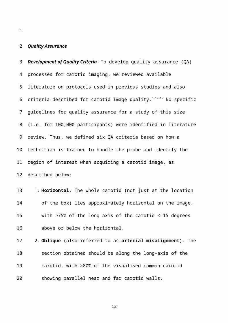
Development of Quality Criteria - To develop quality assurance (QA) processes for carotid
imaging, we reviewed available literature on protocols used in previous studies and also
criteria described for carotid image quality.5,12–15 No specific guidelines for quality assurance
for a study of this size (i.e. for 100,000 participants) were identified in literature review.
Thus, we defined six QA criteria based on how a technician is trained to handle the probe and
identify the region of interest when acquiring a carotid image, as described below:
1. Horizontal. The whole carotid (not just at the location of the box) lies approximately
horizontal on the image, with >75% of the long axis of the carotid < 15 degrees above
or below the horizontal.
2. Oblique (also referred to as arterial misalignment). The section obtained should be
along the long-axis of the carotid, with >80% of the visualised common carotid
showing parallel near and far carotid walls.
3. Box location. The distal edge of the box for automated CIMT measurement should be
positioned 1cm proximal to the flow divider of the carotid bulb, and there should be
no focal plaque within the box.
4. White-black-white. The CIMT should have distinct borders that define a clear
“white-black-white” pattern on the monitor.
5. CIMT tracking. The automated CIMT measurement was recorded at the correct time
in the cardiac cycle (end-diastole) and > 50% of the CIMT within the measurement
box has been tracked. All areas of CIMT tracked should accurately align with the B-
mode image.
6. Angle. The acquisition angle of the image should have been recorded within 30
degrees of the selected angle category (i.e., for images selected to represent 120
degrees, the acceptable range is 90-150 degrees).
9
1
2
3
4
5
6
7
8
9
10
11
12
13
14
15
16
17
18
19
20
21
22
23
24
Based on these six quality criteria, we developed a single Pass/Fail QA flag for images. For
an image to pass the overall QA, an image needed to pass all of the individual criteria of box
position and CIMT tracking, but could fail one of horizontal, oblique or white-black-white
criteria. For each participant, a carotid study consisted of four images showing the CIMT
measurement acquired at predefined angles of acquisition (i.e., right 150o, right 120o, left
210o, and left 240o). A separate QA flag was generated for each angle of acqusition.
Assessment of Quality Criteria - To evaluate our approach, we used a test dataset of 100
carotid studies to estimate both inter and intra-observer reproducibility of the quality criteria,
after which we evaluated the process as a whole in the Pilot Phase (2567 subjects, of whom
2560 had a QA assessment at each angle of acquisition). To ensure consistency in application
of these criteria, we developed a web-based interface to allow the rapid semi-automated
review of images. The images were loaded into a simple custom designed grading tool
(Intelligent Ultrasound Ltd, Abingdon, United Kingdom), and each image was assessed twice
(on separate occasions) by three clinicians experienced in grading carotid ultrasound images,
henceford called graders. When assessing inter- and intra-observer reliability or agreement,
measures that adjust the raw percentage agreement by that expected due to chance are used,
such as Cohen’s kappa. However, Cohen’s kappa and its direct extensions tend to show
paradoxical behaviour at the extremes of prevalence (for example, as in our case, if a large
majority of images passed QA assessment), with very high agreement between observers
sometimes even leading to negative kappas.16,17 We therefore used Gwet’s first order
agreement coefficient, referred to as the AC1 statistic, to assess reliability. This, like Cohen’s
kappa, is 0 in cases of no agreement and 1 in cases of perfect agreement, but incorporates
observer uncertainty into the model and tends to be less affected by prevalence.17,18
Application of Quality Assurance and Quality Audit in Pilot Phase - Participant age was
provided and analysed according to decade of birth, with age groups of ≤50, 51-60, 61-70,
10
1
2
3
4
5
6
7
8
9
10
11
12
13
14
15
16
17
18
19
20
21
22
23
24
25
and ≥71 years at the time of carotid ultrasound. Once the QA criteria were finalised, all
studies acquired were reviewed for quality assurance (QA) purposes by a trained UK
Biobank senior radiographer and information fed back to the sonographers on a weekly basis.
Images from each of the four acquired angles were graded as Pass/Fail, according to the
developed criteria and based on an overall assessment of each image. A random selection of
approximately 10% of these images, as well as any borderline images, were reviewed by
expert staff in the Oxford Cardiovascular Clinical Research Facility. Results from the
external review were fed back to the imaging centre on a monthly basis to complete the audit
cycle, with teleconferences as required to discuss any specific issues.
Statistical Analysis - At the end of the pilot phase, we used the available data on CIMT,
quality assurance and the age and sex of participants to investigate the overall validity of the
acquired carotid measurements. Specifically, we studied associations between CIMT and age
and sex, reasoning that measured CIMT should be higher in older age groups and in male
participants. We used multivariate linear regression, with predictor variables of age (as an
ordered categorical variable based on decade) and sex. We then assessed the impact of the
quality assurance process on carotid measures by studying how the model fit varied if images
that failed QA were excluded. We did this based on exclusion of all studies in which one
image failed QA. All statistical analyses were performed using R version 3.2.2.19
RESULTS
Assessment of Quality Criteria - 588 images from 100 participants acquired during the pilot
phase of the UK Biobank Imaging Enhancement were graded in the semi-automated tool
(Figure 2A) by three graders on two separate occasions. More than one image from each
angle of acquisition was eligible for assessment during this phase of the QA process. Overall,
there was a very good agreement between evaluations performed by different graders
11
1
2
3
4
5
6
7
8
9
10
11
12
13
14
15
16
17
18
19
20
21
22
23
24
25
26
(Supplementary Figure 1, Supplementary Table 1). All criteria showed excellent intra-
observer reliability (minimum Gwet AC1 88%), with good inter-observer reliability
(minimum Gwet AC1 78%). Examples of images passing and failing QA are shown in Figure
2B-G.
Application of Quality Assurance and Quality Audit - Images from the first 2567
participants after implementation of the QA protocol were evaluated. Pass/Fail QA flags for
all four angles of acquisition were available for 2560 participants, and, of these, 2519 (98%)
had at least one image passing QA. 523 (20%) studies had at least one image failing QA but
the proportion of images failing QA decreased over time, with an average across views of
16.2% failing QA in the first two month period compared to 6.4% failing in the last two
month period analysed (Supplementary Figure 2).
Evaluation of validity of carotid datasets
Data for evaluation of associations were available for 2558 of the 2567 participants. Based on
the entire dataset, mean CIMT was higher in older, compared with younger, age groups and
was higher in men than in women (Table 1, Supplementary Figure 3). A linear regression
model (Table 2) showed a 61µm (95% CI: 55-67µm) increase in CIMT per decade, and a
48µm (95% CI: 39-57µm) increase in male compared to female participants compared to the
average CIMT of 553µm (95% confidence interval (95% CI): 542-564µm) in 45-50 year old
females in the model (adjusted R2 0.191, p<2x10-16). Studies with one image that failed QA
tended to have on average a 46µm greater CIMT (Supplementary Figure 3) and a linear
regression model with a QA flag (indicating any image failing QA) had slightly higher
adjusted R2 than the model without the QA flag (Table 2). However, the confidence interval
for reference CIMT in younger females, as well as those of older age and male sex, was very
12
1
2
3
4
5
6
7
8
9
10
11
12
13
14
15
16
17
18
19
20
21
22
23
similar under both models, suggesting that the impact of including ‘failed’ images on the
overall assessment is small.
DISCUSSION
We describe a protocol for carotid image acquisition and semi-automated measurement of
carotid IMT within a limited timeframe for application within a large cohort study.
Furthermore, we describe development of quality assurance criteria to allow evaluation of
quality of images and audit control. Application of these procedures reduced the number of
quality fails within scans and helped to ensure that 98% of participants had at least one CIMT
measure. Analysis of the dataset confirms that the CIMT measures within the UK Biobank
Imaging Enhancement pilot phase associate with age and sex in the expected way, implying
face validity of the measures.
A key feature of the UK Biobank imaging protocol is that it needs to be performed within a
limited time frame in participants who also undergo abdominal, cardiac and brain magnetic
resonance imaging, dual energy X-ray absorptiometry (DEXA), blood sampling and an
extensive questionnaire. During the study visit, only 10 minutes is available for carotid
imaging, including time for patient registration. In addition to this time constraint, we expect
that data acquisition will be performed by multiple technicians at three separate imaging
assessment centres over the course of the Imaging Enhancement Study (which will take 6-7
years). Our protocol was therefore developed to achieve a relatively detailed image
acquisition in a highly reproducible manner. A recent meta-analysis highlights the variability
in CIMT measurements across studies.20 The use of semi-automated measurement techniques
is recommended in international consensus statements to reduce inter-observer variability,12,13
and we therefore chose an ultrasound system that implements these. In addition, the
13
1
2
3
4
5
6
7
8
9
10
11
12
13
14
15
16
17
18
19
20
21
22
23
24
25

CardioHealth Station system we used has been shown to have excellent reproducibility even
in less-experienced users.6,21,22
A potential limitation of the protocol at this time is the lack of quantification of the plaque
that may be present in the images. Focal plaque has been shown to provide additional
discrimination above that provided by CIMT measurement alone.23,24 However, the additional
discrimination is modest (area under the receiver operating curve 0.64 for plaque vs 0.61 for
CIMT as a predictor of myocardial infarction over an average of eight years in a large meta-
analysis).24 As the assessment of plaque is much more subjective, it is both operator
dependent and time consuming. Therefore it was not considered practical to provide
evaluation of plaque at the time of image acquisition. However, the image acquisition
protocol provides both a short axis 2D sweep along the length of the carotid as well as
longitudinal views to above the bifurcation. Retrospective evaluation of stored image datasets
will therefore be possible to assess plaque characteristics and other novel carotid parameters
– these images will be available to external researchers. In addition, these stored raw image
datasets have the potential to be used for future novel metric evaluations including, for
example, assessment of geometric variation or measurements of intima media thickness in
other locations within the vessel. By linking these measures to the available imaging
modalities or later clinical outcomes, potentially new carotid biomarkers of cerebral or
cardiovascular health could be identified.
The within and between rater reliability of our QA criteria suggest they can be applied with
little inter- or intra-observer variability. A number of these criteria, such as the “White-Black-
White” criterion, are integral to CIMT measurement, while others, such as the exclusion of
14
1
2
3
4
5
6
7
8
9
10
11
12
13
14
15
16
17
18
19
20
21
22
23
24

focal plaque from the measurement box, were chosen to increase reproducibility. The
“Angle” criterion has relatively relaxed requirements for the angle of acquisition, as we
decided that the exact angle of acquisition was not as important as repeated measurements of
the two carotids in this single time point study, and therefore should not lead to failure of the
entire carotid study. With the development of a standardised interface for grading the quality
of the images acquired and a larger number of studies acquired it may be possible to develop
image analysis techniques that would automate the QA process further.
The impact of inclusion or exclusion of QA criteria on the model fit was very small and
therefore data released by UK Biobank includes all CIMT measures irrespective of the QA
flag. There was a higher rate of QA fails with older age, and the provision of QA flag may
therefore be of scientific interest. There is a distinct possibility that future analyses will reveal
that images that fail QA have predictive value in themselves. For example, it is possible that
highly tortuous carotid arteries would be more likely to fail QA, and that the tortuosity is
predictive of future cardiovascular events. The QA flag for each image is therefore provided
along with the CIMT measure on the UK Biobank data showcase (available at
biobank.ctsu.ox.ac.uk/showcase).
A major advantage of the QA process is that real-time feedback is possible during training.
The number of images failing QA during the course of the pilot phase more than halved
following such feedback based on QA measures, with a rapid fall on implementation of the
QA audit process and maintenance of lower levels thereafter. Interestingly, both left sided
angles of acquisition had a higher failure rate initially, and appeared to have a more rapid
improvement compared to right sided angles of acquisition. As we did not record handedness
15
1
2
3
4
5
6
7
8
9
10
11
12
13
14
15
16
17
18
19
20
21
22
23
24

of operators, we were unable to investigate this further. The audit process involved feedback
to the technicians of both number of image fails and reasons for image quality failures.
Furthermore, as each image could be identified by time and operator, the advice could be
personalised for individual technicians to improve their carotid imaging technique. Several
operators performed carotid imaging during the pilot phase and new operators started during
that period. The QA criteria were introduced as part of the training for new technicians and
each was required to show competency against the QA criteria in test cases before being
allowed to scan participants. Therefore the maintenance of a low failure rate provides
reassurance that the use of the QA criteria within training processes was also robust.
In summary, carotid images and semi-automated CIMT measures are now available from the
UK Biobank Imaging Enhancement Study. This is the first published description of the
protocol used for acquisition of these images and the rationale for the design. Furthermore,
we provide a detailed evaluation of the quality assurance process, description of a semi-
automated online tool for rapid image evaluation and demonstrate that the data generated fits
expected patterns of association with age and gender.
16
1
2
3
4
5
6
7
8
9
10
11
12
13
14
15
16

Conflicts of interest
SL is an employee of, and RB and RM are consultants to Panasonic Corporation. JP is an
employee of, and AN, MY and PL are consultants to Intelligent Ultrasound Ltd. SH, SG, NA,
CS, RC, PM, and PL are employees of or have received support from UK Biobank.
17
1
2
3
4
5
6
7

REFERENCES
1. Sudlow C, Gallacher J, Allen N, et al. UK Biobank: An Open Access Resource for Identifying the Causes of a Wide Range of Complex Diseases of Middle and Old Age. PLoS Med 2015; 12: e1001779.
2. Petersen SE, Matthews PM, Bamberg F, et al. Imaging in population science: cardiovascular magnetic resonance in 100,000 participants of UK Biobank - rationale, challenges and approaches. J Cardiovasc Magn Reson 2013; 15: 46.
3. Naqvi TZ, Lee M-S. Carotid Intima-Media Thickness and Plaque in Cardiovascular Risk Assessment. JACC Cardiovasc Imaging 2014; 7: 1025–1038.
4. Polak JF, Pencina MJ, Pencina KM, et al. Carotid-wall intima-media thickness and cardiovascular events. N Engl J Med 2011; 365: 213–21.
5. Nambi V, Chambless L, Folsom AR, et al. Carotid intima-media thickness and presence or absence of plaque improves prediction of coronary heart disease risk: the ARIC (Atherosclerosis Risk In Communities) study. J Am Coll Cardiol 2010; 55: 1600–7.
6. Vanoli D, Wiklund U, Lindqvist P, et al. Successful novice’s training in obtaining accurate assessment of carotid IMT using an automated ultrasound system. Eur Heart J Cardiovasc Imaging 2014; 15: 637–42.
7. Herder M, Johnsen SH, Arntzen K a., et al. Risk Factors for Progression of Carotid Intima-Media Thickness and Total Plaque Area: A 13-Year Follow-Up Study: The Tromsø Study. Stroke 2012; 43: 1818–1823.
8. Clarke R, Du H, Kurmi O, et al. Burden of carotid artery atherosclerosis in Chinese adults: Implications for future risk of cardiovascular diseases. Eur J Prev Cardiol 2017; 24: 647–656.
9. Petersen SE, Matthews PM, Francis JM, et al. UK Biobank’s cardiovascular magnetic resonance protocol. J Cardiovasc Magn Reson 2015; 18: 8.
10. Wilman HR, Kelly M, Garratt S, et al. Characterisation of liver fat in the UK Biobank cohort. PLoS One 2017; 12: e0172921.
11. Miller KL, Alfaro-Almagro F, Bangerter NK, et al. Multimodal population brain imaging in the UK Biobank prospective epidemiological study. Nat Neurosci 2016; 19: 1523–1536.
12. Touboul P-J, Hennerici MG, Meairs S, et al. Mannheim Carotid Intima-Media Thickness and Plaque Consensus (2004-2006-2011). Cerebrovasc Dis 2012; 34: 290–296.
13. Stein JH, Korcarz CE, Hurst RT, et al. Use of Carotid Ultrasound to Identify Subclinical Vascular Disease and Evaluate Cardiovascular Disease Risk: A Consensus Statement from the American Society of Echocardiography Carotid Intima-Media Thickness Task Force Endorsed by the Society for Vascular. J Am Soc Echocardiogr 2008; 21: 93–111.
14. Bots ML, den Ruijter HM. Should We Indeed Measure Carotid Intima-Media Thickness for Improving Prediction of Cardiovascular Events After IMPROVE? J Am Coll Cardiol 2012; 60: 1500–1502.
18
1
234
567
89
1011
12131415
161718
192021
222324
2526
2728
293031
323334
3536373839
404142

15. Nambi V, Chambless L, He M, et al. Common carotid artery intima-media thickness is as good as carotid intima-media thickness of all carotid artery segments in improving prediction of coronary heart disease risk in the Atherosclerosis Risk in Communities (ARIC) study. Eur Heart J 2012; 33: 183–90.
16. Feinstein AR, Cicchetti D V. High agreement but low kappa: I. The problems of two paradoxes. J Clin Epidemiol 1990; 43: 543–9.
17. Gwet KL. Computing inter-rater reliability and its variance in the presence of high agreement. Br J Math Stat Psychol 2008; 61: 29–48.
18. Feng GC. Factors affecting intercoder reliability: a Monte Carlo experiment. Qual Quant 2013; 47: 2959–2982.
19. R Core Team. R: A Language and Environment for Statistical Computinghttps://www.r-project.org/ (2016).
20. Liao X, Norata GD, Polak JF, et al. Normative values for carotid intima media thickness and its progression: Are they transferrable outside of their cohort of origin? Eur J Prev Cardiol 2016; 23: 1165–1173.
21. Singh S, Nagra A, Maheshwari P, et al. Rapid Screening for Subclinical Atherosclerosis by Carotid Ultrasound Examination: The HAPPY (Heart Attack Prevention Program for You) Substudy. Glob Heart 2013; 8: 83–9.
22. Vanoli D, Lindqvist P, Wiklund U, et al. Fully automated on-screen carotid intima-media thickness measurement: a screening tool for subclinical atherosclerosis. J Clin Ultrasound 2013; 41: 333–9.
23. Baldassarre D, Hamsten A, Veglia F, et al. Measurements of Carotid Intima-Media Thickness and of Interadventitia Common Carotid Diameter Improve Prediction of Cardiovascular Events. J Am Coll Cardiol 2012; 60: 1489–1499.
24. Inaba Y, Chen JA, Bergmann SR. Carotid plaque, compared with carotid intima-media thickness, more accurately predicts coronary artery disease events: A meta-analysis. Atherosclerosis 2012; 220: 128–133.
19
1234
56
78
910
1112
131415
161718
192021
222324
252627
28
29

Table 1. Carotid intima-media thickness by age.
Age (years) n
Overall mean (sd)
Female
n (%)
Female
mean (sd)
Male
mean (sd)
Poor QA
n (%)
≤ 50 170 588 (84) 95 (56%) 578 (76) 600 (93) 23 (14%)
51-60 787 628 (107) 451 (57%) 607 (83) 657 (126) 119 (15%)
61-70 1180 701 (123) 596 (51%) 675 (104) 728 (134) 238 (20%)
≥ 71 421 763 (145) 175 (42%) 739 (126) 781 (155) 143 (34%)
“Poor QA” refers to studies where at least one of the four angles of acquisition was deemed to
not pass quality assurance criteria. CIMT is measured in micrometres. Abbreviations: CIMT,
carotid intima-media thickness; sd, standard deviation.
20
1
2
3
4
5
6
7
8
9
Table 2. Linear regression, with carotid intima media thickness as outcome variable.
Basic model Incorporating QA
n 2558 2558
Adjusted R2 0.19 0.21
Average CIMT for ≤ 50 year old females
553 (542 - 564) 551 (539 - 562)
Increase per decade 61 (55 - 67) 58 (52 - 63)
Increase in males 48 (39 - 57) 45 (36 - 54)
Increase if study had any image failing QA
46 (35 - 57)
CIMT is measured in µm, with the 95% confidence interval shown in brackets. The average
CIMT for ≤ 50 year old females is based on the linear regression model intercept. Abbreviations:
CIMT, carotid intima media thickness; QA, quality assurance.
21
1
2
3
4
5
6
7
8
9
10

Figure 1. Overview of UK Biobank carotid ultrasound protocol.
Outputs from the protocol are shown on the right of the figure. Four separate sets of CIMT
measurements were obtained: right carotid at 150o, right carotid at 120o, left carotid at 210o, and left
carotid at 240o. Numeric values stored were maximum, mean, and minimum CIMT.
Abbreviations: CIMT, carotid intima-media thickness.
22
1
2
3
4
5
6
7
8

Figure 2. Quality assurance grading process.
Figure showing the grading interface (A) and example images (lower panel). The grading
interface shows an image not meeting the “White-black-white” quality assurance (QA) criterion
(indicated by the lack of a tick in this box). The example images show a good quality image (B),
23
1
2
3
4
5
and images failing quality assurance criteria: Horizontal (C), Oblique (D), Box location (E), CIMT
tracking (F), and Angle (G). Assessment of the Angle criterion was facilitated by the use of an
automated transducer position identifier, as shown in 2G.
24
1
2
3