abstract - mineralogical society of america102 in this study, these techniques were combined, for...
TRANSCRIPT
1
2
3
4
, 5
REVISION 1
Non-destructive, multi-method, internal analysis of multiple inclusions in a single diamond:
first occurrence of mackinawite (Fe,Ni)1+xS
Giovanna Agrosì 1, Gioacchino Tempesta 1, Daniela Mele 1, Ignazio Allegretta 2, Roberto Terzano2
Steven B. Shirey 3, Graham D. Pearson 4, Fabrizio Nestola 56
7
1 Dipartimento di Scienze della Terra e Geoambientali, Università degli Studi “Aldo Moro”, Via 8
Orabona, 4, 70125 Bari, Italia 9 2 Dipartimento di Scienze del suolo, della pianta e degli alimenti (Di.S.S.P.A.), Università degli 10
Studi “Aldo Moro” Via Amendola, 165/A 70126 - Bari – Italia 11 3 Department of Terrestrial Magnetism, Carnegie Institution for Science, 5241 Broad Branch Road, 12
NW, Washington, DC 20015, USA 13 4 Department of Earth and Atmospheric Sciences, University of Alberta, Edmonton, Alberta 14
Canada T6G 2E3 15 5 Dipartimento di Geoscienze, Università degli Studi di Padova, Via G. Gradenigo 6, 35131 Padova, 16
Italia 17 18
Abstract 19
A single gem lithospheric diamond with 5 sulfide inclusions from the Udachnaya kimberlite 20
(Siberia, Russia) has been analyzed non-destructively to track the growth conditions of the 21
diamond. Sulfides are the most abundant mineral inclusions in many lithospheric diamond crystals 22
and are the most favorable minerals to date diamond crystals by Re-Os isotope systematics. Our 23
investigation used non-destructive, micro-techniques, combining X-ray tomography, X-ray 24
fluorescence, X-ray powder diffraction and Raman spectroscopy. This approach allowed us to 25
determine the spatial distribution of the inclusions, their chemical and mineralogical composition on 26
the micro- scale and, finally, the paragenetic association, leaving the diamond host completely 27
unaffected. The sample was also studied by X-ray diffraction topography to characterize the 28
structural defects of the diamond and to obtain genetic information about its growth history. The X-29
ray topographic images show that the sample investigated exhibits plastic deformation. One set of 30
{111} slip lamellae, corresponding to polysynthetic twinning, affects the entire sample. Chemical31
data on the inclusions still trapped within the diamond show they are monosulfide solid solutions of 32
Fe, Ni and indicate a peridotitic paragenesis. Micro X-ray diffraction reveals that the inclusions 33

mainly consist of a polycrystalline aggregate of pentlandite and pyrrothite. A thorough analysis of 34
the Raman data suggests the presence of a further Fe,Ni sulfide, never reported so far in diamonds: 35
mackinawite. The total absence of any oxides in the sulfide assemblage clearly indicates that 36
mackinawite is not simply a “late” alteration of pyrrhotite and pentlandite due to secondary 37
oxidizing fluids entering diamond fractures after the diamond transport to the surface. Instead, it is 38
likely formed as a low-temperature phase that grew in a closed system within the diamond host. It is 39
possible that mackinawite is a more common phase in sulfide assemblages within diamond crystals 40
than has previously been presumed, and that the percentage of mackinawite within a given sulfide 41
assemblage could vary from diamond to diamond and from locality to locality. 42
43
Keywords: Diamond, sulfide, mackinawite, non-destructive analyses 44
45
Introduction 46
The study of diamond and mineral inclusions trapped within them may provide critical insights into 47
aspects of deep mantle mineralogy as well as the origin of the cratonic lithosphere and its evolution 48
(Sobolev 1977; Stachel and Harris 2008; Shirey et al. 2013 and references therein). Over the last 30 49
years (i.e. since Richardson et al. 1984), these studies have also helped to constrain our 50
understanding of the temporal evolution of the lithospheric mantle and the water content of cratonic 51
lithosphere and the Earth’s mantle transition zone (Pearson et al. 2014; Novella et al. 2015; Nestola 52
and Smyth 2016; Jean et al. 2016; Taylor et al. 2016). To obtain information about the physico-53
chemical conditions under which the crystallization of diamond occurred, scientists have mainly 54
investigated the growth history of the diamond crystals, the geochemical and crystallographic 55
features of their inclusions and the relationships between the inclusions and their diamond hosts. 56
These studies have been commonly performed by destructive methods involving crushing (Sobolev 57
et al. 1970; Gurney et al. 1984; Gurney 1989; Aulbach et al. 2009) or ion/laser ablating (Seitz et al. 58

2003; Gallou et al. 2012) the diamond samples in order to expose the inclusions for conventional 59
geochemical analyses. 60
Recently the trend has been to cut plates to obtain images of diamond internal growth zoning by 61
cathodoluminescence (Howell et al. 2015 and reference therein). However, in all these approaches, 62
some crucial information about the growth conditions of diamond, such as the entrapment pressure 63
of the inclusions, their original crystallographic orientation with respect to the diamond host (e.g. 64
Nestola et al. 2011; Fedortchouk et al. 2011; Nestola et al. 2012; Nestola 2015; Borges et al. 2016), 65
is lost, or their original volatile content could be lost. In situ investigation of diamond with the 66
inclusions still trapped in it, using non-destructive techniques is the best way to preserve this 67
information. For this reason, in recent years, the scientific community in diamond research has 68
developed a different methodological approach to investigate diamond crystals without destroying 69
the samples. Among different methods, Raman spectroscopy (eg. Sobolev et al. 2000; Pearson et al. 70
2014; Smith et al. 2016) and quantitative birefringence analysis using the MetriPolTM system 71
(Howell 2012 and references therein) represent the prevalent techniques to measure the elastic 72
effects derived from the differences in thermo-elastic properties between mineral inclusions and 73
host diamond. From measuring these elastic effects, an indication about the entrapment pressure of 74
the inclusions may be obtained and from this the depth (or at least the minimum depth) in the 75
mantle of diamond formation. More recently, non-destructive X-ray diffraction (and micro-X-ray 76
diffraction) analysis (XRD) was adopted to develop “elastic geobarometry” as a way to determine 77
the depth of crystallization of inclusion-bearing diamonds. Moreover, this technique furnished key 78
information on the diamond-inclusion reciprocal crystallographic orientations useful to constrain 79
protogenesis versus syngenesis (Nestola et al. 2014; Angel et al. 2014; Angel et al. 2015a,b,c; 80
Milani et al. 2016; Nestola et al. 2017). 81
In this study , with the aim to “map” spatial and chemical information that relate to the origin of the 82
diamond while completely preserving the diamond, we have used a multi-technique approach, 83
adding to the aforementioned micro-Raman spectroscopy and XRD the following non-destructive 84

methods: X-ray topography (XRDT), Micro-computed X-ray tomography analysis (CXRT) and 85
Micro X-ray fluorescence (XRF). In the past, the capabilities of each of these non-destructive 86
techniques were already experienced in the diamond research, using mainly synchrotron source. 87
XRF was previously used to obtain direct chemical analysis of inclusions trapped in diamond, 88
providing, in some cases, also 3D reconstruction of maps (e.g. Brenker et al. 2005; Sitepu et al. 89
2005; Silversmidt 2011; Pearson et al. 2014; La Force et al. 2014). CXRT has been successfully 90
adopted to locate mineral inclusions in insufficiently transparent diamond crystals (Kovalenko et al. 91
2012; Nestola et al. 2012; Nimis et al. 2016). 92
XRDT method, extensively used, in the past, to screen the crystalline quality of natural and 93
synthetic crystals used as electronic devices (Agrosì et al. 2009 and 2011), has been successfully 94
employed in Earth Sciences research to study the growth history of tourmalines, garnets and beryls 95
(Agrosì et al. 2006; Agrosì et al. 2011; Tempesta et al. 2011; Pignatelli et al. 2015). Recently, this 96
method has been applied to provide minerogenetic information on diamond in non-destructive way. 97
The results obtained in two previous studies by XRDT on diamond samples from Finsch mine, 98
South Africa (Agrosì et al. 2013), and from the Udachnaya kimberlite, Siberia (Agrosì et al. 2016), 99
revealed significant petrogenetic relationships between the mineral inclusions and their diamond 100
hosts. 101
In this study, these techniques were combined, for the first time, to investigate one diamond from 102
Udachnaya kimberlite (Siberia, Russia) with the inclusions still trapped in it, using only 103
conventional sources. 104
It is worth noting that this methodological approach not only allowed to study the structural defects, 105
define the spatial distribution of the inclusions, determine their chemical composition and identify 106
mineral species, but also allowed to find the first occurrence of mackinawite as an inclusion in 107
diamond. To our knowledge, the only previous suggestion of the possible presence of mackinawite 108
was reported by Thomassot (2006) where an analysis was made of a multi-phase sulfide inclusion 109
within a diamond. However, unfortunately, in this case, the identification of mackinawite was only 110

made on the basis of chemical analysis and stoichiometry, which alone cannot be used for such a 111
definitive identification. No structural data were reported. So at present, our work reports the first 112
definitive evidence of mackinwaite in diamond. 113
Experimental methods 114
Sample 115
The diamond studied in this work is a brown specimen of type IaAB from Udachnaya kimberlite 116
(Siberia, Russia). The crystal exhibits an octahedral morphology, flattened along the (110) plane, 117
with stepped development of the {111} faces (Fig. 1a). Optical observations revealed that the 118
diamond has anomalous birefringence and contains cleavage planes (CP) and fractures partially 119
healed by dark microinclusions (Fig. 1a). The presence of healed fractures renders it difficult, if not 120
impossible, to observe, using optical microscopy alone, the large included solid phases inside the 121
inner part of diamond. 122
123
XRDT 124
XRDT represents a helpful method to obtain images of extended lattice defects with a resolution 125
limit of a few µm. This method is particularly suitable to study structural defects in diamond 126
because the low attenuation coefficient of the X-ray beam makes this mineral highly transparent to 127
X-rays, allowing investigation of the structural defects of the whole sample, instead of only the128
surface of sample, as is the case with cathodoluminescence. Indeed, the topographic method used 129
here works in transmission mode and provides images of the strain fields associated with defects, 130
without the necessity to cut the sample in slices. 131
The topographs, taken with Laue geometry, were carried out using a Rigaku camera with 132
monochromatic radiation (MoKα1) and with a micro-focus X-ray tube. The 1 mm thickness of the 133
sample allows the optimum kinematic diffraction condition μt ≈ 1 (μ = linear absorption coefficient; 134
t = crystal thickness), minimizing X-ray absorption. Spatial resolution using these conditions is 135
about 1-2 μm. Diffraction contrast was recorded on high-resolution photographic films (SR Kodak) 136

that provide better contrast resolution rather than other recording methods. Characterization of the 137
structural defects was performed by applying the extinction criteria to their diffraction contrasts, 138
according to kinematic and dynamic X-ray diffraction theories (Authier and Zarka 1994). Detailed 139
XRDT procedures used in this study are given in Agrosì et al. (2016). 140
CXRT141
This technique is a powerful, non-destructive method for obtaining 3D information on internal 142
structures of a large variety of objects (Cnudde and Boone 2013; Howarth et al. 2015). It is able to 143
distinguish highly-X-ray-absorbing mineral inclusions from highly X-ray transparent diamond, 144
providing a 3D reconstruction of the sample and the spatial distribution of the inclusions trapped in 145
it as well as a visualization of their morphologies (see Nestola et al. 2012 for an example of 146
application to diamond). 147
In this study, the instrument utilized was a SkyScan 1172 microtomograph equipped with a W tube. 148
A 45 kV X-ray source was used with a current of 218 A. A total of 1200 absorption radiographs 149
were acquired over a 360° rotation with an angular step of 0.3°. Random movement of the vertical 150
axis and multiple-frame averaging to minimize the Poisson noise in the projection images were 151
used. Beam hardening was reduced by the presence of a 0.5 mm Al-filter between source and 152
detector. The nominal spatial resolution for the resulting model was 4.75 m. The raw data were 153
reconstructed into two-dimensional slice images using the software “NRecon, SkyScan, Belgium”. 154
Corrections for the beam-hardening effect and ring artefacts were also applied during the 155
reconstruction process; CXRT datasets were analyzed using the software “CT-analyser, Skyscan, 156
Belgium”. 157
XRF158
In this study, chemical analyses were obtained by conventional source using an M4 Tornado μXRF 159
(Bruker Nano GmbH, Berlin) equipped with a Rh tube with policapillary optics (50 kV, 600 μA, 30 160
W) having a spot size of 25 μm. Two XFlash® silicon drift detectors with an active area of 30 mm2161
were used. The resolution was lower than 140 eV for both detectors. For elemental maps, one 162

spectrum was acquired every 10 μm with both detectors and acquisition time set at 10 ms per pixel. 163
Elemental analyses on inclusions within the diamond were performed with only one detector in 164
order to exclude diffraction peaks which can overlap heavy metal K lines. A live time of 60 s was 165
used. All analyses were performed under vacuum (20 mbar) to avoid Ar absorption and to detect 166
light elements. Spectral quantification was performed in standardless mode using M4 Tornado 167
software. NIST SRM 611 and NIST SRM 613 standards were analyzed in order to periodically test 168
the performance of the instrument. 169
XRD170
X-ray diffraction measurements were performed using a Rigaku Oxford Diffraction SuperNova171
single-crystal diffractometer, equipped with a Dectris Pilatus 200K area detector and a Mova X-ray 172
microsource (beam spot ~ 0.12 mm; Nestola et al. 2016). For the measurements, MoKα-radiation, 173
operating at 50 kV and 0.8 mA was used. The sample to detector distance was 68 mm. Data 174
reduction was performed using the CrysAlis software (Rigaku Oxford Diffraction). The instrument 175
is able to perform in “micro powder diffraction mode” to acquire X-ray diffractograms on 176
polycrystalline grains entrapped in diamond crystals with size down to 20-10 m. 177
Micro-Raman spectroscopy 178
Micro-Raman spectroscopy was performed on the sample in backscattered geometry with a Jobin-179
Yvon Horiba ‘‘XploRATM’’ apparatus, equipped with a microscope with 10 and 50 objectives 180
and a motorized x–y stage. The 532 nm line of a class 1 laser used for excitation was coupled with a 181
1200 line/mm grating and a high-sensitivity CCD air cooled detector with a 1024 pixel. A filter was 182
used to reduce the laser power on the sample. The frequency calibration was performed against the 183
Raman peak of silicon. The instrument allowed a spatial resolution of about 1 µm, with a spectral 184
resolution of 1.8 cm-1. The peak positions were obtained from a multipoint baseline corrected185
spectrum using the computer program GRAMS/AI 8.0 (Thermo Electron # 2001). 186
187
Results 188

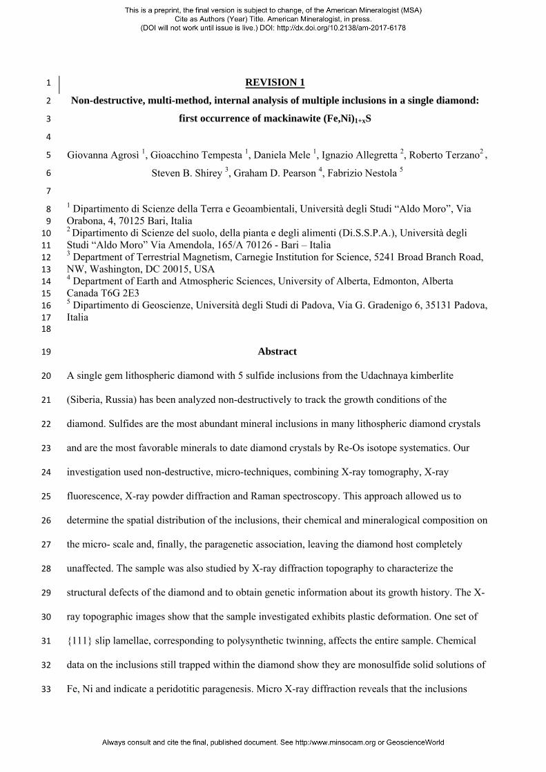
Optical observations reveal that the diamond crystal, although transparent, contains several 189
microfractures healed by dark and opaque material making difficult the optical study of the 190
inclusions (Fig. 1a). The crystal exhibits anomalous birefringence and a trace of cleavage parallel to 191
the (111) plane passing through the whole crystal (Fig. 1a, 1b, 1c). 192
In order to visualise the inclusions, the crystal was studied by CXRT. The inclusions could be 193
easily distinguished from the host diamond crystal by simply thresholding the grey value histogram 194
of the reconstructed images (Fig. 1b). 195
3D tomographic surface rendering (see Fig. 1b) shows five highly absorbing large inclusions on the 196
core of the diamond. Three of the inclusions that extend near the cleavage trace exhibit an apparent 197
diamond-imposed morphology, whereas two other inclusions show irregular, polyhedral 198
morphologies. Some cracks propagating through the diamond from each of the inclusions may be 199
ascribed to their different thermo-elastic behaviour with respect to the diamond host. Some healed 200
fractures, optically observed, do not cause any contrasts in the tomographic images. Only one 201
healed fracture, labelled F, in the lower part of the figure can be observed (Fig. 1b). The 202
tomographic study reveals no connection between this fracture and the inclusions and consequently, 203
it is not related to the trapped inclusions. 204
The X-ray topographic images (Fig. 1c and Fig. 2) show that the diamond exhibits micro-205
laminations (µL) parallel to the (111) plane (Fig. 1c and Fig. 2a, 2c). Such laminations represent the 206
polysynthetic twinning of micro-lamellae commonly found in diamond crystals (Titkov et al. 2012). 207
The diffraction contrasts observed in these images reveal regions slightly disoriented relative to one 208
another. Indeed, in Figure 2 it can be observed that under the same diffraction vector, and thus 209
under the same Bragg angle, it is necessary to place the diamond in two different positions differing 210
by a few arc-seconds to obtain the diffraction of the whole sample. In Figure 2d the diffraction 211
contrast corresponding to the diamond core can be observed (see the dashed red line). In the core, 212
corresponding to early stage of diamond crystal growth, the large inclusions are out of contrast. The 213
topographic images show, additionally, that the fracture previously observed by optical microscopy 214

and by X-ray tomography and labelled as cleavage plane, forms an angle of about 70 degrees with 215
respect to the direction of the microlaminations. This confirms that this discontinuity coincides with 216
a typical octahedral cleavage along the (1-11) plane. It is worth noting that the cleavage plane 217
misaligns the microlaminations and thus it is post-twinning feature. 218
The chemical composition of the inclusions was detected by XRF. Semi-quantitative chemical 219
analyses obtained on the large inclusions (Table 1) suggest that these inclusions are essentially an 220
aggregate of Fe,Ni sulfides with small amounts of Cu and Cr. Due to the microcrystalline nature of 221
the aggregate (see micro X-ray diffraction results) and the relatively large beam diameter (about 25 222
m), it was not possible to obtain a reliable chemical composition for each of the three identified223
sulfides at the 1-2 m scale. However, we were able to obtain a bulk chemical composition that 224
corresponds with a formula: (Fe0.83Ni0.31Cu0.01Cr0.01)1.16S. 225
Chemical maps obtained on the whole sample were compared with a CXRT image (Fig. 3). To 226
verify the consistency between the chemical maps and the contrast observed by micro-tomography, 227
an image showing the same inclination of the sample observed on the maps was obtained from the 228
CXRT reconstruction. The sample shape visible from the maps is deformed due to the instrument229
geometry; the [110] axis appears to be inclined with respect to the images shown in Figures 1 and 2, 230
where the sample is placed exactly perpendicular to the [110] direction. The correlation between 231
the -XRF images and the properly oriented tomographic image confirms the content of Fe, Ni and 232
Cu in the inclusions and reveals that the aforementioned inclination produces the following 233
artefacts: inclusions 2 and 3 appear to be overlapping and the fracture F appears connected to these 234
inclusions (Fig. 3). Moreover, the maps reveal that the contrast of this fracture observed on the 235
tomographic images is due to a secondary filling of a Fe-rich material. 236
No additional information about the nature of the dark phases that healed the other fractures could 237
be acquired. The lack of tomographic contrast and the chemical information corresponding to these 238

secondary microinclusions could be due to the fact that they are likely dominated by light elements, 239
as well as carbon, decorating the pathways of late stage fluids. 240
The mineralogical composition of the inclusions was established by means of XRD and micro-241
Raman spectroscopy. The X-ray diffractogram (Figure 4) confirms that the inclusions mainly 242
consist of a microcrystalline aggregate of Fe-Ni sulfides (no diffraction spots, typical of single 243
crystal, were found during the measurements). In detail, the main peaks were ascribed to pentlandite 244
and pyrrhotite (Fig. 4). However, the Raman results (see below) indicate the presence of a third 245
sulphide, mackinawite. Unfortunately, the diffraction peaks of mackinawite are totally overlapped 246
with those of pentlandite and thus it is not trivial to definitively confirm its presence solely by 247
diffraction. Using the software HighScore Plus TM (Panalytical), we attempted to obtain a phase248
quantification of the three sulfides (a reference pattern of mackinawite was added to the 249
diffractogram based on the Raman results). Considering that the most intense diffraction peak of 250
mackinawite is the 001 at about 5.05 Å (Figure 4), it is evident that such a phase would represent a 251
very minor fraction of the aggregate. In detail, as visible in the pie graph in Figure 4, we obtained: 252
pyrrhotite = 54%, pentlandite = 44% and mackinawite = 2% (the reference codes from the ICSD 253
database are: 98-000-5868 for pyrrhotite, 98-001-7595 for pentlandite and 98-004-8846 for 254
mackinawite). 255
Micro-Raman spectroscopy confirms clearly the presence of mackinawite. Actually, if the x-ray 256
diagrams of mackinawite, pentlandite and pyrrhotite show a number of overlapped peaks, the 257
corresponding Raman spectra are very different (Figure 5a). In Figure 5b, we show four spectra 258
taken on the inclusions, which were compared to the Raman Ruff database (Lafuente et al. 2015), 259
obtaining a very satisfactory match with mackinawite (ID: R060388), a sulphide of Fe and Ni, with 260
reported chemical formula (Fe,Ni)1.00-1.07S. Nevertheless, a further comparison carried out between 261
our spectra and other literature spectra measured on natural and synthetic mackinawite (Boughriet 262
et al. 1997; Bourdoiseau et al. 2008; Genchev et al. 2016; and Hansson et al. 2006) definitively 263
confirms the presence of mackinawite trapped in the diamond investigated here. 264

265
Discussion 266
Structural defects 267
The diamond studied exhibits micro-laminations that correspond to polysynthetic twinning. The 268
development of one system of micro-lamellae along the whole sample confirms that the diamond is 269
a single crystal and not an aggregate of different grains, as indicated also by the morphological 270
evidence. The micro-lamellae are likely formed by slip-plane development, dislocation generation 271
and motion during plastic deformation and correspond to {111} micro-twins (De Vries 1975; 272
Shiryaev et al. 2007; Titkov et al. 2012; Gainutdinov et al. 2013). These kinds of defects were 273
commonly related to the brown and pink colour of diamond crystals even though the same features 274
were recently found also in a colorless IaAB diamond from Finsch mine (South Africa; Agrosì et al. 275
2013) and, conversely, not found in some pink diamond samples (Howell et al. 2015). The 276
formation of the twinned micro-lamellae is an important mechanism for deformation 277
accommodation in diamond and represents the first step of the process. Such accommodation occurs 278
by means of significant rearrangements of point defects as impurities, vacancies and carbon 279
interstitials between the twin planes under applied stress and by ˂110˃ {111} dislocation glide 280
(Tkach and Vishnevsky 2004; Gaillou et al. 2010). 281
The quantitative estimation of the mechanical conditions under which micro-twinning occurs is 282
very difficult to determine. Previous high-pressure and high-temperature experiments performed on 283
synthetic diamonds in order to evaluate the mechanisms of deformation under confined pressure 284
showed that {111} micro-twins begin to form at T ≥ 1000°C and P = 3.5 GPa for polycrystalline 285
diamond crystals (Yu et al. 2012) and T ≥ 1700 °C and P = 5.1 GPa for single crystals (Howell et 286
al. 2012). However, it is worth noting that during the last experiment, the temperature was enhanced 287
to obtain shorter durations for the experiments (such temperature is significantly above the typical 288
temperature at the base of the lithosphere, being about 1350 °C; see Mather et al. 2011). Moreover, 289
in our case, these T/P values should be considered only approximate because the mechanism of 290

plastic deformation in natural diamond crystals also depends on the contribution of crystalline 291
growth defects, which are the major factors affecting material’s strength. Unfortunately, it is very 292
difficult to re-establish the early state of crystalline defects in plastically deformed natural diamond 293
crystals. Nevertheless, even if the HP-HT experiments carried out on synthetic diamond samples 294
cannot indicate specific values of stress/strain for natural diamond crystals, at least they suggest that 295
the plastic deformation of our diamond occurred under a typical range of P and T conditions for the 296
upper mantle and are not related to the late stage of diamond ascent. 297
Inclusions still trapped within diamond 298
The analysis of the inclusions still trapped within the diamond focused on 5 large inclusions located 299
in the core, that represent the early stage of diamond growth. The micro-tomography reconstruction 300
reveals clear diamond-imposed morphology for at least three inclusions, whereas two of them 301
appear to have irregular shapes. Unfortunately, the post growth plastic deformation masks any 302
information about the entrapment mechanism of inclusions. 303
Chemical analyses of these aggregates reveal that they are Fe-Ni sulfides with small amounts of Cu 304
and Cr. The results obtained allow us to assign the paragenetic suite to this diamond: the mean 305
content of Cr (≥ 0.19 wt%) and Ni (≥ 18wt%) (Table 1), in agreement with literature data, indicate a 306
peridotitic origin (Stachel and Harris 2008; Taylor and Liu 2009; Thomassot et al. 2009). As we 307
mentioned above, non-destructive analyses by X-ray diffraction and micro-Raman spectroscopy 308
allowed us to identify the sulfide assemblage, consisting of pyrrhotite, pentlandite and mackinawite. 309
Sulfides are the most dominant mineral class residing as inclusions within diamonds world-wide 310
(e.g. Gurney et al. 1979; Stachel and Harris 2008). It is generally accepted that diamond can 311
crystallize from metasomatic C-H-N-O-S fluids, carbonatitic fluids (Haggerty 1986; Westerlund et 312
al. 2004; Thomassot et al. 2007, Thomassot et al. 2009; Aulbach et al. 2009), from a melt, or under 313
sub-solidus conditions (Stachel and Luth 2015). Experimental data applied to mantle-derived 314
diamond genesis demonstrates that sulfide melts with dissolved carbon are capable of forming a 315
limited mass of diamond crystals with specific mineralogical and physical properties (Shuskanova 316
and Litvin 2008). 317
It is generally agreed that sulfide inclusions are encapsulated as a monosulfide solid solution in the 318
Fe-Ni-S system, with a minor amount of Cu. The different thermal expansion properties of mono-319
sulfides and diamond creates a series of cracks radiating from the sulfides after encapsulation. 320
During cooling, the monosulfide solid solution become unstable and exsolution to a fine-grained 321
intergrowth of pyrrhotite, pentlandite, chalcopyrite and sometimes pyrite, occur. Chalcopyrite, 322
especially, migrates into the minute cracks in the diamond crystals (Taylor and Liu 2009). A 323
protogenetic origin has been invoked for the origin of sulfide inclusions in the diamond by Spetsius 324
et al. (2002), Thomassot et al. (2009) and Jacob et al. (2016) while Wiggers De Vries et al. (2013) 325
provided evidence for a co-genetic nature. 326
Recently, Bataleva et al. (2016) synthesized diamond crystals, in the SiO2-(Mg,Ca)CO3-(Fe,Ni)S 327
system at 321 6.3 GPa and ~ 1700{degree sign}C, and obtained stones with a wide variety of 328
syngenetic inclusions, including quenched sulphide melt with elongated “bottle” or “bullet” shapes. 329
The shape of the inclusions found in this study do not match with the Bataleva et al. (2016) sulfide 330
morphologies, suggesting a different origin. The faceted morphology could be ascribed to a 331
protogenetic origin, even if we have insufficient experimental evidence to determine definitively 332
whether our inclusions are syngenetic or protogenetic. Indeed, the results obtained by XRDT clearly 333
indicate that the studied inclusions were trapped in the early stage of diamond growth. 334
Mackinawite 335
A further important discovery of our work is the presence of mackinawite in the sulfide assemblage. 336
This phase represents a metastable tetragonal iron sulfide and was formally defined as a mineral by 337
Evans et al. (1964) from the Mackinaw Mine, Washington. Previously, Kuovo et al. (1963) had 338
described a tetragonal iron sulfide from the Outokumpu Mine, Finland. In both of these 339
occurrences, mackinawite was associated with a high temperature phase assemblage apparently 340
related to the monosulfide-solid solution. Subsequently, mackinawite has been found commonly in 341

other high temperature mineral associations (Rickard et al. 2006 and references therein) as well as 342
in serpentinized ultramafic rocks or in low-temperature aqueous systems, associated with pyrrhotite, 343
pentlandite, cubanite and chalcopyrite (Ostwald 1978; Lennie et al. 1995; Wolthers et al. 2005). 344
Mackinawite has also been occasionally found in iron and carbonaceous chondrite meteorites 345
(Buchwald 1977). To date, the compositional range and stability of this sulfide are not fully 346
established. Bishop et al. (1975), in a review on sulfides from a spinel Lherzolite from the De Beers 347
pool mines, ascribed mackinawite formation to two different mechanisms operating at low 348
temperature: (a) an initial late-stage replacement of pentlandite and subsequently (b) exsolution 349
producing an intergrowth of lamellae of different Fe/Ni sulfides. 350
Previous studies of mackinawite stability in highly reducing and anoxic low temperature 351
environments report the phase as a low-temperature precursor of pyrite, greigite, valleriite and in 352
some cases marcasite at temperatures < 300°C (Schoonen and Barnes 199la, 1991b, 199lc; Lennie 353
et al. 1995; Wolthers st al. 2003; Rickard et al. 2006; Li et al. 2008; Wang et al. 2015). These 354
studies also investigated the thermal stability of mackinawite reporting that on heating synthetic 355
mackinawite an irreversible transition to hexagonal pyrrhotite occurred in the temperature range of 356
120°-200°C. Additionally, Csákberényi-Malasics et al. (2012) established that mackinawite was the 357
first phase to crystallize from the amorphous Fe monosulfide in aqueous solutions at ambient 358
temperatures and pressures. In the same anaerobic environment, the formation of mackinawite was 359
recently attributed also to the activities of prokaryotes (Rickard et al, 2017). Despite these numerous 360
studies, the thermal stability of mackinawite containing significant amounts of Co, Ni, or Cu, and 361
its role in the exsolution mechanism of monosulfide solid solution are not satisfactorily known 362
especially with regard to the pressure/temperature history of the diamond host during crystallization 363
and sampling+transport in the kimberlite eruption. 364
The finding of mackinawite trapped in diamond in this study allows for two different hypotheses: it 365
is either of secondary origin or it is primary. In the first case, a metasomatic fluid could have 366
penetrated along the fractures generating a replacement of the phases trapped initially. This 367

hypothesis is rejected because it would be unusual that such a secondary replacement would have 368
produced only mackinawite with no other associated phases like djerfisherite, hematite, magnetite, 369
ilmenite (Sharygin et al. 2003) or others phases commonly found in altered Fe/Ni sulfides. None of 370
these phases are observed. The second hypothesis proposes a primary origin of the mackinawite. 371
This hypothesis is supported by the texture of the sulphide inclusion. The presence of pentlandite 372
and pyrrhotite in a very small assemblage with mackinawite suggests a typical exsolution process 373
from a monosulfide melt. Mackinawite could be formed during the later stages of the exsolution 374
process. 375
Mackinawite is very difficult to identify when it belongs to a complex intergrowth of pyrrhotite and 376
pentlandite. During routine chemical analyses performed on sulfides released from diamond 377
samples it would be practically impossible to find the mineral, especially if it is present in very low 378
concentrations, as observed here. The extraction of inclusions is usually carried out by crushing and 379
polishing diamond crystals or by their combustion. All of these processes may contribute to the 380
presence of very small amounts of mackinawite being destroyed or over-looked. 381
To determine exactly the phases exsolved from mono-sulphide solid solution it would be necessary 382
to know some crucial factors such as the bulk starting composition (e.g., the Ni content), the cooling 383
history, pressure and the kinetics of the exsolution process. 384
The presence of “primary” mackinawite in a sulfide assemblage within a diamond raises further 385
possibilities for the fractionation of Re from Os and hence the development of complex Re-Os 386
isotope systematics during the cooling and exsolution of sulfides included in diamonds. The precise 387
dating sulfides within diamond (Pearson et al. 1998) requires the full recovery of all sulfide phases 388
associated with an exsolved sulfide aggregate (e.g., Richardson et al. 2001), to minimise 389
experimental “error” in the measurement of the Re/Os ratio. 390
While we do not know the systematic partitioning behaviour of Re and Os between mackinawite 391
and other sulfides in detail, it will likely fractionate Re from Os differently from other co-existing 392
sulfide phases. and Hhence it means that it is critical, if applying Re-Os geochronology to sulfide 393

grains, to recover the entire sulfide for analysis, otherwise the Re/Os system will not be accurately 394
analysed. This further emphasises the need for great care in recovering as much of the sulfide as 395
possible in Re-Os geochronology and highlights the very significant problems encountered when 396
using laser-ablation analysis for diamond sulfide Re-Os geochronology, because this method uses 397
polished sulfides where always some portion of the sulfide has been lost. 398
399
Implications 400
Sulfides represent the most abundant mineral inclusions in lithospheric diamond crystals (Stachel 401
and Harris 2008). They cover a key role in determining (a) the paragenesis of their diamond hosts as 402
well as (b) their age by Re-Os systematics. In order to avoid serious mistakes in both (a) and (b) it is 403
evident that the chemical system must remain uncorrupted since the time of the diamond formation. 404
Our non-destructive multi-analytical approach definitively shows a reliable experimental protocol to 405
study sulfides still trapped within diamond preserving the “entirety” of inclusions. The set of 406
different methods used in this study reports the presence of small amount of mackinawite as part of 407
the typical polycrystalline aggregate of pentlandite and pyrrothite found as inclusion within a 408
diamond. Although this is the first report of mackinawite as inclusion in diamond, we consider such 409
sulfide not so rare as it is likely that previous works focused on sulfides could have overlooked it. 410
Actually, the possibility that mackinawite is a more common phase in sulfide assemblages within 411
diamond than previously presumed, and that the percentage of mackinawite within a given sulfide 412
assemblage could vary from diamond to diamond and from locality to locality, cannot be ruled out. 413
The genetic implications of the presence of such new sulfide within a diamond as a primary phase 414
could have some influences in the Re-Os fractionation and, in turn, in the diamond dating. This 415
study provides evidence to address correctly future studies on sulfide inclusions in diamond and 416
could open new scenarios on the thermal evolution of the diamond host. 417
418
419

420
421
Acknowledgements 422
The authors are very grateful to National Project PONa3_00369 “SISTEMA” and to Laboratories 423
network "Micro X-ray Lab" of the University of Bari “A. Moro” for the analyses by micro-CT and 424
micro-XRF, respectively. The research was supported by ERC Starting Grant INDIMEDEA (grant 425
number 307322) awarded to Fabrizio Nestola, University of Padova (Italy). 426
427
References cited 428
Agrosì, G., Bosi, F., Lucchesi, S., Melchiorre, G. and Scandale, E. (2006) Mn-tourmaline crystals 429
from island of Elba (Italy): growth history and growth marks. American Mineralogist, 91, 430
944-952.431
Agrosì, G., Capitani, G. C., Scandale, E. and Tempesta, G. (2011) Near-atomic images of interfaces 432
between twin-related lamellae in a synthetic 6H-SiC sample. Physics and chemistry of 433
minerals, 38 (2), 101-109. Doi: 10.1007/s00269-010-0387-y 434
Agrosì, G., Nestola F., Tempesta G., Bruno M., Scandale, E. and Harris, J.W. (2016) X-ray 435
topographic study of a diamond from Udachnaya: Implications for the genetic nature of 436
inclusions. Lithos, 248 (25), 153–159. 437
Agrosì, G., Scandale, E. and Tempesta, G. (2011) Growth marks of titanian-andradite crystals from 438
Colli Albani (Italy). Periodico Di Mineralogia, 80, 89–104. 439
Agrosì, G., Tempesta, G., Scandale, E. and Harris, J.W. (2013) Growth and post-growth defects of a 440
diamond from Finsch mine (South Africa). European Journal of Mineralogy, 25 (4), 551-441
559. 442
Agrosì, G., Tempesta, G., Capitani, G.C., Scandale, E. and Siche, D. (2009) Multi-analytical study 443
of syntactic coalescence of polytypes in a 6H–SiC sample. Journal of Crystal Growth, 311, 444
4784–4790. 445
Angel, R.J., Mazzucchelli, M.L., Alvaro, M., Nimis, P., and Nestola, F. (2014) Geobarometry from 446
host-inclusion systems: The role of elastic relaxation. American Mineralogist, 99, 2146–447
2149. 448
Angel, R.J., Milani, S., Alvaro, M., and Nestola, F. (2015a). OrientXplot: a program to analyse and 449
display relative crystal orientations. Journal of Applied Crystallography, 48. 450

---, R.J., Nestola, F., and Mazzucchelli, M.L. (2015b) Diamond thermoelastic properties and 451
implications for determining the pressure of formation of diamond inclusion systems. 452
Russian Geology and Geophysics, 56, 225–234. 453
---, R.J., Nimis, P., Mazzucchelli, M.L., Alvaro, M., and Nestola, F. (2015c) How large are 454
departures from lithostatic pressure? Constraints from host–inclusion elasticity. Journal of 455
Metamorphic Geology, 33, 801–813. 456
Aulbach, S., Stachel, T., Creaser, R. A., Heaman, L. M., Shirey, S. B., Muehlenbachs, K., 457
Eichenberg, D. and Harris, J. W. (2009) Sulphide survival and diamond genesis during 458
formation and evolution of Archaean subcontinental lithosphere: A comparison between the 459
Slave and Kaapvaal cratons. Lithos, 112S, 747–757. 460
Authier, A. and Zarka, A. (1994) X-ray topographic study of the real structure of minerals, in A.S. 461
Marfunin (Ed.), Composition, Structure and Properties of Mineral Matter. Springer -Verlag, 462
Berlin, p. 221-233. 463
Bataleva, Y.V., Palyanov, N. Y., Borzdov, Y. M., Kupriyanov, I. N. and Sokol, A. G. (2016) 464
Synthesis of diamonds with mineral, fluid and melt inclusions. Lithos, 265, 292–303. 465
Bishop, F.C., Smith, J.V. and Dawson, J.B. (1975) Pentlandite-magnetite intergrowth in De Beers 466
spinel Lherzolite: review of sulfide in nodules. Physics and Chemistry of the Earth, 9, 323-467
337. 468
Borges, M.P.A.C., Moura, M.A., Lenharo, S.L.R., Smith , C.B. and Araujo, D.P. (2016) 469
Mineralogical characterization of diamonds from Roosevelt Indigenous Reserve, Brazil, 470
using non-destructive methods. Lithos, 265, 182–198. 471
Boughriet, A., Figueiredo, R., Laureyns, J. and Recourt, P. (1997) Identification of newly generated 472
iron phases in recent anoxic sediments: 57Fe Mossbauer and microRaman spectroscopic 473
studies. Journal of Chemical Society Faraday Transactions 93, 3209-3215. 474
Bourdoiseau, J. A., Jeannin, M., Sabot, R., Remazeilles, C. and Refait, P. (2008) Characterisation of 475
mackinawite by Raman spectroscopy: Effects of crystallisation, drying and oxidation. 476
Corrososion Science, 50, 3247-3255. doi.org/10.1016/j.corsci.2008.08.041 477
Brenker, F. E., Vincze, L., Vekemans, B., Nasdala, L., Stachel, T., Vollmer, C., Kersten, M., 478
Somogyi, A., Adams, F., Joswig, W. and Harris, J.W. (2005) Detection of a Ca-rich 479
lithology in the Earth's deep (>300 km) convecting mantle. Earth and Planetary Science 480
Letters, 236, issue 3-4, 579-587. 481

Buchwald, V.F. (1977) The mineralogy of iron meteorites. Philosophical Transactions of the Royal 482
Society. London, A. 286, p. 453–491. 483
Cnudde, V. and Boone, M.N. (2013) High-resolution X-ray Computed Tomography in 484
Geosciences: a Review of the Current Technology and Applications. Earth-science Reviews, 485
123, 1–17. 486
Csákberényi-Malasics D., Rodriguez-Blanco, J. D., Kovács Kis V., Rečnik, A., Benning, L. G. and 487
Pósfai, M. (2012) Structural properties and transformations of precipitated FeS. Chemical 488
Geology, 294-295, 249–258. 489
De Vries, R.C. (1975) Plastic deformation and ‘‘work-hardening’’ of diamond. Materials Research 490
Bulletin, 10, 1193–1200. 491
Evans, H. T., Milton, C., Chao, E. C. T., Adler, I., Mead, C., Ingram, B. and Berner, R. A. (1964) 492
Valleriite and the new iron sulfide, mackinawite, U.S. Geological Survey Professional 493
Paper, 475-D, 64-69. 494
Fedortchouk, Y., Manghnani, M.H., Hushur, A., Shiryaev, A., and Nestola, F. (2011) An atomic 495
force microscopy study of diamond dissolution features: The effect of H2O and CO2 in the 496
fluid on diamond morphology. American Mineralogist, 96, 1768–1775. 497
Gaillou, E., Post, J.E., Bassim, N.D., Zaitsev, A.M., Rose, T., Fries, M.D., Stroud, R.M. and Butler, 498
J.E. (2010) Spectroscopic and microscopic characterizations of color lamellae in natural 499
pink diamonds. Diamond and Related Materials, 19 (10), 1207-1220. 500
Gaillou, E., Post, J.E., Rose, T. and Butler, J.E. (2012) Cathodoluminescence of natural, plastically 501
deformed pink diamonds Microscopy and Microanalysis, 18 (6), 1292-1302. 502
Gainutdinov, R.V., Shiryaev, A.A., Boyko, V.S. and Fedortchouk, Y. (2013) Extended defects in 503
natural diamonds: An Atomic Force Microscopy investigation. Diamond and Related 504
Materials, 40, 17-23. 505
Genchev, G. and Erbe, A. (2016) Raman Spectroscopy of Mackinawite FeS in anodic iron sulfide 506
corrosion Products. Journal of The Electrochemical Society, 163 (6) C333-C338. 507
Gurney, J.J. (1989) Diamonds. In J. Ross, Ed., Kimberlite and Related Rocks: Their Mantle/Crust 508
Setting, Diamonds, and Diamonds Exploration. Geological Society of Australia Special 509
Publication, 14, 935-965. 510
Gurney, J.J., Harris, J.W., and Richardson, R.S. (1979) Silicate and oxide inclusions in diamonds 511
from the Finsch kimberlite pipe. In F.R. Boyd and H.O.A. Meyer, Eds. Kimberlites, 512

diatremes, and diamonds: Their geology, petrology, and geochemistry, p. 1-15. American 513
Geophysical Union, Washington, D.C. 514
Gurney, J.J., Harris, J.W. and Rickard, R.S. (1984) Silicate and oxide inclusions in diamonds from 515
the Orapa mine, Botswana. Kimberlites II: The Mantle and Crust-Mantle Relationships, p. 516
3-9517
Haggerty, S. E. (1986) Diamond genesis in a multiply constrained model. Nature, 320, 34-38. 518
Hansson, E. B., Odziemkowski, M. S. and Gillham, R. W. (2006) Formation of poorly crystalline 519
iron monosulfides: Surface redox reactions on high purity iron, spectroelectrochemical 520
studies. Corrosion Science, 48, 3767. 521
Howell, D. (2012) Strain-induced birefringence in natural diamond: a review. European Journal of 522
Mineralogy, 24, 575–585. 523
Howell, D., Piazolo, S., Dobson, D.P., Wood, I.G., Jones A.P., Walte, N., Frost, D.J., Fisher, D., 524
Griffin, W.L. (2012) Quantitative characterization of plastic deformation of single diamond 525
crystals: A high pressure high temperature (HPHT) experimental deformation study 526
combined with electron backscatter diffraction (EBSD) Diamond & Related Materials, 30, 527
20–30. 528
Howell, D., Fisher, D., Piazolo S., Griffin W.L. and Sibley, S. J. (2015) Pink color in Type I 529
diamonds: Is deformation twinning the cause? American Mineralogist, 100, 1518–1527. 530
Howarth, G.H., Sobolev, N.V., Pernet-Fisher, J.F., Ketcham, R.A., Maisano, J.A., Pokhilenko, 531
L.N., Taylor, D. and Taylor, L.A. (2015) 3-D X-ray tomography of diamondiferous mantle532
eclogite xenoliths, Siberia: A review. Journal of Asian Earth Sciences, 101, 39-67. 533
Jacob, D. E., Piazolo, S., Schreiber, A. and Trimby, P. (2016) Redox-freezing and nucleation of 534
diamond via magnetite formation in the Earth’s mantle. Nature Communications, 7, 11891 535
DOI: 10.1038/ncomms11891 536
Jean, M.M., Taylor, L.A., Howarth, G.H., Peslier, A.H., Fedele, L., Bodnar, R.J., Guan, Y., Doucet, 537
L.S., Ionov, D.A., Logvinova, A.M., Golovin, A.V. and Sobolev, N.V.( 2016) Olivine538
inclusions in Siberian diamonds and mantle xenoliths: Contrasting water and trace-element 539
contents. Lithos, 265, 31-41. 540

Kovalenko, A., Petráková, V., Ashcheulov, P., Záliš S., Nesládek, M., Kraus, I. and Kratochvílová, 541
I. (2012) Parameters affecting the luminescence of nanodiamond particles: quantum542
chemical calculations. Physica Status Solidi, A 209, 1769-1773. 543
Kouvo, O., Yrjo, V. and Long, J.V.P. (1963) A tetragonal iron sulfide. American Mineralogist, 48, 544
511-524.545
La Force, B., Schmitz, S., Vekemans, B., Rudloff, J., Garrevoet, J., Tucoulou, R., Brenker, F., 546
Martinez-Criado and G., Vincze L. (2014) Nanoscopic X-ray Fluorescence imaging of 547
meteoritic particles and diamond inclusions. Analytical Chemistry, 86, 12369-12374. 548
doi.org/10.1021/ac503764h 549
Lafuente, B., Downs, R.T., Yang, H., and Stone, N. (2015) The power of databases: the RRUFF 550
project. In: Highlights in Mineralogical Crystallography, T Armbruster and R M Danisi, eds. 551
Berlin, Germany, W. De Gruyter, p. 1-30. 552
Lennie, A.R., England, K.E.R. and Vaughan, D.J. (1995) Transformation of synthetic mackinawite 553
to hexagonal pyrrhotite: A kinetic study. American Mineralogist, 80, 960-967. 554
Li, Y., van Santen, R.A. and Webe, Th. (2008) High-temperature FeS–FeS2 solid-state transitions: 555
Reactions of solid mackinawite with gaseous H2S. Journal of Solid State Chemistry, 181, 556
3151–3162. 557
Mather, K.A., Pearson, D.G., McKenzie, D., Kjarsgaard, B. and Priestley, K. (2011) Constraining 558
the depth and the thermal history of cratonic lithosphere using peridotite xenolith and 559
xenocryst thermobarometry and seismology. Lithos, 125, 729-742. 560
Milani S., Nestola F., Angel R.J., Nimis P. and Harris J.W. (2016) Crystallographic orientations of 561
olivine inclusions in diamonds. Lithos, 265, 312-316. 562
Nestola F. and Smyth J.R. (2016) Diamonds and water in the deep Earth: a new scenario. 563
International Geology Review, 58, 3, 263-276. 564
Nestola, F., 2015. The crucial role of crystallography in diamond research. Rendiconti Lincei, 26, 565
225-233.566
Nestola, F., Merli, M., Nimis, P., Parisatto, M., Kopylova, M., Stefano, A. De, Longo, M., Ziberna, 567
L., and Manghnani, M. (2012a) In situ analysis of garnet inclusion in diamond using single-568
crystal X-ray diffraction and X-ray micro-tomography. European Journal of Mineralogy, 24, 569
599–606. 570

---, F., Nimis, P. and Angel, R.J. (2012b) Diamonds, the mantle petrologist’s best friends. European 571
Journal of Mineralogy, 24, 561–562. 572
Nestola, F., Nimis, P., Ziberna, L., Longo, M., Marzoli, A., Harris, J.W., Manghnani, M.H., and 573
Fedortchouk, Y. (2011) First crystal-structure determination of olivine in diamond: 574
Composition and implications for provenance in the Earth’s mantle. Earth and Planetary 575
Science Letters, 305, 249–255. 576
Nestola, F., Jung H. and Taylor, L. A. (2017) Mineral inclusions in diamonds may be synchronous 577
but not syngenetic. Nature Communications, 8, 14168 doi: 10.1038/ncomms14168 578
Nestola, F., Burnham, A.D., Peruzzo, L., Tauro, L., Alvaro, M., Walter, M.J., Gunter, M., Anzolini, 579
C. and Kohn, S.C. (2016) Tetragonal Almandine-Pyrope Phase, TAPP: finally a name for it, 580
the new mineral jeffbenite. Mineralogical Magazine, 80, 1219-1232. 581
Nimis, P., Alvaro, M., Nestola, F., Angel, R. J., Marquardt, K., Rustioni, G., Harris, J. W. and 582
Marone, F. (2016) First evidence of hydrous silicic fluid films around solid inclusions in 583
gem-quality diamonds. Lithos, 260, 384-389. 584
Novella, D., Bolfan-Casanova, N., Nestola, F. and Harris, J.W. (2015) H2O in olivine and garnet 585
inclusions still trapped in diamonds from the Siberian craton: Implications for the water 586
content of cratonic lithosphere peridotites. Lithos, 230, 180-183. 587
Ostwald, J. (1978) A note on the occurrences of nickeliferous and cupriferous mackinawite. 588
Mineralogical Magazine, 42, 516–517. 589
Pearson, D. G., Shirey, S. B., Harris, J. W., and Carlson, R. W. (1998) A Re-Os isotope study of 590
sulfide diamond inclusions from the Koffiefontein kimberlite, S.Africa: constraints on 591
diamond crystallisation ages and mantle Re-Os systematics: Earth Planetary Science Letters, 592
160, 311-326. 593
Pearson, D.G., Brenker, F.E., Nestola, F., McNeill, J., Nasdala, L., Hutchison, M.T., Matveev, S., 594
Mather, K., Silversmit, G., Schmitz, S., Vekemans, B. and Vincze, L. (2014) Hydrous 595
mantle transition zone indicated by ringwoodite included within diamond. Nature, 13, 596
507(7491), 221-224. 597
Pignatelli, I., Giuliani, G., Ohnenstetter, D., Agrosì, G., Mathieu, S., Morlot, C. and Branquet, Y. 598
(2015) Colombian trapiche emeralds: recent advances in understanding their formation. 599
Gems and Gemology, 51 (3), 222-259. 600

Richardson S. H., Gurney J. J., Erlank A. J. and Harris J.W. (1984) Origin of diamonds in old 601
enriched mantle. Nature, 310, 198- 202; doi:10.1038/310198a0. 602
Richardson, S.H., Shirey, S.B., Harris, J.W. and Carlson, R.W. (2001) Archean subduction recorded 603
by Re–Os isotopes in eclogitic sulfide inclusions in Kimberley diamonds. Earth and 604
Planetary Science Letters, 191, 257-266. 605
Rikcard, D., Griffith, A., Oldroyd, A., Butler, I.B., Lopez-Capel, E., Manning, D.A.C. and 606
Apperley, D.C. (2006) The composition of nanoparticulate mackinawite, tetragonal iron(II) 607
monosulfide. Chemical Geology, 235, 286–298. 608
Rickard, D., Mussmann, M., Steadman, J.A. (2017) Sedimentary sulphides. Elements, 13, 119-124. 609
Schoonen, M.A.A. and Barnes, H.L. (199la) Reactions forming pyrite and marcasite from solution: 610
I. Nucleation of FeS2 below 100 C. Geochimica et Cosmochimica Acta, 55 (6), 1495-1504. 611
Schoonen, M.A.A. and Barnes, H.L. (199lb) Reactions forming pyrite and marcasite from solution: 612
II. Via FeS precursors below 100 C. Geochimica et Cosmochimica Acta, 55 (6), 1505-1514. 613
Schoonen, M.A.A. and Barnes, H.L. (199lc) Mechanisms of pyrite and marcasite formation from 614
solution: III. Hydrothermal processes. Geochimica et Cosmochimica Acta 55 (12), 3491-615
3504. 616
Seitz, H.M., Brey, G., Stachel, T. and Harris, J. (2003) Li abundances in inclusions in diamonds 617
from the upper and lower mantle. Chemical Geology, 201, 307-318. 618
Sharygin, V.V., Golovin, A.V. and Pokhilenko, N.P. Academician of the RAS Sobolev, N.V., 619
(2003) Djerfisherite in unaltered kimberlites of the Udachnaya-East Pipe, Yakutia. Doklady 620
Earth Sciences 390, 4, 554–557. 621
Shirey, S.B., Cartigny, P., Frost, D.J., Keshav, S., Nestola, F., Nimis, P., Pearson, D.G., Sobolev, 622
N.V. and Walter, M.J. (2013) Diamonds and the geology of mantle carbon, in: Hazen, R.M., 623
Jones, A.P., and Baross, J.A., (Eds.), Carbon in Earth: Reviews in Mineralogy & 624
Geochemistry, 75, p. 335-421. 625
Shiryaev, A.A., Frost, D.J. and Langenhorst, F. (2007) Impurity diffusion and microstructure in 626
diamonds deformed at high pressures and temperatures. Diamond and Related Materials, 16, 627
503–511. 628
Shuskanova, A.V. and Litvin, Y. A. (2008) Diamond nucleation and growth in sulfide-carbon 629
melts: an experimental study at 6.0–7.1 GPa. European Journal of Mineralogy, 20 (3), 349-630
355. 631
Silversmidt, G., Vekemans, B., Appel, K., Schmitz, S., Schoonjans, T., Brenker, F.E., Kaminsky, F. 632
and Vincze, L. (2011) Three-Dimensional Fe Speciation of an Inclusion Cloud within an 633
Ultradeep Diamond by Confocal μ-X-ray Absorption Near Edge Structure: Evidence for 634
Late Stage Overprint. Analytical Chemistry, 83(16), 6294-9. DOI: 10.1021/ac201073s 635
Sitepu, H., Kopylova, M.G., Quirt, D.H., Cutler, J.N. and Kotzer, T.G. (2005) Synchrotron micro-636
X-ray fluorescence analysis of natural diamonds: first steps in identification of mineral637
inclusions in situ. American Mineralogist, 90, 1740–1747. 638
Smith, E.M., Shirey, S.B., Nestola, F., Bullock, E.S., Wang, J., Richardson, S.H. and Wang, W. 639
(2016) Large gem diamonds from metallic liquid in Earth's deep mantle. Science, 354, 1403-640
1405. 641
Sobolev, N. V., Bartoshinskiy, Z.V., Yefimova, E.S., Lavrent’ev and Y. G. Pospelova L.N. (1970) 642
Olivine-garnet-chrome diopside assemblage from Yakutian diamond. Doklady Akademii 643
Nauk SSSR, 192, 1349-1352. 644
Sobolev, N.V. (1977) Deep-seated inclusions in kimberlites and the problem of the composition of 645
the upper mantle, American Geophysical Union, Washington, D.C., 279 p. 646
Sobolev, N. V., Fursenko, B.A., Goryainov, S.V., Shu, J., Hemley, R.J., Mao, H. and Boyd, F.R., 647
(2000) Fossilized high pressure from the Earth’s deep interior: The coesite-in-diamond 648
barometer. PNAS, 97 (22), 11875-11879. doi:10.1073/pnas.220408697. 649
Spetsius, Z.V., Belousova, E.A., Griffin, W.L., O'Reilly, S.Y. and Pearson, N.J. (2002) Archean 650
sulfide inclusions in Paleozoic zircon megacrysts from the Mir kimberlite, Yakutia: 651
implications for the dating of diamonds. Earth and Planetary Science Letters, 199, 111-126 652
Stachel, T., and Harris, J.W. (2008) The origin of cratonic diamonds - Constraints from mineral 653
inclusions. Ore Geology Reviews, 34, 5–32. 654
Stachel, T. and Luth, R.W. (2015) Diamond formation — Where, when and how? Lithos, 220–223, 655
200–220. http://dx.doi.org/10.1016/j.lithos.2015.01.028 656
Taylor, L.A. and Liu, Y. (2009) Sulfide inclusions in diamonds: not monosulfide solid solution.657
Russian Geology and Geophysics, 50, 1201–1211. 658

Taylor, L., A., Logvinova, A.M., Howarth, G.H., Liu, Y., Peslier, A.H., Rossman, G.R., Guan, Y., 659
Chen, Y. and Sobolev, N.V. (2016) Low water contents in diamond mineral inclusions: 660
Proto-genetic origin in a dry cratonic lithosphere. Earth and Planetary Science Letters, 433, 661
125–132. doi.org/10.1016/j.epsl.2015.10.042. 662
Tempesta, G., Scandale, E. and Agrosì, G. (2011) Striations and hollow channels in rounded beryl 663
crystals. Periodico di Mineralogia, 79/1, 75-87. 664
Thomassot, E. (2006) Origine et formation des diamants dans le manteau supérieur terrestre : apport 665
d’une systématique multi-isotopique (carbone, azote et soufre). PhD thesis. 666
Thomassot, E., Cartigny, P., Harris J.W., Lorand J.P., Rollion-Bard C. and Chaussidon, M. (2009) 667
Metasomatic diamond growth: A multi-isotope study (13C, 15N, 33S, 34S) of sulphide 668
inclusions and their host diamonds from Jwaneng (Botswana). Earth and Planetary Science 669
Letters, 282, 79–90. 670
Thomassot, E., Cartigny, P., Harris, J.W. and Viljoen, K.S. (2007) Methane-related diamond 671
crystallization in the Earth's mantle: stable isotope evidences from a single diamond-bearing 672
xenolith. Earth and Planetary Science Letters, 257, 362–371. 673
Titkov, S.V., Krivovichev, S.V. and Organova, N.I. (2012) Plastic deformation of natural diamonds 674
by twinning: evidence from X-ray diffraction studies. Mineralogical Magazine, 76, 143–675
149. 676
Tkach, V.N. and Vishnevsky, A.S., 2004. Investigation of diamond single crystals of various origin 677
using Kossel method. in ‘‘Superhard materials. Synthesis and applications. Vol. 2, Structure 678
and properties of superhard materials, methods of investigation’’, Alkon, Kiev, 288–296. 679
Wang, M., Chou M., Lu, W. and De Vivo B., (2015) Effects of CH4 and CO2 on the sulfidization 680
of goethite and magnetite: an in situ Raman spectroscopic study in high-pressure capillary 681
optical cells at room temperature. European Journal of Mineralogy, 27, 193–201. 682
Wiggers de Vries, D.F., Pearson, D.G., Bulanova, G.P., Smelov, A.P., Pavlushin, A.D. and Davies, 683
G.R. (2013) Re–Os dating of sulphide inclusions zonally distributed in single Yakutian 684
diamonds: Evidence for multiple episodes of Proterozoic formation and protracted 685
timescales of diamond growth. Geochimica et Cosmochimica Acta, 120, 363–394. 686
Westerlund, K.J., Gurney, J.J., Carlson, R.W., Shirey, S.B., Hauri, E.H. and Richardson, S.H. 687
(2004) A metasomatic origin for late Archean eclogitic diamonds: Implications from 688
internal morphology of diamonds and Re-Os and S isotope characteristics of their sulfide 689
inclusions from the late Jurassic Klipspringer kimberlites. South African Journal of 690
Geology, 107, 119-130, doi:10.2113/107.1-2.119. 691
Wolthers, M., Charlet, L., Van der Linde, P. R., Rickard, D. and Van der Weijden C. H. (2005) 692
Surface Chemistry of Disordered Mackinawite (FeS). Geochimica Cosmochimica Acta, 693
69(14), 3483-3492. 694
Yu, X., Raterron, P., Zhang, J., Xhijun L., Liping, W. and Zhao, Y. (2012) Constitutive Law and 695
Flow Mechanism in Diamond Deformation. Scientific Reports, 2, 876, Published online 696
2012 Nov 19. doi: 10.1038/srep00876 697
698
Figure captions 699
Figure 1. a) Optical micrograph of sample. The Miller indices of the faces are shown. b) 3D micro-700
tomographic reconstruction of the sample. c) X-Ray traverse topograph taken using MoKα1 701
radiation. Arrow shows the diffraction vector projection g. The large inclusions, with the 702
corresponding numbers in b), can be observed only in the tomographic and topographic images. 703
CP: cleavage plane. F: fracture. µL: micro-laminations. 704
Figure 2. X-Ray traverse topographs taken using MoKα1 radiation. Arrows show the diffraction 705
vector projection g. a) and b) g=22̅0; c) and d) g=311̅̅ ̅̅ ̅. µL: micro-laminations. Dashed red line706
surrounds the core of diamond. 707
Figure 3. Element maps of Fe, Ni and Cu obtained by micro-XRF compared to 3D micro-708
tomographic image of the sample, exhibiting the same orientation of the maps. CP: cleavage plane. 709
F: fracture. 710
Figure 4. X-Ray Diffractogram. The grey vertical lines belong to pyrrhotite (from literature), the 711
red vertical lines are the refined peak positions by HighScore Plus software, the green vertical lines 712
are pentlandite (from literature), the blue vertical lines are mackinawite (from literature). The grey 713
vertical bands indicate that with all the three phases all peaks are assigned. The most intense 714
diffraction peak of mackinawite (001) at about 5.05 Å is shown. The pie graph shows the semi-715
quantitative mineralogical composition of the inclusions: pyrrhotite = 54%, pentlandite = 44% and 716
mackinawite = 2% (the reference codes from the ICSD database are: 98-000-5868 for pyrrhotite, 717
98-001-7595 for pentlandite and 98-004-8846 for mackinawite).718
Figure 5. micro-Raman spectra. a) Comparisons among the Raman spectra of Ruff database 719
corresponding to mackinawite, pentlandite and pyrrhotite. Note the difference. b) Comparisons 720
among spectra acquired on 4 different sites of interest of the 1 inclusion and the spectrum of 721
mackinawite, ID: R060388 of Ruff database, (Lafuente et al., 2015). Note the analogy. 722

723
Table caption 724
Table 1. Mean values of the semi-quantitative analyses by micro-XRF taken on the inclusions 1, 2 725
and 3 of Figure 1b. 726
727
728


Table 1. Mean values of the semi- quantitative analyses by micro-XRF taken on the inclusions 1, 2 and 3.
Incl. 1 (wt%) Err Incl. 2 (wt%) Err Incl. 3 (wt%) Err S 33.13 2.56 26.49 0.69 28.36 1.28 Cr 0.19 0.01 0.26 0.01 0.27 0.01 Fe 47.68 0.43 49.89 0.31 50.43 0.31 Ni 18.59 0.07 22.81 0.07 20.42 0.05 Cu 0.41 0.01 0.54 0.01 0.52 0.01