absorption spectra of benzoic acid in water at different
TRANSCRIPT
Absorption Spectra of Benzoic Acid in Water at Different pH and in the Presence of Salts: Insights from the Integration
of Experimental Data and Theoretical Cluster Models
Journal: Physical Chemistry Chemical Physics
Manuscript ID CP-ART-12-2019-006728.R1
Article Type: Paper
Date Submitted by the Author: 10-Feb-2020
Complete List of Authors: Karimova, Natalia; University of California Irvine, ChemistryLuo, Man; University of California San DiegoGrassian, Vicki; University of California San Diego, Gerber, Robert; The Hebrew University of Jerusalem , Chemistry; University of California, Irvine,
Physical Chemistry Chemical Physics
1
Absorption Spectra of Benzoic Acid in Water at Different pH and in the
Presence of Salts: Insights from the Integration of Experimental Data and
Theoretical Cluster Models
Natalia V. Karimova1, Man Luo2, Vicki H. Grassian2,3, R. Benny Gerber1,4
1Department of Chemistry, University of California, Irvine, CA 92697, USA
2Department of Chemistry and Biochemistry, University of California, San Diego, CA 92093, USA
3Department of Nanoengineering and Scripps Institution of Oceanography, University of California, San
Diego, CA 92093, USA
3Institute of Chemistry and Fritz Haber Research Center, Hebrew University of Jerusalem, Jerusalem
91904, Israel
Abstract
The absorption spectra of molecular organic chromophores in aqueous media are of considerable
importance in environmental chemistry. In this work, the UV-vis spectra of benzoic acid (BA), the
simplest aromatic carboxylic acid, in aqueous solutions at varying pH and in the presence of salts
are measured experimentally. The solutions of different pH provide insights into the contributions
from both the non-dissociated acid molecule and the deprotonated anionic species. The
microscopic interpretation of these spectra is then provided by quantum chemical calculations for
small cluster models of benzoic species (benzoic acid and benzoate anion) with water molecules.
Calculations of the UV-vis absorbance spectra are then carried out for different clusters such as
C6H5COOH∙(H2O)n and C6H5COO-∙(H2O)n , where n =0 - 8. The following main conclusions from
Page 1 of 26 Physical Chemistry Chemical Physics
2
these calculations and the comparison to experimental results can be made: (i) the small water
cluster yields good quantitative agreement with observed solution experiments; (ii) the main peak
position is found to be very similar at different levels of theory and is in excellent agreement with
the experimental value, however, a weaker feature about 1 eV to lower energy (red shift) of the
main peak is correctly reproduced only by using high level of theory, such as Algebraic
Diagrammatic Construction (ADC); (iii) dissociation of the BA into ions is found to occur with a
minimum of water molecules of n=8; (iv) the deprotonation of BA has an influence on the
computed spectrum and the energetics of the lowest energy electronic transitions; (v) the effect of
the water on the spectra is much larger for the deprotonated species than for the non-dissociated
acid. It was found that to reproduce experimental spectrum at pH 8.0, additional continuum
representation for the extended solvent environment must be included in combination with explicit
solvent molecules (n ≥ 3); (vi) salts (NaCl and CaCl2) have minimal effect on the absorption
spectrum and; (vii) Experimental results showed that B-band of neutral BA is not sensitive to the
solvent effects whereas the effect of the water on the C-band is significant. The water effects blue-
shift this band up to ~0.2 eV. Overall, the results demonstrate the ability to further our
understanding of the microscopic interpretation of the electronic structure and absorption spectra
of BA in aqueous media through calculations restricted to small cluster models.
1. Introduction
Organic acids are widely present in natural waters.1 These acids can form complexes with transient
metals or are components of large complex organic clusters. For example, BA is a simple but
important moiety within the large, more complex naturally occurring humic substances found in
soils, humic-like substances found in atmospheric aerosols and chromophoric dissolved organic
matter (cDOM) found in the sea surface microlayers and, more recently found within sea spray
Page 2 of 26Physical Chemistry Chemical Physics

3
aerosol.2–5 All of these are effective photosensitizer6,7, exhibit strong absorption extending into the
visible spectral region,8–11 that can play a significant role in the Earth’s radiative energy balance.
Therefore, systematic investigation of small molecules can provide some initial understanding of
the optical properties of these more complex light absorbing substances.
Aromatic organic acids have their own strong absorption bands in the UV range, and are subject
to photochemical transformation under solar radiation.12–15 BA and BA derivatives have three
characteristic absorption bands: an A-band around 190 nm (6.5 eV), a B-band around 230 nm (5.5
eV) and a C-band around 280 nm (4.5 eV).12–14,16 In this paper, we will focus on a study of the 200
to 300 nm region of the optical absorption spectrum (B- and C-bands). There is agreement on
several aspects of the absorption spectrum from previous studies including the general shape and
position of these bands with the B-band is a strong sharp peak, whereas the C-band is weak and
broad.12–14,16 In addition, the C-band is about 1 eV lower energy from the main B-band. Kamath
and co-workers ascribed the B-band to an intramolecular charge transfer or electronic transfer
absorption, whereas the C-band was considered as the shifted benzene band.13 There are some
differences in the shape of the curve and the position of the peak maxima for the C-band in polar
solvents and alcoholic solutions.12 Smith et al. performed an experimental and made a theoretical
study of optical absorption spectra of BA and eight benzoic acid derivatives at different pH in
water solutions.14
Photoactivation processes of benzoic acid derivatives have also been studied.17–21 Results show
that BA and its derivatives frequently undergo excited state configurational changes such as
conformational isomerization,17 photolysis,18,19 and proton transfer.20,21 Nevertheless,
investigation of the photoexcitation of BA and BA derivatives is important for further
understanding of the processes in larger systems. The photophysical and photochemical properties
Page 3 of 26 Physical Chemistry Chemical Physics
4
of these relatively small species in solution are complex, even for single benzoic acid due to: (i)
medium effects – many solvent molecules may be involved in influencing the structure and
chromophoric activity22 and; (ii) contribution of the speciated forms (neutral molecules and their
deprotonated anions) to the optical absorption spectrum can be significant. An important source of
information is pH, which changes the equilibrium between neutral and anionic species.23,24 In such
cases, the combination of the experimental study with theoretical simulations can provide deeper
insight into processes of photoactivation of molecules, for example, benzoic acid.
In this work, experimental spectroscopic measurements are combined with theoretical calculations
to study the optical absorption spectra of BA in aqueous solution at different pH, which therefore
throws the light both on the non-dissociated molecule and on the anionic speciated form of BA.
Experimental measurements were performed for BA with concentration 0.1 mM in the solution of
water, 0.5 M NaCl and 0.167 M CaCl2. As benzoic acid has two speciated forms, the UV-vis
experiments are carried out at pH = 2.5 and pH = 8.0 which correspond to the two different species:
protonated and deprotonated forms, respectively (pHs are adjusted using 1 N HCl and 1 N NaOH).
Theoretical calculations were performed for several small clusters of the formulae
C6H5COOH∙(H2O)n and C6H5COO-∙(H2O)n (where n = 0 ÷ 8) in gas phase and with additional the
extended solvent environment (polarizable continuum model). Ab initio Molecular Dynamic
simulations in the ground state were performed to include all possible speciated forms into the
total optical absorption spectrum. To calculate the excitation energies and oscillator strength,
methods ADC(n) (where n = 2 and 3)25,26 and TDDFT27 were applied. Additionally, solvent effects
on the simulated optical absorption spectra of neutral and ionized speciated forms of BA were
studied. Solvent effects were included using two approaches. The first involves only explicit water
molecules were introduced into our model systems (calculations were performed in gas phase).
Page 4 of 26Physical Chemistry Chemical Physics

5
The second where explicit water molecules are combined with continuum representation for the
extended solvent environment (isolated and hydrated BA clusters were calculated using
polarizable continuum model). This systematic investigation of the photoexcitation of BA is very
important for further understanding of more complex organic substances.
Additionally, we want to state main achievements of this study. Introduction of the high level of
theory such ADC allowed to obtain additional information about systems of interest. Specifically,
we were confirmed with appropriate interpretation of the weak low-energy peak (C-band).
Investigation of strength and limitations of cluster model for simulation of BA in solution at
different pH were obtained. Now we understand the relationships between cluster model and
continuum model.
2. Experimental Data
Figure 1. Experimental UV-vis spectra of 0.1 mM benzoic acid in aqueous solutions: (a) in water
at pH 2.5 and 8; (b) in water compared to in 0.5 M NaCl and 0.167 M CaCl2 solutions at pH 2.5;
and (c) in water compared to in 0.5 M NaCl and 0.167 M CaCl2 solutions at pH 8.0.
The UV-vis spectra between 200 nm to 300 nm for benzoic acid in water at acidic and basic pH is
shown in Figure 1a. Based on the pKa of 4.2 for benzoic acid28, the absorption spectrum at pH 2.5
Page 5 of 26 Physical Chemistry Chemical Physics

6
represent that for neutral benzoic acid as the system consists above 98% of neutral species at this
pH and the absorption spectrum at pH 8 represent that for anion species as the system consists
above 99.9% of anionic species at pH 8. The neutral benzoic acid at acidic pH obtain higher
absorbance and red shift on both B-band and C-band compare to anion species. The B-band peak
maximum for neutral and anion species were found to be centered at 230 nm and 225 nm
respectively. The C-band is broad with the peak maximum centered at around 274 nm and 269 nm
for neutral and anion species, respectively. These results are in agreement with previous
studies.14,16 The effect of salt ions that commonly found in the marine environment, such as NaCl
and CaCl2, on the absorption spectra of benzoic acid were also tested, which is shown in Figure
1b and 1c. No obvious difference observed between the spectra for benzoic acid in water and in
other salt solutions at both acidic and slightly basic pH.
3. Theoretical Methods and Models
Theoretical calculations were performed for a neutral molecule of benzoic acid and its anion in
the gas phase and stabilized by water molecules. All calculations were performed using Q-Chem
program.29 Geometry optimizations employ the B3LYP functional30 and basis set 6-31+G*.
Additionally, for hydrated species, dispersion corrections from Grimme’s DFT-D2 method were
used.31
Models for BA at pH 2.5. It was mentioned that at pH 2.5 the system consists above 98% of neutral
species of BA. Recent theoretical study32 showing that proton transfer from a neutral benzoic acid
molecule to solvent water molecules occurs only when at least eight water molecules are attached
to the benzoic acid, forming a hydrated cluster. Our AIMD calculations for C6H5COOH∙(H2O)8
cluster also detected the proton transfer from neutral BA to the water cluster (and vice versa):
𝐶6𝐻5𝐶𝑂𝑂𝐻 ∙ (𝐻2𝑂)8 ⇄ 𝐶6𝐻5𝐶𝑂𝑂− ∙ 𝐻3𝑂
+ ∙ (𝐻2𝑂)7
Page 6 of 26Physical Chemistry Chemical Physics

7
Partial charge analysis along the AIMD trajectories showed that half of the time BA exists in a
neutral form C6H5COOH∙(H2O)8 and another half of the time it is ionized as C6H5COO–
∙H3O+∙(H2O)7 (Figure S1). This ionization process plays a significant influence on the quality of
the final optical absorption spectrum (Figure S2).
Therefore, to avoid the proton transfer in our model the only clusters of BA with up to 7 water
molecules will be considered (C6H5COOH∙(H2O)n where n = 0 ÷ 7). The initial ground state
geometries of these clusters were implemented from the theoretical study32.
Models for BA at pH 8.0. This system consists above 99.9% of anionic species (C6H5COO ̶) at this
pH. For this system, the proton transfer issue was not detected for systems even with 8 water
molecules. Thus, for simulation of the optical spectrum at current pH, model systems C6H5COO-
∙(H2O)n (where n = 0 ÷ 8) were considered. The initial ground state geometries of these clusters
were obtained by removing the proton from BA in the neutral clusters C6H5COOH∙(H2O)n.
Optical absorption spectra. Excitation spectra are calculated using the Time-dependent Density
Functional Theory (TDDFT)27 method such as B3LYP33,34 which was assessed with standard
People (6-31+G*, 6-311+G*, 6-311++G**) and Dunning (aug-cc-pVTZ and aug-cc-PVQZ) basis
sets.35,36 High-level calculations such as the Algebraic Diagrammatic Construction (ADC)26,37,38
were performed as the benchmark TDDFT. Shemesch et al.38 demonstrated that ADC level of
theory can be successfully applied for study of photoinduced reactions of carboxylic acids, such
as acrylic acid. The ADC(3) and ADC(2) methods with 6-31+G*, 6-311+G*, 6-311++G** basis
sets were applied for simulation of the optical absorption spectrum of the global minimum of the
isolated BA molecule.
Only a moderate basis set effect was found for all considered methods (the maximum difference
in the position of the peaks is ~ 0.04 eV). However, the closest results to aug-cc-pVQZ basis set
Page 7 of 26 Physical Chemistry Chemical Physics
8
obtained using aug-cc-pVTZ and 6-311++G**. The basis sets assessment is given in the
Supporting Information (Table S1). Therefore, the 6-311++G** basis set was chosen for further
calculations, including the molecular orbital analysis and UV spectra calculations.
All presented in the current work experimental spectra were obtained in water solution. Thus, it is
very important to take solvent effects into account for calculations of the UV spectra. To do this,
the explicit solvent molecules (water clusters in our suggested models) were combined with the
polarizable continuum model (C-PCM)39. The C-PCM was employed in combination with
B3LYP/6-311++G** method. Solute cavities are constructed from a union of atom-centered
spheres whose radii are 1.2 times the atomic van der Waals radii suggested by Bondi.40
To describe the nature of the excited states, the hole/particle Natural Transition Orbitals
(NTOs)41,42 pairs were calculated for every excited state involved into formation of B- and C-
bands.
AIMD simulations. To include all possible speciated forms into the total optical spectrum the
combinations of AIMD with TDDFT was suggested. To explore this, we applied ab initio
molecular dynamics (AIMD)43,44 method for systems C6H5COOH∙(H2O)7 and C6H5COO-∙(H2O)8
at constant energy in the ground state, with potentials at the B3LYP/6-31+G* level of theory.
Initial velocities were sampled for the equilibrium structure of interest from a Boltzmann
distribution at 298 K. A total of 10 reactive trajectories per system were propagated for up to 7.0
ps using a time-step of 0.4 fs. From each trajectory, we extracted a structure every 40 fs of the
simulation, and their vertical excitation energies and oscillator strengths were calculated with the
B3LYP/6-311++G** (with C-PCM). For each excitation energy, the vertical transitions were
convoluted with a Lorentzian line shape with a width of 20 nm, and all of the resulting Lorentzians
Page 8 of 26Physical Chemistry Chemical Physics

9
were added to yield the excitation spectrum. This approach was previously used by Shemesh and
co-workers45 for simulation of β-hydroxyalkyl nitrates.
4. Results and Discussion
Spectra of isolated benzoic acid species. Theoretical optical absorption spectrum (in gas phase) of
neutral BA calculated with B3LYP/6-311++G**, ADC(2)/6-311++G** and ADC(3)/6-311++G**
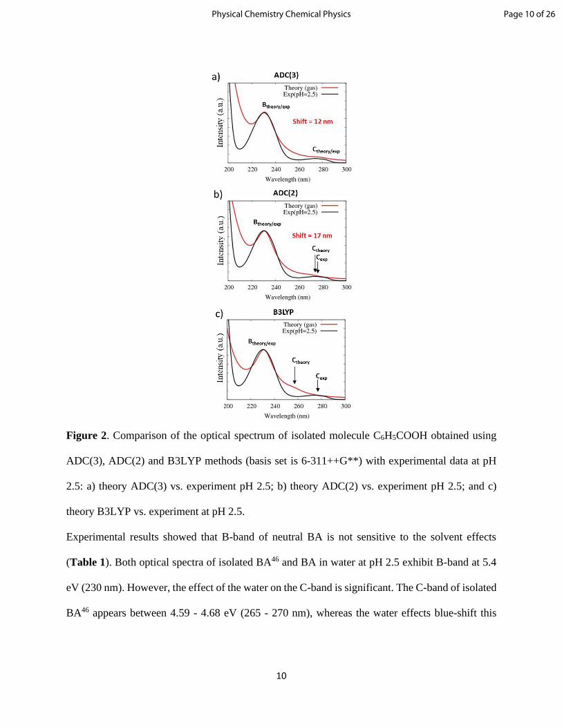
is compared with experimental spectrum in water at acidic pH of 2.5 (Table 1, Figure 2).
Table 1. Experimental (in water at pH 2.5), experimental and theoretical optical absorption
spectral data for benzoic acid (where the ∆(EB-EC) is the energy gap between the maximums of
C- and B-bands).
Method Peak Maximum
∆(EB-EC) C-band B-band
Experiment in water (pH 2.5) 4.35 - 4.50 eV
(275 - 285 nm)
5.40 eV
(230 nm) 0.90 - 1.05 eV
Experiment isolated BA46 4.59 - 4.68 eV
(265 - 270 nm)
5.40 eV
(230 nm) 0.72 - 0.81 eV
ADC(3)/6-311++G** 4.70 eV
(264 nm)
5.69 eV
(218 nm) 0.99 eV
ADC(2)/6-311++G** 4.95 eV
(250 nm)
5.84 eV
(213 nm) 0.89 eV
B3LYP/6-311++G** 4.85 eV
(256 nm)
5.36 eV
(231 nm) 0.51 eV
Page 9 of 26 Physical Chemistry Chemical Physics
10
Figure 2. Comparison of the optical spectrum of isolated molecule C6H5COOH obtained using
ADC(3), ADC(2) and B3LYP methods (basis set is 6-311++G**) with experimental data at pH
2.5: a) theory ADC(3) vs. experiment pH 2.5; b) theory ADC(2) vs. experiment pH 2.5; and c)
theory B3LYP vs. experiment at pH 2.5.
Experimental results showed that B-band of neutral BA is not sensitive to the solvent effects
(Table 1). Both optical spectra of isolated BA46 and BA in water at pH 2.5 exhibit B-band at 5.4
eV (230 nm). However, the effect of the water on the C-band is significant. The C-band of isolated
BA46 appears between 4.59 - 4.68 eV (265 - 270 nm), whereas the water effects blue-shift this
Page 10 of 26Physical Chemistry Chemical Physics
11
band up to ~0.2 eV (Table 1). The maximum energy gap between experimental B- and C-bands
are around 0.8 and 1 eV for isolated BA and for BA in water at pH 2.5, respectively.
All considered theoretical methods exhibit both absorption bands (B- and C-) for benzoic acid in
the range of 200-300 nm (Table 1, Figure 2). As shown in Figure 2a-b, offsetting the
theoretically predicted peaks by 0.2 eV (~12 nm) for ADC(3) and 0.45 eV (17 nm) for ADC(2)
methods leads to a very good agreement with the experimental data. It is important to mention,
that the energy gap between theoretical B- and C-bands is between 0.9 – 1 eV (both ADC
methods), which is in good agreement with the experiment (Table 1).
TDDFT (B3LYP/6-311++G**) optical absorption spectrum of isolated benzoic acid showed that
the theoretical B-band has the exact position as in the experiment and is localized around 231 nm
(5.36 eV) (Figure 2c, Table 1). The C-band is significantly blue shifted with respect to
experiment: the energy difference between B- and C-bands is around 0.51 eV only (which is
smaller by ~0.3 eV than the experimental value for isolated BA46). Smith and co-worker,14
calculated optical absorption spectrum of neutral BA using different exchange–correlation (XC)
functionals such as meta-GGA hybrid M06-2X, double hybrid B2PLYPD, and range-separated
functionals CAM-B3LYP, ωB97XD, and LC-ωPBE, but all tested functionals could not reproduce
the correct energy gap between B- and C-bands as well.
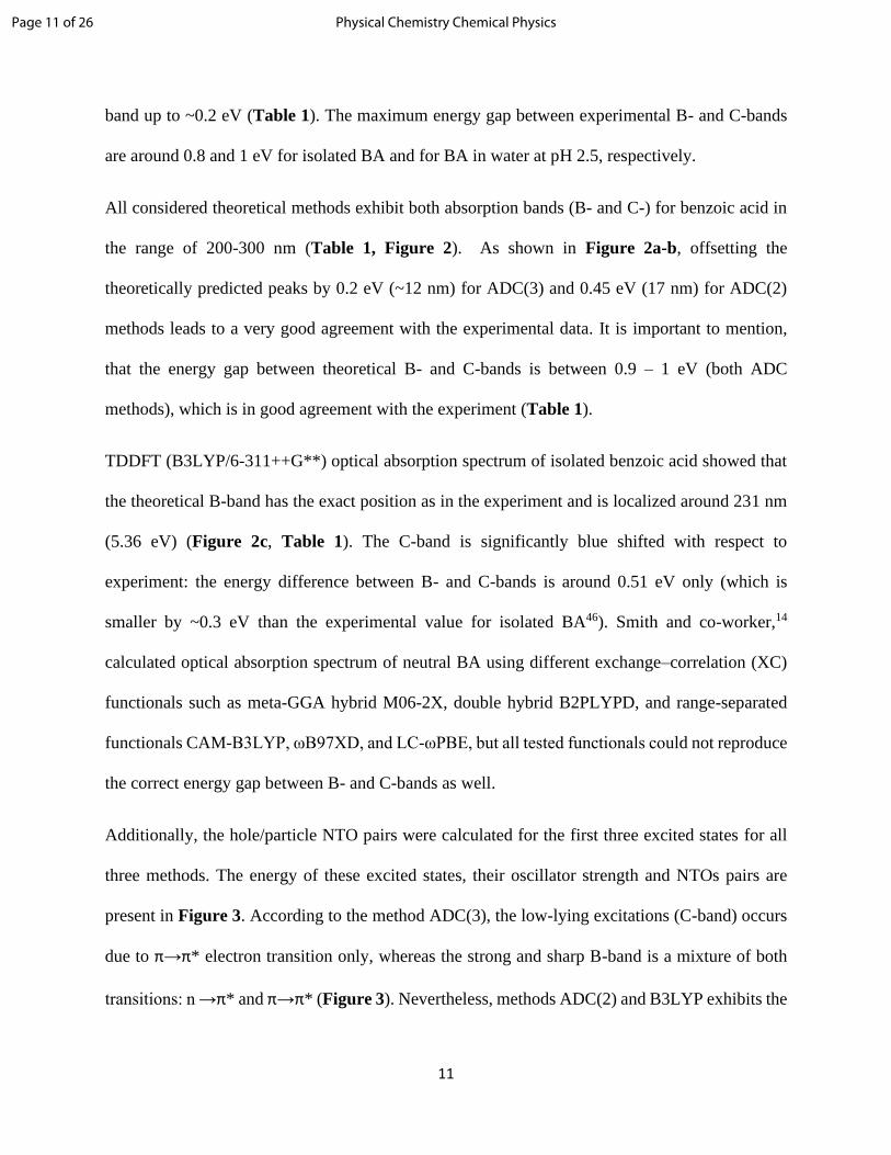
Additionally, the hole/particle NTO pairs were calculated for the first three excited states for all
three methods. The energy of these excited states, their oscillator strength and NTOs pairs are
present in Figure 3. According to the method ADC(3), the low-lying excitations (C-band) occurs
due to π→π* electron transition only, whereas the strong and sharp B-band is a mixture of both
transitions: n →π* and π→π* (Figure 3). Nevertheless, methods ADC(2) and B3LYP exhibits the
Page 11 of 26 Physical Chemistry Chemical Physics
12
opposite results: C-band is a mixture of n →π* and π→π* electron transitions, and the B-band
arises due to π→π* electron transition only. It should be noticed, that in all methods, the oscillator
strength of the excited states which assign with n →π* transitions are very weak (amplitudes are
10-5÷10-6) (Figure 3). Therefore, we can expect that contributions to the formation of the B-and
C-bands of neutral BA are mainly from π→π* electron transitions.
Figure 3. Natural transition orbitals of the first three excited states (formed B and C-bands) of BA
molecule calculated at the ADC(3), ADC(2) and B3LYP level of theory with basis set 6-
311++G**. Orbitals are represented with a contour value of 0.035.
Page 12 of 26Physical Chemistry Chemical Physics

13
These theoretical results allowed us to conclude that, TDDFT reproduces correctly the shape and
maximum position of the strong B-band (good agreement with experiment). However, in the case
of C-band, TDDFT cannot reproduce the correct position of this band (it is blue-shifted by ~0.3-
0.4 eV with respect to experimental C-band), but TDDFT reproduces the nature of involved
orbitals with ADC(2) level accuracy. In addition, TDDFT is computationally less expensive with
respect to ADC(n) levels of theory. Thus, in this work, TDDFT level of theory will be used to
study the nature of B-band only.
Cluster Size and Predicted Spectra: C6H5COOH∙(H2O)n and C6H5COO-∙(H2O)n (n = 0-8). To
check the effect of the solvent effects on the optical properties of the BA at different pH two
speciated forms of BA stabilized by water cluster of different sizes were tested:
C6H5COOH∙(H2O)n and C6H5COO-∙(H2O)n (n = 0-8). Optical absorption spectra of these clusters
were calculated using B3LYP/6-311++G** method in the gas phase and with the additional
inclusion of a continuum representation for the extended solvent environment (C-PCM).
Benzoic Acid with Water Molecules (C6H5COOH∙(H2O)n). The isolated neutral molecule of BA
and BA hydrated clusters were used for simulations of the experimental spectrum of BA at low
pH = 2.5. The ground state minimum energy equilibrium structures C6H5COOH·(H2O)n (n = 0 -
7) and their optical spectra are shown in Figure 4. Theoretical results demonstrated that the shape
of the optical spectrum of neutral BA form is not significantly sensitive to the number of water
molecules in the considered clusters. Results obtained in the gas phase calculations (red curve)
showed that all considered systems exhibit the B-band around 230 nm (excellent agreement with
experiment). The maximum shift of theoretical B-band (around 0.1 eV (5 nm)) obtained for the
larges system C6H5COOH·(H2O)7. However, note, that this cluster is just one possible structure of
Page 13 of 26 Physical Chemistry Chemical Physics

14
the system and when we consider the contribution to the total optical absorption spectrum of all
structures from MD trajectories, the position of theoretical B-band can be improved.
The additional solvent effects such as the polarizable continuum model (C-PCM) does not make
any significant changes in the shape of the optical spectra of these neutral clusters. However, the
B-band is red-shifted with respect to the experiment by 0.2 eV (~10 nm). The reason for this shift
can be the interaction of the hydrophobic Ph-group with the continuum.
Figure 4. Experimental optical absorption spectrum at pH 2.5 (black line), theoretical optical
absorption spectra of neutral clusters (equilibrium structures) C6H5COOH·(H2O)n (where n = 0-7)
Page 14 of 26Physical Chemistry Chemical Physics

15
in gas phase (red line) and with including solvent effects (C-PCM) (blue line). Level of theory is
B3LYP/6-311++G**.
Page 15 of 26 Physical Chemistry Chemical Physics

16
Figure 5. Experimental optical absorption spectrum at pH 8.0 (black line), theoretical optical
absorption spectra of ionized clusters (equilibrium structures) C6H5COO ̶·(H2O)n (where n = 0÷8)
in gas phase (red line) and with including solvent effects (C-PCM) (blue line). Level of theory is
B3LYP/6-311++G**.
System C6H5COO-∙(H2O)n. To simulate the experimental spectrum of BA at high pH = 8.0, the
ionized form of BA was considered with different number of water molecules C6H5COO-∙(H2O)n
(n=0 ÷ 8). The experimental B-band of this system arises around 225 nm. The obtained results
showed that the spectrum of C6H5COO- is significantly sensitive to solvent effects (Figure 5). For
example, the theoretical optical absorption spectrum of isolated C6H5COO– does not exhibit any
peaks between 200 – 300 nm (Figure 5). Stabilization of the BA anion with one water molecules
allowed us to localize one shoulder peak around 218 nm (Figure 5) in gas phase.
Significant improvements with the position of the B-band maximum and the curve shape were
obtained when a model system with m ≥ 3 water molecules was considered and additional internal
solvent effects were included (CPCM). For example, the largest system C6H5COO-∙(H2O)8 exhibit
the sharp peak around 5.41 eV (229 nm), the difference with experiment is just 4 nm now (Figure
5).
Comparison spectra of neutral and ionized BA forms. To understand the difference between the
optical properties of BA at different pH (two speciated forms), the optical absorption spectra of
two largest clusters (equilibrium structures) C6H5COOH∙(H2O)7 and C6H5COO ̶·(H2O)8 were
compared. The theoretical optical absorption spectra together with the oscillator strength of the
excited states for C6H5COOH∙(H2O)7 and C6H5COO ̶·(H2O)8 clusters are presented in Figure 6
(method is B3LYP/6-311++G** with including C-PCM algorithm). In the cluster
Page 16 of 26Physical Chemistry Chemical Physics

17
C6H5COOH∙(H2O)7, the B-band arises due to one strong excited state at 5.16 eV (240 nm, f =
0.370), Figure 6a. In the case of C6H5COO–∙(H2O)8, the B-band has a maximum at 5.39 eV (229
nm) and it is more complex with respect to neutral BA cluster: it is formed by two excited states
at 5.12 eV (242 nm, f = 0.0014) and 5.39 eV (229 nm, f = 0.230), Figure 6b.
Figure 6. Theoretical optical absorption spectra calculated using B3LYP/6-311++G** with
including solvent effects (C-PCM): (a) of neutral cluster C6H5COOH·(H2O)7; (b) clusters
C6H5COO ̶·(H2O)8; and (c) comparison these two theoretical spectra.
The NTOs pairs involved in the transitions of the B-band both clusters are shown in Figure 7. The
orbitals have similar features but there are subtle differences as well. For example, in the optical
absorption spectrum of C6H5COOH∙(H2O)7 system, the B-band arises due to π→π* only (Figure
7a). However, in the cluster C6H5COO–∙(H2O)8, non-bonding HOMO orbitals are involved as well:
n→π* and π→π* transitions (Figure 7b). Comparison of the orbitals of C6H5COOH∙(H2O)7
cluster with orbitals of the isolated C6H5COOH molecule in the gas phase showed, that in both
cases the orbitals involved in the electron transitions of the B-band localized on the BA fragment
(Figure 3 and 7a).
Additionally, obtained theoretical data reproduce the relative intensities of B-band at different pH
(Figure 1a and Figure 6c). Experimental B-band at pH 2.5 (Iexp(2.5)) much intense than at pH 8.0
Page 17 of 26 Physical Chemistry Chemical Physics

18
(Iexp(8.0)). The ratio of experimental B-band intensities is 𝐼exp(8.0)
𝐼exp(2.5)= 0.76. Theoretical ratio of B-
band intensities for considered clusters is very close to the experimental value and equivalent to
𝐼𝑃ℎ𝐶𝑂𝑂−∙(𝐻2𝑂)7𝐼𝑃ℎ𝐶𝑂𝑂𝐻∙(𝐻2𝑂)8
= 0.80.
Figure 7. Natural transition orbitals of the excited states (for B-band) of two speciated forms of
BA: a) C6H5COOH∙(H2O)7 and b) C6H5COO ̶·(H2O)8 calculated at the B3LYP/6-311++G** (with
including solvent effects by C-PCM). Orbitals are represented with a contour value of 0.035.
AIMD simulations and final optical spectra of C6H5COOH∙(H2O)7 and C6H5COO-∙(H2O)8 . To
consider the contribution to the total optical absorption spectrum of all possible structures, AIMD
trajectories were simulated and used to calculate total TDDFT optical absorption spectrum of
largest systems: C6H5COOH∙(H2O)7 and C6H5COO-∙(H2O)8. It was found (Figures 4 and 5) that
in the case of C6H5COO- that additional explicit solvent molecules are needed to be included in
combination with a continuum representation for the extended solvent environment to be able to
reproduce the experimental spectrum correctly. Method for calculation of vertical excited states
Page 18 of 26Physical Chemistry Chemical Physics

19
along AIMD trajectories is B3LYP/6-311++G** with including additional solvent effects by C-
PCM.
Figure 8. Comparison of the theoretical and experimental spectral data. Theoretical spectrum
presented here is an average spectrum of all structures along AIMD trajectories. Optical absorption
spectra of (a) C6H5COOH∙(H2O)7 versus the experimental data at pH 2.5; and (b) C6H5COO–
∙(H2O)8 versus the experimental data at pH = 8.0. Method B3LYP/6-311++G** (with including
solvent effects by C-PCM). Theory – red curve, experiment – black.
Overall, the total theoretical optical absorption spectra of C6H5COOH∙(H2O)7 and C6H5COO-
∙(H2O)8 systems from the AIMD simulations are presented in Figure 8. The maximum of the B-
band was found at 5.16 eV (240 nm) for C6H5COOH∙(H2O)7 and 5.41 eV (229 nm) for C6H5COO-
∙(H2O)8. As shown in Figure 8a-b, offsetting the theoretically predicted peaks by 4 (for ionized
BA) and 10 (neutral BA) nm leads to a very good agreement with the experimental data. In the
case of the neutral structure C6H5COOH∙(H2O)7, the 10 nm shift is related to the effect of the
extended solvent environment model (C-PCM). Additional calculations of total (AIMD) optical
absorption spectrum for C6H5COOH∙(H2O)7 in the gas phase allowed us to localize B-band at 234
nm (experimental value is 230 nm), Figure S3.
Page 19 of 26 Physical Chemistry Chemical Physics
20
5. Concluding Remarks
In the present work, experimental spectroscopic measurements are combined with theoretical
calculations to study the optical absorption spectra of BA in aqueous solution at different pH. The
experimental results for absorption spectra of benzoic acid in water at acidic and basic pH are in
agreement with early studies. There is no obvious effect on the benzoic acid absorption spectra
found due to the presence of salt ions such as NaCl and CaCl2. Experimental results showed that
B-band of neutral BA is not sensitive to the solvent effects whereas the effect of the water on the
C-band is significant.46 The water effects blue- shift this band up to ~0.2 eV
Small theoretical clusters of BA with different numbers of water molecules demonstrated that
solvent effects are very important for reproducing the correct optical spectra of BA species:
especially for the deprotonated form of BA such as C6H5COO-∙(H2O)n (where n = 0 - 8). Only
systems with n ≥ 3 water molecules combined with the extended solvent environment provide a
good agreement with the experiment, in particular, the shape and the maximum position of the B-
band.
Small theoretical clusters can be successful in describing chromophores in solution. Theoretical
results showed that B-band of neutral BA (experiment at pH 2.5) arises from the π→π* transitions
only, whereas in the case of ionized BA (experiment at pH 8.0) this band appears due to the
combination of n→π* and π→π* transitions. NTOs analysis of the theoretical clusters allowed us
to see that the orbitals involved in the electron transitions of the B-band are mainly localized on
the BA molecule, which indicates that the excitations have a contribution from BA and ion of BA.
Overall, we can conclude that the combination of the excited states quantum chemistry with
molecular dynamics provides a very good interpretation of the experiment, and the results
Page 20 of 26Physical Chemistry Chemical Physics
21
demonstrate the possibility of microscopic understanding of the absorption spectra of organic
molecules in water by calculations restricted to small cluster models.
Supporting Information
The Supporting Information is available free of charge on the ACS Publications website.
• Proton transfer process between benzoic acid and water molecules: NBOs charges of the
fragments C6H5COOH/(C6H5COO∙∙∙H) stabilized by 8 water molecules. Theoretical
optical absorption spectra of C6H5COOH∙(H2O)8 versus the experimental data at pH 2.5
(theoretical spectrum presented here is an average spectrum of all structures along AIMD
trajectories. It shows the effect of the proton transfer on the quality of the theoretical optical
spectrum for the system with neutral BA). Table of experimental (in water at pH 2.5) and
theoretical spectral data for isolated benzoic acid (different methods and basis sets).
Theoretical optical absorption spectra (gas phase) of C6H5COOH∙(H2O)7 versus the
experimental data at pH 2.5 (theoretical spectrum presented here is an average spectrum of
all structures along AIMD trajectories).
• Additional data related to this paper can be accessed from the NSF-CAICE Data Repository
(doi: 10.6075/J0RN367K) or may be requested from the authors.
Acknowledgements
The authors would like to gratefully acknowledge supported by the National Science Foundation
through the Center for Aerosol Impacts on Chemistry of the Environment funded under the Centers
for Chemical Innovation Program Grant CHE1801971. Additionally, this work used the Extreme
Science and Engineering Discovery Environment (XSEDE),47 which is supported by grand
Page 21 of 26 Physical Chemistry Chemical Physics

22
number grant number TG-CHE17006. The authors would also like to thank Professor Juan Navea
and Dr. Dorit Shemesh for helpful discussions.
Author contributions
Natalia V. Karimova performed theoretical simulations; Man Luo made experimental
measurements. Vicki H. Grassian (experimental aspects) and R. Benny Gerber (theoretical
aspects) participated in the analysis of results and conclusions and are corresponding authors.
References
1 E. M. Thurman, Organic Geochemistry of Natural Waters, Springer Netherlands,
Dordrecht, 1985.
2 W. S. Wan Ngah, M. A. K. M. Hanafiah and S. S. Yong, Adsorption of humic acid from
aqueous solutions on crosslinked chitosan–epichlorohydrin beads: Kinetics and isotherm studies,
Colloids Surf B Biointerfaces, 2008, 65, 18–24.
3 D. Kwon, M. J. Sovers, V. H. Grassian, P. D. Kleiber and M. A. Young, Optical Properties
of Humic Material Standards: Solution Phase and Aerosol Measurements, ACS Earth Space
Chem., 2018, 2, 1102–1111.
4 F. Nasser and I. Lynch, Secreted protein eco-corona mediates uptake and impacts of
polystyrene nanoparticles on Daphnia magna, J. Proteomics, 2016, 137, 45–51.
5 S. Jayalath, H. Wu, S. C. Larsen and V. H. Grassian, Surface Adsorption of Suwannee
River Humic Acid on TiO 2 Nanoparticles: A Study of pH and Particle Size, Langmuir, 2018, 34,
3136–3145.
6 J. V. Trueblood, M. R. Alves, D. Power, M. V. Santander, R. E. Cochran, K. A. Prather
and V. H. Grassian, Shedding Light on Photosensitized Reactions within Marine-Relevant Organic
Thin Films, ACS Earth Space Chem., 2019, 3, 1614–1623.
7 K. McNeill and S. Canonica, Triplet state dissolved organic matter in aquatic
photochemistry: reaction mechanisms, substrate scope, and photophysical properties, Environ. Sci.
Proc. Imp., 2016, 18, 1381–1399.
8 C. Baduel, D. Voisin and J.-L. Jaffrezo, Seasonal variations of concentrations and optical
properties of water soluble HULIS collected in urban environments, Atmos. Chem. Phys., 2010,
10, 4085–4095.
9 A. Hoffer, A. Gelencsér, P. Guyon, G. Kiss, O. Schmid, G. P. Frank, P. Artaxo and M. O.
Andreae, Optical properties of humic-like substances (HULIS) in biomass-burning aerosols,
Atmos. Chem. Phys., 2006, 6, 3563–3570.
10 Y.-Ping. Chin, George. Aiken and Edward. O’Loughlin, Molecular Weight, Polydispersity,
and Spectroscopic Properties of Aquatic Humic Substances, Environ. Sci. Technol., 1994, 28,
1853–1858.
11 D. P. Veghte, S. China, J. Weis, L. Kovarik, M. K. Gilles and A. Laskin, Optical Properties
Page 22 of 26Physical Chemistry Chemical Physics

23
of Airborne Soil Organic Particles, ACS Earth Space Chem., 2017, 1, 511–521.
12 H. E. Ungnade and R. W. Lamb, The Absorption Spectra of Benzoic Acid and Esters, J.
Am. Chem. Soc., 1952, 74, 3789–3794.
13 B. V. Kamath, J. D. Mehta and S. L. Bafna, Ultraviolet absorption spectra: Some
substituted benzoic acids, J. Appl. Chem. Biotechnol., 2007, 25, 743–751.
14 H.-B. Guo, F. He, B. Gu, L. Liang and J. C. Smith, Time-Dependent Density Functional
Theory Assessment of UV Absorption of Benzoic Acid Derivatives, J. Phys. Chem. A, 2012, 116,
11870–11879.
15 M. Karabacak, Z. Cinar, M. Kurt, S. Sudha and N. Sundaraganesan, FT-IR, FT-Raman,
NMR and UV–vis spectra, vibrational assignments and DFT calculations of 4-butyl benzoic acid,
Spectrochim. Acta A, 2012, 85, 179–189.
16 C. E. Lund Myhre and C. J. Nielsen, Optical properties in the UV and visible spectral
region of organic acids relevant to tropospheric aerosols, Atmos. Chem. Phys., 2004, 4, 1759–1769.
17 S. Amiri, H. P. Reisenauer and P. R. Schreiner, Electronic Effects on Atom Tunneling:
Conformational Isomerization of Monomeric Para -Substituted Benzoic Acid Derivatives, J. Am.
Chem. Soc., 2010, 132, 15902–15904.
18 I. P. Pozdnyakov, V. F. Plyusnin and V. P. Grivin, Photophysics and Photochemistry of 2-
Aminobenzoic Acid Anion in Aqueous Solution, J. Phys. Chem. A, 2009, 113, 14109–14114.
19 A. L. Sobolewski and W. Domcke, Photophysics of intramolecularly hydrogen-bonded
aromatic systems: ab initio exploration of the excited-state deactivation mechanisms of salicylic
acid, Phys. Chem. Chem. Phys., 2006, 8, 3410.
20 D. M. Friedrich, Z. Wang, A. G. Joly, K. A. Peterson and P. R. Callis, Ground-State Proton-
Transfer Tautomer of the Salicylate Anion, J. Phys. Chem. A, 1999, 103, 9644–9653.
21 D. Vulpius, G. Geipel and G. Bernhard, Excited-state proton transfer of 3-hydroxybenzoic
acid and 4-hydroxybenzoic acid, Spectrochim. Acta A, 2010, 75, 558–562.
22 M. C. Almandoz, M. I. Sancho and S. E. Blanco, Spectroscopic and DFT study of solvent
effects on the electronic absorption spectra of sulfamethoxazole in neat and binary solvent
mixtures, Spectrochim. Acta A, 2014, 118, 112–119.
23 P. C. Mowery, R. H. Lozier, Q. Chae, Y.-W. Tseng, M. Taylor and W. Stoeckenius, Effect
of acid pH on the absorption spectra and photoreactions of bacteriorhodopsin, Biochemistry, 1979,
18, 4100–4107.
24 O. Bajjou, A. Bakour, M. Khenfouch, M. Baitoul, B. Mothudi, M. Maaza and E. Faulques,
pH and concentration effect on the optical absorption properties of Sn(V) tetrakis (4-pirydyl)
porphyrin functionalized graphene oxide., J. Phys. Conf. Ser., 2018, 984, 012004.
25 M. Wormit, D. R. Rehn, P. H. P. Harbach, J. Wenzel, C. M. Krauter, E. Epifanovsky and
A. Dreuw, Investigating excited electronic states using the algebraic diagrammatic construction
(ADC) approach of the polarisation propagator, Molecular Physics, 2014, 112, 774–784.
26 P. H. P. Harbach, M. Wormit and A. Dreuw, The third-order algebraic diagrammatic
construction method (ADC(3)) for the polarization propagator for closed-shell molecules:
Efficient implementation and benchmarking, J. Chem. Phys., 2014, 141, 064113.
27 E. Runge and E. K. U. Gross, Density-Functional Theory for Time-Dependent Systems,
Phys. Rev. Lett., 1984, 52, 997–1000.
28 W. M. Haynes, D. R. Lide and CRC Press, Eds., CRC handbook of chemistry and physics:
a ready-reference book of chemical and physical data, CRC Press, Boca Raton, Fla., 91. ed., 2010–
2011., 2010.
29 Y. Shao, Z. Gan, E. Epifanovsky, A. T. B. Gilbert, M. Wormit, J. Kussmann, A. W. Lange,
Page 23 of 26 Physical Chemistry Chemical Physics
24
A. Behn, J. Deng, X. Feng, D. Ghosh, M. Goldey, P. R. Horn, L. D. Jacobson, I. Kaliman, R. Z.
Khaliullin, T. Kuś, A. Landau, J. Liu, E. I. Proynov, Y. M. Rhee, R. M. Richard, M. A. Rohrdanz,
R. P. Steele, E. J. Sundstrom, H. L. Woodcock, P. M. Zimmerman, D. Zuev, B. Albrecht, E.
Alguire, B. Austin, G. J. O. Beran, Y. A. Bernard, E. Berquist, K. Brandhorst, K. B. Bravaya, S.
T. Brown, D. Casanova, C.-M. Chang, Y. Chen, S. H. Chien, K. D. Closser, D. L. Crittenden, M.
Diedenhofen, R. A. DiStasio, H. Do, A. D. Dutoi, R. G. Edgar, S. Fatehi, L. Fusti-Molnar, A.
Ghysels, A. Golubeva-Zadorozhnaya, J. Gomes, M. W. D. Hanson-Heine, P. H. P. Harbach, A.
W. Hauser, E. G. Hohenstein, Z. C. Holden, T.-C. Jagau, H. Ji, B. Kaduk, K. Khistyaev, J. Kim,
J. Kim, R. A. King, P. Klunzinger, D. Kosenkov, T. Kowalczyk, C. M. Krauter, K. U. Lao, A. D.
Laurent, K. V. Lawler, S. V. Levchenko, C. Y. Lin, F. Liu, E. Livshits, R. C. Lochan, A. Luenser,
P. Manohar, S. F. Manzer, S.-P. Mao, N. Mardirossian, A. V. Marenich, S. A. Maurer, N. J.
Mayhall, E. Neuscamman, C. M. Oana, R. Olivares-Amaya, D. P. O’Neill, J. A. Parkhill, T. M.
Perrine, R. Peverati, A. Prociuk, D. R. Rehn, E. Rosta, N. J. Russ, S. M. Sharada, S. Sharma, D.
W. Small, A. Sodt, T. Stein, D. Stück, Y.-C. Su, A. J. W. Thom, T. Tsuchimochi, V. Vanovschi,
L. Vogt, O. Vydrov, T. Wang, M. A. Watson, J. Wenzel, A. White, C. F. Williams, J. Yang, S.
Yeganeh, S. R. Yost, Z.-Q. You, I. Y. Zhang, X. Zhang, Y. Zhao, B. R. Brooks, G. K. L. Chan, D.
M. Chipman, C. J. Cramer, W. A. Goddard, M. S. Gordon, W. J. Hehre, A. Klamt, H. F. Schaefer,
M. W. Schmidt, C. D. Sherrill, D. G. Truhlar, A. Warshel, X. Xu, A. Aspuru-Guzik, R. Baer, A.
T. Bell, N. A. Besley, J.-D. Chai, A. Dreuw, B. D. Dunietz, T. R. Furlani, S. R. Gwaltney, C.-P.
Hsu, Y. Jung, J. Kong, D. S. Lambrecht, W. Liang, C. Ochsenfeld, V. A. Rassolov, L. V.
Slipchenko, J. E. Subotnik, T. Van Voorhis, J. M. Herbert, A. I. Krylov, P. M. W. Gill and M.
Head-Gordon, Advances in molecular quantum chemistry contained in the Q-Chem 4 program
package, Mol. Phys., 2015, 113, 184–215.
30 A. D. Becke, Density‐functional thermochemistry. III. The role of exact exchange, J.
Chem. Phys., 1993, 98, 5648–5652.
31 S. Grimme, Semiempirical GGA-type density functional constructed with a long-range
dispersion correction, J. Comp. Chem., 2006, 27, 1787–1799.
32 P. Krishnakumar and D. K. Maity, Microhydration of a benzoic acid molecule and its
dissociation, New J. Chem., 2017, 41, 7195–7202.
33 T. L. Eliason, D. K. Havey and V. Vaida, Gas phase infrared spectroscopic observation of
the organic acid dimers (CH3(CH2)6COOH)2, (CH3(CH2)7COOH)2, and (CH3(CH2)8COOH)2,
Chem. Phys. Lett., 2005, 402, 239–244.
34 J.-P. Cornard and C. Lapouge, Absorption Spectra of Caffeic Acid, Caffeate and Their 1:1
Complex with Al(III): Density Functional Theory and Time-Dependent Density Functional
Theory Investigations, J. Phys. Chem. A, 2006, 110, 7159–7166.
35 T. H. Dunning, Gaussian basis sets for use in correlated molecular calculations. I. The
atoms boron through neon and hydrogen, The Journal of Chemical Physics, 1989, 90, 1007–1023.
36 R. A. Kendall, T. H. Dunning and R. J. Harrison, Electron affinities of the first‐row atoms
revisited. Systematic basis sets and wave functions, J. Chem. Phys., 1992, 96, 6796–6806.
37 S. A. Epstein, D. Shemesh, V. T. Tran, S. A. Nizkorodov and R. B. Gerber, Absorption
Spectra and Photolysis of Methyl Peroxide in Liquid and Frozen Water, J. Phys. Chem. A, 2012,
116, 6068–6077.
38 D. Shemesh and R. B. Gerber, Molecular Dynamics of Photoinduced Reactions of Acrylic
Acid: Products, Mechanisms, and Comparison with Experiment, J. Phys. Chem. Lett., 2018, 9,
527–533.
39 T. N. Truong and E. V. Stefanovich, A new method for incorporating solvent effect into
Page 24 of 26Physical Chemistry Chemical Physics
25
the classical, ab initio molecular orbital and density functional theory frameworks for arbitrary
shape cavity, Chemical Physics Letters, 1995, 240, 253–260.
40 A. Bondi, van der Waals Volumes and Radii, J. Phys. Chem., 1964, 68, 441–451.
41 F. Plasser, B. Thomitzni, S. A. Bäppler, J. Wenzel, D. R. Rehn, M. Wormit and A. Dreuw,
Statistical analysis of electronic excitation processes: Spatial location, compactness, charge
transfer, and electron-hole correlation, J. Comput. Chem., 2015, 36, 1609–1620.
42 S. A. Mewes, F. Plasser, A. Krylov and A. Dreuw, Benchmarking Excited-State
Calculations Using Exciton Properties, J. Chem. Theory Comput., 2018, 14, 710–725.
43 D. Marx, Ab initio molecular dynamics: the virtual laboratory approach, Wiley - VCH,
Weinheim, 2007.
44 R. B. Gerber, D. Shemesh, M. E. Varner, J. Kalinowski and B. Hirshberg, Ab initio and
semi-empirical Molecular Dynamics simulations of chemical reactions in isolated molecules and
in clusters, Phys. Chem. Chem. Phys., 2014, 16, 9760–9775.
45 D. E. Romonosky, L. Q. Nguyen, D. Shemesh, T. B. Nguyen, S. A. Epstein, D. B. C.
Martin, C. D. Vanderwal, R. B. Gerber and S. A. Nizkorodov, Absorption spectra and aqueous
photochemistry of β-hydroxyalkyl nitrates of atmospheric interest, Mol. Phys., 2015, 113, 2179–
2190.
46 E. Roth, A. Chakir and A. Ferhati, Study of a Benzoylperoxy Radical in the Gas Phase:
Ultraviolet Spectrum and C 6 H 5 C(O)O 2 + HO 2 Reaction between 295 and 357 K, J. Phys. Chem.
A, 2010, 114, 10367–10379.
47 J. Towns, T. Cockerill, M. Dahan, I. Foster, K. Gaither, A. Grimshaw, V. Hazlewood, S.
Lathrop, D. Lifka, G. D. Peterson, R. Roskies, J. R. Scott and N. Wilkens-Diehr, XSEDE:
Accelerating Scientific Discovery, Comput. Sci. Eng., 2014, 16, 62–74.
Page 25 of 26 Physical Chemistry Chemical Physics

Table of Contents Entry: The microscopic interpretation of the electronic structure and absorption
spectra of BA in aqueous media through calculations restricted to small cluster models
Page 26 of 26Physical Chemistry Chemical Physics