absorption, distribution and excretion · pdf filecan cross that membrane. • be able to...
TRANSCRIPT
89
ABSORPTION, DISTRIBUTION AND EXCRETION R.C. Shank, Ph.D.
Department of Community and Environmental Medicine, University of California, Irvine, USA
OBJECTIVE You should:
• know the structure of the mammalian cell membrane and how xenobiotics can cross that membrane.
• be able to distinguish between water-soluble and fat (lipid)-soluble xenobiotics.
• know how xenobiotics enter the body by absorption through the skin, lungs, and gastrointestinal tract.
• know how xenobiotics move through the body.
• know the importance of the rates of absorption and excretion in determining the concentration of xenobiotics in the blood and tissues.
INTRODUCTION
The biological response to a toxic chemical is directly dependent upon the dose of the chemical delivered to the target organ.
There is an important difference between exposure and dose.
Exposure is the presentation of the xenobiotic (chemical foreign to the body) to the individual. Units of exposure to xenobiotics are usually parts per million (ppm) or a unit of weight per cubic meter of air, liter of water, or kilogram of food (diet). Dermal exposures are usually expressed as concentrations of the solution in contact with a surface area, (for example, micrograms per square centimeter).
Dose is the amount of xenobiotic that reaches the target organ to elicit a chemical reaction that takes place between the toxicant and an endogenous compound in or on the target cell. Units of dose are usually given as a unit of weight administered per kilogram body weight or square meter of body surface area (same as exposure).
When an exposure occurs, the toxicant must first cross from the environment and enter the body; transfer across cells composing the body surfaces (skin, lungs, gastrointestinal tract) is called absorption, more specifically, absorption from the environment into the blood or lymphatic circulatory systems.
From these circulatory systems, the toxicant passes to some, several, or all of the tissues in the body; this process is called distribution.
Topic: Principles of Toxicology
Copyright 2002 by Chulabhorn Research Institute

90
The transfer of the toxicant from the circulatory system into the tissues is also called absorption; it is similar to the process for the transfer of a chemical from the body surface into the circulation. Therefore, one must always consider two aspects of absorption: 1) transport from the body surface into the blood (or lymph) and 2) from the blood into the tissues. The removal of toxicant from the body is excretion, and this process is usually accomplished by specific action of the kidney (formation of urine), liver (formation of bile), and lung (exhalation of volatile compounds). Polarity and Solubility Polar solutes are soluble in polar solvents because solute molecule can form hydrogen bonds and align their charges with solvent molecules. Nonpolar solutes are not soluble in polar solvents because the solute molecules do not have charges. Nonpolar solutes are soluble in nonpolar solvents because the solute molecules can align the van der Waals forces with the nonpolar solvent molecules. This is illustrated in the scheme below.
The Cell Membrane To understand the process of absorption of chemicals from the body surfaces into the blood and from the blood into the tissues, it is helpful to examine the structure and chemical nature of the cell membrane, because it is, in most cases, this membrane through which the toxicant must pass to reach the target site and elicit the biological (toxic) response.
Topic: Principles of Toxicology
Copyright 2002 by Chulabhorn Research Institute

91
Figure 1 is a schematic representation of a typical mammalian cell, showing a few of the many subcellular organelles; a small portion of the cell membrane is enlarged in Figure 2 to illustrate the configuration of phospholipids and proteins that make up this membrane.
endoplasmicreticulum
nucleusmitochondrion
Figure 2
Figure 1. Schematic representation of a mammalian cell
+ - + + - - +
- + + Figure 2. Schematic representation of a portion of a mammalian cell membrane
In Figure 2 the phospholipid molecule is represented by the shaded oval with two tails and membrane proteins are represented by twisting lines carrying positive and negative charges. Figure 3, on the next page, is an illustration of a phospholipid molecule, which makes up the bulk of the cell membrane. In this illustration, phosphatidylcholine distearate is used as an example (there are several other similar molecules in cell membranes) and the polar, water-soluble head and non-polar, lipid-soluble tails of the molecule are indicated.
Figure 3. A phospholipid molecule
Topic: Principles of Toxicology
Copyright 2002 by Chulabhorn Research Institute
92
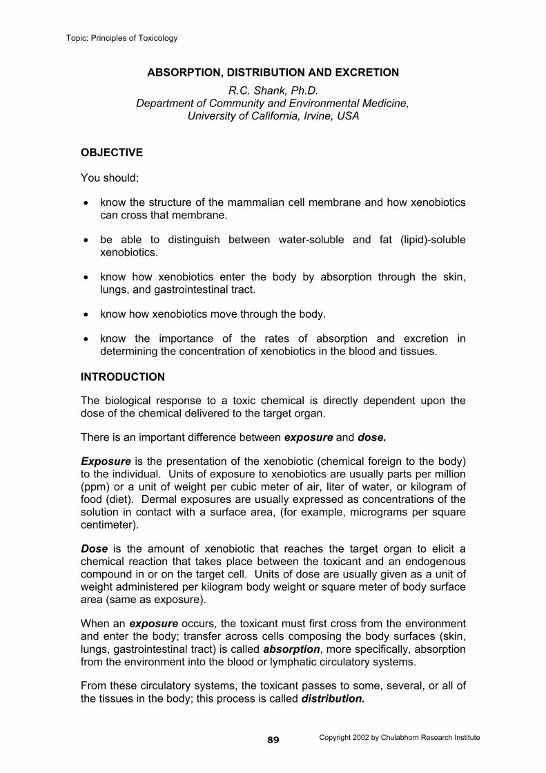
There are special features about this membrane that make the processes of absorption and excretion much easier to understand. The membrane behaves as a film of oil (fluid lipid) in an aqueous (water) environment. Globular proteins in the fluid mosaic of the membrane are free to move along the plane of the membrane (Figure 4).
Figure 4. Structure of mammalian cell membrane Some of these proteins completely traverse the membrane, providing aqueous channels through the lipid membrane. Small water-soluble molecules and ions can diffuse through these channels, while lipid-soluble molecules diffuse freely through the phospholipid component of the cell membrane. Large water-soluble molecules cannot readily cross these membranes except by special transport mechanisms. Proteins can cross, both in absorption and secretion, by a special process called pinocytosis. Specialized transport mechanisms for absorption will not be discussed in this lecture.
Because the majority of the surface area of the cell membrane is phospholipid, lipid-soluble compounds cross the cell membrane much faster (i.e., at greater rates) than do water-soluble compounds which are restricted to crossing the membrane only where protein channels occur. Thus, on the basis of the structure of the cell membrane, a generalization regarding absorption can be made: lipid-soluble compounds are absorbed from the body's surfaces at faster rates (usually much faster) than water-soluble compounds, unless the water-soluble compound crosses the cell membrane by a specialized transport mechanism. The major routes by which environmental toxicants enter the body are through the skin, the lungs, and the gastrointestinal tract. Some xenobiotics can act directly on the exterior surface of the plasma membrane, they bind to a specialized protein (receptor) in the membrane. Reaction with that membrane receptor can cause an endogenous compound to move from the plasma membrane to other organelles in the cell, such as the nucleus, to effect a biological response.
surfaceprotein
transmembraneprotein
phospholipids
Topic: Principles of Toxicology
Copyright 2002 by Chulabhorn Research Institute

93
RATE OF ABSORPTION Recalling the importance of the dose-response relationship, the toxic effect depends heavily on the concentration of the toxicant in the blood; this concentration drives the distribution of the toxicant into the tissues where the chemical can act on targets and receptors to initiate the toxic response. Here, the rate of absorption is important. As the rate of absorption increases, the concentration of the toxicant in the blood and tissues increases. The rate of absorption of a chemical through a cell membrane and through a tissue depends of both the size of the molecule and the lipid:water partition coefficient for that molecule. The lipid:water partition coefficient is determined
by dissolving the chemical in a mixture of lipid and water and measuring the concentration of the chemical in each solvent. Lipid is usually represented by the solvent octanol. The lipid:water partition coefficient is the ratio of the concentration of the chemical in octanol to the concentration in water: coeff. = [conc. in octanol] ÷ [conc. in water]. The logarithm of the coefficient is called the log Kow. The relation between rate of absorption for a chemical and the log Kow for that chemical is given in Figure 5 on the next page.
If the concentration of a chemical in the octanol phase is 10 grams per liter, and the concentration of that same chemical in the water phase is 0.1 gram per liter, then the lipid:water partition coefficient for that chemical is 10 ÷ 0.1, or 100 and the log pKow is 2. This chemical, if its molecular weight is less than several hundred, would be expected to be absorbed into cells rapidly. Again: the magnitude of the toxic effect is a function of the concentration of the toxicant at the site where the action is initiated. rate of absorption log pKow
Figure 5. Rate of absorption through cell membranes In most cases in environmental toxicology where the concentration of the xenobiotic in the air, water, food, etc is low, absorption takes place by passive diffusion, and therefore, at a rate proportional to the concentration gradient
1 3 5 7
Topic: Principles of Toxicology
Copyright 2002 by Chulabhorn Research Institute
94
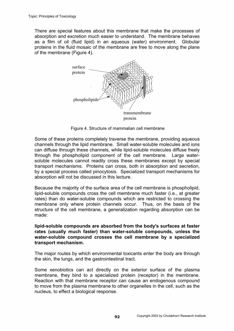
between the site of absorption and the blood. In these cases, the rate of absorption is described by exponential or first-order kinetics.
ln Ct = ln Co – kat
where Co = initial concentration of xenobiotic at the absorption site Ct = concentration of xenobiotic at time t ka = rate constant for absorption, equal to 0.693 ÷ t½ t½ = time taken for 50% of Co to be absorbed; time when Ct/Co = ½. The rate constant, ka, is the natural log of the proportion of the xenobiotic that has been absorbed in one unit of time. If half of the xenobiotic is absorbed in one hour, the concentration at the site of absorption will have decreased by a factor of 2; in this case, ka is ln 2 ÷ 1 hr, or 0.693/hr. Therefore, ka = (0.693) ÷ t½ . For example, the concentration of an ingested toxicant in the stomach determines, for the most part, the rate at which the toxicant is absorbed from the stomach into the blood; as the concentration of the toxicant in the stomach decreases due to absorption into the blood, the rate at which more toxicant is absorbed also decreases. For most toxicants at high dose, the concentration of the xenobiotic at the site of absorption may be so high that the amount being absorbed per unit time has little effect on the concentration at time t, Ct. The rate of absorption will be independent of Ct until the concentration decreases to a much lower value. Under these conditions, the rate of absorption is constant and follows zero-order kinetics. Figure 6 compares the rate of absorption of a xenobiotic under two conditions of zero-order and first-order kinetics, plotting the data as a linear function of the plasma concentration of the xenobiotic. Figure 7 takes the same data and plots the plasma concentration as a log function.
Figure 6. Rate of absorption from blood, linear plot
0102030405060708090
100
1 2 3 4 5 6
Time, hr.
Plas
ma
conc
.
ZeroFirst
Topic: Principles of Toxicology
Copyright 2002 by Chulabhorn Research Institute
95
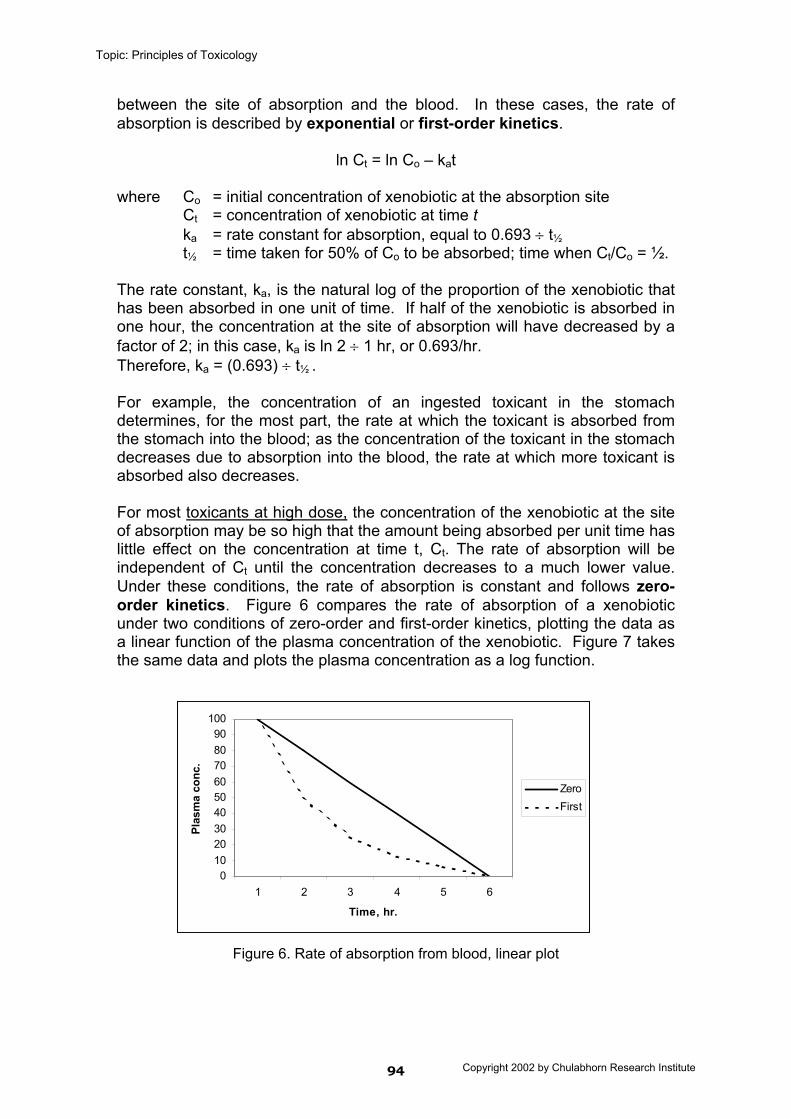
Figure 7. Rate of absorption from blood, log plot
The rate of absorption can be determined as the slope of the line in the linear plot for zero-order kinetics and in the log plot for first-order kinetics. ABSORPTION OF TOXICANTS THROUGH THE SKIN
stratum corneum
stratum lucidum
stratum granulosum
stratum spinosum
stratum basale
basement membranemelanocytes
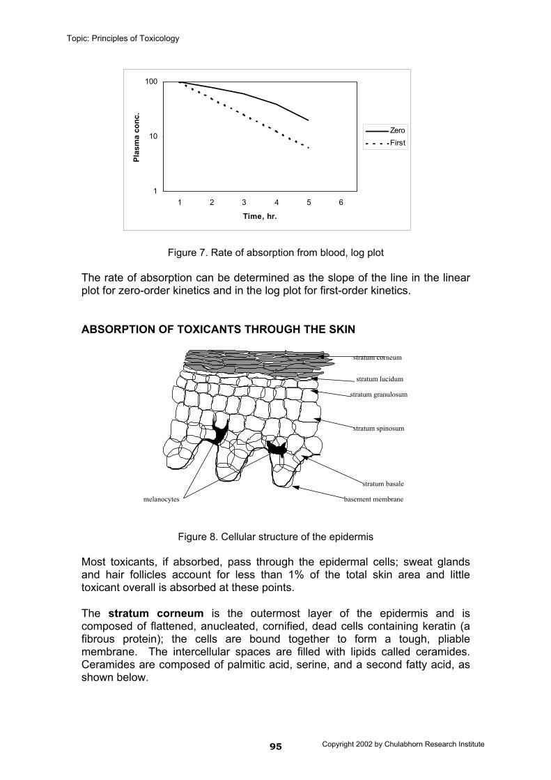
Figure 8. Cellular structure of the epidermis Most toxicants, if absorbed, pass through the epidermal cells; sweat glands and hair follicles account for less than 1% of the total skin area and little toxicant overall is absorbed at these points. The stratum corneum is the outermost layer of the epidermis and is composed of flattened, anucleated, cornified, dead cells containing keratin (a fibrous protein); the cells are bound together to form a tough, pliable membrane. The intercellular spaces are filled with lipids called ceramides. Ceramides are composed of palmitic acid, serine, and a second fatty acid, as shown below.
1
10
100
1 2 3 4 5 6
Time, hr.
Plas
ma
conc
.ZeroFirst
Topic: Principles of Toxicology
Copyright 2002 by Chulabhorn Research Institute

96
The epidermis is the rate-limiting barrier to absorption. Polar toxicants appear to diffuse through the outer surface of the keratin filaments of the hydrated stratum corneum. Non-polar toxicants dissolve in and diffuse through the non-aqueous lipid matrix between protein filaments; the rate of diffusion is related to lipid solubility and inversely to molecular weight. For a toxicant to be absorbed through the skin, into the circulation, it must pass through several layers of cells. The rate of clearance of toxicants from the dermis into the systemic circulation depends on skin thickness, effective blood flow, interstitial fluid movement, lymphatics, and other factors. The faster the absorption, the higher the blood concentration, the greater the distribution and diffusion pressure to drive the toxicant into the cells of the body. Regional differences in rates of absorption for human skin are shown in Figure 9.
Areas of the skin where absorption of lipid-soluble toxicants is usually slow: 1. arch of the foot; plantar region 2. ball of the foot 3. palm of the hand 4. ventral forearm 5. back 6. abdomen Areas of the skin where absorption of lipid-soluble toxicants is usually fast: 7. scalp 8. axilla 9. forehead 10. angle of the jaw (in men who shave) 11. ear canal 12. scrotum
Figure 9. Regional differences in the rate of absorption through human skin
1 2
3
4
5
6
7
8
9
1011
12
CH NH
CH2
CH C
OH
CH R
OH
CH
O
CH3(CH2)12
fatty acid
Ceramide
Topic: Principles of Toxicology
Copyright 2002 by Chulabhorn Research Institute
97
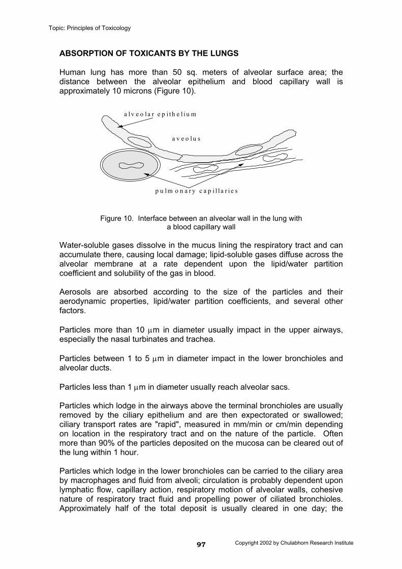
ABSORPTION OF TOXICANTS BY THE LUNGS Human lung has more than 50 sq. meters of alveolar surface area; the distance between the alveolar epithelium and blood capillary wall is approximately 10 microns (Figure 10).
Figure 10. Interface between an alveolar wall in the lung with a blood capillary wall
Water-soluble gases dissolve in the mucus lining the respiratory tract and can accumulate there, causing local damage; lipid-soluble gases diffuse across the alveolar membrane at a rate dependent upon the lipid/water partition coefficient and solubility of the gas in blood. Aerosols are absorbed according to the size of the particles and their aerodynamic properties, lipid/water partition coefficients, and several other factors. Particles more than 10 µm in diameter usually impact in the upper airways, especially the nasal turbinates and trachea. Particles between 1 to 5 µm in diameter impact in the lower bronchioles and alveolar ducts. Particles less than 1 µm in diameter usually reach alveolar sacs. Particles which lodge in the airways above the terminal bronchioles are usually removed by the ciliary epithelium and are then expectorated or swallowed; ciliary transport rates are "rapid", measured in mm/min or cm/min depending on location in the respiratory tract and on the nature of the particle. Often more than 90% of the particles deposited on the mucosa can be cleared out of the lung within 1 hour. Particles which lodge in the lower bronchioles can be carried to the ciliary area by macrophages and fluid from alveoli; circulation is probably dependent upon lymphatic flow, capillary action, respiratory motion of alveolar walls, cohesive nature of respiratory tract fluid and propelling power of ciliated bronchioles. Approximately half of the total deposit is usually cleared in one day; the
p u l m o n a r y c a p i l l a r i e s
a v e o l u s
a l v e o l a r e p i t h e l i u m
Topic: Principles of Toxicology
Copyright 2002 by Chulabhorn Research Institute

98
remainder is cleared in days or years, depending on the compound. The least soluble particles in ductal fluid are cleared slowest. Soluble particles which lodge in the alveolus diffuse directly into the pulmonary blood circulation; insoluble particles may slowly penetrate into the interstitial spaces and reach the blood via the lymphatic system. ABSORPTION OF TOXICANTS BY THE GASTROINTESTINAL TRACT Absorption can take place from the mouth to the rectum; generally, compounds are absorbed in the portion of the gut in which the compound exists at its highest concentration and in its most lipid-soluble form. Toxicants very similar in structure to nutrients and electrolytes may be actively transported into blood (for example, 5-fluoracil by pyrimidine transport; lead by calcium transport). Particles of several hundred Angstroms or Å (1 Å = 10-8 cm = 0.1 nanometer) in diameter enter the interstitial epithelium, are carried through the cytoplasm within intact vesicles, are discharged into the interstices of the lamina propria and then gain entrance into the lymphatics of the mucosa, much like fat absorption (for example, azo dye particles, several hundred Å in diameter; latex particles, up to 2,200 Å in diameter, botulinum toxin); see Figure 11.
Figure 11. Passage of particles through intestinal epithelium (villus) Important biochemical transformations can take place in the GI tract that can alter absorption or toxicity. These transformations occur in tract bacteria (flora) and/or in epithelial cells lining the tract. Many toxicants are weak acids or weak bases and exist in solution as a mixture of ionized (protonated) and nonionized forms. The less polar, nonionized forms are usually more lipid-soluble and are the forms that will diffuse rapidly across a lipoid membrane. The proportion of a toxicant that exists in the nonionized form depends on the dissociation constant of the compound and on the pH of the solution in which it is dissolved.
m icro v illi
ly m p h a tic lu m en
in te s tin a l lu m en
in te rce llu la rsp ace
p a rtic le
Topic: Principles of Toxicology
Copyright 2002 by Chulabhorn Research Institute
99
The relationship is given by the Henderson-Hasselbach equation: for a weak acid: pKa - pH = log [nonionized] [ionized] for a weak base: pKa - pH = log [ionized] [nonionized] Think of an acid as a hydrogen ion (proton; H+) donor and a base as a hydrogen ion receiver. weak acid: CH3COOH (nonionized); CH3COO- (ionized) weak base: CH3CH2NH2 (nonionized); CH3CH2NH3
+ (ionized) When a weak acid is half-ionized, the concentration of the ionized form, [ionized], is equal to the concentration of the nonionized, [nonionized], so that the log expression above becomes the log of 1, which is zero; therefore, at half-ionization the pKa is equal to the pH. The following chart summarized the absorption of weak acids and bases. It is the nonionized form of the compound that is less polar and therefore more rapidly absorbed. The nonionized form, indicated by the shaded boxes, predominate at low pH for weak acids and at high pH for weak bases. Therefore, weak acids are usually absorbed faster from the acidic gastric juice and slower in the lower, less acid, intestine. The reverse is true for weak bases.
low pH high pH weak acid nonionized ionized weak base ionized nonionized
DISTRIBUTION OF TOXICANTS IN THE BODY The distribution and excretion of toxicants depends on: 1. water compartments in the tissues of the body 2. lipid compartments in the tissues of the body 3. macromolecular binding 4. passage through placenta 5. passage in brain and cerebrospinal fluid 6. rate of pulmonary excretion 7. rate of renal excretion 8. rate of biliary excretion 9. rate of metabolism 10. lactation, perspiration, salivation, lachrymation; reproductive tract
secretions
Topic: Principles of Toxicology
Copyright 2002 by Chulabhorn Research Institute
100
Protein binding of toxicants is analogous to enzyme-substrate binding and drug-receptor binding, except there is no decomposition of the substrate nor any biological response to the process. There are no covalent bonds involved; the bonds are ionic and therefore the process is readily reversible. (Covalent binding is usually referred to as an alkylation of arylation and is not considered here.) Binding takes place on both plasma and tissue proteins; not all proteins bind each toxicant to the same extent, and the degree of binding depends on the type and number of binding sites on the protein, and the pH of the solution (controls ionization). Albumin is most important (constitutes 50% of the plasma protein); it has approximately 100 positive and 100 negative potential binding sites:
NH2+ NH2+NH3+SOCOO
At pH 7.4 albumin has more negative charges than positive charges; at pH 5 about 100 each per molecule. The biological response caused by the toxicant is dependent upon and parallels the concentration of the unbound toxicant in the plasma. Toxicants that form stable bonds with blood proteins will accumulate in the body and can be potentially dangerous. The plasma-bound compounds can be released suddenly by exposure to new compounds which can compete for the same binding sites. for example: in some newborns the liver may lack the enzyme, glucuronyl transferase which conjugates bilirubin, a water-insoluble hemoglobin breakdown product; as a result, large amounts of bilirubin accumulate on the plasma albumin (hyperbilirubinemia); if such infants are given sulfonamides or vitamin K which displace the bilirubin from albumin, enough bilirubin can be freed in a short period of time and enter the brain to cause kernicterus (widespread destruction of nerve cells in the brain). EXCRETION OF TOXICANTS The process of excretion is basically the same as that of absorption, transfer of chemicals across biological membranes according to chemical concentration gradients; the chemicals move from compartments of high concentration to compartments of low concentration. Water-soluble compounds can be rapidly excreted by the kidney via the urine (compounds with molecular weights usually less than 400) and by the liver via the bile (compounds with molecular weights usually greater than 300). Both urine and bile are aqueous (water) systems.
Topic: Principles of Toxicology
Copyright 2002 by Chulabhorn Research Institute

101
As in absorption, there are specialized processes that can act against concentration gradients and can move water-soluble compounds rapidly across lipid membranes; the secretion of bile (an aqueous fluid) by the liver is an example of a tissue removing from cells chemical compounds which are water-soluble. Lipid-soluble compounds can be excreted only slowly, if at all, into the body's aqueous waste routes, urine and bile; therefore, lipid-soluble compounds are retained in the body for long times or until they are metabolized to water-soluble derivatives. Lipid-soluble compounds which are filtered from the blood by the kidney are rapidly reabsorbed back into the blood by the kidney before the urine leaves that organ. The kidney can eliminate only water-soluble compounds and, in special cases, compounds which are characterized chemically as organic anions or cations, for which there are active transport systems to move these compounds from the plasma into the urine. Volatile compounds (chemicals with high vapor pressures) can be excreted by the lung in the expired air. Some lipid-soluble compounds can leave the body as components of lactation fluid (mother’s milk), dead skin cells, hair, and seminal fluid.
SUMMARY Exposure to a xenobiotic via air, food, or water, or by contact with the skin can lead to transfer of that xenobiotic to the blood and circulation to the tissues of the body. Lipid-soluble compounds are usually absorbed faster than water-soluble xenobiotics because the cell (plasma) membrane is more lipoid than aqueous. Xenobiotics can be held in the body by being stored in the fatty tissues and by being bound to proteins. The body can excrete only water-soluble xenobiotics in the urine and bile; lipid-soluble xenobiotics can be excreted via the lung only if they are highly volatile. The concentration of a xenobiotic in the blood determines in large part the concentration of that xenobiotic in most tissues. Fast rates of absorption into the blood and slow rates of excretion from the body can lead to high concentrations of xenobiotics in the body.
Topic: Principles of Toxicology
Copyright 2002 by Chulabhorn Research Institute
102
REFERENCES S.J. Singer and G.L. Nicolson. The fluid mosaic model of the structure of cell membranes. Science 175:720, 1972. K. K. Rozman and C.D. Klaassen. Absorption, distribution and excretion of toxicants. IN: Casarett and Doull’s Toxicology. The Basic Science of Poisons. editor, Curtis D. Klaassen; editors emeriti, Mary O. Amdur, John Doull; 5th ed. Pergamon Press, New York; 1996; pp 91-112.
SELF ASSESSMENT QUESTIONS A factory worker inhales arsine gas, AsH3, which is slightly soluble in water; follow the course the arsine would take from inhalation to excretion in the urine. Which of the following xenobiotics are highly water-soluble
CH3-CH2-CH2-CH2-CH2-CH3 CH3-OH
CH2=C=OCOOH
benzenemethanoln-hexane
ketene carbontetrachloride
benzoic acid
Cl-C-ClCl
Cl
Which of the above xenobiotics are highly lipid-soluble? Why are lipid-soluble xenobiotics only slowly excreted, if at all, in the urine? The most important factors in achieving high concentrations of water soluble xenobiotics in the blood are fast rates of absorption and slow rates of excretion. What is the next most important factor?
Topic: Principles of Toxicology
Copyright 2002 by Chulabhorn Research Institute