aborted myocardial infarction in a patient with rapid progression of wellens syndrome
TRANSCRIPT
The Journal of Emergency Medicine, Vol. 43, No. 3, pp. e181–e184, 2012Copyright � 2012 Elsevier Inc.
Printed in the USA. All rights reserved0736-4679/$ - see front matter
doi:10.1016/j.jemermed.2011.05.070
RECEIVED: 17 OACCEPTED: 29 M
ClinicalCommunications: Adults
ABORTED MYOCARDIAL INFARCTION IN A PATIENT WITH RAPID PROGRESSIONOF WELLENS SYNDROME
Ivan Stankovic, MD, Alja Vlahovic-Stipac, MD, PHD, Ivan Ilic, MD, Biljana Putnikovic, MD, PHD,and Aleksandar N. Neskovic, MD, PHD
Department of Cardiology, Clinical Hospital Center Zemun, Faculty of Medicine, University of Belgrade, Belgrade, SerbiaReprint Address: Ivan Stankovic, MD, Department of Cardiology, Clinical Hospital Center Zemun, Vukova 9, Belgrade 11080, Serbia
, Abstract—Background: Wellens syndrome refers toa distinct electrocardiographic pattern of deeply invertedor biphasic T waves in the anterior precordial leads, in thepresence of critical proximal stenosis of the left anteriordescending coronary artery (LAD). The natural history ofthe syndrome is an extensive myocardial infarction withinweeks of hospital admission. Case Report: This report de-scribes a 63-year-old man in whom typical electrocardio-graphic signs of Wellens syndrome advanced to persistentST-segment elevation within 7 min of presentation. Exten-sive anterior myocardial infarction (AMI) was aborted byprimary percutaneous coronary intervention of a sub-occluded proximal LAD. Conclusion: Given the large areaof the left ventricle supplied by a sub-occluded LAD, devas-tating AMI could have been expected and may have resultedin serious ventricular dysfunction and death. Therefore,early recognition of Wellens syndrome is essential and canbe lifesaving. � 2012 Elsevier Inc.
, Keywords—Wellens syndrome; aborted myocardialinfarction; rapid progression
INTRODUCTION
Acute chest pain is one of the most frequent complaints inemergency departments (EDs). Wellens syndrome isa type of unstable angina in which negative or minimallyelevated cardiac serum markers are associated withcharacteristic electrocardiogram (ECG) changes and
ctober 2010; FINAL SUBMISSION RECEIVED: 29 Janay 2011
e181
critical stenosis of the proximal left anterior descending(LAD) coronary artery (1). As the natural history of thesyndrome is an extensive myocardial infarction withinweeks of hospital admission, early recognition and inter-vention are of the utmost importance. We present themost rapid documented evolution of Wellens syndrometo an acute anterior wall myocardial infarction, whichwas aborted by primary percutaneous coronary interven-tion (PCI).
CASE REPORT
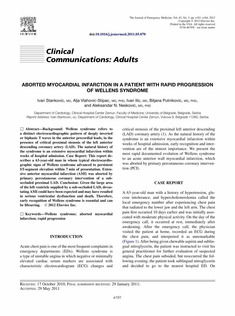
A 63-year-old man with a history of hypertension, glu-cose intolerance, and hypercholesterolemia called thelocal emergency number after experiencing chest painthat radiated to the lower jaw and the left arm. The chestpain first occurred 10 days earlier and was initially asso-ciated with moderate physical activity. On the day of theemergency call, it occurred at rest, immediately afterawakening. After the emergency call, the physicianvisited the patient at home, recorded an ECG duringthe chest pain, and interpreted it as unremarkable(Figure 1). After being given chewable aspirin and sublin-gual nitroglycerin, the patient was instructed to visit hisgeneral practitioner for further evaluation of suspectedangina. The chest pain subsided, but reoccurred the fol-lowing evening; the patient took sublingual nitroglycerinand decided to go to the nearest hospital ED. On
uary 2011;
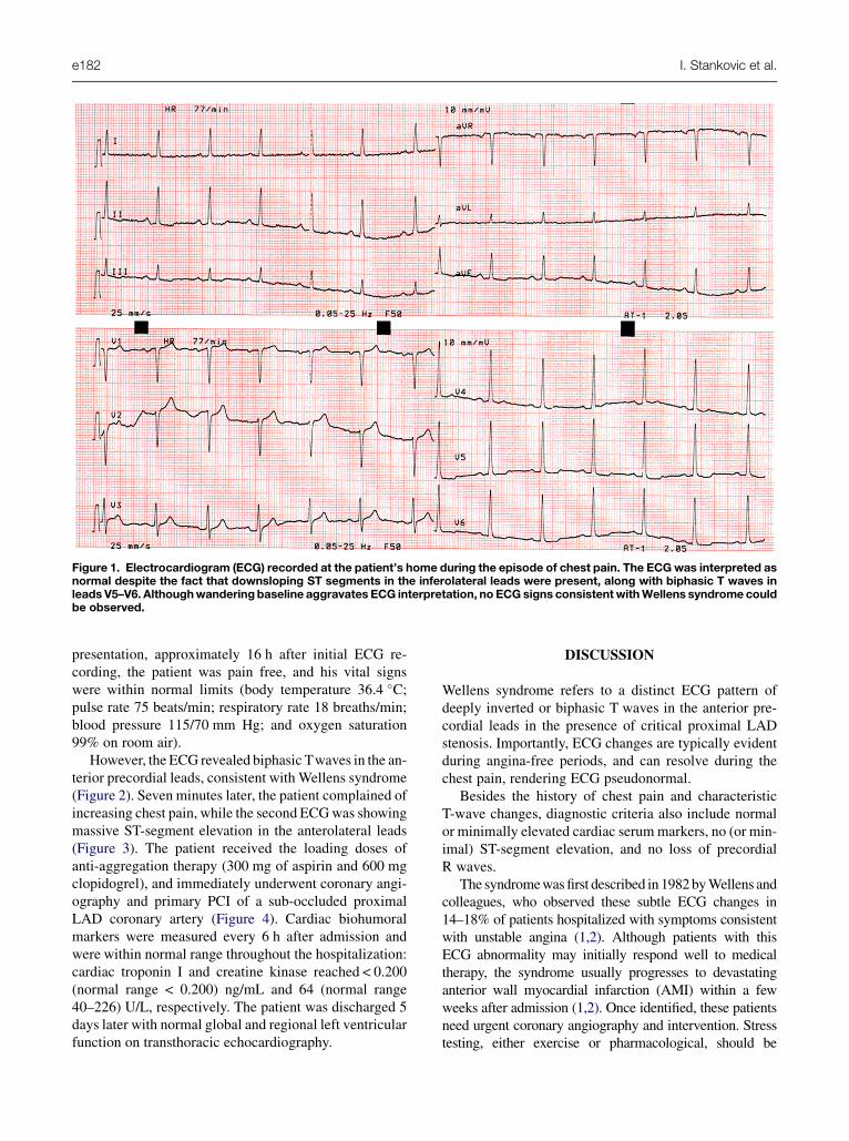
Figure 1. Electrocardiogram (ECG) recorded at the patient’s home during the episode of chest pain. The ECGwas interpreted asnormal despite the fact that downsloping ST segments in the inferolateral leads were present, along with biphasic T waves inleads V5–V6. Althoughwandering baseline aggravates ECG interpretation, no ECG signs consistentwithWellens syndrome couldbe observed.
e182 I. Stankovic et al.
presentation, approximately 16 h after initial ECG re-cording, the patient was pain free, and his vital signswere within normal limits (body temperature 36.4 �C;pulse rate 75 beats/min; respiratory rate 18 breaths/min;blood pressure 115/70 mm Hg; and oxygen saturation99% on room air).
However, the ECG revealed biphasic Twaves in the an-terior precordial leads, consistent with Wellens syndrome(Figure 2). Seven minutes later, the patient complained ofincreasing chest pain, while the second ECGwas showingmassive ST-segment elevation in the anterolateral leads(Figure 3). The patient received the loading doses ofanti-aggregation therapy (300 mg of aspirin and 600 mgclopidogrel), and immediately underwent coronary angi-ography and primary PCI of a sub-occluded proximalLAD coronary artery (Figure 4). Cardiac biohumoralmarkers were measured every 6 h after admission andwere within normal range throughout the hospitalization:cardiac troponin I and creatine kinase reached < 0.200(normal range < 0.200) ng/mL and 64 (normal range40–226) U/L, respectively. The patient was discharged 5days later with normal global and regional left ventricularfunction on transthoracic echocardiography.
DISCUSSION
Wellens syndrome refers to a distinct ECG pattern ofdeeply inverted or biphasic T waves in the anterior pre-cordial leads in the presence of critical proximal LADstenosis. Importantly, ECG changes are typically evidentduring angina-free periods, and can resolve during thechest pain, rendering ECG pseudonormal.
Besides the history of chest pain and characteristicT-wave changes, diagnostic criteria also include normalor minimally elevated cardiac serummarkers, no (or min-imal) ST-segment elevation, and no loss of precordialR waves.
The syndromewasfirst described in 1982byWellens andcolleagues, who observed these subtle ECG changes in14–18% of patients hospitalized with symptoms consistentwith unstable angina (1,2). Although patients with thisECG abnormality may initially respond well to medicaltherapy, the syndrome usually progresses to devastatinganterior wall myocardial infarction (AMI) within a fewweeks after admission (1,2). Once identified, these patientsneed urgent coronary angiography and intervention. Stresstesting, either exercise or pharmacological, should be

Figure 2. Initial 12-lead electrocardiogram recorded at presentation to the Emergency Department (at 4:22), showing biphasicT waves in precordial leads V2–V3, consistent with Wellens syndrome.
Figure 3. Electrocardiogram (ECG) recorded during the attack of chest pain (at 4:29), 7 min after the initial ECG. Note massiveST-segment elevation in anterolateral leads, consistent with acute anterior ST-segment elevation myocardial infarction.
Rapid Progression of Wellens Syndrome e183

Figure 4. (A) Coronary angiogram demonstrating sub-occlusive lesion of the proximal left anterior descending coronary arterycoronary artery (arrow). (B) Final result on culprit lesion (arrow) after primary percutaneous coronary intervention.
e184 I. Stankovic et al.
avoided as it may provoke severe ischemia and may havefatal consequences.
We present the most rapid documented progression ofWellens syndrome to acute anterior ST-segment elevationmyocardial infarction (STEMI), which was aborted byprimary PCI. Until now, the most rapid reported progres-sion of Wellens syndrome to STEMI occurred within55 min (3).
Given the large area of the left ventricle supplied bysub-occluded LAD, extensive AMI could be expectedand could have resulted in serious ventricular dysfunc-tion, congestive heart failure, and death.
Therefore, timely recognition of Wellens syndrome isessential and can be lifesaving. However, the most recentAmerican and European guidelines for the diagnosis andtreatment of non-ST-segment elevation acute coronarysyndromes failed to notice the importance of Wellenssyndrome (4,5).
CONCLUSION
The current case highlights the importance of recognizingthe high-risk patients with Wellens syndrome amongpatients with unstable angina. Characteristic ECGchanges in a patient with Wellens syndrome indicatenot only the site of critical stenosis, but also the patient’sgrim prognosis if left untreated.
We believe that recognition of this ECG pattern shouldbe part of residency curriculum and be underlined duringtraining of physicians involved in the evaluation ofpatients presenting with chest pain, because diagnosticerrors, if they occur, are, as a rule, deleterious for thepatient and may pose serious medicolegal issues.
REFERENCES
1. De Zwaan C, Bar WHM, Wellens HJJ. Characteristic electrocardio-graphic pattern indicating a critical stenosis high in the left anteriordescending coronary artery in patients admitted because of impend-ing myocardial infarction. Am Heart J 1982;103:730–6.
2. de Zwaan C, Bar FW, Janssen JH, et al. Angiographic and clinicalcharacteristics of patients with unstable angina showing an ECGpattern indicating critical narrowing of the proximal LAD coronaryartery. Am Heart J 1989;117:657–65.
3. Donahue B, Chan SB, Bhandarkar S. Rapid progression of Wellenssyndrome in the Emergency Department. J Emerg Med. 2010 May22: [Epub ahead of print], doi:10.1016/j.jemermed.2010.04.004.
4. Anderson JL, Adams CD, Antman EM, et al. ACC/AHA 2007 guide-lines for the management of patients with unstable angina/non–ST-elevation myocardial infarction: a report of the American College ofCardiology/American Heart Association Task Force on PracticeGuidelines (Writing Committee to Revise the 2002 Guidelines fortheManagement of PatientsWith Unstable Angina/Non–ST-ElevationMyocardial Infarction). Circulation 2007;116:e148–304.
5. Task Force for Diagnosis and Treatment of Non-ST-SegmentElevation Acute Coronary Syndromes of European Society ofCardiology; Bassand JP, Hamm CW, Ardissino D, et al. Guidelinesfor the diagnosis and treatment of non-ST-segment elevation acutecoronary syndromes. Eur Heart J 2007;28:1598–660.