abdominal aspecifies one cell type in drosophilaby...
TRANSCRIPT

INTRODUCTION
The Hox/homeotic genes encode homeodomain transcriptionfactors that generate morphological diversity along the majorbody axis during animal development (Lewis, 1978; Wakimotoand Kaufman, 1981; McGinnis and Krumlauf, 1992; Mann andMorata, 2000). Work in Drosophilasuggests that they fulfil thisrole by regulating a large overall number of downstream loci(reviewed by Akam, 1998; Graba et al., 1997). Studiesfocusing on the individuation of serially homologousappendages indicate that very different subsets of thedownstream gene pool are likely to be deployed in eachdevelopmental context. For example, during the formationof a dorsal appendage, the haltere, Ultrabithorax (Ubx)suppresses wing development by regulating Serrate, spalt(sal), wingless(wg), vestigial, blistered, achaeteand probablymany other downstream genes (Weatherbee et al., 1998). In thecase of leg development, the major function of Antennapedia(Antp) in distal and medial regions appears to be to repress theantennal selector gene homothorax (hth) but in more proximalterritory other, as yet unknown, Antptargets are involved(Casares and Mann, 2001). In the abdomen, leg developmentis completely blocked as the resident Hox genes suppresscomponents of the ventral appendage programme itself, suchas Distal-less(Vachon et al., 1992).
Implicit in the above examples is the idea that Hox genesfunction to modify an underlying metameric pattern or groundstate (Lewis, 1978; Struhl, 1983). We use this term in adevelopmental context to refer to the body plan formed without
any Hox inputs. At present, the mechanisms that link the inputsfrom Hox genes and ground state genes to final morphologicalreadouts are unknown. This is largely because it has not yetbeen possible to characterise a complete battery of Hox targetssufficient for any one patterning process. In order to identifysuch a target set in a relatively simple and well-defined system,we have initiated a single-cell resolution study of the larvaloenocyte, a specialised secretory cell that is restricted to thelarval abdominal segments (Bodenstein, 1950; Gould et al.,2001). Two recent reports have shown that oenocytes arederived from the dorsal embryonic ectoderm by a localinduction involving epidermal growth factor receptor (EGFR)activation within the presumptive oenocyte itself (Elstob et al.,2001; Rusten et al., 2001). The relevant ligand, secreted Spitz(sSpi), is made by a nearby cell of the peripheral nervoussystem: a chordotonal organ precursor called C1. Importantly,the zinc-finger transcription factor encoded by the sal gene isrequired to prepattern the responding ectoderm so thatinduction results in an oenocyte-specific EGFR output.
Here we investigate why oenocyte formation is restricted toabdominal segments. We use genetic analysis to show that theformation of this cell type requires an input from abdAthat cannot be substituted for by the closely related Ubx gene. Usingthe GAL4/UAS system, we show that abdAplays no direct roleduring oenocyte differentiation but acts transiently in C1during the induction phase. We then employ various Hoxmutant rescue assays to demonstrate that this non-cellautonomous role of abdA is mediated by only one principaltarget gene rhomboid (rho),required in C1 for processing the
2957Development 129, 2957-2963 (2002)Printed in Great Britain © The Company of Biologists Limited 2002DEV7963
The Hox/homeotic genes encode transcription factorsthat generate segmental diversity during Drosophiladevelopment. At the level of the whole animal, they arebelieved to carry out this role by regulating a large numberof downstream genes. Here we address the unresolved issueof how many Hox target genes are sufficient to define theidentity of a single cell. We focus on the larval oenocyte,which is restricted to the abdomen and induced in responseto a non-cell autonomous, transient and highly selectiveinput from abdominal A (abdA). We use Hox mutant rescueassays to demonstrate that this function of abdA can bereconstituted by providing Rhomboid (Rho), a processing
factor for the EGF receptor ligand, secreted Spitz. Thus, inorder to make an oenocyte, abdA regulates just oneprincipal target, rho, that acts at the top of a complexhierarchy of cell-differentiation genes. These studiesstrongly suggest that, in at least some contexts, Hox genesdirectly control only a few functional targets within eachnucleus. This raises the possibility that much of the overallHox downstream complexity results from cascades ofindirect regulation and cell-to-cell heterogeneity.
Key words: Hox/homeotic, EGFR, Drosophila
SUMMARY
abdominal A specifies one cell type in Drosophila by regulating one principal
target gene
Véronique Brodu, Philip R. Elstob and Alex P. Gould*
Medical Research Council, National Institute for Medical Research, Mill Hill, London NW7 1AA, UK*Author for correspondence (e-mail: [email protected])
Accepted 7 April 2002

2958
sSpi signal. This function of AbdA prolongs sSpi productionuntil the Hox-independent oenocyte prepattern has been fullyassembled in the responding ectoderm. Thus, in this context, asingle principal Hox target is sufficient only because all cell-type specificity information is present in the ground state andabdA merely provides the permissive inducing signal touncover it.
MATERIALS AND METHODS
Fly stocksen-GAL4, sal-GAL4, UAS-rho, UAS-sspi, svp-lacZ,rho-lacZ,ato1 and Df(3R)p13 were as described (Elstob et al., 2001).Other stocks were: AntpRW10, abdAM1, Scr4 Antp25, Ubxbxd100,ato-GAL4 10 (Hassan et al., 2000), UAS-abdA.M (Michelson,1994), UAS-Ubx1a.C (Castelli-Gair et al., 1994), UAS-EGFRACT(Queenan et al., 1997), also called UAS-EGFRtop4.2.exdB108mutants, derived from germline clones, were made asdescribed (Rauskolb et al., 1993). Crosses were made at 25°Cexcept for those shown in Fig. 2F-H, Fig. 3F,J,K, and Fig. 4A-D,F, which were made at 29°C.
ImmunolabellingImmunolabelling and confocal microscopy were as described(Elstob et al., 2001). Fig. 2 shows single confocal sections,other figures show projections of several confocal sections.Primary antibodies were as described (Elstob et al., 2001)with the following additions: anti-Ato (Jarman et al., 1995) at1:2,000, mouse anti-AbdA (Kellerman et al., 1990) at 1:1,000or rat anti-AbdA (Macias et al., 1990) at 1:500, and mouseanti-Ubx (White and Wilcox, 1984) at 1:20. RNA in situhybridisation used an Alas probe, as described previously(Ruiz de Mena et al., 1999).
RESULTS
A selective, transient and non-cellautonomous requirement for abdAOenocytes are present in clusters of approximately sixcells in each of the abdominal segments A1-A7 (Fig.1A). In the thorax, there is no EGFR induction aroundC1 and no specific serial homologue of the oenocyte.In order to score unambiguously the presence ofoenocytes in a range of different genetic backgrounds,we identified a panel of seven immediate-early, earlyand late markers (Fig. 5; E. Gutierrez and A. G.,unpublished). To determine why oenocyte formation isrestricted to the abdomen, embryos lacking various Hoxgenes or extradenticle(exd), which encodes a Hox co-factor (Mann and Chan, 1996), were examined. Theseexperiments indicate that oenocyte formation requiresexdand abdAbut not two other Hox genes that are alsoexpressed in the abdomen: Antp and Ubx(Fig. 1B-E).To assess whether oenocytes form in the absence of allHox functions, we examined the T1 segment inembryos lacking Sex combs reduced(Scr) and Antpactivities (Struhl, 1983; Macias and Morata, 1996). Nooenocytes are produced in this context, and thereforethese cells are not part of the ground state (Fig. 1F).However, the ground state does contain both thesignalling and responding cell types involved in
oenocyte induction: C1 and the Sal-positive dorsal ectoderm(data not shown).
Two types of GAL4/UAS assay (Brand and Perrimon, 1993)were used to test the potential of genes to form oenocytes. Theectopic assay reveals whether gene products can triggeroenocyte formation in the T1-T3 thoracic segments (Fig. 1, Fig.3) while the rescue assay tests the potential of genes to
V. Brodu, P. R. Elstob and A. P. Gould
Fig. 1.abdAis necessary and sufficient to specify oenocytes. In this andsubsequent figures, oenocytes are labelled with anti-Sal unless otherwisestated. (A,B) Anti-β-galactosidase immunostaining of late embryos carryingsvp-lacZshowing oenocyte clusters present in A1-A7 (indicated) in a wild-type background (A) but missing in an exdB108mutant (B). (C-F) Lateembryos homozygous for AntpRW10(C) or Ubxbxd100(D) display normaloenocyte clusters whereas those homozygous for abdAM1 (E) do not. Scr4
Antp25 (F) double mutants show a wild type oenocyte pattern. (G,H) Using en-GAL4to drive UAS-Ubx(G) or UAS-abdA(H) indicates that AbdA but notUbx can specify oenocytes in T1-T3 (indicated).

2959abdA regulates rho to make oenocytes
overcome the oenocyte deficit in abdAmutants (Fig. 4). First,we used en-GAL4to express AbdA or Ubx in ectodermal stripesthat include the oenocyte precursors (Elstob et al., 2001). In this
ectopic assay, only AbdA could produce oenocytes in T1-T3(Fig. 1G,H). Together with the preceding results, this indicatesthat abdA provides a highly selective patterning input and isboth necessary and sufficient for the oenocyte fate.
At the time of oenocyte induction during stage 11, we finda transient burst of AbdA expression in both C1 and oenocyteprecursors (Fig. 2A-D). To ascertain where abdA function isrequired, we used two drivers that, unlike en-GAL4, havecomplementary expression in the oenocyte precursors (sal-GAL4, Fig. 2E), or in the C1 lineage (ato-GAL4, Fig. 2F).Driving AbdA with ato-GAL4 is sufficient to induce a lateoenocyte marker in thoracic segments (Fig. 2G,H) and also torescue oenocyte formation in abdAmutants (see Fig. 4A,H).In each assay, using sal-GAL4to drive AbdA in the dorsalectoderm fails to produce oenocytes and using both driverstogether does not augment the numbers of oenocytes formedwith ato-GAL4 alone (Fig. 2H, see Fig. 4H). Theseexperiments demonstrate clearly that abdAis required in theC1 lineage but not in the presumptive oenocyte itself, despitebeing transiently expressed there. It therefore follows thatalthough abdAswitches on an extensive hierarchy of early-to-late differentiation genes within the oenocyte, all thisregulation must be indirect.
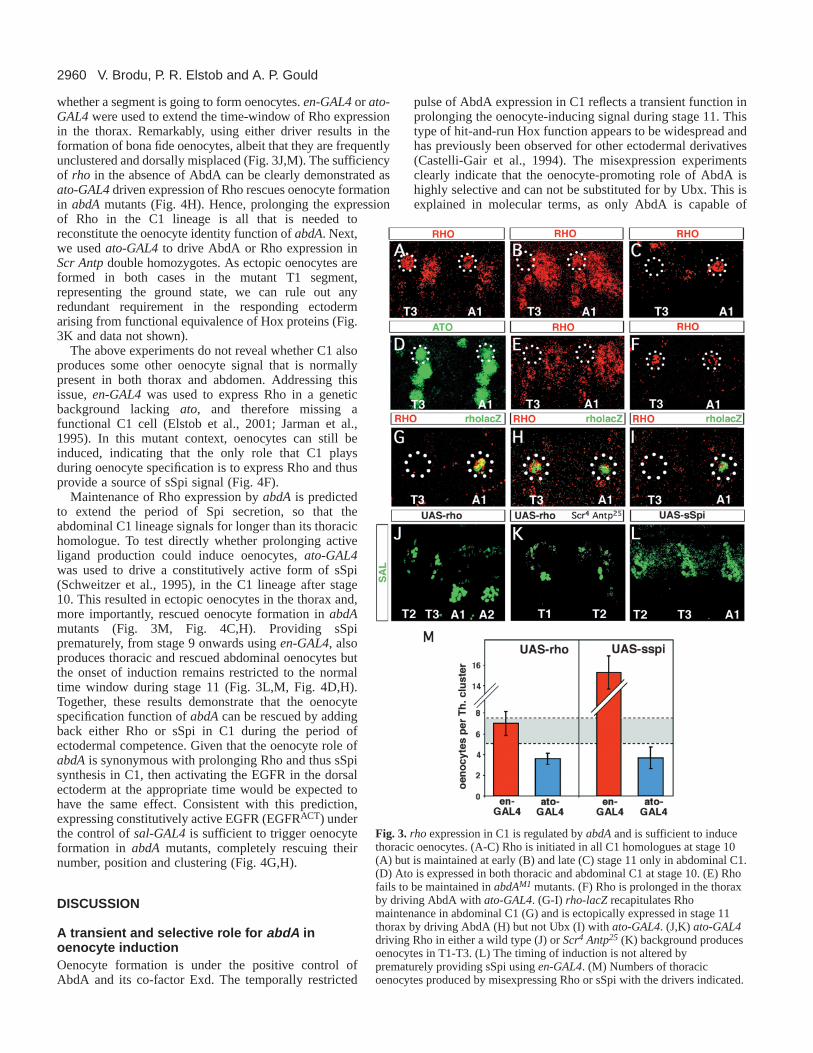
abdA maintains the transcription of rhomboid in C1We looked for potential abdAtargets from amongst the genesknown to play a role in the specification or function of C1. Thisparticular sensory organ precursor produces a type of stretchreceptor, the chordotonal organ, that is defined by the proneuralgene atonal(ato) (Jarman et al., 1993). ato is also required foroenocyte formation (Elstob et al., 2001; Rusten et al., 2001)but it is similarly expressed in thoracic and abdominal C1, isnot regulated by abdAand is downregulated prior to oenocyteinduction (Fig. 3D and data not shown). We then examined rho,a gene downstream of atoand rate-limiting for the productionof sSpi by cleavage from an inactive membrane-boundprecursor (mSpi) in the Golgi apparatus (Lee et al., 2001). Likeato, rho is also required for oenocyte formation (Elstob et al.,2001). Rho protein is first expressed in C1 at stage 10, after ithas delaminated from the dorsal ectoderm. As with Ato at thisstage, early Rho is present at similar levels in thoracic andabdominal C1 precursors and is not under abdAcontrol (Fig.3A). During stage 11, however, thoracic Rho becomesextinguished while abdominal Rho persists at a similar level inthe C1 lineage (Fig. 3B,C). Unlike the early expression, thislate phase correlates with the time of oenocyte induction andis missing in abdAmutants (Fig. 3E). Furthermore, drivingAbdA in the C1 lineage during stage 11, either in the thoraxof a wild-type embryo, or in the abdomen of an abdAmutant,is sufficient to prolong Rho expression (Fig. 3F, Fig. 4B).Together, these results indicate that the maintenance but not theestablishment of Rho expression is under abdA control.Analysis of a rho-lacZline, expressed at stage 11 but not stage10, suggests that this late regulation is at the transcriptionallevel and is mediated by a different enhancer than thatcontrolling the early phase of expression (Fig. 3G-I).
Maintaining Rho is sufficient to rescue oenocyteformation in abdA mutantsNext, we asked whether the rather simple Rho timing differencebetween the thorax and the abdomen is responsible for deciding
Fig. 2.AbdA misexpression in the thoracic C1 lineage inducesoenocytes. (A,B) Oenocyte precursors strongly express AbdA at stage11 (A) but not stage 13 (B). (C) C1 (circled here and subsequently),labelled with anti-Rho, transiently expresses AbdA at stage 11.(D) Cartoon representing oenocyte precursors (red), chordotonalorgan precursors C1-C3 (green), the tracheal pit (tp, green) and thedorsal Sal domain (pink) (Elstob et al., 2001) (E,F) Marking the C1-lineage with anti-Rho, reveals that sal-GAL4and ato-GAL4drivecomplementary expression of UAS-nlslacZin the dorsal ectodermincluding the oenocyte precursors (E) or in the C1 lineage (F)respectively. (G)ato-GAL4driving UAS-abdAproduces Alas-positiveoenocytes in the thorax. (H) Numbers of thoracic oenocytes producedby misexpressing AbdA with the drivers indicated. This andsubsequent graphs show the mean±1 s.d. for experimental (error bars)and wild-type abdominal counts (grey zone).
2960
whether a segment is going to form oenocytes. en-GAL4or ato-GAL4were used to extend the time-window of Rho expressionin the thorax. Remarkably, using either driver results in theformation of bona fide oenocytes, albeit that they are frequentlyunclustered and dorsally misplaced (Fig. 3J,M). The sufficiencyof rho in the absence of AbdA can be clearly demonstrated asato-GAL4driven expression of Rho rescues oenocyte formationin abdAmutants (Fig. 4H). Hence, prolonging the expressionof Rho in the C1 lineage is all that is needed toreconstitute the oenocyte identity function of abdA. Next,we used ato-GAL4to drive AbdA or Rho expression inScr Antpdouble homozygotes. As ectopic oenocytes areformed in both cases in the mutant T1 segment,representing the ground state, we can rule out anyredundant requirement in the responding ectodermarising from functional equivalence of Hox proteins (Fig.3K and data not shown).
The above experiments do not reveal whether C1 alsoproduces some other oenocyte signal that is normallypresent in both thorax and abdomen. Addressing thisissue, en-GAL4was used to express Rho in a geneticbackground lacking ato, and therefore missing afunctional C1 cell (Elstob et al., 2001; Jarman et al.,1995). In this mutant context, oenocytes can still beinduced, indicating that the only role that C1 playsduring oenocyte specification is to express Rho and thusprovide a source of sSpi signal (Fig. 4F).
Maintenance of Rho expression by abdA is predictedto extend the period of Spi secretion, so that theabdominal C1 lineage signals for longer than its thoracichomologue. To test directly whether prolonging activeligand production could induce oenocytes, ato-GAL4was used to drive a constitutively active form of sSpi(Schweitzer et al., 1995), in the C1 lineage after stage10. This resulted in ectopic oenocytes in the thorax and,more importantly, rescued oenocyte formation in abdAmutants (Fig. 3M, Fig. 4C,H). Providing sSpiprematurely, from stage 9 onwards using en-GAL4, alsoproduces thoracic and rescued abdominal oenocytes butthe onset of induction remains restricted to the normaltime window during stage 11 (Fig. 3L,M, Fig. 4D,H).Together, these results demonstrate that the oenocytespecification function of abdAcan be rescued by addingback either Rho or sSpi in C1 during the period ofectodermal competence. Given that the oenocyte role ofabdAis synonymous with prolonging Rho and thus sSpisynthesis in C1, then activating the EGFR in the dorsalectoderm at the appropriate time would be expected tohave the same effect. Consistent with this prediction,expressing constitutively active EGFR (EGFRACT) underthe control of sal-GAL4is sufficient to trigger oenocyteformation in abdAmutants, completely rescuing theirnumber, position and clustering (Fig. 4G,H).
DISCUSSION
A transient and selective role for abdA inoenocyte inductionOenocyte formation is under the positive control ofAbdA and its co-factor Exd. The temporally restricted
pulse of AbdA expression in C1 reflects a transient function inprolonging the oenocyte-inducing signal during stage 11. Thistype of hit-and-run Hox function appears to be widespread andhas previously been observed for other ectodermal derivatives(Castelli-Gair et al., 1994). The misexpression experimentsclearly indicate that the oenocyte-promoting role of AbdA ishighly selective and can not be substituted for by Ubx. This isexplained in molecular terms, as only AbdA is capable of
V. Brodu, P. R. Elstob and A. P. Gould
Fig. 3. rhoexpression in C1 is regulated by abdAand is sufficient to inducethoracic oenocytes. (A-C) Rho is initiated in all C1 homologues at stage 10(A) but is maintained at early (B) and late (C) stage 11 only in abdominal C1.(D) Ato is expressed in both thoracic and abdominal C1 at stage 10. (E) Rhofails to be maintained in abdAM1 mutants. (F) Rho is prolonged in the thoraxby driving AbdA with ato-GAL4. (G-I) rho-lacZrecapitulates Rhomaintenance in abdominal C1 (G) and is ectopically expressed in stage 11thorax by driving AbdA (H) but not Ubx (I) with ato-GAL4. (J,K) ato-GAL4driving Rho in either a wild type (J) or Scr4 Antp25 (K) background producesoenocytes in T1-T3. (L) The timing of induction is not altered byprematurely providing sSpi using en-GAL4. (M) Numbers of thoracicoenocytes produced by misexpressing Rho or sSpi with the drivers indicated.

2961abdA regulates rho to make oenocytes
maintaining the transcription of rhoin the C1 lineage. Suchselectivity contrasts with the equivalent biological activities ofUbx and AbdA proteins in promoting haltere formation(Casares et al., 1996). In this regard, we note that exdisrequired to make an oenocyte but not a haltere (Gonzalez-Crespo and Morata, 1995) and therefore may allow these twoHox proteins to discriminate between different targets, as hasbeen suggested previously (Mann and Chan, 1996).
The oenocyte function of abdA is mediated by oneprincipal target, rhoThe results reported here allow us to add sSpi to the growing listof intercellular signalling molecules that are known to be targetsof the Hox genes (Immergluck et al., 1990; Reuter et al., 1990;Szuts et al., 1997; Szuts et al., 1998; Wiellette and McGinnis,1999). For example, during abdominal denticle patterning, it hasbeen shown that Ubx and abdApositively regulate Serratesignalling, in turn expanding rho expression and creating anadditional row of denticulate cells (Szuts et al., 1997; Wielletteand McGinnis, 1999). In another context, the visceral mesoderm,these same two Hox genes directly or indirectly regulate theproduction of at least three signals required to induce appropriatelevels of Labial in the adjacent endoderm: Decapentaplegic, Wgand Vein (Yu et al., 1996; Szuts et al., 1998). Interestingly,different levels of Wg can induce two alternative cell fates in thegut, copper cells or large flat cells, thus indicating that thissignalling input plays an instructive rather than a permissive role(Hoppler and Bienz, 1995).
Although many different signalling molecules and also awide range of other types of gene product are all known to beHox targets, a set of these sufficient for any one patterningprocess had not yet been clearly defined. For the first time, we
have presented a stringent proof of sufficiency by rescuing acellular Hox phenotype with the gene products of the relevantdownstream targets. In the context of the oenocyte, this hasrevealed that only one target gene, rho, is sufficient to executeall aspects of abdAfunction. Our experiments do notdistinguish whether the transcriptional maintenance of rho byabdA in C1 is direct or indirect. Either way, the complexdownstream genetic cascade triggered by Rho-dependentactivation of sSpi is sufficient to substitute for an input fromabdA. Importantly, even late differentiation markers such asalas are switched on in the oenocyte by providing abdAfunction specifically in the C1 lineage. Hence, abdA,via itsone principal target rho, plays a non-cell autonomous role inpromoting the differentiation of the complex oenocyte fate.
As the numerical deficit of chordotonal organs that is foundin abdA mutants (Heuer and Kaufman, 1992) can also berescued with sSpi (Fig. 4E), it appears that rho may be theprincipal target of abdAin this system too. Although no othersingle-cell functions of Hox genes have yet been clearlydefined by rescue, we think it likely that in most, if not all,developmental contexts, Hox genes directly control only a fewcritical targets within each nucleus at any one time. In thisscenario, the overall downstream complexity that has beenobserved previously would largely arise from cell-to-cellheterogeneity and cascades of indirect regulation. Both of thesefactors will need to be given careful consideration whenevergenome-wide approaches, such as microarrays, are employedfor the identification of biologically relevant Hox targets.
A prepattern for a segment-specific cell type is HoxindependentIn the absence of any Hox input, oenocytes are completely
Fig. 4. Activating the EGFR pathwayrescues the oenocyte deficit in abdAmutants. (A,B) AbdA driven by en-GAL4(A) or ato-GAL4(B) rescuesoenocyte formation or Rhomaintenance respectively in abdAM1
homozygotes. (C-E) sSpi driven byato-GAL4(C) or en-GAL4(D) in anabdAM1 background rescues oenocyteformation. In addition, anti-Futsch/22C10 labelling reveals that adorsal or lateral array of 5-7chordotonal organs (arrowheads) isproduced with en-GAL4(E), insteadof the dorsal triplet found in abdAmutants (Heuer and Kaufman, 1992).(F) The lack of oenocytes inato1/Df(3R)p13 transheterozygotes(Elstob et al., 2001), is rescued bydriving Rho with en-GAL4.(G) Providing EGFRACT with sal-GAL4 in an abdAM1 backgroundproduces a normal oenocyte pattern.(H) Numbers of oenocytes perabdominal cluster produced with theGAL4-driver/UAS combinationsindicated.

2962
missing and therefore are not an overt part of the ground state.At first sight, it might seem that for cell types that have nomorphological representation in the ground state, such asoenocytes, Hox genes must necessarily play a classicinstructive role in defining the appropriate pathway ofdifferentiation. However, as we will now argue, this is not theonly way that Hox genes can direct the formation of segment-specific cell types.
Previously, we described two lines of evidence that the sSpisignal from C1 is permissive in the sense that it does not itselfcontain any oenocyte specificity information (Elstob et al.,2001). First, providing ectopic sSpi signal outside of arestricted dorsal zone around C1 fails to induce oenocytes. Andsecond, the degree of sSpi signalling influences the number ofinduced cells rather than their identity. In contrast, it has beendemonstrated that all of the cell-type specificity information isencoded in the dorsal ectoderm as an oenocyte prepattern(Elstob et al., 2001). One crucial component of this prepatternis encoded by sal. The Sal zinc-finger transcription factor actsto prime the EGFR response in favour of the oenocyte fate. Inits absence, there is a fate switch and sSpi signalling nowinduces secondary chordotonal organs (Elstob et al., 2001;Rusten et al., 2001). Thus, it has been shown that oenocytespecificity is provided by the sal-dependent prepattern and notby the sSpi-inducing signal.
We have analysed the segmental restriction of oenocyteinduction and provide evidence supporting a model wherethere is no Hox input into the prepattern but the timing of thesSpi-inducing signal is controlled by abdA (Fig. 5). Togetherwith our previous finding that sSpi signalling is permissive, wenow conclude that abdAdoes not directly specify the oenocyteidentity, rather it determines which segments will formoenocytes. This involves modifying the signalling properties ofC1, a serially reiterated cell type that is part of the ground state.In turn, this provides a permissive trigger that uncovers acryptic oenocyte identity also present in the ground state.Hence ato and sal, two of the genes that contribute to theground state, are essential for specifying the C1 cell typeand the complete oenocyte prepattern respectively. Anotherimportant feature of our model is that the dorsal ectoderm isnot competent for oenocyte induction until stage 11. Thismakes the prediction that if competence were to be acquiredearlier, when C1 expresses Rho in both the thorax andabdomen, then oenocytes would be produced in all trunksegments independently of Hox genes.
We thank B. Hassan, H. Bellen, M. Calleja, G. Morata, E. Bier, A.Jarman, R. Garesse, M. Freeman, A. Michelson, E. Wieschaus, R.Barrio, R. White, I. Duncan, J. Casanova and the Bloomington andUmea stock centres for flies, probes and antibodies. We also thank J.-P. Vincent, D. Stemple, I. Salecker, C. Alexandre, and also E. Gutierrez,B. Bello and other members of the Gould laboratory for advice andcritical reading of the manuscript. This work was supported by theMedical Research Council and a EMBO long term fellowship to V. B.
REFERENCES
Akam, M. (1998). Hoxgenes: from master genes to micromanagers.Curr.Biol. 8, R676-R678.
Bodenstein, D. (1950). The postembryonic development of Drosophila. InBiology of Drosophila(ed. M. Demerec). New York: John Wiley & Sons.
Brand, A. H. and Perrimon, N. (1993). Targeted gene expression as a meansof altering cell fates and generating dominant phenotypes.Development118,401-415.
Casares, F., Calleja, M. and Sanchez-Herrero, E.(1996). Functionalsimilarity in appendage specification by the Ultrabithorax and abdominal-A Drosophila HOX genes.EMBO J. 15, 3934-3942.
Casares, F. and Mann, R. S.(2001). The ground state of the ventralappendage in Drosophila.Science293, 1477-1480.
Castelli-Gair, J., Greig, S., Micklem, G. and Akam, M.(1994). Dissectingthe temporal requirements for homeotic gene function.Development120,1983-1995.
Elstob, P. R., Brodu, V. and Gould, A. P.(2001). spalt-dependent switchingbetween two cell fates that are induced by the Drosophila EGF receptor.Development128, 723-732.
Gonzalez-Crespo, S. and Morata, G.(1995). Control of Drosophilaadultpattern by extradenticle. Development121, 2117-2125.
Gould, A. P., Elstob, P. R. and Brodu, V.(2001). Insect oenocytes: a modelsystem for studying cell-fate specification by Hox genes.J. Anat. 199, 25-33.
Graba, Y., Aragnol, D. and Pradel, J.(1997). Drosophila Hox complexdownstream targets and the function of homeotic genes.BioEssays19, 379-388.
Hassan, B. A., Bermingham, N. A., He, Y., Sun, Y., Jan, Y. N., Zoghbi, H.Y. and Bellen, H. J.(2000). atonalregulates neurite arborization but doesnot act as a proneural gene in the Drosophilabrain.Neuron25, 549-561.
Heuer, J. G. and Kaufman, T. C. (1992). Homeotic genes have specificfunctional roles in the establishment of the Drosophilaembryonic peripheralnervous system.Development115, 35-47.
Hoppler, S. and Bienz, M. (1995). Two different thresholds of winglesssignalling with distinct developmental consequences in the Drosophilamidgut.EMBO J. 14, 5016-5026.
V. Brodu, P. R. Elstob and A. P. Gould
Fig. 5.Permissive model for oenocyte specification by abdA. Atstage 10, Ato activates rhotranscription in C1. Rho may or may not(?) process mSpi to sSpi in the Golgi apparatus at this stage, eitherway the response in the dorsal ectoderm is blocked (cross). By stage11, the dorsal ectoderm acquires the full oenocyte prepattern andbecomes competent for induction. At this time, Ato is no longerpresent but AbdA and its co-factor Exd maintain rho transcription,thus keeping sSpi available to activate the EGFR and in turn thehierarchy of oenocyte differentiation genes including: pointed (pnt),argos, spalt (sal), seven up (svp), ventral veins lacking (vvl),Hepatocyte nuclear factor 4 (Hnf4) and delta-aminolevulinatesynthase (Alas).
Stage 10 Stage 11
rho
sSpi
EGFR
dorsalectoderm
late
early
immediateearly
Alas
Hnf4
sal svp vvlpnt argos
Ras/MAPK
stage 10 stage 11
Stage 10 Stage 11
rho
AbdA ExdAto
C1
Rho
mSpi
?mSpi
Rho

2963abdA regulates rho to make oenocytes
Immergluck, K., Lawrence, P. A. and Bienz, M.(1990). Induction acrossgerm layers in Drosophila mediated by a genetic cascade.Cell 62, 261-268.
Jarman, A. P., Grau, Y., Jan, L. Y. and Jan, Y. N.(1993). atonalis aproneural gene that directs chordotonal organ formation in the Drosophilaperipheral nervous system.Cell 73, 1307-1321.
Jarman, A. P., Sun, Y., Jan, L. Y. and Jan, Y. N.(1995). Role of theproneural gene, atonal, in formation of Drosophilachordotonal organs andphotoreceptors.Development121, 2019-2030.
Kellerman, K. A., Mattson, D. M. and Duncan, I. (1990). Mutationsaffecting the stability of the fushi tarazuprotein of Drosophila.Genes Dev.4, 1936-1950.
Lee, J. R., Urban, S., Garvey, C. F. and Freeman, M.(2001). Regulatedintracellular ligand transport and proteolysis control EGF signal activationin Drosophila.Cell 107, 161-171.
Lewis, E. B.(1978). A gene complex controlling segmentation in Drosophila.Nature276, 565-570.
Macias, A., Casanova, J. and Morata, G.(1990). Expression and regulationof the abd-Agene of Drosophila.Development110, 1197-1207.
Macias, A. and Morata, G. (1996). Functional hierarchy and phenotypicsuppression among Drosophila homeotic genes: the labialand emptyspiraclesgenes.EMBO J. 15, 334-343.
Mann, R. and Chan, S.-K.(1996). Extra specificity from extradenticle: thepartnership between HOX and PBX/EXD homeodomain proteins.TrendsGenet. 12, 258-262.
Mann, R. S. and Morata, G. (2000). The developmental and molecularbiology of genes that subdivide the body of Drosophila.Annu. Rev. CellDev. Biol. 16, 243-271.
McGinnis, W. and Krumlauf, R. (1992). Homeobox genes and axialpatterning.Cell 68, 283-302.
Michelson, A. M. (1994). Muscle pattern diversification in Drosophila isdetermined by the autonomous function of homeotic genes in the embryonicmesoderm.Development120, 755-768.
Queenan, A. M., Ghabrial, A. and Schupbach, T.(1997). Ectopic activationof torpedo/Egfr, a Drosophilareceptor tyrosine kinase, dorsalizes both theeggshell and the embryo.Development124, 3871-3880.
Rauskolb, C., Peifer, M. and Wieschaus, E.(1993). extradenticle, a regulatorof homeotic gene activity, is a homolog of the homeobox-containing humanproto-oncogene pbx1.Cell 74, 1101-1112.
Reuter, R., Panganiban, G. E. F., Hoffmann, F. M. and Scott, M. P.(1990).Homeotic genes regulate the spatial expression of putative growth factors in
the visceral mesoderm of Drosophilaembryos.Development110, 1031-1040.
Ruiz de Mena, I., Fernandez-Moreno, M. A., Bornstein, B., Kaguni, L. S.and Garesse, R.(1999). Structure and regulated expression of the delta-aminolevulinate synthase gene from Drosophila melanogaster. J. Biol.Chem. 274, 37321-37328.
Rusten, T. E., Cantera, R., Urban, J., Technau, G., Kafatos, F. C. andBarrio, R. (2001). Spalt restricts EGFR mediated induction of chordotonalprecursors in the embryonic PNS of Drosophila.Development128, 711-722.
Schweitzer, R., Shaharabany, M., Seger, R. and Shilo, B. Z.(1995).Secreted Spitz triggers the DER signaling pathway and is a limitingcomponent in embryonic ventral ectoderm determination.Genes Dev. 9,1518-1529.
Struhl, G. (1983). Role of the esc+ gene product in ensuring the selectiveexpression of segment-specific homeotic genes in Drosophila.J. Embryol.Exp. Morphol. 76, 297-331.
Szuts, D., Freeman, M. and Bienz, M.(1997). Antagonism between EGFRand Wingless signalling in the larval cuticle of Drosophila.Development124, 3209-3219.
Szuts, D., Eresh, S. and Bienz, M.(1998). Functional intertwining of Dppand EGFR signaling during Drosophilaendoderm induction.Genes Dev. 12,2022-2035.
Vachon, G., Cohen, B., Pfeifle, C., McGuffin, M. E., Botas, J. and Cohen,S. M. (1992). Homeotic genes of the Bithorax complex repress limbdevelopment in the abdomen of the Drosophila embryo through the targetgene Distal-less.Cell 71, 437-450.
Wakimoto, B. T. and Kaufman, T. C.(1981). Analysis of larval segmentationin lethal genotypes associated with the Antennapedia gene complex inDrosophila melanogaster. Dev. Biol. 81, 51-64.
Weatherbee, S. D., Halder, G., Kim, J., Hudson, A. and Carroll, S.(1998).Ultrabithorax regulates genes at several levels of the wing-patterninghierarchy to shape the development of the Drosophilahaltere.Genes Dev.12, 1474-1482.
White, R. A. and Wilcox, M. (1984). Protein products of the bithoraxcomplex in Drosophila.Cell 39, 163-171.
Wiellette, E. L. and McGinnis, W. (1999). Hoxgenes differentially regulateSerrateto generate segment-specific structures.Development126, 1985-1995.
Yu, X., Hoppler, S., Eresh, S. and Bienz, M. (1996). decapentaplegic, a targetgene of the winglesssignalling pathway in the Drosophila midgut.Development122, 849-858.