ab126556 - brdu cell proliferation elisa kit (colorimetric) · pdf fileab126556 - brdu cell...
TRANSCRIPT

ab126556 - BrdU Cell
Proliferation ELISA Kit
(colorimetric)
Instructions for Use
For the quantitative measurement of BrdU incorporation into newly synthesized DNA of actively proliferating cells. This product is for research use only and is not intended for diagnostic use.

1
Table of Contents
1. Introduction 2
2. Principle of Assay 3
3. Assay Summary 4
4. Kit Contents 7
5. Storage and Handling 8
6. Additional Materials Required 8
7. Protocol 9
8. Typical Data 14
2
1. Introduction
A non-isotopic enzyme immunoassay for the quantification of DNA
synthesis and cell proliferation.
Evaluation of cell cycle progression is essential for investigations in
many scientific fields. Measurement of [3H] thymidine incorporation
as cells enter S phase has long been the traditional method for the
detection of cell proliferation. Subsequent quantification of [3H]
thymidine is performed by scintillation counting or autoradiography.
This technology is slow, labor intensive and has several limitations
including the handling and disposal of radioisotopes and the
necessity of expensive equipment.
A well-established alternative to [3H] thymidine uptake has been
demonstrated by numerous investigators. In these methods
bromodeoxyuridine (BrdU), a thymidine analog replaces [3H]
thymidine. BrdU is incorporated, into newly synthesized DNA strands
of actively proliferating cells. Following partial denaturation of double
stranded DNA, BrdU is detected immunochemically allowing the
assessment of the population of cells, which are actively
synthesizing DNA.
ab126556 involves incorporation of BrdU into cells cultured in
microtiter plates using the cell layer as the solid phase. The resultant
assay is sensitive, rapid, easy to perform and applicable to high
sample throughput. In addition to evaluation of cell proliferation,
3
information such as cell number, morphology and analysis of cellular
antigens can be obtained from a single culture.
2. Principle of the Assay
ab126556 involves incorporation of BrdU into cells cultured in
microtiter plates using the cell layer as the solid phase. During the
final 2 to 24 hours of culture BrdU is added to wells of the microtiter
plate. BrdU will be incorporated into the DNA of dividing cells. To
enable antibody binding to the incorporated BrdU cells must be fixed,
permeabilized and the DNA denatured. This is all done in one step
by treatment with Fixing Solution. Detector anti-BrdU monoclonal
antibody is pipetted into the wells and allowed to incubate for one
hour, during which time it binds to any incorporated BrdU. Unbound
antibody is washed away and horseradish peroxidase-conjugated
goat anti-mouse antibody is added, which binds to the Detector
Antibody.
The horseradish peroxidase catalyzes the conversion of the
chromogenic substrate tetra-methylbenzidine (TMB) from a colorless
solution to a blue solution (or yellow after the addition of stopping
reagent), the intensity of which is proportional to the amount of
incorporated BrdU in the cells. The colored reaction product is
quantified using a spectrophotometer.
4
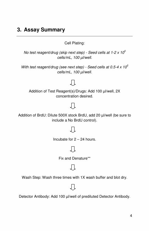
3. Assay Summary
Cell Plating:
No test reagent/drug (skip next step) - Seed cells at 1-2 x 10⁵
cells/mL, 100 µl/well.
With test reagent/drug (see next step) - Seed cells at 0.5-4 x 10⁵
cells/mL, 100 µl/well.
Addition of Test Reagent(s)/Drugs: Add 100 µl/well, 2X
concentration desired.
Addition of BrdU: Dilute 500X stock BrdU, add 20 µl/well (be sure to
include a No BrdU control).
Incubate for 2 – 24 hours.
Fix and Denature**
Wash Step: Wash three times with 1X wash buffer and blot dry.
Detector Antibody: Add 100 µl/well of prediluted Detector Antibody.
5
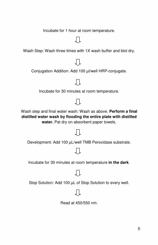
Incubate for 1 hour at room temperature.
Wash Step: Wash three times with 1X wash buffer and blot dry.
Conjugation Addition: Add 100 µl/well HRP-conjugate.
Incubate for 30 minutes at room temperature.
Wash step and final water wash: Wash as above. Perform a final
distilled water wash by flooding the entire plate with distilled
water. Pat dry on absorbent paper towels.
Development: Add 100 µL/well TMB Peroxidase substrate.
Incubate for 30 minutes at room temperature in the dark.
Stop Solution: Add 100 µL of Stop Solution to every well.
Read at 450/550 nm.

6
**Adherent and Suspension Cells (No-Spin Procedure wells):
• Aspirate (or flick) the media from the cell wells.
• Add 200 µl/well Fixing Solution.
• Incubate 30 minutes at room temperature.
• Aspirate the Fixing Solution and blot the plates dry.
**Suspension Cells (Spin Procedure):
• Spin the plates for 5 minutes at 1000 rpm.
• Aspirate media; add 200 µl/well Fixing Solution.
• Incubate for 30 minutes at room temperature.
• Aspirate the Fixing Solution and blot the plates dry.
7
4. Kit Contents
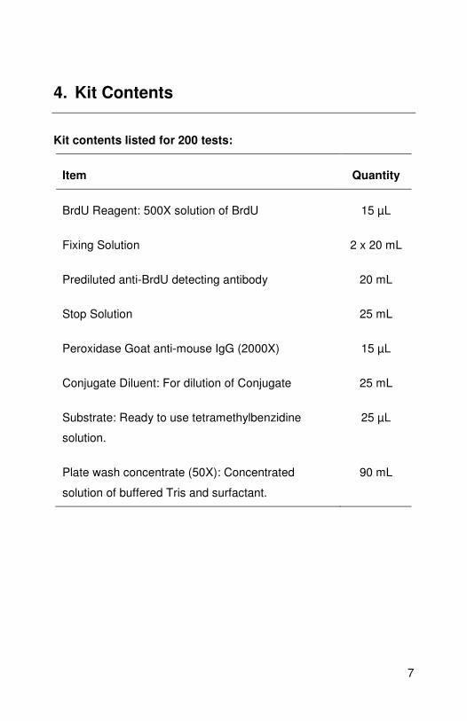
Kit contents listed for 200 tests:
Item Quantity
BrdU Reagent: 500X solution of BrdU 15 µL
Fixing Solution 2 x 20 mL
Prediluted anti-BrdU detecting antibody 20 mL
Stop Solution 25 mL
Peroxidase Goat anti-mouse IgG (2000X) 15 µL
Conjugate Diluent: For dilution of Conjugate 25 mL
Substrate: Ready to use tetramethylbenzidine
solution.
25 µL
Plate wash concentrate (50X): Concentrated
solution of buffered Tris and surfactant.
90 mL
8
5. Storage and Handling
Upon receipt, store entire kit at 4-8°C.
Before first use:
Remove the Fixative/Denaturing Solution and place at room
temperature for at least 4 hours prior to use. Precipitates that may
occur while cold should go back into solution.
6. Additional Materials Required
• 2-20 µl, 20-200 µl and 200-1000 µl precision pipettors with
disposable tips.
• Wash bottle or multichannel dispenser for washing.
• 2000 mL graduated cylinder.
• PBS (137 mM NaCl, 2.7 mM KCl, 4.3 mM Na2HPO4-7H2O,
1.4 mM KH2PO4).
• Deionized or distilled H2O
• Spectrophotometer capable of measuring absorbance in 96-
well plates using dual wavelength of 450-540 or 450-595 nm
or a single read at 450 nm.
9
• Tissue culture microtiter plate (96 well culture dish).
• Sterile reagent troughs.
• Micro syringe filter (0.2 µm).
• Syringe
7. Protocol
Notes: Do not expose reagents to excessive light. Do not mix
reagents from different kits. The buffers and reagents used in this kit
contain anti-microbial and anti-fungal reagents. Care should be
taken to prevent direct contact with these products.
Recommended Controls:
Two types of controls are recommended to insure validity of the
experiment.
i. Blank: Add only tissue culture media (no cells).
ii. Background: Cells are present in the wells but do not add
the BrdU Reagent.
1. Cell Plating
Seed cells using a sterile 96-well tissue culture plate, cells are
plated at 2 x 10⁵ cells/mL in 100 µl/well of appropriate cell
10
culture media. Some of the wells on the plate should be set
aside for several controls. These should include wells that do not
receive cells (media alone), and wells which contain cells but will
not receive the BrdU reagent (assay background).
2. Addition of Test Reagent
The test reagent can be a cell proliferation enhancer or
alternatively, can induce growth inhibition or arrest. The test
reagent is diluted to twice the desired final concentration (2X) in
the cell media. 100 µl/well is added on top of the cell wells. The
test reagent should be titered in the assay to determine optimum
concentration for inducing cell proliferation or growth arrest. The
length of time for test reagent incubation should also be
determined for your system (time course study). BrdU addition
(see step 3 below) will occur 2-24 hours prior to the end of the
test.
3. Addition of BrdU
BrdU will be incorporated into proliferating cells and should be
added at least 2 hours prior to the end of the test reagent
incubation period. Better sensitivity and signal to noise ratios are
obtained when longer BrdU labelling times are used. Dilute the
500X concentrated stock 1:500 by adding 8 µl of BrdU stock to
4 mL of cell media. Pipette 20 µl of the diluted BrdU label to the
appropriate wells. Reminder: a series of wells should be set
aside that do NOT receive the BrdU label (- BrdU control for
determining assay background). Incubate the assay 2-24 hours.
11
4. Fix and Denature Step and Storage of Fixed Plates
For detection of the BrdU label by the anti-BrdU monoclonal
antibody, it is necessary to fix the cells and denature the DNA
using a solution provided in this kit (Fixing Solution). There is no
need to spin the cells prior to addition of the fixing solution.
However, if suspension cells are being used, better precision is
obtained if the cell plates are spun in a centrifuge prior to the
fix/denature step. Plates may be fixed (see steps 5-6) and stored
at 4°C for assay at a later time. Place dried plates in a sealed
dry plastic bag, zip-lock type bags or heat sealed plastic bags
are suitable for this purpose. Plates are stable for at least one
month when properly stored.
5. Adherent and Suspension Cells (No-Spin Procedure)
Aspirate the media from the cell wells (this can be done
mechanically or plate can be inverted over appropriate reservoir
and blotted on absorbent paper towels). Add 200 µl/well Fixing
Solution and incubate at room temperature for 30 minutes.
Aspirate the Fixing Solution and blot the plate dry. Note: Fixed
plates can be stored for up to 1 month at 4°C if stored in a heat
sealed or zip-lock bag. If storing your plates for future use, make
sure the plates are blotted well and are very dry (NO Fixing
Solution should be left in the wells).
6. Suspension Cells (Spin Fix/Denature Procedure)
Spin the plates in the centrifuge (using appropriate centrifuge
microtiter plate holders) for 5 minutes at 1000 rpm. Aspirate the

12
media and add 200 µl/well Fixing Solution. Incubate for 30
minutes at room temperature. Aspirate the Fixing Solution and
blot the plates dry. The assay can be run immediately or plates
may be stored for future use (see note above).
7. Wash Step
Dilute the 50X Wash Buffer 1:50 by adding 40 mL to 1.96 L of
distilled water. A microtiter plate washer may be used for all
wash steps OR a squirt bottle for manual plate washing may
also be used. In either case, the wells should be filled completely
with wash buffer. Wash the plate three times with 1X Wash
Buffer prior to adding Detector Antibody. Aspirate the wash
solution after the final wash and blot dry on paper towels.
8. Addition of Detector Antibody
The anti-BrdU monoclonal Detector Antibody is provided as a
prediluted solution. Add 100 µl/well and incubate for 1 hour at
room temperature.
9. Wash Step
Wash as in Step 7 above.
10. Preparation and Addition of the Peroxidase Goat Anti-
Mouse IgG Conjugate
The Peroxidase Goat Anti-Mouse IgG Conjugate is provided as
a concentrated stock solution. Dilute the Conjugate 1:2000 by
adding 6 µl to 12 mL of Conjugate Diluent provided. Once

13
diluted, this solution should be filtered using a 0.22 µm
syringe filter. This lowers the assay background and improves
precision. Pipette 100 µl/well and incubate for 30 minutes at
room temperature.
11. Wash Step and Final Water Wash
Wash as in Step 7 above. Perform a final water wash by
flooding the entire plate with distilled water. Pat dry on
absorbent paper towels.
12. Addition of Substrate
For 200 tests (Two 96 well plates):
Pipette 100 µL/well TMB Peroxidase substrate and incubate for
30 minutes at room temperature in the dark. Positive wells will
be visible by a blue color, the intensity of which is proportional to
the amount of BrdU incorporation in the proliferating cells.
13. Addition of Stop Solution and Reading of the Plate
Stop the reaction by pipetting 100 µL of Stop Solution provided
to every well. The color of positive wells will change from blue to
bright yellow. Read the plate using a spectrophotometric
microtiter plate reader set at a dual wavelength of 450/550 nm
(alternatively, 450/540 nm or 450/595 nm may be used or a
single read at 450 nm).
14
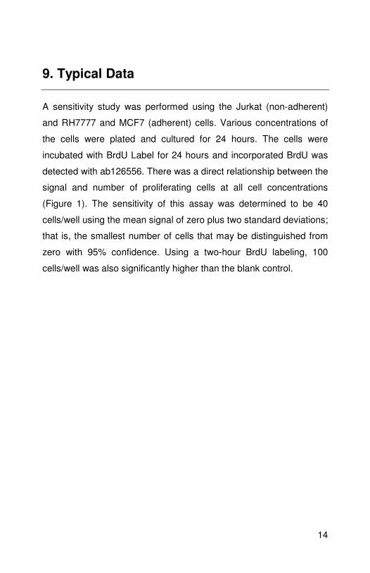
9. Typical Data
A sensitivity study was performed using the Jurkat (non-adherent)
and RH7777 and MCF7 (adherent) cells. Various concentrations of
the cells were plated and cultured for 24 hours. The cells were
incubated with BrdU Label for 24 hours and incorporated BrdU was
detected with ab126556. There was a direct relationship between the
signal and number of proliferating cells at all cell concentrations
(Figure 1). The sensitivity of this assay was determined to be 40
cells/well using the mean signal of zero plus two standard deviations;
that is, the smallest number of cells that may be distinguished from
zero with 95% confidence. Using a two-hour BrdU labeling, 100
cells/well was also significantly higher than the blank control.
15
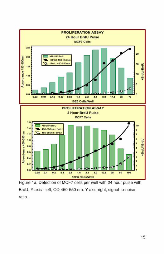
Figure 1a. Detection of MCF7 cells per well with 24 hour pulse with
BrdU. Y axis - left, OD 450-550 nm. Y axis-right, signal-to-noise
ratio.

16
Figure 1b. Detection of Jurkat cells (non-adherent) per well with 24
hour pulse with BrdU. Y axis - left, OD 450-550 nm. Y axis-right,
signal-to-noise ratio.
17
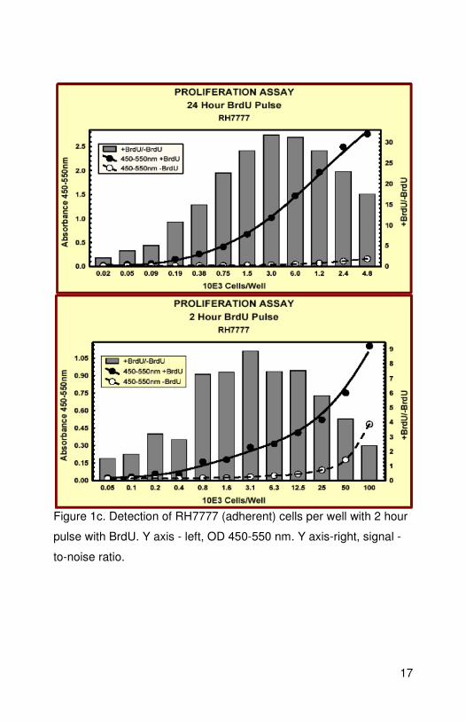
Figure 1c. Detection of RH7777 (adherent) cells per well with 2 hour
pulse with BrdU. Y axis - left, OD 450-550 nm. Y axis-right, signal -
to-noise ratio.

18

19
UK, EU and ROW Email: [email protected] Tel: +44 (0)1223 696000 www.abcam.com US, Canada and Latin America Email: [email protected] Tel: 888-77-ABCAM (22226) www.abcam.com China and Asia Pacific Email: [email protected]
Tel: 108008523689 (中國聯通)
www.abcam.cn Japan Email: [email protected] Tel: +81-(0)3-6231-0940 www.abcam.co.jp
Copyright © 2012 Abcam, All Rights Reserved. The Abcam logo is a registered trademark.
All information / detail is correct at time of going to print.