aapm annual conference, 2019, san antonio, tx · 2019-10-08 · a boria*, c perez-torres, purdue...
TRANSCRIPT

AAPM Annual Conference, 2019, San Antonio, TX
Alumni Dinner
Student Presentations

Riverwalk

World Heritage Sites
Mission Alamo (Mission San Antonio de Valero)
Mission San José
Mission San Juan
Mission Espada

From Left to Right: Sana Tabbassum, Fatimah Almomen, Keith Stantz (photographer), Minsong Cao, Daniel McIlrath, Andrew Boria, Eric Cameron, Mychaela Coyne, Katherine Tapp, Allison Roth, Connor Holloway, Robert Stewart, George Sandison, Genevieve Wu
Purdue Alumni Breakfast
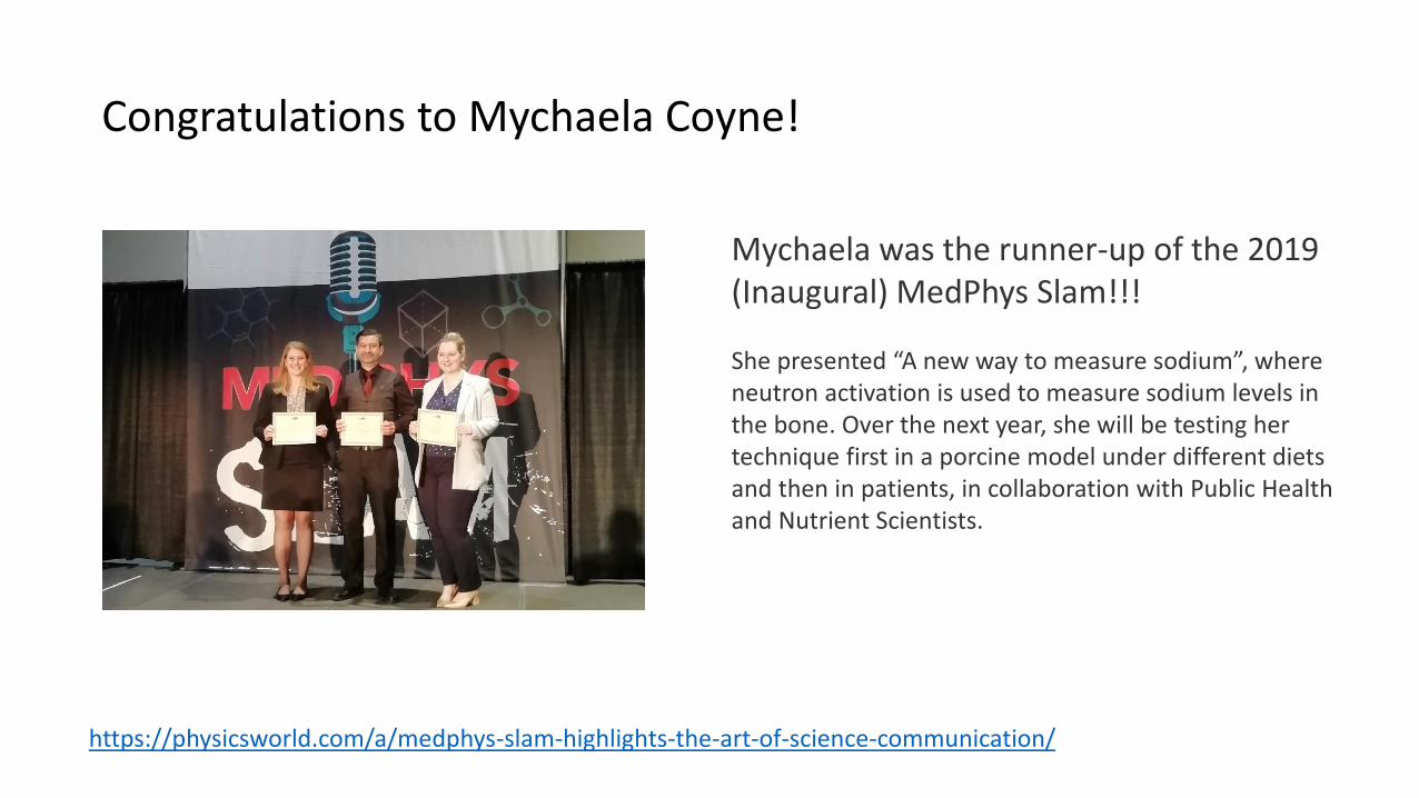
Congratulations to Mychaela Coyne!
https://physicsworld.com/a/medphys-slam-highlights-the-art-of-science-communication/
Mychaela was the runner-up of the 2019 (Inaugural) MedPhys Slam!!!
She presented “A new way to measure sodium”, where neutron activation is used to measure sodium levels in the bone. Over the next year, she will be testing her technique first in a porcine model under different diets and then in patients, in collaboration with Public Health and Nutrient Scientists.

PO-GePV-T-397 Minimal Difference Between Fractionated and Single-Fraction Exposure in a Murine Model of Radiation NecrosisA Boria*, C Perez-Torres, Purdue University, West Lafayette, IN
Purpose: To investigate the level of sparing provided by fractionated radiation therapy in comparison to
single fraction radiation therapy in the mouse brain via irradiation with an X-Rad 320 cabinet irradiator.
Methods: PXi Precision X-Ray’s X-Rad 320 unit was used to conduct mouse irradiations at a dose rate of 2
Gy/min. Mice were anaesthetized with isoflurane, and received radiation to a sub-hemispheric portion of
the brain sized 0.5 cm by 0.5 cm. Four fractionation schemes were evaluated: 5 fractions of 20 Gy, 10
fractions of 10 Gy, 5 fractions of 18 Gy, and 10 fractions of 9 Gy. The results of these fractionated
irradiations were compared to results from single fraction irradiations with doses ranging from 50 Gy to
100 Gy. The development of radiation necrosis was tracked out to six weeks, with a 7T Bruker MRI using
T2 and post-contrast T1 imaging. Histology was obtained after final imaging was performed and scored for lesion severity.
Results: All four fractionation schemes had single fraction equivalent doses less than 50 Gy. Though our
single fraction irradiations of 50 Gy did not produce radiation necrosis within 26 weeks, all fractionated
regimes led to measurable radiation necrosis on MRI and histology. The two 100 Gy total fractionation
schemes had lesion volumes most similar to single fraction irradiations of 90 Gy, while the two 90 Gytotal fractionation schemes were most similar to single fraction irradiations of 80 Gy.
Conclusion: Radiation necrosis occurring with these fractionated schemes suggest that the sparing effects
of fractionation in our radiation necrosis model are minimal. Fractionation may not be as important a parameter in our murine model compared to parameters such as total dose.

PO-GePV-T-405 Murine Radiation-Induced Stomach Pathology for Whole Thoracic IrradiationD McIlrath1*, C Perez-Torres2 , (1) Purdue Univ, West Lafayette, IN, (2) Purdue University, West Lafayette, IN
Purpose: Radiation-induced lung injury is a common side effect in the treatment of lung and breast cancers. There is a large focus in the field on leveraging murine models of radiation-induced lung injury to find novel treatments for the condition. While attempting to irradiate mice lungs for purposes of creating a radiation-induced pulmonary fibrosis model, noticeable declines in health were observed at much earlier timepoints than recorded for lung pathology. This was later attributed to stomach pathology observed in CT images and ex-vivo dissection.
Methods: For this study, we used longitudinal microCT to characterize male C57Bl/6 mice irradiated with a single dose of 20 Gy to the whole thoracic area delivered by an X-Rad cabinet irradiator. CT was performed with respiratory gating at 2 to 4 week timepoints to construct a timeline of pathology leading up to fibrosis and to quantify severity of fibrosis afterwards. However, several mice imaged at the 4 week timepoint showed evidence of stomach distention. These mice were sacrificed and their stomachs removed. Histology was performed on the stomachs using H&E staining.
Results: On the CT images, we observed a large, spherical volume of hypointense signal, caudal to the lungs (Figure 1). This correlates with a distended stomach caused by constipation and gas build-up within the stomach. Mice sacrificed and dissected showed unpassed bolus as contents of the stomach, and histology showed cell necrosis of the stomach walls.
Conclusion: The histology indicated an inability for food to be digested and moved into the small intestine. This lead to a blockage and ensuing stomach distention. Given the severity of the pathology’s consequences, it lead to the mice's imminent mortality. Shields or any beam-contouring devices need careful placement to ensure protection of the stomach given its higher radiosensitivity in contrast to the lungs.

PO-GePV-T-406Thoracic Radiation Injury Murine Model for Pulmonary FibrosisD McIlrath1*, C Perez-Torres2 , (1) Purdue Univ, West Lafayette, IN, (2) Purdue University, West Lafayette, IN
Purpose: Radiation-induced lung injury (RILI) is a common sequelae in the setting of lung and breast cancer. Often, patients who suffer from RILI experience pneumonitis and pulmonary fibrosis months after treatment. These pathologies have commonly been modeled using mice and observing their deterioration until mortality, then quantifying pathology on histological sections.
Methods: With this study, we attempt to use longitudinal microCT to characterize male C57Bl/6 mice irradiated with a single dose of 20 Gy to the whole thoracic area delivered by an X-Rad cabinet irradiator. CT was performed with a 4 minute respiratory gating sequence at 2 to 4 week timepoints to construct a timeline of pathology leading up to fibrosis and, additionally, to quantify severity of fibrosis afterwards. Images were analyzed on ITK-SNAP to segment anatomy and pathology using their machine learning algorithm. Histology was later performed using H&E and Trichrome stains to provide ex-vivo verification of pathology.
Results: At the 4-6 week timepoint, observable physical pathology occurred – most notably alopecia and erythemea. CT images showed little lung pathology at that time, but later timepoints – closer to 12 weeks –showed areas within the lung of hyperintense nodules, expanding over time. Figure 1 shows CT images of a mouse who exhibited this pathology 18 weeks post-irradiation.
Conclusion: The hyperintense nodules were believed to be fibrotic tissue, and histology confirmed collagen production localized to tissue near the bronchi – correlated with the hyperintense areas on the CT images. Mice that survived to later timepoints showed exponential deterioration of their health and exponential progression of pulmonary fibrosis. Mice eventually exhibited extreme lethargy as a precursor to death if not euthanized before mortality.

WE-C1030-GePD-F6-1 A Comparative Study to Optimize the In-Vivo Neutron Activation Analysis SystemS Tabbassum1*, H Nie2 , (1) PURDUE UNIVERSITY, W Lafayette, IN, (2) Purdue Univ, West Lafayette, IN
Purpose: To develop a highly sensitive in-vivo neutron activation analysis system with a neutron fusion source, to quantify trace elements in human.
Methods: In this work, we have developed an irradiation assembly for in-vivo measurement of trace elements in the human hand using FLUKA, and compare the results with similar model in Monte Carlo N-Particle (MCNP). Design of irradiation assembly include: 3.5 cm high density polyethylene moderator, neutron multiplier made of Beryllium, a fast neutron filter based on single crystal Sapphire (Al2O3) and reflectors. In order to assess the sensitivity of in-vivo system for thermal neutron activation analysis, we have maximized thermal neutron flux inside the irradiation cave (i.e. where the sample is placed), minimized the fast neutrons flux to reduce the dose, without substantial decrease in total flux. The thermal neutron flux and thermal neutron spectrum inside the irradiation cave and dose was calculated.
Results: For an irradiation cave of dimension 20x10x20 cm, thermal neutron yield is maximized to 6.71x10^5 n /s for DD fast neutron yield of 1X10^9 n/s. Fast neutron yield inside the irradiation cave is 3.8X10^4 n/s. Comparison between MCNP and FLUKA shows a good general agreement. Thermal neutron flux (below 0.1ev) inside radiation cave is 3 times more than fast neutron (above 0.1ev). However, FLUKA shows higher total of flux 9x10^5 n/s inside the cave as compared with MCNP of 6.9x10^5 n/s, mainly due to different physics models. Radiation dose rate inside the irradiation cave is 3.3mSv/hr. from the neutrons.
Conclusion: Optimized assembly for the in-vivo neutron activation analysis showed a high thermal flux resulting in higher sensitivity and low fast neutron flux. Results from both FLUKA and MCNP validates the feasibility of designed irradiation assembly, however differences between two simulation codes require experimental validation to benchmark a reference system.

WE-FG-303-7 Photoacoustic CT to Characterize the Effects of Antiangiogenic Drugs On Acute and Chronic HypoxiaD Miles*, K Stantz , Purdue University, West Lafayette, IN
Purpose: Tumor angiogenesis drives unpredictable changes in blood perfusion and diffusion, which can have a major impact on tumor growth and metastasis. Anti-angiogenic drugs (AA) are designed to combat these effects, often by disrupting neovasculature formed during angiogenesis, but their therapeutic effects are poorly understood. In this work, we use photoacoustic CT spectroscopy (PCT-S) to assess the impact of various AA agents in syngeneic murine tumors.
Methods: Twenty-six immunocompetent mice were seeded with MMTV-PyMT tumor fragments and treated with one of four conventional AA agents (IgG4, DC101, Ang2, DC101-plus-Ang2) after growing to a volume of 250 mm³. Seven days after treatment (3-treatments over 1-week), photoacoustic scans were acquired, sampling at ten NIR wavelengths. Longitudinal scans were acquired for individual tumors every 15 minutes over the span of two hours. PCT-S images were co-registered to assess regional changes in hemoglobin oxygen saturation (SaO2) over time.
Results: Longitudinal PCT-S data revealed intratumor regions of consistently low (chronic) and oscillating (acute) SaO2 in all tumors. All treatment groups observed comparable levels of acute hypoxia. DC101-treated tumors showed a slight elevation in normoxia (nss). Mice treated with Ang2 presented a significant increase in the abundance of chronic hypoxia relative to DC101, suggesting more-efficient disruption of neovasculature. Combination of DC101+Ang2 yielded a higher average chronically-hypoxic fraction than Ang2 alone, but this enhancement was not statistically significant.
Conclusion: Varying conventional AA agents induce distinct changes in the tumor microenvironment that can be measured using PCT-S. Spatial-temporal responses has the potential to discern vascular functional changes to different dose combinations. Histological analysis of hypoxia-specific factors (CA9, HIF2alpha) are currently being evaluated and will be presented.