aaapppppprrroooaaaccchhh tttooo hhhuuummmaaannn...
TRANSCRIPT

91

92
Scientific Staff : Dr. Arun Kumar Jain, Dr. S Sriramachari,
Dr. Jagjit Kaur Sindhu, Dr. Madhu Bhatnagar In collaboration with : Dr. S K Raza, DRDO, Gwalior Dr. Sudha Salhan, SJ Hospital, New Delhi
Technical Staff : Mr. S D Joshi, Mrs. Kamlesh Sharma Duration : 2002-2005
Aims, Objectives & Background
Of late there has been a lot of awareness about pollution, its adverse effects and
sources and efforts are being made to control and bring down the levels of pollutants in the
environment. Use of pesticides and fertilizers for agriculture, industrial effluents and the
increasing numbers of automobiles on road have been worldwide recognized to be the
major contributors to environmental pollution. For the implementing agencies to take any
decision, it is mandatory that background data on the levels of polluting chemicals should
be available through various monitoring programmes. In order to get dynamic information
about the changes in the environment, regular monitoring of pollutants at various levels of
ecosystem and biosphere assumes great significance. While some attempts are being made
to assess levels of pollutants in air, water or vegetables, there have been negligible attempts
at Human Environmental Bio-monitoring in India or elsewhere in the world for want of a
suitable monitoring model. The limitations faced by HEBM Program and advantages of
using Human Placenta for HEBM have already been enumerated in the last report.
This is an extramural research project sanctioned by Department of Biotechnology
with an aim to establish the utility of human placenta as a tool for comprehensive bio-
monitoring for organic pollutants and to demonstrate the feasibility of monitoring region
specific organic pollutants in placenta. It also aims to establishing standard operating
procedures (SOP) for the above two objectives.
111... MMMooonnniiitttooorrriiinnnggg ooofff OOOrrrgggaaannniiiccc CCChhheeemmmiiicccaaalll PPPooolll llluuutttaaannntttsss iiinnn PPPlllaaaccceeennntttaaalll TTTiiissssssuuueee::: AAA NNNeeewww AAApppppprrroooaaaccchhh tttooo HHHuuummmaaannn EEEnnnvvviiirrrooonnnmmmeeennntttaaalll BBBiiiooo---mmmooonnniiitttooorrriiinnnggg

93
Work done during the year
This project had been initiated for comprehensive bio-monitoring of Organic
Chemical Pollutants in human population using placenta as tool for HEBM. During the
year under report approximately 4000 more pregnant women attending Antenatal Out
Patient Department Clinic of Safdarjung Hospital were screened for possible exposure to
pollutants. A total of 150 women who reported exposure to agricultural chemicals during
pregnancy due to their involvement in agricultural activity by themselves or their family
members were selected and followed up for subsequent collection of placental sample at
the time of delivery. So far a total of 100 samples have been collected which include 51
random samples and 41 samples from high-risk population. In addition fifty samples of
blood and milk have also been collected.
During the period under report, recovery experiments were conducted using two
different methods, viz., Soxhlet Extraction, extraction using vortex mixer for extraction of
pollutants. Both the methods were tried without addition of TCA and after addition of TCA
in the placental homogenate. Further all the four extractions procedures were tried with
four solvents namely n-Hexane, Dichloromethane, Acetonitrile and methanol again both
individually as well as sequentially. It was found that Soxhlet extraction caused more
background peaks under GC-MS as compared to extraction using vortex mixer. Further
TCA extraction did not gave any additional advantage. Of the four solvents tried it was
observed that both hexane as well as DCM gave good recovery 80 to 90%. Accordingly,
Standard Operating Procedure for collection of placental sample, extraction of organic
chemical pollutants from the placental tissue, cleanup and concentration by nitrogen
purging has been finalized. Extracts have been prepared for all the 100 placental samples as
per the finalized SOP and 50 extracts were concentrated by gentle nitrogen purge
technique.
Further mixtures of pesticide standards consisting of more than 100 pollutants were
procured and used for standardizing the separation of each pollutant under GC-MS. The
operating conditions for GC-MS analysis by Trace GC-MS (Thermo-Finnigan) have also
been finalized (Table 1). The retention times in GC and predominant mass fragments for
94
all the pesticide/pollutant standard were recorded (Tables 2 and 3) and used for
interpretation of retention time and Mass Spectra recorded for the placental samples
On the basis of matching retention time with the known pesticide standards, the Gas
chromatography has revealed possibility of presence of pesticide residues in the placental
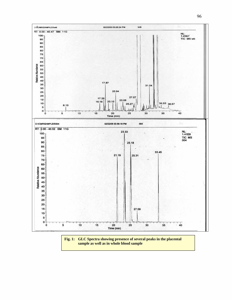
extracts. Figures 1 shows representative GC spectra for two samples. These show presence
of peaks at retention time 16.18 to 34.53 minutes, which match with the retention time of
several of the pollutants as listed in table 4.
Some of the GC-Mass spectra and Molecular structural formulae for a few representative
pesticides and poly aromatic hydrocarbons are given in Table 5.
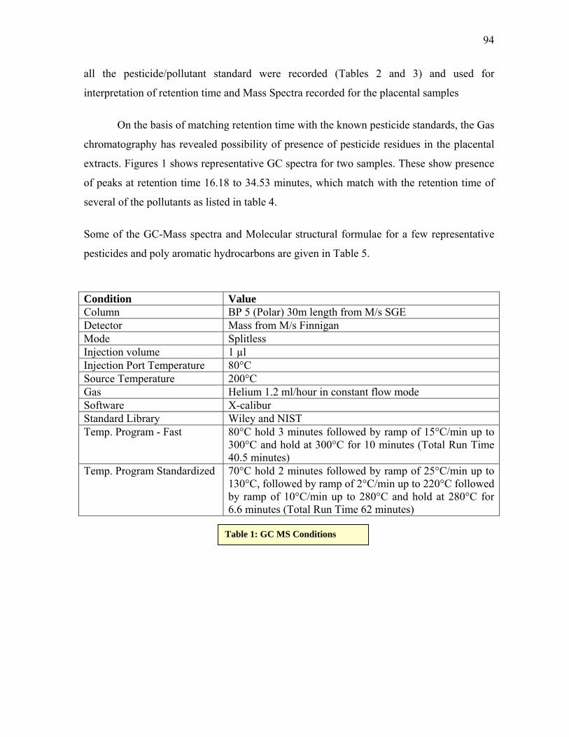
Condition Value Column BP 5 (Polar) 30m length from M/s SGE Detector Mass from M/s Finnigan Mode Splitless Injection volume 1 µl Injection Port Temperature 80°C Source Temperature 200°C Gas Helium 1.2 ml/hour in constant flow mode Software X-calibur Standard Library Wiley and NIST Temp. Program - Fast 80°C hold 3 minutes followed by ramp of 15°C/min up to
300°C and hold at 300°C for 10 minutes (Total Run Time 40.5 minutes)
Temp. Program Standardized 70°C hold 2 minutes followed by ramp of 25°C/min up to 130°C, followed by ramp of 2°C/min up to 220°C followed by ramp of 10°C/min up to 280°C and hold at 280°C for 6.6 minutes (Total Run Time 62 minutes)
Table 1: GC MS Conditions
95
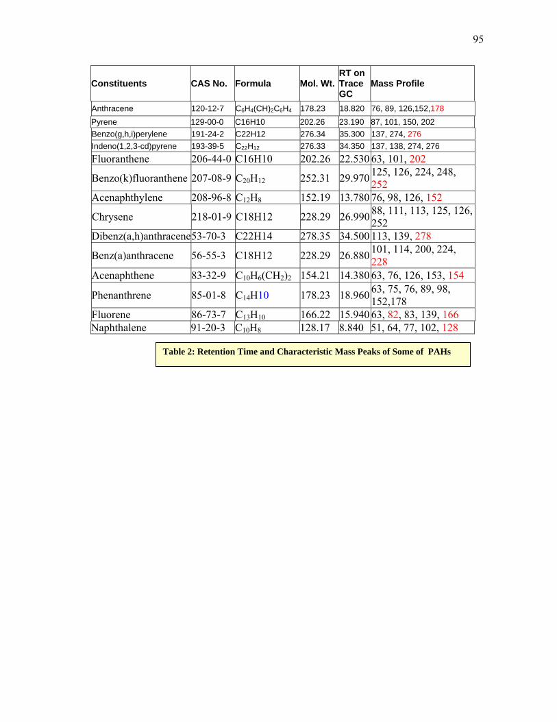
Constituents CAS No. Formula Mol. Wt.RT on Trace GC
Mass Profile
Anthracene 120-12-7 C6H4(CH)2C6H4 178.23 18.820 76, 89, 126,152,178 Pyrene 129-00-0 C16H10 202.26 23.190 87, 101, 150, 202 Benzo(g,h,i)perylene 191-24-2 C22H12 276.34 35.300 137, 274, 276 Indeno(1,2,3-cd)pyrene 193-39-5 C22H12 276.33 34.350 137, 138, 274, 276 Fluoranthene 206-44-0 C16H10 202.26 22.530 63, 101, 202
Benzo(k)fluoranthene 207-08-9 C20H12 252.31 29.970 125, 126, 224, 248, 252
Acenaphthylene 208-96-8 C12H8 152.19 13.780 76, 98, 126, 152
Chrysene 218-01-9 C18H12 228.29 26.990 88, 111, 113, 125, 126, 252
Dibenz(a,h)anthracene53-70-3 C22H14 278.35 34.500 113, 139, 278
Benz(a)anthracene 56-55-3 C18H12 228.29 26.880 101, 114, 200, 224, 228
Acenaphthene 83-32-9 C10H6(CH2)2 154.21 14.380 63, 76, 126, 153, 154
Phenanthrene 85-01-8 C14H10 178.23 18.960 63, 75, 76, 89, 98, 152,178
Fluorene 86-73-7 C13H10 166.22 15.940 63, 82, 83, 139, 166 Naphthalene 91-20-3 C10H8 128.17 8.840 51, 64, 77, 102, 128
Table 2: Retention Time and Characteristic Mass Peaks of Some of PAHs
96
Fig. 1: GLC Spectra showing presence of several peaks in the placental sample as well as in whole blood sample

97
Constituents CAS No. Formula Mol. Wt.
RT on Trace GC
Mass Profile
Dichlorvos 62-73-7 C4H7Cl2O4P 220.98 10.270 79, 109, 145, 185 Methyl parathion 298-00-0 C8H10NO5PS 263.21 20.270 109, 125, 233, 263 Chlorpyrifos-methyl
5598-13-0 C7H7Cl3NO3PS 322.60 20.275 109, 125, 286
Fenitrothion 122-14-5 C9H12NO5PS 277.24 20.970 109,125, 260, 277
Phosalone 2310-17-0 C12H15ClNO4PS2367.81 27.765 97, 111, 121, 182, 367
α-BHC 319-84-6 C6H6Cl6 290.83 13.960 109, 181, 219 g-BHC 58-89-9 C6H6Cl6 290.83 16.060 109, 181, 219
Aldrin 309-00-2 C12H8Cl6 364.91 21.390 66, 79, 91, 101, 263, 293
Heptachlor 76-44-8 C10H5Cl7 373.32 20.490 100, 237, 272
α-Chlordane 5103-71-9 C10H6Cl8 409.78 22.980 75, 109, 237, 272, 373
o,p’-DDE 3424-82-6 C14H8Cl4 318.02 23.090 105, 176, 210, 246,
316 p,p’-DDE 72-55-9 C14H8Cl4 318.02 23.850 176, 210, 246, 316
p,p’-DDD 72-54-8 C14H10Cl4 320.04 24.860 88, 101, 165, 176, 199, 235,
p,p’-DDT 50-29-3 C14H9Cl5 354.48 25.730 165,199, 235,
Simazine 122-34-9 C7H12ClN5 201.66 18.200 68, 96, 138, 173, 186, 201
Atrazine 1912-24-9 C8H14ClN5 215.68 18.340 58, 68, 173, 200, 215
Terbuthylazine 5915-41-3 C9H16ClN5 229.71 18.730 173, 214, 229
Pentachloroaniline 527-20-8 C6H2Cl5N 265.35 19.830 263, 265, 267,
Cyanazine 21725-46-2 C9H13ClN6 240.70 21.530 68, 172, 198, 225, 240
Alachlor 15972-60-8 C14H20ClNO2 269.77 20.500 146, 160, 188
Metazachlor 67129-08-2 C14H16ClN3O 277.75 22.320 81, 133, 209
Bentazon 25057-89-0 C10H12N2O3S 240.30 21.780 92, 119, 161, 198
Endosulfan I 959-98-8 C9H6Cl6O3S 406.92 24.665 85, 170, 195, 207, 241, 267

98
Endosulfan II 33213-65-9 C9H6Cl6O3S 406.92 23.280 85, 160, 170, 195, 207
Endosulfan sulfate 1031-07-8 C9H6Cl6O4S 422.92 25.695 229, 237, 272, 387,
422
Piperonyl butoxide 51-03-6 C19H30O5 338.44 26.240 147, 176, 221, 281, 355, 429
Bromopropylate 18181-80-1 C17H16Br2O3 428.12 26.920 155, 183, 341
Dieldrin 60-57-1 C12H8Cl6O 380.91 23.925 79, 108, 237, 263, 345Endrin 72-20-8 C12H8Cl6O 380.90 24.470 67, 81, 263, 281, 317
trans-Permethrin 51877-74-8 C21H20Cl2O3 391.29 29.130 163, 165, 183
cis-Permethrin 54774-45-7 C21H20Cl2O3 391.31 29.290 163, 165, 183
Retention Time of the sample
Retention Time of the standard Standard
16.18 16.23 2,3,5,6-Tetrachloronitrobenzene 17.87 17.89 Hexachlorobenzene 20.10 20.08 2,4,4'-Trichlorobiphenyl 20.94 20.97 2,2',5,5'-Tetrachlorobiphenyl & Fenitrothion 21.19 21.239 Malathion 23.08 23.09 o,p’-DDE 23.33 23.3 Endosulfan II 25.18 25.17 2,2',3,4,4',5'-Hexachlorobiphenyl 26.27 26.24 Piperonyl butoxide 27.07 27.06 Methoxychlor 34.53 34.5 Dibenz(a,h)anthracene
Table 3: Retention Time and Characteristic Mass Peaks of Some of Organo-Phosphorus, Organo-Chlorine and Pyrethroid Pesticides
Table 4: GC Retention Times of the samples that matched with the GC retention times of the standards under similar conditions

99
Constituents Structural Diagram Mass Spectrum
trans-Permethrin
Endosulfan sulfate
Dursban
Methyl parathion
2,3,5,6-Tetrachloro-
nitro-benzene
Terbuthylazine

100
Cyanazine
Phenanthrene
Naphthalene
p,p’-DDT
g-BHC
GC- MS analysis of the placental extracts showed presence of some normal
biological constituents such as cholesterol (Fig.2). In few cases the GC-MS analysis
revealed presence of pollutants like Cycloheptatrienylium Bromide (Fig. 3) and
Naphthalene. In the latter case the peaks matched both the GC retention time as well as
Mass Fragments Profile (Fig. 4). Further confirmation of these residues by GC-MS is still
under progress.
Table 5: Molecular Structural Diagrams and GC-Mass Spectrums of a few representative pesticides and PAHs

101
Fig. 2: GC- MS spectrum from another sample showing presence of Cycloheptatrienylium Bromide Peak
Fig. 3: GC-MS spectrum from a placental extract shows presence of cholesterol a normal constituent of the tissue

102
Fig 4: GC MS Spectrum form a sample of human placenta showing presence of characteristic peak for Naphthalene
103
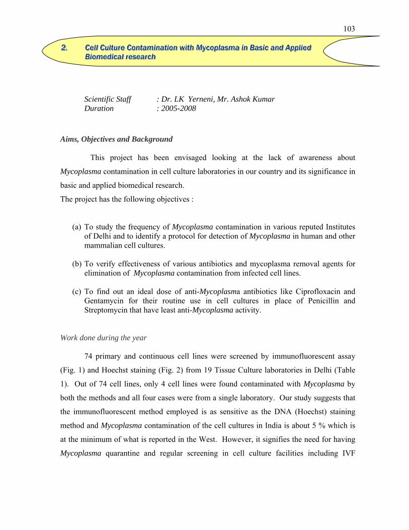
Scientific Staff : Dr. LK Yerneni, Mr. Ashok Kumar Duration : 2005-2008
Aims, Objectives and Background This project has been envisaged looking at the lack of awareness about
Mycoplasma contamination in cell culture laboratories in our country and its significance in
basic and applied biomedical research.
The project has the following objectives :
(a) To study the frequency of Mycoplasma contamination in various reputed Institutes of Delhi and to identify a protocol for detection of Mycoplasma in human and other mammalian cell cultures.
(b) To verify effectiveness of various antibiotics and mycoplasma removal agents for
elimination of Mycoplasma contamination from infected cell lines.
(c) To find out an ideal dose of anti-Mycoplasma antibiotics like Ciprofloxacin and Gentamycin for their routine use in cell cultures in place of Penicillin and Streptomycin that have least anti-Mycoplasma activity.
Work done during the year
74 primary and continuous cell lines were screened by immunofluorescent assay
(Fig. 1) and Hoechst staining (Fig. 2) from 19 Tissue Culture laboratories in Delhi (Table
1). Out of 74 cell lines, only 4 cell lines were found contaminated with Mycoplasma by
both the methods and all four cases were from a single laboratory. Our study suggests that
the immunofluorescent method employed is as sensitive as the DNA (Hoechst) staining
method and Mycoplasma contamination of the cell cultures in India is about 5 % which is
at the minimum of what is reported in the West. However, it signifies the need for having
Mycoplasma quarantine and regular screening in cell culture facilities including IVF
222... CCCeeelll lll CCCuuullltttuuurrreee CCCooonnntttaaammmiiinnnaaatttiiiooonnn wwwiiittthhh MMMyyycccoooppplllaaasssmmmaaa iiinnn BBBaaasssiiiccc aaannnddd AAApppppplll iiieeeddd BBBiiiooommmeeedddiiicccaaalll rrreeessseeeaaarrrccchhh

104
facilities across our country. Further analysis of Mycoplasma contamination is being
undertaken by PCR.
Positive Negative Institution No.of cultures IFA Hoechst IFA Hoechst
AIIMS 58 4 4 54 54 Lab 1 17 - - 17 17 Lab 2 7 - - 7 7 Lab 3 5 - - 5 5 Lab 4 2 - - 2 2 Lab 5 11 - - 11 11 Lab 6 5 - - 5 5 Lab 7 5 - - 5 5 Lab 8 6 4 4 2 2 JNU 8 - - 8 8
Lab 1 2 - - 2 2 Lab 2 1 - - 1 1 Lab 3 1 - - 1 1 Lab 4 1 - - 1 1 Lab 5 1 - - 1 1 Lab 6 1 - - 1 1 Lab 7 1 - - 1 1 Lab 8 1 - - 1 1
VP Chest 1 - - 1 1 IGIB 1 1 1
Inst. of Path 7 - - 7 7 Lab1 1 - - 1 1 Lab2 6 - - 6 6
Table 1: The incidence of Mycoplasma contamination in cell culturesfrom various reputed Institutes in Delhi.

105
Negative control Positive control
Moderately infected T3 culture Heavily infected T8culture Fig. 1: Mycoplasma contamination in cell culture as detected by Immunofluorescent assay
Negative control Heavily infected T8 culture Fig. 2: Mycoplasma contamination in cell culture as detected by Hoechst (33258) DNA staining.
106
Future Plan of Action
Application of human epidermal sheets cultured from autologous epidermal stem
cells in burns patients. Submitted to DBT under the programme entitled “Cell Based
Therapy”. In this project, it was proposed to undertake a study on the evaluation of
proliferative potential of human sera on human epidermal stem cells by growth curves and
BrdU labeling for flow cytometric analysis in addition to undertaking clinical application
of cultured epidermis on 50 patients in phased manner. It is proposed to collaborate with
Burns Division, Safdarjung hospital, New Delhi and Research & Referral hospital.
I . Immunohistochemical Evaluation of Estrogen receptor and estrogen induced proteins in the ejaculated spermatozoa of infertile subjects
Scientific Staff : Dr. S. Jayaraman, Mr. Varun Kapur, Mr. Shakaut,
Mr.Sajad and Ms. Deepali Mathur In collaboration with : Dr. MM Misro, NIHFW. New Delhi. Technical Staff : Mr. Satya Pal Singh Kasana. Duration : 2005 (Till August).
i) Immunohistochemical profiles of Estrogen Receptor α, Estrogen Receptor β, Andogen Receptor, Estrogen induced / related proteins like Progesterone Receptors, Cathepsin D, HSP-27 and Aromatase P 450 in the ejaculated spermatozoa from infertile subjects
Immunohistochemical evaluation carried out with ejaculated spermatozoa from
the subjects attending the infertility clinics indicated that:
a) 23/73 (31.5%) samples demonstrated the presence of most of Estrogen Receptor
α Estrogen Receptor β, Andogen Receptor, Estrogen induced / related proteins
like Progesterone Receptors, HSP-27 in the ejaculated spermatozoa, however,
these proteins were totally absent in 38/73 (52.05%) samples, while a variable
percentage of spermatozoa expressed these receptors in the rest of the samples.
333... PPPaaattthhhoooppphhhyyysssiiiooolllooogggiiicccaaalll RRRooollleee ooofff EEEssstttrrrooogggeeennn iiinnn mmmaaallleeesss

107
b) A number of subjects in the third group mentioned above showed only one or
two of the above mentioned proteins in the spermatozoa
c) Cathepsin D was consistently absent in the spermatozoa in all the groups.
d) A significant correlation(P>0.01) between the percentage of spermatozoa
showing forward motility and the spermatozoon presence of Estrogen Receptor
α Estrogen Receptor β, Aromatase P 450- - indicating their diagnostic and
prognostic significance with fertility potential of the spermatozoa. The
observations also suggest that their inhibitors may act as useful vaginal
contraceptives.
II) Flowcytometric evaluation of Estrogen Receptor (ER) and estrogen induced proteins in the rat testicular germ cells.
Scientific Staff : Dr. S. Jayaraman, Mr.Varun Kapur, Mr.Shakaut,
Mr.Sajad and Ms. Deepali Mathur. Technical Staff : Mr.Satya Pal Singh Kasana. Duration : 2005 (Till August).
Investigations were undertaken to confirm the earlier immunohistochemical
observations on the presence of ER α in rat testicular cells using and to evaluate
the presence of Estrogen induced proteins in the rat testicular germ cells using flow
cytometry. . Rat testicular cells were collected and differential centrifugation
procedures to separate the different germ cells and to carry out the flow cytometric
evaluation of the estrogen receptors as well that of different estrogen induced
proteins. How ever the studies could not be completed as the number of testicular
germ cell types following differential centrifugation procedures using Percol
gradients were insufficient following staining. Hence the results of
immunohistochemical localization of the same were carried out in the isolated
testicular germ cells.
The studies indicated that Estrogen α receptors were presented in primary
spermatogonia, primary spermatocytes, round and elongated spermatids. The ER β
in the spermatogonia round and elongated spermatids. The most important
observation was that Cathepsin D could be localized only in the round spermatids,

108
while the Progesterone receptors, IGF2, HSP 27, PS2 were present in one or more
of the rat germ cells.
The studies confirm the important role of Estrogen in proliferation,
differentiation, maturation and perhaps in apoptosis as well. The localization of
Cathapsin D is the first report in the literature as per our knowledge.
III) Detection of mRNA transcripts of Estrogen Receptor α in the
ejaculated spermatozoa from fertile individuals.
Dr .S. Jayaraman and Mr. Pankaj Sharma. Our earlier results indicated the presence splice variants of ERα and Progesterone
receptor from the ejaculated spermatozoa from infertile individuals. However presence
of wild type of ERα could not be demonstrated in the ejaculated spermatozoa from
subjects attending infertile clinic but the spermatozoan characteristics were of ‘fertile
range’. Hence studies were undertaken in the mRNA isolated from the spermatozoa
from known fertile individuals. The results are presented in Fig.1.
The results confirm our earlier hypothesis that presence or absence of some
spermatozoan proteins may be playing a role , perhaps in the causation of male
infertility.
The presence of wild type ER alpha mRNA transcripts fro human ejaculated spermatozoa from fertile individuals

109
Scientific Staff : Dr. Ranvir Singh, Dr. S Jayaraman In collaboration with : Dr. Sumita Saluja, SJ Hospital, New Delhi Duration : 2004-2005
Aims, Objectives and Background A number of evidences indicate the association of HTLV-1 with various diseases
including adult T-cell leukemia / non-Hodgkin’s lymphoma, HTLV-1 associated
myelopathy (HAM) / TROPICAL spastic paraparesis (TSP), development of inflammatory
diseases in various organs such as eyes, lungs and joints, inflammatory ocular diseases
such as endogenous uveitis, episcleritis, retinitis pigmentosa and degenerative choroiditis,
etc. In these conditions, the presence of HTLV-1 can be associated as an accompaniment
of repeated blood transfusions. However, recently in a pilot study, Ramalingam et al
(2001) have reported a strong association of HTLV-1 with haematological malignancies
(8/86 patients) and evidence for both horizontal and vertical transmission of the infection in
South-Indian population.
In view of the reported association between HTLV-1 and haematological
malignancies in a comparatively smaller number of subjects (n=86), the current
investigations were planned to study the association between HTLV-1 and haematologic
malignancies. Such a study carried out in another population namely the North-Indian
subjects will confirm the above mentioned serological evidences and if confirmed may lead
to the possible introduction of HTLV-1 monitoring policy in the National Blood
Transfusion Services.
Work done during the year A total of 186 blood samples, comprising 157 sera from blood donors and 29 sera
from patients with haematological malignancies, were collected and tested for anti-HTLV-
1 antibodies through Particle Agglutination test (PAT). No sample was found to be having
anti-HTLV-1 antibodies. Blood samples from patients with haematological malignancies
444... EEEvvvaaallluuuaaatttiiiooonnn ooofff HHHuuummmaaannn TTT---ccceeelll lll LLLyyymmmppphhhoootttrrrooopppiiiccc VVViiirrruuusss TTTyyypppeee---111 (((HHHTTTLLLVVV---111))) IIInnn BBBlllooooooddd DDDooonnnooorrrsss aaannnddd PPPaaatttiiieeennntttsss WWWiiittthhh LLLeeeuuukkkeeemmmiiiaaa///LLLyyymmmppphhhooommmaaa fffrrrooommm DDDeeelllhhhiii

110
and of blood donors were received from Safdarjang Hospital, New Delhi and Institute of
Pathology, New Delhi, respectively.
Future plan of action Nearly 160 blood donors and 30 patients with hematologic malignancies will be
screened for detection of anti-HTLV-1 antibodies through PAT. Sera found positive by
PAT will be confirmed through Line Immunoassay (LIA).
Scientific Staff : Dr. S Jayaraman, Dr. Usha Agarwal In collaboration with : Dr. S L Kapoor Duration : 2004-2006
Aims, Objectives and Background
Immunohistochemistry is routinely employed for diagnosis/prognosis as well as for
therapeutic monitoring. It encompasses a wide variety of hormone receptors, apoptotic
markers, tumour and proliferation markers, etc. The staining intensity obtained is
quantified by a scoring system into grades of mild, moderate and severe. The staining is
directly related to the presence of antigen. The direct relationship between the intensity of
staining and the number of hormonal receptors in histological specimens has been
accepted. Similarly, the use of DNA content in defining the prognosis of lung cancer
patients has been recognized. However, the stoichiometric relationship between the antigen
concentration and intensity of immunohistochemical staining is yet to be demonstrated.
The diagnostic, prognostic as well as therapeutic management of the patient may vary if
there is a relationship between the intensity of immunostaining and the concentration of
antigen. Investigations were planned with an objective of developing an image analysis
system for specifically recognizing and quantitating the area as well as the intensity of
immunostain.
555... QQQuuuaaannntttiiitttaaatttiiiooonnn ooofff IIImmmmmmuuunnnooohhhiiissstttoooccchhheeemmmiiicccaaalll ssstttaaaiiinnniiinnnggg

111
Work done during the year
The results achieved so far include identifying the Area of Interest (AOI), count of
number of cells, giving area% of five different grades of brown present in the image.
However, the quantification of the DAB product by analyzing the pixel values was
observed to be affected by the haematoxylin. So attempts to develop a defined colour
palette, which can be used as a universal standard, are being undertaken. The results of the
collaborative studies suggest that we may be in a position to achieve the objective in the
near future.