a young woman with first-trimester metrorrhagia, fever and
TRANSCRIPT

ABSTRACT
Inflammatory bowel disease commonly affects women withchild-bearing potential, and clinical activity extent is most relevantat the time of conception. Below, we report on the case of a 19-year-old woman who was admitted for first-trimester metrorrhagiaand fever, with various extraintestinal manifestations, mainly in-cluding erythema nodosum and episcleritis during the course ofdisease. The differential diagnosis of these manifestations led tothe diagnosis of Crohn’s disease, which involved the whole colon.
Key words: Inflammatory bowel disease. Erythema nodosum.Extraintestinal manifestations. Pregnancy.
Lalueza Blanco A, Rodríguez Alcalde D, González Gómez C,López G, Martínez Montiel P, Sanz García E, Solís HerruzoJA. A young woman with first-trimester metrorrhagia, feverand skin lesions. Rev Esp Enferm Dig 2006; 98: 204-215.
CASE REPORT PRESENTATION
Dr. C. González Gómez. Department of Internal Medi-cine: A 19-year-old gypsy woman presented in our Emer-gency Department with first-trimester metrorrhagia andfever.
Initially she had hypogastric pain and fever up to 39 ºC,which was clinically and hemodynamically well tolerated.
A scraping procedure was performed, following whichabdominal pain improved; however, high temperaturepersisted despite this procedure and the use of ampicillin,gentamycin, and metronidazole. Baseline laboratoryworkup revealed moderate leukocytosis, and microcyticanemia.
Given the patient status her admission to the internalmedicine ward was decided upon. On new history takingthe patient reported a previous pregnancy that yielded ahealthy, currently 16-months-old child, and having lost15 kg of weight in the absence of hyperorexia since then.She reported having taken no oral contraceptives, and noother medications or toxics. For 15 days before admis-sion she had two or three soft stools daily with no patho-logic products, but she had spontaneously recovered be-fore visiting the emergency room.
During the review of systems she reported no othersystemic symptoms, except a number of painful lesionsin the oral mucosa that she had never consulted for.
During her stay at the Gynecology Department a num-ber of erythematous, papulonodular lesions in variousstages developed on her skin; these were widespread butpredominated in the lower limbs, particularly in thepretibial area. Subsequently she had an episode ofmonoarthritis in her right knee with inflammatory, sterilesynovial fluid.
Physical examination
Blood pressure: 93/50. Temperature 38 ºC. Severethinness overall. Head and neck: normal venous pressure.No lesions in the oral mucosa. Conjunctival elevatedwhite-yellowish lesion with hyperemia on the medial as-pect of both eyes, with a foreign-body sensation (Fig. 1).Chest: cardiopulmonary auscultation, rhythmic at 130beats per minute; rest, normal. Abdomen: moderatepainful hepatomegaly; tenderness in right iliac fossa and
A young woman with first-trimester metrorrhagia, fever and skinlesions
A. Lalueza Blanco, D. Rodríguez Alcalde1, C. González Gómez, G. López2, P. Martínez Montiel1, E. SanzGarcía3 and J. A. Solís Herruzo1
Departments of Internal Medicine, 1Gastroenterology, 2Pathology and 3Radiology. Hospital Universitario 12 de Octubre.Madrid, Spain
1130-0108/2006/98/3/204-215REVISTA ESPAÑOLA DE ENFERMEDADES DIGESTIVASCopyright © 2006 ARÁN EDICIONES, S. L.
REV ESP ENFERM DIG (Madrid)Vol. 98. N.° 3, pp. 204-215, 2006
Recibido: 10-10-05.Aceptado: 19-10-05.
Correspondencia: Antonio Lalueza Blanco. Servicio de Medicina Interna.Hospital Universitario 12 de Octubre. Avda. de Córdoba, s/n. 28041 Ma-drid. e-mail: [email protected]
CLINICOPATHOLOGICAL CONFERENCE
CCP LALUEZA 6/4/06 13:27 Página 204

hypogastrium, with no peritonism or palpable masses.Limbs: no osteoarticular symptoms. Skin: papulous,nodular, erythematous, hyperemic lesions in variousstages, predominating in the lower limbs (Fig. 2).
Laboratory tests
Blood test: hemoglobin 8.2 g/dl; hematocrit 26%;MCV 70 fl; MCH 22 pg; platelets 551,000/mm3; leuko-cytes 8690/mm3 (N 60%; L 30%; M 6%); reticulocytes0.55%; absolute reticulocyte count 20,800/mm3; ESR 100mm/h. Coagulation: prothrombin activity 85%; INR1.12; APTT 31.2 s; fibrinogen 504 mg/dl. Chemistry:glucose 87 mg/dl; LDH 107 IU/l; GOT 18 IU/l; GPT 16IU/l; gamma-GT 118 IU/l; AP 344 IU/l; total bilirubin0.31 mg/dl; total proteins 6.4 g/dl; albumin 2.85 g/dl; cre-atinine 0.62 mg/dl; calcium 8.7 mg/dl; phosphorus 3.9mg/dl; uric acid 3.1 mg/dl; cholesterol 118 mg/dl; triglyc-erides 100 mg/dl; sodium 137 mEq/l; potassium 4.6mEq/l. Thyroid hormones: free T4 1.3 ng/dL; TSH 1.8µIU/mL. Iron profile: iron 25 µg/dl; ferritin 226 ng/ml;transferrin 153 mg/100; TIBC 216 mcg/100; SAT-trans-ferrin 12%. Urinalysis: density 1020; pH 7; leukocytes+++; rest, negative; with some bacteria, 4 RBCs per field,25 leukocytes per field and some amorphous phosphates;folic acid: 2.73 ng/ml. Vitamin B-12: 1.85 pg/ml.Clostridium difficile toxin: positive. Repeat coprocul-tures: negative. Repeat blood cultures: sterile. Mantoux:negative. HIV: negative. Chest x-rays on admission wasnormal, and abdomen x-rays showed an abnormal lu-minogram in the right iliac fosse with thickened submu-cosal structure.
COMMENTS AND CLINICAL COURSE
Dr. A. Lalueza Blanco. Department of Internal Medi-cine: In summary, this is a young woman who, since de-livering her first child 16 months ago, had a constitutionalsyndrome with severe weight loss and clear protein-ener-gy malnutrition signs. Fifteen days before admission shehad a transient episode of scarce diarrhea with no abnor-mal products and in association with hypogastric pain thatspontaneously subsided with no treatment. She presentedto hospital with first-trimester metrorrhagia and fever upto 39 ºC, and thus underwent scraping, which managed toimprove abdominal pain while fever persisted despite an-tibiotic therapy. A sole microbiologic evidence had beenavailable that far, namely the presence of Clostridium dif-ficile toxin in her stools, which was adequately managed.Severe hepatomegaly associated with bilateral episcleritisand elevated skin lesions predominating in the lowerlimbs, which suggested erythema nodosum, was seen dur-ing admission.
These findings suggest the presence of a systemic in-flammatory process involving various organs, althoughthe presence of infection –mainly subacute– accountingfor the patient’s illness cannot be excluded at the presenttime.
Before proceeding with differential diagnosis, I wouldlike to know whether the histologic study of skin lesionsconfirmed a diagnosis of erythema nodosum.
Fig. 1.- Elevated reddish lesions of varying size in the lower limbs, con-sistent with erythema nodosum.Lesiones sobreelevadas y rojizas, de tamaño variable, en miembros in-feriores, compatibles con eritema nodoso.
Fig. 2. Episcleritis.Epiescleritis.
Vol. 98. N.° 3, 2006 A YOUNG WOMAN WITH FIRST-TRIMESTER METRORRHAGIA, 205FEVER AND SKIN LESIONS
REV ESP ENFERM DIG 2006; 98(3): 204-215
CCP LALUEZA 6/4/06 13:27 Página 205
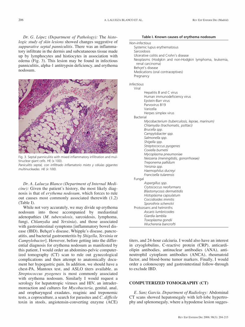
Dr. G. López (Department of Pathology): The histo-logic study of skin lesions showed changes suggestive ofsuppurative septal panniculitis. There was an inflamma-tory infiltrate in the dermis and subcutaneous tissue madeup by lymphocytes and histiocytes in association withedema (Fig. 3). This lesion may be found in infectiouspanniculitis, alpha-1 antitrypsin deficiency, and erythemanodosum.
Dr. A. Lalueza Blanco (Department of Internal Medi-cine): Given the patient’s history, the most likely diag-nosis is that of erythema nodosum, which forces to ruleout causes most commonly associated therewith (1,2)(Table I).
While not very accurately, we may divide up erythemanodosum into those accompanied by mediastinaladenopathies (M. tuberculosis, sarcoidosis, lymphoma,fungi, Chlamydia and Yersinia), and those associatedwith gastrointestinal symptoms [inflammatory bowel dis-ease (IBD), Behçet’s disease, Whipple’s disease, pancre-atitis, and bacterial gastroenteritis by Shigella, Yersinia orCampylobacter]. However, before getting into the differ-ential diagnosis for erythema nodosum as manifested bythis patient, I would order an abdomino-pelvic computer-ized tomography (CT) scan to rule out gynecologicalcomplications and then attempt to anatomically docu-ment her hypogastric pain. In addition, we should have achest-PA, Mantoux test, and ASLO titers available, asStreptococcus pyogenes is most commonly associatedwith erythema nodosum. Similarly I would request aserology for hepatotropic viruses and HIV, an intrader-moreaction and cultures for Mycobacteria, genital, anal,and oropharyngeal exudates, reaginic and treponemictests, a coproculture, a search for parasites and C. difficiletoxin in stools, angiotensin-converting enzyme (ACE)
titers, and 24-hour calciuria. I would also have an interestin cryoglobulins, C-reactive protein (CRP), anticardi-olipin antibodies, antinuclear antibodies (ANA), anti-neutrophil cytoplasm antibodies (ANCA), rheumatoidfactor, and blood-borne tumor markers. Finally, I wouldorder a colonoscopy and gastrointestinal follow-throughto exclude IBD.
COMPUTERIZED TOMOGRAPHY (CT)
E. Sanz García. Department of Radiology: AbdominalCT scans showed hepatomegaly with left-lobe hypertro-phy and splenomegaly, where a hypodense lesion sugges-
206 A. LALUEZA BLANCO ET AL. REV ESP ENFERM DIG (Madrid)
REV ESP ENFERM DIG 2006; 98(3): 204-215
Fig. 3. Septal panniculitis with mixed inflammatory infiltration and mul-tinuclear giant cells. HE (x 100).Paniculitis septal, con infiltrado inflamatorio mixto y células gigantesmultinucleadas. HE (x 100).
Table I. Known causes of erythema nodosum
Non-infectiousSystemic lupus erythematosusSarcoidosisUlcerative colitis and Crohn’s diseaseNeoplasms (Hodgkin and non-Hodgkin lymphoma, leukemia,
renal carcinoma)Behçet’s diseaseMedications (oral contraceptives)Pregnancy
InfectiousViral
Hepatitis B and C virusHuman immunodeficiency virusEpstein-Barr virusParvovirus B19VaricellaHerpes simplex virus
BacterialMycobacterium (tuberculosis, leprae, marinum)Chlamydia (trachomatis, psittaci)Brucella spp.Campylobacter spp.Salmonella spp.Shigella spp.Streptococcus pyogenesCoxiella burnetiiMycoplasma pneumoniaeNeisseria (meningitidis, gonorrhoeae)Treponema pallidumYersinia spp.Haemophilus ducreyiFrancisella tularensis
FungalAspergillus spp.Crytococcus neoformansBlastomycosis dermatitidisHistoplasma capsulatumCoccidioides immitisSporothrix schenckii
Protozoans and helminthsAscaris lumbricoidesGiardia lambliaToxoplasma gondiiWuchereria bancrofti
CCP LALUEZA 6/4/06 13:27 Página 206
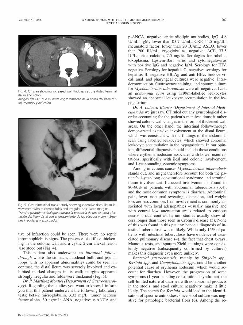
tive of infarction could be seen. There were no septicthrombophlebitis signs. The presence of diffuse thicken-ing in the colonic wall and a cystic 2-cm anexal lesionalso stood out (Fig. 4).
This patient also underwent an intestinal follow-through where the stomach, duodenal bulb, and jejunalloops with no apparent abnormalities could be seen; incontrast, the distal ileum was severely involved and ex-hibited marked changes in its wall; margins appearedstrongly irregular and folds were thickened (Fig. 5).
Dr. P. Martínez Montiel (Department of Gastroenterol-ogy): Regarding the studies you want to know, I informyou that this patient underwent the following laboratorytests: beta-2 microglobulin, 3.32 mg/L; tumor necrosisfactor alpha, 30 ng/mL; ANA, negative; c-ANCA and
p-ANCA, negative; anticardiolipin antibodies, IgG, 4.8U/mL; IgM, lower than 0.07 U/mL; CRP, 11.5 mg/dL;rheumatoid factor, lower than 20 IU/mL; ASLO, lowerthan 200 IU/mL; cryoglobulins, negative; ACE, 37.5IU/L; urine calcium, 7.5 mg/%. Serologies for rubella,toxoplasma, Epstein-Barr virus and cytomegaloviruswith positive IgG and negative IgM. Serology for HIV,negative. Serology for hepatitis C, negative; serology forhepatitis B: negative HBsAg and anti-HBc. Endocervi-cal, anal, and pharyngeal cultures were negative. Intra-dermoreaction, fluorescence staining, and sputum culturefor Mycobacterium tuberculosis were all negative. Last,an abdominal scan using Tc99m-labelled leukocytesshowed an abnormal leukocyte accumulation in the hy-pogastrium.
Dr. A. Lalueza Blanco (Department of Internal Medi-cine): As we just saw, CT ruled out any gynecological dis-order accounting for the patient’s manifestations; it rathershowed colonic wall changes in the form of thickened wallareas. On the other hand, the intestinal follow-throughdemonstrated extensive involvement at the distal ileum,which was consistent with the findings of the abdominalscan using labelled leukocytes, which showed abnormalleukocyte accumulation in the hypogastrium. In our opin-ion, differential diagnosis should include those conditionswhere erythema nodosum associates with bowel manifes-tations, specifically with ileal and colonic involvement,and 1-year-standing systemic symptoms.
Among infectious causes Mycobacterium tuberculosisstands out, and might therefore account for both the pa-tient’s 1-year-long constitutional syndrome and terminalileum involvement. Ileocecal involvement is found in80-90% of patients with abdominal tuberculosis (3,4),and the most common symptom is diarrhea. Abdominalpain, fever, nocturnal sweating, distension, and weightloss are less common. Ileal involvement is commonly as-sociated with local adenopathies –usually massive andwith central low attenuation areas related to caseousnecrosis; dual-contrast barium studies usually show ul-cers longer than those seen in Crohn’s disease (5). Noneof this was found in this patient; hence a diagnosis of in-testinal tuberculosis was unlikely. While only 15% of pa-tients with intestinal tuberculosis have evidence of asso-ciated pulmonary disease (4), the fact that chest x-rays,Mantoux tests, and sputum Ziehl stainings were consis-tently negative (subsequently confirmed by cultures)makes this diagnosis even more unlikely.
Bacterial gastroenteritis, mainly by Shigella spp.,Yersinia spp. and Campylobacter spp., could be anotherpotential cause of erythema nodosum, which would ac-count for diarrhea. However, the progression of somesymptoms (1-year-standing constitutional syndrome), theself-limited nature of diarrhea with no abnormal productsin the stools, and stool culture negativity make it littlelikely. The search for Yersinia would lead to the identifi-cation of specific antibodies, since stool culture was neg-ative for pathologic bacterial flora (6). Among the re-
Vol. 98. N.° 3, 2006 A YOUNG WOMAN WITH FIRST-TRIMESTER METRORRHAGIA, 207FEVER AND SKIN LESIONS
REV ESP ENFERM DIG 2006; 98(3): 204-215
Fig. 4. CT scan showing increased wall thickness at the distal, terminalileum and colon.Imagen del TAC que muestra engrosamiento de la pared del íleon dis-tal, terminal y del colon.
Fig. 5. Gastrointestinal transit study showing extensive distal ileum in-volvement with thickened folds and irregular, spiculated margins.Tránsito gastrointestinal que muestra la presencia de una extensa afec-tación del íleon distal con engrosamiento de los pliegues y con márge-nes irregulares y espiculados.
CCP LALUEZA 6/4/06 13:27 Página 207

maining bacterial causes for gastroenteritis, the only onethat should be considered is Neisseria gonorrhoeae, eventhough the prolonged course, and the fact that antibioticscovering this pathogen had been received with no clinicalimprovement, as well as microbial culture negativitywould rule it out.
Sarcoidosis may explain arthritis, uveitis, and skin in-volvement. While exceptional, ileal involvement hasbeen described in 17 cases of intestinal sarcoidosis in theabsence of respiratory symptoms (7), which greatly com-plicates differential diagnosis versus Crohn’s disease.The fact that chest x-rays was strictly normal, togetherwith calciuria and normal ACE titers, make the diagnosisof sarcoidosis difficult (8). In patients with sarcoidosisACE sensitivity for this disease ranges from 58 to 86%,depending on disease activity; in contrast, this test’sspecificity reaches 90%. They may be present in tubercu-losis, silicosis, primary biliary cirrhosis, and lymphoma,but not in Crohn’s disease (7-11).
Among systemic causes Behçet’s disease and sys-temic lupus erythematosus (SLE) should be ruled out.The former disease may account for erythema nodosum,for gastrointestinal symptoms, and potentially for oralaphthae as reported in the patient’s history. Behçet’s dis-ease may affect any portion of the bowel, but the ileoce-cal region is most commonly compromised (12). Histo-logic lesions and similar extraintestinal manifestationsmay render the distinction between Behçet’s disease andCrohn’s disease a difficult task (13-15). Pain is the mostcommonly seen abdominal symptom, and is present insome series in up to 90% of patients, followed by diar-rhea and gastrointestinal bleeding (12). However, thispatient does not meet the criteria allowing a diagnosisof Behçet’s disease, mainly the major criteria (recurrentoral ulers with at least 3 episodes in 12 months), andonly has one of the minor criteria (skin lesions includ-ing erythema nodosum). She reported no recurrent geni-tal ulcers, ocular lesions or pathergy (16). Colonoscopyusually demonstrates deep isolated ulcers in the ileoce-cal region (12). LSE, while a most uncommon cause oferythema nodosum (17), may explain skin lesions; how-ever, the patient’s immune profile and the absence ofadditional clinical or blood data make it little likely; inaddition, diarrhea is not easily explained by this condi-tion.
IBD may justify the constitutional syndrome’s pro-gression, as well as abdominal pain, radiographic bowelfindings, and fever at admission; however, the fact thatgastrointestinal symptoms were scarce is outstanding. Inthis disease, up to 35% of patients have extraintestinalmanifestations. In our case report, the patient had erythe-ma nodosum. This finding occurs in 15% of patients –itusually develops during flare-ups and is closely linked toperipheral joint disease. At admission the patient hadtype-1 peripheral arthritis symptoms involving the rightknee. Episcleritis is the most commonly occurring ocu-lar symptom in Crohn’s disease, and affects 5% of pa-
208 A. LALUEZA BLANCO ET AL. REV ESP ENFERM DIG (Madrid)
REV ESP ENFERM DIG 2006; 98(3): 204-215
Fig. 6. Colonic mucosa where irregular, flat ulcers may be seen. Theneighboring mucosa was preserved, and exhibited a normal submuco-sal capillary pattern.Mucosa del colon en la que se observan úlceras planas e irregulares. Lamucosa de la vecindad estaba conservada y en ella era posible recono-cer el dibujo normal de los capilares submucosos.
Fig. 7. Similar to the previous image, with ulcers and edematous muco-sal islets.Imagen similar a la anterior, con úlceras e islotes de mucosa edemato-sa.
CCP LALUEZA 6/4/06 13:27 Página 208

tients (18); its severity is also parallel to that of boweldisease (19-23).
Among erythemas nodosa of tumoral origin Hodgkinand non-Hodgkin lymphomas stand out, as do leukemias;however, no laboratory parameters warrant such diag-
noses. From a pathological standpoint, a cecal carcinomaor carcinoid tumor might also be considered.
To clear doubts I truly believe that colonoscopy –plusileoscopy if possible– in association with the sampling oflesions encountered is essential.
Dr. D. Rodríguez Alcalde (Department of Gastroen-terology): Colonoscopy showed a patchy involvement ofthe whole colon with numerous fibrin-covered ulcers,some of them serpiginous, that bled on touch; the biggestulcers were up to 3-3.5 cm in size, alternating with appar-ently normal mucosal areas (Figs. 6 and 7). In the vicinityof ulcers, and within some of them, there were millimet-ric pseudopolypoid formations consistent with damagedmucosal areas, whereas mucosal regions far removedfrom ulcers had a normal macroscopic appearance with awell preserved vascular pattern. Involvement was moresevere in the descending colon and sigma, while only afew aphthae were found in the rectum. Ileoscopy onlymanaged to show the most distal 5-7 cm, but no lesionswere seen in that ileal portion. All of this was tronglysuggestive of IBD, specifically Crohn’s disease with pan-colonic involvement in a moderate flare-up.
Dr. G. López (Department of Pathology): Colorectalbiopsy showed a severely distorted glandular architectureand densely packed, mixed inflammatory infiltrationreaching beyond the muscularis mucosae and into thesubmucosa. Microabscesses and cryptitis were apparent.It was all strongly suggestive of idiopathic inflammatorydisease, most likely Crohn’s disease with high inflamma-tory activity (Figs. 8 and 9).
Dr. C. González Gómez (Department of Internal Medi-cine): Indeed, with a presumed diagnosis of Crohn’s dis-ease involving the whole colon and terminal ileum treat-ment was empirically started with mesalazine 800 mgand intravenous methylprednisolone 1 mg/kg/day. Feverregressed within 48 hours, and both extraintestinal mani-festations and laboratory changes gradually improved tothe point that the patient could be discharged.
Dr. A. Lalueza Blanco (Department of Internal Medi-cine): Chronologically, a fact to point out in this patient isthe development of insidious manifestations includingweight loss in the wake of her first pregnancy, with a sub-sequent episode of severe extraintestinal manifestationsduring the second pregnancy. These are the two most in-teresting aspects of this case: extraintestinal manifesta-tions and then the association of pregnancy with IBD.
During pregnancy, there is seemingly no higher riskfor spontaneous abortion, and pregnancy is no risk fac-tor for IBD relapse. Also, fertility has not been seen todecrease except in patients with proctocolectomy orileoanal anastomosis. In this respect it must be stressedthat sulfasalazine induces reversible hypomotility andoligospermia in 80% of males receiving this drug(24,25).
We may pose a couple of questions here –on the onehand whether pregnancy affects IBD activity; on the oth-er hand how may this disease impact on pregnancy. Re-
Vol. 98. N.° 3, 2006 A YOUNG WOMAN WITH FIRST-TRIMESTER METRORRHAGIA, 209FEVER AND SKIN LESIONS
REV ESP ENFERM DIG 2006; 98(3): 204-215
Fig. 8. Colorectal mucosa with marked architectural distortion and se-vere infiltration in the lamina propria by lymphocytes and plasma cells,which reaches beyond the muscularis mucosae and involves the sub-mucosa. HE (x 40).Mucosa colorrectal con marcada distorsión arquitectural e intensa infil-tración de la lámina propia por un infiltrado linfoplasmocitario quetraspasa la musculares mucosae y afecta a la submucosa. HE (x 40).
Fig. 9. Colorectal mucosa with a dense, mixed inflammatory infiltrate.Close-up of cryptitis and a microabscess. HE (x 100).Mucosa colorrectal con denso infiltrado inflamatorio mixto. Detalle decriptitis y microabsceso. HE (x 100).
CCP LALUEZA 6/4/06 13:27 Página 209

210 A. LALUEZA BLANCO ET AL. REV ESP ENFERM DIG (Madrid)
REV ESP ENFERM DIG 2006; 98(3): 204-215
garding the former question, there is a clear associationwith activity extent during pregnancy: if the condition isstable at pregnancy onset, it will probably remain likethat until the end of pregnancy. Inflammatory disease re-lapses during pregnancy in one third of patients, but thispercentage does not differ from that of relapse riskamong non-pregnant women (26). On the contrary, ifIBD is active when pregnancy starts, this flare-up willprobably remain for the rest of pregnancy (70% of pa-tients). IBD complications that may occur during preg-nancy do not differ from those seen in non-pregnant pa-tients. No differences exist during the postpartum periodeither (27). In the presence of perineal involvement aCaesarean section is often advised; when IBD is inactiveor during mild-to-moderate flare-ups vaginal delivery ispossible, but episiotomy is advised against given the sub-sequent fistulization risk (28,29).
Crohn’s disease has a greater impact on fetal develop-ment when compared to ulcerative colitis; in fact, ulcera-tive colitis is thought not to influence pregnancy out-come, except for a risk of preterm delivery for womenfirst hospitalized during pregnancy. Crohn’s disease isconsidered a risk factor for preterm delivery and lowbirth weight. This risk is similar to that seen in smokingmothers (30). In no case is there a higher risk of congeni-tal abnormalities (31).
REFERENCES
1. Brodell RT, Mehrabi D. Underlying causes of erythema nodosum.Postgrad Med 2000; 108: 147-9.
2. Requena L, Requena C. Erythema nodosum. Dermatol Online J2002; 8: 4.
3. Engin G, Acunas B, Acunas G, Tunaci M. Imaging of extrapul-monary tuberculosis. Radiographics 2000; 20: 471-88.
4. Vanhoenacker FM, De Backer AI, Op de Beeck B, Maes M, VanAltena R, Van Beckevoort D, et al. Imaging of gastrointestinal andabdominal tuberculosis. Eur Radiol 2004; 14: 103-15.
5. Boudiaf M, Zidi SH, Soyer P, et al. Tuberculous colitis mimickingCrohns disease: utility of computed tomography in the differentia-tion. Eur Radiol 1998; 8: 1221-3.
6. Baert F, Knockaert D, Bobbaers H. Bilateral hilar lymphadenopa-thy associated with Yersinia enterocolitica infection. Clin InfectDis 1994; 19: 197-8.
7. Storch I, Rosoff L, Katz S. Sarcoidosis and inflammatory boweldisease. J Clin Gastroenterol 2001; 4: 345.
8. Dumot JA, Karim A, Petras RE, Lashner BA. Sarcoidosis present-ing as granulomatous colitis. Am J Gastroenterol 1998; 10: 1949-51.
9. Bulger K, O’Riordan M, Purdy S, et al. Gastrointestinal sarcoidosisresembling Crohn’s disease. Am J Gastroenterol 1988; 83: 1415-7.
10. Adenis A, Wallaert B, Colombel JF, Cortor A, Marchandise X,Janin A, et al. Intestinal involvement in sarcoidosis. Gastroenterol-ogy 1993; 104: 355-6.
11. Clague RB. Sarcoidosis or Crohn’s disease? Br Med J 1972; 3:804.
12. Lee CR, Kim WH, Cho YS, Kim MH, Kim JH, Park IS, Bang D.Colonoscopic findings in intestinal Behçet´s disease. InflammBowel Dis 2001; 7: 243-9.
13. Terrin G, Borrelli O, Di Nardo G, Pacchiarotti C, Cucchiara S. Achild with aptthae and diarrhoea. Lancet 2002; 359: 316.
14. Kallinowski B, Noldge G, Stiehi A. Crohn´s disease with Behçet´ssyndrome appearance. A case report. Z Gastroenterol 1994; 32:642-4.
15. Yazici H, Yurdakul S, Hamurydan V. Behçet syndrome. Curr OpinRheumatol 1999; 11: 53-7.
16. Internacional study group for Behçet´s disease. Lancet 1990; 335:1078-80.
17. Ajubi N, Nossent JC. Panniculitis as the first manifestation of sys-temic lupus erythematosus: description of two cases. Neth J Med1993; 42: 25-9.
18. Petrelli EA, Mc Kinley M, Troncale FJ. Ocular manifestations ofinflammatory bowel disease. Ann Ophthalmol 1982; 14: 356-60.
19. Levine JB, Lukawski-Trubish D. Extraintestinal considerations ininflammatory bowel disease. Gastroenterol Clin North Am 1995;24: 633-46.
20. Veloso FT, Carvalho J, Magro F. Immune-related systemic mani-festations of inflammatory bowel disease. A prospective study of792 patients. J Clin Gastroenterol 1996; 23: 29-34.
21. Greenstein AJ, Janowitz HD, Sachar DB. The extraintestinal com-plications of Crohn’s disease and ulcerative colitis. A study of 700patients. Medicine 1976; 55: 401.
22. Orchard TR, Wordsworth BP, Jewell DP. Peripheral arthropathiesin inflammatory bowel disease: their articular distribution and nat-ural history. Gut 1998; 42: 387-91.
23. Juillerat P, Mottet C, Froehlich F, Felley C, Vader JP, Burnand B,et al. Extraintestinal manifestations of Crohn´s disease. Digestion2005; 71: 31-6.
24. Birnie GG, McLeod TI, Watkinson G. Incidence of sulphasalazine-induced male infertility. Gut 1981; 22: 452-5.
25. Toovey S, Hudson E, Hendry WF, Levi AJ. Sulphasalazine andmale infertility: reversibility and possible mechanism. Gut 1981;22: 445-51.
26. Nielsen OH, Andreasson B, Bondesen S, Jarnum S. Pregnancy inulcerative colitis. Scand J Gastroenterol 1983; 18: 735-42.
27. Rogers RG, Katz VL. Course of Crohn´s disease during the preg-nancy and its effect on pregnancy outcome: a retrosprective re-view. Am J Perinatol 1995; 12: 262-4.
28. Fonager K, Sorensen HT, Olsen J. Pregnancy outcome for womenwith Crohn´s disease: a follow-up study based on linkage betweennational registries. Am J Gastroenterol 1998; 93: 2426-30.
29. Norgard B, Fonager K, Sorensen HT, Olsen J. Birth outcomes ofwomen with ulcerative colitis: a nationwide Danish cohort study.Am J Gastroenterol 2000; 95: 3165-70.
30. Katz J. Pregnancy and inflammatory bowel disease. Curr OpinGastroenterol 2004; 20: 328-32.
31. Mottet C, Juillerat P, Gonvers J, Froehlich F, Burnand B, Vader J,et al. Pregnancy and Crohn´s disease. Digestion 2005; 71: 54-61.
CCP LALUEZA 6/4/06 13:27 Página 210