a walk through tau therapeutic strategies
TRANSCRIPT
Jadhav et al. Acta Neuropathologica Communications (2019) 7:22 https://doi.org/10.1186/s40478-019-0664-z
REVIEW Open Access
A walk through tau therapeutic strategies
Santosh Jadhav1,10, Jesus Avila2,3, Michael Schöll4,5,6,7, Gabor G. Kovacs8, Enikö Kövari9, Rostislav Skrabana10,Lewis D Evans11, Eva Kontsekova10, Barbara Malawska12, Rohan de Silva13, Luc Buee14* and Norbert Zilka10*Abstract
Tau neuronal and glial pathologies drive the clinical presentation of Alzheimer’s disease and related humantauopathies. There is a growing body of evidence indicating that pathological tau species can travel from cell tocell and spread the pathology through the brain. Throughout the last decade, physiological and pathological tauhave become attractive targets for AD therapies. Several therapeutic approaches have been proposed, including theinhibition of protein kinases or protein-3-O-(N-acetyl-beta-D-glucosaminyl)-L-serine/threonine Nacetylglucosaminylhydrolase, the inhibition of tau aggregation, active and passive immunotherapies, and tau silencing by antisenseoligonucleotides. New tau therapeutics, across the board, have demonstrated the ability to prevent or reduce taulesions and improve either cognitive or motor impairment in a variety of animal models developing neurofibrillarypathology. The most advanced strategy for the treatment of human tauopathies remains immunotherapy, whichhas already reached the clinical stage of drug development. Tau vaccines or humanised antibodies target a varietyof tau species either in the intracellular or extracellular spaces. Some of them recognise the amino-terminus orcarboxy-terminus, while others display binding abilities to the proline-rich area or microtubule binding domains.The main therapeutic foci in existing clinical trials are on Alzheimer’s disease, progressive supranuclear palsy andnon-fluent primary progressive aphasia. Tau therapy offers a new hope for the treatment of many fatal braindisorders. First efficacy data from clinical trials will be available by the end of this decade.
Keywords: Alzheimer’s disease, Tau vaccines, Therapeutic interventions, Immunotherapy, Tauopathies, PET imaging,Aggregation
IntroductionTau protein is considered to be one of the most peculiarproteins in the central nervous system. It is located in sev-eral cell compartments, including the axon, dendrites, nu-cleus, nucleolus, cell membrane and synapses [310].However, tau is also present in the interstitial fluid [284,370], and can pass into cerebrospinal fluid (CSF), where itis found at concentrations of 10-25 pg/ml (pT181-tau) or300-400 pg/ml (tau) [28, 29, 248]. In physiological condi-tions, extracellular tau may enter neurons either via adynamin-mediated endocytic mechanism or by classicalendocytosis [95]. In neurodegenerative tauopathy, dis-eased modified tau can propagate along neuroanatomi-cally connected brain areas via multiple mechanisms andspread tau pathology throughout the brain [231].
* Correspondence: [email protected]; [email protected] of Lille, Inserm, CHU-Lille, UMRS1172, Alzheimer & Tauopathies,Place de Verdun, 59045 Lille cedex, France10AXON Neuroscience R&D Services SE, Dvorakovo nabrezie 10, 811 02Bratislava, SlovakiaFull list of author information is available at the end of the article
© The Author(s). 2019 Open Access This articInternational License (http://creativecommonsreproduction in any medium, provided you gthe Creative Commons license, and indicate if(http://creativecommons.org/publicdomain/ze
Tau belongs to the group of natively disordered pro-teins, which exist in a highly flexible, unfolded structuralstate, largely devoid of well-defined secondary and tertiarystructure, although they are able to fold after binding totargets [329]. The highly flexible structure of tau proteinallows interaction with multiple partners, suggesting itsinvolvement in numerous signalling pathways [308]. Thedark side of its structural repertoire is its ability to interactwith other tau molecules to form oligomers and filaments[298, 338, 339]. These complexes cause degeneration ofneurons and glial cells [97], manifesting as a group of neu-rodegenerative disorders termed ‘tauopathies’ [312].The most prominent tauopathy is Alzheimer’s disease
(AD), the common cause of dementia in older adults. AD isan incurable, progressive degenerative disease of the brain,characterized by the presence of tau and ß- amyloid (Aß)pathology [286]. There are no disease-modifying drugsavailable for AD; only symptomatic treatments trying tocounterbalance the neurotransmitter disturbance exist. Nosignificant new drug for AD has been approved in the last14 years, despite extensive clinical trials. The pipeline has
le is distributed under the terms of the Creative Commons Attribution 4.0.org/licenses/by/4.0/), which permits unrestricted use, distribution, andive appropriate credit to the original author(s) and the source, provide a link tochanges were made. The Creative Commons Public Domain Dedication waiverro/1.0/) applies to the data made available in this article, unless otherwise stated.

Jadhav et al. Acta Neuropathologica Communications (2019) 7:22 Page 2 of 31
been plagued with significant failures, with more than 400failed clinical trials since the last symptomatic Alzheimer’sdrug was approved [71].Despite the field being aware that tau pathology corre-
lates well with the onset and progression of AD for al-most 40 years [39], it is only now that tau targettedtherapy has become attractive for clinical trials. A multi-tude of tau antibodies and vaccines have been tested inpreclinical studies in the last two decades. Currently,eight humanised tau antibodies and two tau vaccineshave entered clinical trials either for AD or frontotem-poral dementia (FTD) [65, 71](www.alzforum.org). Inlight of the failure of the clinical trials with amyloid tar-geting drugs, tau therapy is manifesting as the frontrun-ner in the search for an effective treatment for AD.
Tour de tau - tau as a protein with multiple facesIn contrast to amyloid precursor protein (APP), the func-tion of tau protein was already known at the time of thediscovery of it as a constituent of neurofibrillary degener-ation. Tau is a microtubule-associated protein (MAP),promoting the polymerization and assembly of microtu-bules [351]. In the adult human brain, there are six iso-forms of tau protein generated by alternative splicing froma single gene located on chromosome 17 [120, 238]. Atthe N-terminal end, they differ by the addition of a 29amino-acid sequence (1 N) or as replicates (2 N - total of58 amino acids) coded by exons 2 and 3. The sequencecoded by exon 3 is only present if the sequence encodedby exon 2 is inserted. Interestingly, the 2 N tau isoformsare weakly expressed in the human brain [119, 214, 295].The microtubule binding region (MTBR), has three (3R:R1, R3, R4) or four repeat domains (4R: R1-R4). The se-quence encoded by exon 10 allows the insertion of a 31amino acid microtubule binding domain (R2) which isinserted after the first repeat R1. Tau isoforms with 3Rand 4R are equally expressed, since their ratio is about 1:1in the human brain [295]. However, some neurons do notexpress 4R tau isoforms. For instance, granular cells of thedentate gyrus only express mRNAs of 3R-tau isoforms[119]. Thus, tau isoforms have different cellular and lam-inar distribution in the human brain [46].The strict classification of tau protein as a MAP may
have delayed research on its other biological functions. Ifsequence homology (70-90%) with other MAPs is evidentin the microtubule binding domains, the N-terminal por-tion of tau is unique. It must therefore have other uniquefunctions [194]. Logically, as a MAP, tau has functions incell trafficking, but it also interacts with dynactin andsynaptogyrin-3, suggesting specific related-functions, suchas synaptic vesicle control [213, 224].The first unexpected functions of tau may be related to
its nuclear localization [201]. These initial findings werewidely discussed, but nowadays, it is clearly established
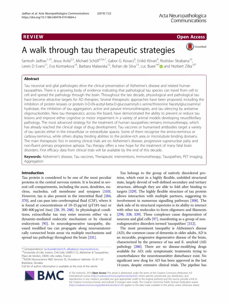
that tau binds to nucleic acids, and may be involved inchromatin remodelling [53, 104, 146, 252, 266, 267]. Thebinding of tau to DNA may allow protection against react-ive oxygen species [316, 349], and binding to RNA maycontribute to ribosome stability and miRNA activity [35].Altogether, these data strongly suggest that tau maymodulate gene expression and RNA stability. Such obser-vations are also supported by tau loss-of-function inpathological conditions. For instance, formation of tauoligomers leads to DNA/RNA damage [337], RNA andribosome instability [225] and changes in nuclearorganization and protein expression [103]. Binding of tauto tRNAs may also initiate tau aggregation by formingdroplets through complex coacervation [378]. Moreover,pathological tau can interact with nucleoporins of the nu-clear pore complex (NPC) and affect their structural andfunctional integrity [93] (Fig. 1).Secondly, tau may also play a role in cell signalling. The
longest brain tau isoform with 441 amino acids (aa) has 85putative sites of phosphorylation. Thus, tau may act as abuffer for cell signalling. For instance, tau may serve as a‘phosphorylation sink’ for the p25-Cdk5 complex, hencesequestering it away from other death-inducing substrates[130]. Tau may also interfere with tyrosine kinase familySrc/Fyn signalling at dendrites [49, 152]. Tau also interactswith phosphatase and tensin homolog (PTEN) and modu-lates insulin signalling. Recent data suggest that loss of taufunction leads to an impaired hippocampal response to in-sulin, caused by altered insulin receptor substrate 1(IRS-1) and PTEN activities [218].Finally, the cytosolic tau protein may also be secreted. This
secretion is stimulated by neuronal activity [263]. Such se-cretion is likely to occur through non-conventional secretorypathways [44]. Recent data suggest that such secretion maybe similar to that of fibroblast growth factor 2 (FGF-2), in-cluding oligomerization, binding to phospho-inositol, andextracellular capture by heparan sulphate proteoglycans[164]. An alternative pathway is the secretion ofpro-interleukin 1, which requires proteolysis. Interestingly,C-terminal-tau fragment Δ422–441 was significantly moresecreted than full length tau [261]. Tau is also secretedwithin extracellular vesicles such as exosomes [346] andectosomes [89]. In pathological conditions, secreted tau mayparticipate to tau seeding and spread (discussed later).To sum up, tau has multiple functions in addition to
axonal microtubule assembly. All of these recently dis-covered tau functions may contribute to the develop-ment of tau pathology and related events (Fig. 1). Thesediscoveries further strengthen the case for tau as thetherapeutic target for AD and tauopathies.
Tau as a driver of neurodegenerationAD is a double proteinopathy, characterized by the pres-ence of both tau-reactive neurofibrillary lesions and

Fig. 1 Yin and Yang of Tau protein
Jadhav et al. Acta Neuropathologica Communications (2019) 7:22 Page 3 of 31
β-amyloid (Aβ) depositions (senile plaques; SPs). Theimportance of both proteins, which are present alsounder physiological circumstances, in the developmentof AD is extensively debated. Numerous clinicopatholog-ical studies were published, favouring both histologicallesions, i.e. NFTs and SPs. However, since the early nine-ties, most studies found a strong correlation betweenneocortical NFT load and cognitive impairment [94].The progression of neurofibrillary pathology begins
in the entorhinal cortex, in contrast to the spreadingof Aβ, where the presence of neocortical SPs precedesthe appearance of hippocampal SPs [39, 91, 320, 327].Aβ pathology is present even in cognitively intact per-sons, so amyloid deposition is not sufficient to explainthe clinical phenotype of AD [77]. In contrast, NFTburden in associative neocortical areas is strongly re-lated with clinically overt dementia. The Braak staging[39] for NFTs, used to define the neuropathologicalseverity of AD in the general neuropathological prac-tice, reveals a strong correlation with cognitive decline[92, 121]. In a study of an oldest-old population, Goldand colleagues [121] found that unlike younger co-horts, Braak stages did not precisely reflect the sever-ity of dementia. Braak stage III correlates poorly withcognitive decline, while Braak stages IV or greater areconsistently associated with at least mild dementia.This discrepancy is most likely due to the increasingprevalence of mixed neuropathologies in theoldest-old, such as a combination of vascular lesionsand AD pathology [156].
As in all neurodegenerative diseases, AD is charac-terised by selective vulnerability of specific brain regions,cortical layers, and neuronal populations. The anatom-ical distribution of tau and neuronal loss reflects the dif-ferent clinical signs of AD well. Anterograde memoryproblems at the beginning of the symptomatology arerelated to tau-burden in the medial temporal lobe [94].During the progression of the clinical presentation, othersigns, such as agnosia, apraxia or speech and behaviouralproblems will add to the memory problems, correspond-ing to the involvement of different associative or limbicregions. The neuropathological background for acalculiaand visuospatial dysfunction is related to the involve-ment of tau pathology in the parietal lobe [94]. Ideomo-tor and dressing apraxia is linked to NFT densities inthe anterior cingulate cortex, while constructionalapraxia relate to NFT densities in the superior parietal,posterior cingulate and occipital cortex [113]. A signifi-cant relationship exists between associative visual agno-sia and tau burden in the secondary visual cortex(Brodmann area 18) and the occipitotemporal visual as-sociation cortex (Brodmann area 37 and ventral 19)[114]. The high NFT density in the superior parietal cor-tex (Brodmann area 7), posterior cingulate cortex (Brod-mann area 23), and CA1 subfield of the hippocampusplays a role in developing temporo-spatial disorientation[115]. Cases with atypical AD, such as posterior corticalatrophy, also underline the importance of tau pathologyin developing clinical signs. Patients presenting mainlywith visual symptomatology have a high NFT burden in
Jadhav et al. Acta Neuropathologica Communications (2019) 7:22 Page 4 of 31
the occipito-parieto-temporal junction and posterior cin-gulate cortex [138]. The anterior brain regions are lessinvolved as compared to the “classic” form of AD.Behavioural problems or speech disorders, more suggestive
of other neurodegenerative diseases such as frontotemporaldementia, could also be present in neuropathologically con-firmed AD. In contrast, prefrontal syndromes are correlatedwith atypical distribution of NFTs in the dorsolateral, medianand orbitofrontal areas [340]. These clinicopathological ob-servations underline the importance of the tau protein in thepathogenesis of AD and its subtypes (amnestic, dysexecutive/behavioural, visuo-spatial, and language presentation).Tauopathies are clinically, biochemically and morpho-
logically heterogeneous neurodegenerative diseases char-acterized by the deposition of abnormal tau (microtubuleassociated protein tau; MAPT) in the brain. Neuropatho-logical phenotypes are distinguished based on the distinctinvolvement of anatomical areas, cell type, and presenceof distinct isoforms of tau in the pathological deposits[172]. If tau protein deposition is the predominant feature,the term primary tauopathy is used. The nomenclatureoverlaps with the classification of frontotemporal lobar de-generation (FTLD). Disorders characterized by tau path-ologies considered having other (possibly diverse) drivingforces (e.g. Creutzfeldt–Jakob disease, Down’s syndrome)are called secondary tauopathies [108].Tauopathies are distinguished based on the ratio of 3 re-
peat (3R)- and 4R-tau and two or three major bands (60,64, and 68 kDa) in Western blot of sarkosyl-insoluble frac-tions [184, 296, 312]. FTLD-tau is grouped based on thetau isoform predominating the morphology. Pick’s disease(PiD) is a 3R tauopathy (60 and 64 kDa bands). 4R tauopa-thies (64 and 68 kDa bands) is comprised of progressivesupranuclear palsy (PSP), corticobasal degeneration(CBD), argyrophilic grain disease (AGD), and globularglial tauopathy (GGT) [172]. Mixed 3R and 4R tauopathy(60, 64 and 68 kDa bands) is the neurofibrillary tangle(NFT)-dementia (discussed also in the frame of primaryage-related tauopathy, PART), and this type of tau path-ology is seen in Alzheimer diseased (AD) brains.Hyperphosphorylated tau is the major constituent of
neuronal and glial inclusions, although there are furtherbiochemical modifications (N- and C-terminal truncation,glycosylation, glycation, nitration of tyrosine residues,transglutamination, deamidation; acetylation; oligomerforms) [173] which are not examined routinely in diagnos-tic practice. Using phospho-dependent tau antibodies sev-eral morphologies of cellular tau immunoreactivity can bedetected [172]. Tau immunoreactivity in neuronscomprises pre-tangles (diffuse cytoplasmic neuronal tauimmunoreactivity), NFTs, Pick bodies (3R-tau immunore-active), spherical inclusions (usually 4R immunoreactive),dystrophic neurites, neuropil threads (axonal), and grains(dendritic). Astrocytic tau pathology includes tufted
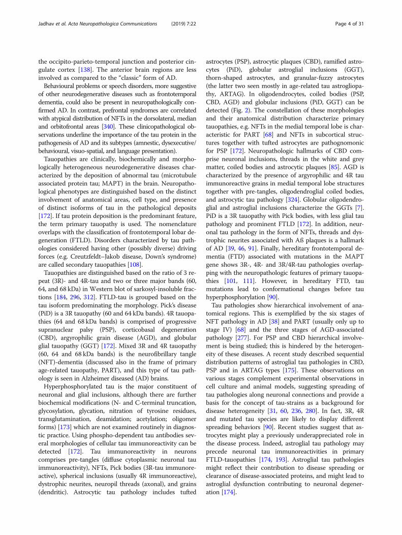
astrocytes (PSP), astrocytic plaques (CBD), ramified astro-cytes (PiD), globular astroglial inclusions (GGT),thorn-shaped astrocytes, and granular-fuzzy astrocytes(the latter two seen mostly in age-related tau astrogliopa-thy, ARTAG). In oligodendrocytes, coiled bodies (PSP,CBD, AGD) and globular inclusions (PiD, GGT) can bedetected (Fig. 2). The constellation of these morphologiesand their anatomical distribution characterize primarytauopathies, e.g. NFTs in the medial temporal lobe is char-acteristic for PART [68] and NFTs in subcortical struc-tures together with tufted astrocytes are pathognomonicfor PSP [172]. Neuropathologic hallmarks of CBD com-prise neuronal inclusions, threads in the white and greymatter, coiled bodies and astrocytic plaques [85]. AGD ischaracterized by the presence of argyrophilic and 4R tauimmunoreactive grains in medial temporal lobe structurestogether with pre-tangles, oligodendroglial coiled bodies,and astrocytic tau pathology [324]. Globular oligodendro-glial and astroglial inclusions characterize the GGTs [7].PiD is a 3R tauopathy with Pick bodies, with less glial taupathology and prominent FTLD [172]. In addition, neur-onal tau pathology in the form of NFTs, threads and dys-trophic neurites associated with Aß plaques is a hallmarkof AD [39, 46, 91]. Finally, hereditary frontotemporal de-mentia (FTD) associated with mutations in the MAPTgene shows 3R-, 4R- and 3R/4R-tau pathologies overlap-ping with the neuropathologic features of primary tauopa-thies [101, 111]. However, in hereditary FTD, taumutations lead to conformational changes before tauhyperphosphorylation [90].Tau pathologies show hierarchical involvement of ana-
tomical regions. This is exemplified by the six stages ofNFT pathology in AD [38] and PART (usually only up tostage IV) [68] and the three stages of AGD-associatedpathology [277]. For PSP and CBD hierarchical involve-ment is being studied; this is hindered by the heterogen-eity of these diseases. A recent study described sequentialdistribution patterns of astroglial tau pathologies in CBD,PSP and in ARTAG types [175]. These observations onvarious stages complement experimental observations incell culture and animal models, suggesting spreading oftau pathologies along neuronal connections and provide abasis for the concept of tau-strains as a background fordisease heterogeneity [31, 60, 236, 280]. In fact, 3R, 4Rand mutated tau species are likely to display differentspreading behaviors [90]. Recent studies suggest that as-trocytes might play a previously underappreciated role inthe disease process. Indeed, astroglial tau pathology mayprecede neuronal tau immunoreactivities in primaryFTLD-tauopathies [174, 193]. Astroglial tau pathologiesmight reflect their contribution to disease spreading orclearance of disease-associated proteins, and might lead toastroglial dysfunction contributing to neuronal degener-ation [174].

Fig. 2 Tau pathologies in diverse tauopathies. Tau pathology in ADand PART comprise dystrophic neurites (a), axonal threads (b),pretangles (c) and NFTs (d). PSP is characterized by pretangles andthreads (e), subcortical tangles (f), tufted astrocytes (g), andoligodendroglial coiled bodies (h). In CBD cases pretangles andthreads (i), globose neuronal CBD-bodies (j), astrocytic plaques (k),and oligodendroglial coiled bodies (l) can be seen. AGD ischaracterized by 4R-tau positive neuronal dendritic grains (m),pretangles (n), granular/fuzzy astrocytes (o), and oligodendroglialcoiled bodies (p). In GGT cases neuronal pretangles (q), sphericalcytoplasmic inclusions (r), globular astroglial (s) and oligodendroglial(t) inclusions are detected. In Pick’s disease neuronal Pick bodies arefrequent in the dentate gyrus (u) and show 3R immunoreactivity (v;here CA1 subregion is shown), furthermore, ramified astrocytes (w)and small globular oligodendroglial inclusions (x) can be noticed aswell. Finally ARTAG comprises thorn shaped astrocytes and granularfuzzy astrocytes here demonstrated in the subependymal (y1),subpial (y2), perivascular (upper part of image 4) and white matter(lower part of image) (y3), and grey matter (y4) areas. All imagesshow immunostaining for the AT8 antibody except (m) and (v)where immunostaining for 4R- and 3R-tau isoform, respectively, wasperformed. The bar in (a) represents 50 μm for a, e, f, g, h, l, m, t, u,v, y1, and y4; 35 μm for b, c, d, j, k, o, p, x; 30 μm for q and r; 40 μmfor w and y2; 100 μm for i; 25 μm for s; and 150 μm for y3
Jadhav et al. Acta Neuropathologica Communications (2019) 7:22 Page 5 of 31
Pet imaging of tau pathologyRecently, the development of positron emission tomog-raphy (PET) radioligands presumably binding to tau hasenabled the in vivo mapping and quantification of taupathology, hitherto largely confirming autopsy findings.The radioligand [18F] Flortaucipir (FTP, previouslyAV1451 or T807), a benzimidazole pyrimidine derivative,is by far the most widely employed to date. It has beenshown to bind with high affinity to mixed 3R- and4R-tau isoforms in paired-helical filaments (PHF) of ADpatients [26, 309, 361]. A recent study furthermoreshowed that in vivo FTP-binding and post mortem PHFload were highly correlated in a subject with a MAPTR406W mutation, which causes AD-like 3R/4R tau path-ology [309]. However, large inter- and intra-individualdifferences were observed in a recent autopsy study ofseveral tauopathies [361], calling for further investigationof FTP binding characteristics.Off-target binding of tau PET ligands is another major
limitation and challenge to be addressed in novel tracerdevelopment [26, 187, 200]. For example, the alleged tauPET ligand [18F]THK5351 demonstrated strong bindingto monoaminoxidase B (MAO-B) in and ex vivo [133,239], with ligand uptake being reduced by up to 50% in se-lected brain regions by the MAO-B inhibitor selegiline,preventing accurate quantification of tau [239]. Amongthe currently available tracers, the binding characteristicsof FTP have been characterized best. FTP off-target bind-ing has been observed in the caudate, putamen, and palli-dum in elderly individuals regardless of their clinicaldiagnosis [20, 42, 205, 333, 354], and has been attributedto, amongst others, iron binding [59]. Its pronouncedbinding to the substantia nigra, also in cases with no ap-parent tau pathology, has been related to neuromelanin[219–221], as has elevated FTP binding in the pituitarygland, retinal pigment epithelial cells, leptomeninges, andmalignant melanocytes in metastatic melanoma [205, 219,221]. High FTP signal in the choroid plexus has been at-tributed to calcification/mineralization [205], binding totangle-like structures corresponding to so-called Biondiring tangles [150], or melanocyte binding [180, 219, 221]and constitutes an issue for the quantification of hippo-campal ligand uptake due to their close proximity. Here,partial volume correction (PVC) might reduce bias fromchoroid plexus signal on hippocampal signal [180, 211,212, 288]. FTP has also been shown to bind to MAO-Aand B in vitro [335], however, no significant differenceswere observed in vivo between FTP scans of patients withand without MAO-B inhibitors [133].A second generation of tau radioligands is supposed to
be affected less by off-target binding issues, however,in vivo data are thus far limited for these ligands, whichinclude, amongst others, [18F]RO6958948 (Roche) [142,359], [18F]MK-6240 (Merck/Cerveau) [24, 199, 255],
Jadhav et al. Acta Neuropathologica Communications (2019) 7:22 Page 6 of 31
[18F]GTP-1 (Genentech) [278, 279, 350], [18F]PI2620(Life Molecular Imaging, formerly Piramal Imaging)[314] and [18F]PM-PBB3 [249, 299].For [18F] FTP, tracer uptake in physiological aging and
AD appears to follow a particular spatial and temporalpattern. Although longitudinal data are limited to this date[153, 311], the distribution appears to begin in the ento-rhinal cortex, to spread into inferolateral temporal lobesand medial parietal lobes, and to eventually cover most ofthe neocortex in disease cases. To capture this highregionality, which is significantly different from e.g. PETimaging of Aβ pathology (often found throughout theneocortex), several approaches have been suggested for A)binary categorization of tau “positivity” [154, 212, 229,344], and B) topographical staging approaches that recap-itulate post mortem findings of tau distribution [211, 288,290]. This regionality of tau PET ligand uptake in thebrain is further emphasized by studies employingdata-driven approaches without prior definition of ana-tomical regions [293, 352]. However, a few studies havesuggested that ligand uptake assessment based on largercomposite regions may be sufficient to captureAD-related tau PET signal and the longitudinal accumula-tion of tau [153, 211]. On a group level, FTP demon-strated clinical usefulness when its discriminative accuracybetween AD dementia and non-AD neurodegenerativedisorders was examined in a large multisite study, yieldingvery high sensitivity and specificity based on medial-basaland lateral temporal cortex ligand uptake [250].In general, elevated tau tracer binding in the medial
temporal lobe (MTL) can be observed in cognitivelyhealthy older adults, whereas widespread binding in neo-cortical regions of any individual commonly is associatedwith the presence of cortical Aβ [58, 124, 161, 198, 211,262, 288, 291, 294]. However, despite an overall correl-ation between brain Aβ and tau [161], the spatial distri-butions of these two aggregated proteins are discordant[161, 198, 294]. Interestingly, the strongest associationcan be observed between global Aβ and entorhinal tauPET signal [333], rendering this region important for thedetection of AD-related tau PET signal.Tau deposition outside the MTL is more common in
individuals with AD; however, elevated tau tracer uptakehas been reported for in neocortical areas in cognitivelynormal and even Aβ negative individuals [204]. WhileAD patients commonly have more widespread and pro-nounced tracer uptake than controls, exceptions havebeen found in AD patients who are Aβ-positive andshow relatively low levels of tau deposition [262, 344].Longitudinal studies have also demonstrated that in-creasing levels of Aβ are associated with more tau de-position in limbic and neocortical Braak regions severalyears later, even in nominally Aβ-negative individuals[179, 325]. Despite the limited availability of longitudinal
data, it appears that tau accumulates over time in thetemporal lobes of cognitively healthy individuals and ADpatients, albeit this seems to be limited to Aβ-positiveindividuals [153, 311].Compared to associations with Aβ, correlations be-
tween tau PET measures and age across healthy elderlyseem to be weaker and confined to MTL regions [212,289]. Greatest differences in FTP uptake betweenhealthy young and elderly subjects are commonly ob-served in the choroid plexus and basal ganglia; however,tracer uptake in these regions most likely representsoff-target binding [205, 206]. The age of symptom onsetamong AD patients clearly affects tau PET uptake pat-terns. Sporadic early-onset AD patients (EOAD) exhibitdistinctly greater parietotemporal and frontal ligand up-take when compared with late onset AD (LOAD) whichexhibits rather confined temporal lobe uptake [289].Data from studies in early-onset familial/autosomal-do-minant AD are limited, suggesting earliest FTP uptakein the medial temporal lobes of Aβ-positive presymp-tomatic mutation carriers but high cortical uptake,spatially comparable to sporadic EOAD cases in latersymptomatic stages [268, 289].Tau has, in contrast to Aβ, long been known to be much
stronger associated with measures of cognitive decline andneurodegeneration [86, 88, 136, 155, 237]. In fact, greaterFTP uptake has been shown to be related to both poorercognitive function cross-sectionally and retrospective longi-tudinal decline in cognition functioning [13, 212]. In cogni-tively healthy elderly, associations are strongest betweenepisodic memory performance and MTL, namely entorhi-nal cortical tracer uptake, whereas associations with globalcognition are either absent or found for wider, less specificneocortical regions. Interestingly, the effect of MTL tau onepisodic memory seems to be independent of global Aβload [211, 288] both in these individuals and in individualsexperiencing subjective cognitive decline [45]. Moreover,MTL tau accumulation in cognitively normal elderly is as-sociated with patterns of neurodegeneration as assessed byboth structural magnetic resonance imaging (MRI) and[18F] Fluorodeoxyglucose (FDG) PET that are topographic-ally similar to the patterns seen in AD patients [2, 74, 125,132, 176], suggesting that early-stage MTL tau might havea pathogenic role even in cognitively healthy individuals.The relationship between tau, cognition, and neurode-
generation is even more pronounced in AD patients, espe-cially in cases of EOAD who frequently exhibit language,visuospatial, or executive dysfunction rather than memoryimpairment and where the spatial distribution of tau depos-ition strongly reflects the clinical phenotype [250, 368]. Inthese patients, tau deposition is also strongly associatedwith the neurodegeneration markers of atrophy and glucosehypometabolism [27, 148, 250, 344], a relationship that can-not be explained by measures of or the distribution of Aβ
Jadhav et al. Acta Neuropathologica Communications (2019) 7:22 Page 7 of 31
[269]. Statistically, cognitive impairment can be related toboth brain atrophy and tau, however, tau remains solelycorrelated with cognitive dysfunction, even when control-ling for atrophy [23].Generally, FTP uptake might be helpful in distinguish-
ing clinical variants of AD, e.g. a recent study employinga data-driven clustering approach demonstrated that themajority of patients with relatively low entorhinal FTPuptake, compared to overall neocortical uptake, have anatypical clinical EOAD presentations, while most pa-tients with high FTP uptake in both entorhinal and neo-cortex present with EOAD and a typical amnesticphenotype, and most with low FTP uptake in both ento-rhinal and neocortex present with typical LOAD [352].In summary, the assessment of tau accumulation with
PET has revealed a pattern of aggregation on a con-tinuum from normal aging through AD that parallelsneuropathological data and now offers the possibility oflongitudinal studies. The strong relationship between tauPET measures and measures of neurodegeneration andcognition, taking in account the relationship betweentau and Aβ, will elucidate how Aβ and tau pathologyinteract in the development of the processes that arelinked to cognitive decline and clinical dementia.
Extracellular and intracellular tau – Two sides ofone coinIn pathological conditions, tau protein undergoespost-translational modifications, such as, truncation [241,242, 357, 358], phosphorylation [127], ubiquitination [32,181], glycation [283, 373], glycosylation [196, 343], nitra-tion [144, 271, 272] and sumoylation [87, 209]. Amongthem, phosphorylation and truncation are the most stud-ied. Many laboratories suggest that tau hyperphosphoryla-tion on Ser and Thr residues facilitates tau aggregation.Tau is posttranslationally modified at Ser/Thr residies byO-linked N-acetylglucosamine (O-GlcNAc), and thus in-creasing tau O-GlcNAcylation may protect against tau ag-gregation. In tau transgenic mouse models, inhibition ofβ-N-acetyl-glucosaminidase, the enzyme responsible forO-GlcNAc removal, is protective [33].It has been shown that truncated tau proteins are con-
tained in the core of the paired helical filaments. Expres-sion of the tau protein in the brain of transgenic rats andmice induced the formation of extensive neurofibrillarypathology, suggesting that truncated tau is a driving forceof neurofibrillary degeneration [98, 381, 382, 384, 385].Therapeutic approaches against tau pathology target
either intracellular or extracellular tau or eventuallyboth. It has been demonstrated that an increase in thelevel of intracellular tau could result in tau secretion intothe extracellular space or in cell death [122, 304]. Toxicextracellular tau could interact with neuronal cell recep-tors such as M1/M3 muscarinic receptors [122, 123], or
with heparin sulfate linked to cell membrane [372]. Theresult of that interaction could be again the onset ofneuron toxicity and intracellular tau secretion. In thisway, tau pathology could be propagated. Thus, possibletherapies involving the use of muscarinic antagonists[131, 334, 336], or agents decreasing heparin sulphation[372], are under discussion for AD therapy.Extracellular tau is found at significant levels in the
interstitial fluid of the central nervous system (CNS), andcan pass into cerebrospinal fluid (CSF) [370]. Initially,extracellular tau was thought to be only passively releasedby dying neurons, with selective vulnerability of neuronaltypes and cellular signals contributing to the disease pro-gression [285]. However, there is now growing evidencethat tau is actively transferred between neurons underpathological and physiological conditions. Aggregated andsoluble tau variants have been shown to transfer betweenanatomically connected regions of the brain [75, 149,197], and trans-synaptically between cells in culture [280,363]. How tau is actively transferred between neurons is amajor focus of dementia research, as attenuating thepathological spread may limit the progression of disease.Active tau transfer is thought to involve discrete steps in-cluding post translational modification (PTM), extracellu-lar release and subsequent tau internalization.Intracellular tau undergoes various PTMs including
phosphorylation and proteolytic cleavage. Levels of totaland phosphorylated tau detected in the CSF are importantbiomarkers for dementia [28]. Several tau modificationsare detected at proportionally higher levels in extracellularcompared with intercellular fractions, implicating specifictau modifications in active neuronal export [248]. Higherlevels of extracellular aberrantly hyperphosphorylated tauare detected in patients with dementia [79]. Hyperpho-sphorylated tau has a lower binding affinity to microtu-bules (MT) [192] and mislocalizes to somatic anddendritic cell compartments [106, 143, 323]; these factorsmay contribute to active export as dissociation from MTswould allow a greater opportunity for tau to interact withcomponents that facilitate protein export. C-terminallytruncated tau (lacking approximately the last 50 aminoacids) is detected at proportionally higher levels in theCSF samples of healthy individuals and dementia patients[284], and in neurons in culture [43, 163]. These tau spe-cies may be more readily detectable or resistant to degrad-ation. Post-translational modifications and exon splicingevents influence intra- and extracellular tau stability.Phosphorylated and 4R-tau isoform peptides have fasterturnover rates than unphosphorylated and 3R-tau isoformpeptides, respectively. Peptides from the N-terminal tomid-domain tau are more stable and have similarhalf-lives both inside and outside of the cell [284]. Not-withstanding these differences in stability, the proportion-ally higher levels of extracellular truncated tau suggest

Jadhav et al. Acta Neuropathologica Communications (2019) 7:22 Page 8 of 31
that physiological active tau release may be regulated byproteolytic cleavage.Distinguishing between active tau release mechanisms
and passive tau release, due to cell death, is challenging.The process of active tau release has been linked with sev-eral cellular mechanisms. In cell culture, monomeric taucan directly interact with the plasma membrane and pro-teoglycans, leading to unconventional secretion of tau [55,164, 304], or the release of ectosomes containing tau [89].Active tau release is also proposed to be regulated byneuronal activity. Depolarization of neurons promotes taurelease [102, 263, 371] and release of exosomes containinghypophosphorylated tau [346]. These mechanisms aren’tmutually exclusive. However, it is unclear how they are as-sociated and whether they relate to all forms of tau.Following release into extracellular space, pathogenic
tau can be taken up by healthy neurons, and promoteseeded aggregation [165]. There have been conflicting re-ports regarding the forms of tau and route of entry of tauinto various cell types. Studies suggest that aggregated tauis the predominant form internalized into cells [105, 362].However, monomeric full-length tau can also be efficientlytaken up by neurons [95]. These reports show that tau istaken up by endocytosis. Levels of the clathrin-mediatedendocytosis component myc box-dependent-interactingprotein 1 (BIN1) negatively correlate with tau uptake [52].Different forms of tau enter neurons via distinct but over-lapping pathways. Monomeric tau can enter neurons viadynamin-dependent endocytosis that is saturable, suggest-ing uptake is dependent on carrier proteins or receptors[95]. Entry of aggregated tau is attenuated by heparin incell culture, indicating that heparan sulphate proteogly-cans serve as receptor for tau uptake [140].Hyperphosphorylated tau isolated from AD brain tissue
is also recognized by the CNS immune system; microgliainternalize and degrade tau in an Fc-dependent manner[210], and the cytosolic Fc receptor – tripartitemotif-containing protein 21 (TRIM21), inhibits seeded tauaggregation [223]. Conversely, it is also suggested that themigration of microglia through the CNS transfers patho-genic species of tau to new areas of the brain [216].It is currently unknown if tau transfer is a disease-specific
phenomenon or physiological process appropriated duringdisease. Physiological tau transfer may be involved in net-work signaling or neuronal maintenance. Independently ofthe ability of pathological tau to seed aggregation, extracel-lular tau itself has been shown to be neurotoxic [84] andextracellular tau from individuals harboring amyloid pre-cursor protein (APP) gene duplication can also cause syn-aptic dysfunction [145]. Tau immunotherapies thatattenuate transfer of tau with the aim of limiting diseaseprogression are under development [43, 64]. Tau antibodieshave been shown to attenuate intracellular tau aggregation[375], while tau-antibody complexes can be internalized
and targeted for degradation [56, 129, 215]. Identifying epi-topes and conformations that distinguish between physio-logical and pathological tau transfer are importantconsiderations when developing immunotherapies that tar-get extracellular tau.
Tau passive immunotherapyIn Alzheimer’s disease, tau protein is burdened by numer-ous post-translational modifications resulting in aggrega-tion and tangle formation. Therefore, a number of passivevaccines for tau immunotherapy raised against variousepitopes or conformation/s of tau have been developed,showing varied degrees of efficacy in attenuating tau path-ology in animals, along with improvement in cognitive ormotor functions. Several animal models have been usedfor testing of the therapeutic efficacy of monoclonal anti-bodies. Tau pathology is localized in various brain areasincluding hippocampus and brainstem. The location oftau pathology is mostly determined by the gene promotor.The clinical presentation is driven by topographic distri-bution of tau pathology, some of rodent models demon-strated cognitive decline while others suffer fromimpairment of sensori-motor functions [383]. The major-ity of preclinical studies have been performed on trans-genic mice expressing mutant tau proteins (Table 1).However, tau mutations are not linked to familial forms ofAD, but can cause frontotemporal dementia.In general, tau therapeutic antibodies target, neutralize
and/or eliminate either monomeric [36, 374, 375], aggre-gated forms [54], phospho-specific, or conformationallyaltered forms of tau protein [36, 56, 72, 129, 167, 342](Table 1) and thus preventing formation neurofibrillary le-sions. Anti-tau antibodies also differ in their binding siteon tau. They recognise either the N-terminus [4, 73, 374,375], the proline rich region [73, 342], the microtubulebinding region [167, 375] or C-terminus [36, 56, 151].The N-terminus of the tau protein has become attract-
ive for preclinical development of tau therapeutic anti-bodies [4, 73, 374, 375]. This can be attributed tofollowing reasons. Firstly, the conformational changes inthe N-terminal region of tau occur very early in the dis-ease pathogenesis in AD, which affects the function of theprotein [62]. Furthermore, the exposure of the N-terminalis associated with early pathological event in human tauo-pathies [63]. The N-terminal fragment containing Gln124displayed stronger ability to stabilize microtubules [78]. Inaddition, only N-terminal fragments were detected in theCSF from AD subjects [160, 284]. Similar results were alsoobtained from cortical neurons cultured from AD brains[43]. Moreover, the N-terminal fragment of tau proteinwas shown to increase amyloid beta production [43], andimpair mitochondrial function, synaptic plasticity, and inturn was detrimental to neurons [9, 10, 34, 100]. Severalstudies focusing on antibodies targeting N-terminal

Table 1 Tau antibodies tested in preclinical efficacy studies
ANTIBODY EPITOPE ANIMALMODEL
IMPROVEMENT EFFICACY REFERENCE
Cognitive Motor NFTs Insoluble tau
PHF1 pS396/404 P301L nd. nd. nd. Reduced [56]
P301S nd. Improved Reduced Reduced
MC1 aa7–9 and aa 313–322 P301L nd. nd. nd. Reduced
P301S nd. Improved Reduced Reduced
MC1 aa7–9 and aa 313–322 P301L nd. nd. Reduced No change [72]
DA31 aa150–190 No change No change
PHF1 pSer396/404 Reduced Reduced
4E6G7 379-408 (pS396/404) P301L nd. nd. Reduced No change [129]
6B2G12
TOMA nd. Tg2576 Improved Improved nd. Reduced [54]
PHF6 pT231 rTg4510 Improved No change nd. No change [281]
PHF13 pS396 rTg4510 Improved No change nd. No change
PS19 Improved nd. Reduced No change
HJ9.3 aa306–320 P301S Improved No change Reduced Reduced [375]
HJ9.4 aa7–13 Moderate change No change Reduced No change
HJ8.5 aa25–30 Moderate change No change Reduced Reduced
HJ8.5 aa25–30 P301S nd. Improved Reduced Reduced [374]
43D aa6–18 3xTg-AD Improved nd. Reduced nd. [73]
77E9 aa184–195 Improved nd. Reduced nd.
AT8 pS202 + pT205 3xTg-AD nd. nd. Reduced nd. [342]
MAb86 pS422 TauPS2APP nd. nd. Reduced nd. [61]
pS404 mAb IgG2 pS404 K3 and pR5 nd. nd. Reduced Reduced [151]
pS409-tau pS409 P301L nd. nd. Reduced Reduced [182]
Armanezumab aa2–18 THY-Tau22 nd. nd. Reduced nd. [4]
PHF1 pS396/404 P301L nd. Improved Reduced No change [36]
Ta9 pS396 tau609 Improved nd. Reduced Reduced [328]
tau784
Ta4 pSer396 tau609 Improved No change Reduced Reduced
tau784
Ta1505 pSer413 tau609 Improved nd. Reduced Reduced
DC8E8 aa268-273, aa299-304, aa330-335, aa362-367 R3/m4 nd. nd. Reduced Reduced [168]
nd Not defined
Jadhav et al. Acta Neuropathologica Communications (2019) 7:22 Page 9 of 31
sequences of tau have reported varied degree yet promis-ing efficacy in reducing tau pathology and improveingcognitive or motor deficits during preclinical trials [4, 14,73, 374, 375].On the other hand, it has been shown that the majority
of tau in the AD brain is truncated, mostly at theN-terminus [384]. A recent study showed that high mo-lecular weight tau species from AD brain extract demon-strated strong immuno-positivity to C-terminal specificantibodies, and were weakly stained with N-terminal spe-cific antibodies, indicating substantial lack of N-terminal
sequences in oligomers and fibrils from the AD brain[380]. In concordance with this study, two recent papersdemonstrated that N-terminal tau antibodies do not rec-ognise truncated tau and the whole spectrum of aggre-gated forms of tau in Alzheimer’s disease brain. Theymainly decorate a triplet of hyperphosporylatedfull-length tau – A68 [183]. This means that a large por-tion of pathological tau is not recognised by N-terminaltau antibodies [67, 331, 380]. By using a seeded aggrega-tion cell model, N-terminal antibodies (PT26, aa 23-26;PT93, aa27-32; hTau10, aa29-36) showed incomplete

Jadhav et al. Acta Neuropathologica Communications (2019) 7:22 Page 10 of 31
depletion of human-derived seeds even at the concentra-tion, which was sufficient for complete depletion of tauseeds from P301S transgenic model (300 nM) [331]. Simi-larly, two tested N-terminal antibodies (aa15-24, aa 25-30)and MC1 (which recognises both N-terminus and micro-tubule binding domain) failed to fully prevent seeding ofAD tau in a seeded aggregation cell model [67] andin vivo [8]. In contrast, Nobuhara and colleagues [240]demonstrated that N-terminal antibody C13 (aa2-18) effi-ciently removed tau from rTg4510 brain extracts and hu-man AD high molecular weight tau (HMW). Moreover,the antibody reduced tau uptake of pathological mouseand human AD HMW tau in a sensitive FRET-based inmouse primary neurons. It is important to note that theantibodies targeting the N-terminus on tau are not spe-cific to diseased tau, and they possibly reduce the level ofphysiological tau.While beneficial effects of N-terminal antibodies on
reduction of tau uptake or inhibition of seeding activityare still a matter of discussion, the development of noveltherapeutic tau antibodies has shifted to the mid domainof tau protein. In the mid region, phosphorylation of tauat the position pS202 and pT205 was reported as anintracellular and extracellular marker for tau pathologyin AD [39], and is potentially involved in neuronal apop-tosis [166]. Moreover, phosphorylation of tau at T231was also reported as an early event in AD [207, 208].Several mid-domain tau antibodies (PT51, aa153-158,PT79, aa131-140, PT89, aa173-178) demonstratedcomplete depletion of mouse transgenic tauP301S-derived tau seeds. However, incomplete depletionof human derived seeds even at maximal concentrationof 300 nM [331], suggests the different composition ofmouse and human tau seeds. On the other hand, theantibody 6C5 (aa125-131) efficiently removed tau (> 85%reduction) from both mouse transgenic (Tg4510) brainextracts and human AD HMW tau (82% reduction). Fur-thermore, the antibody was the most effective in redu-cing tau uptake of pathological mouse tau (> 90%reduction) and human AD HMW tau (> 75% reduction)as well in a sensitive FRET-based assay in mouse pri-mary neurons [240]. Similarly, the antibody recognisingaa235-250, fully neutralised seeding activity of AD andPSP tau in a seeded aggregation cell model with an IC50of 2.9 nM and 5.6 nM, respectively [67]. These resultsdemonstrate that antibodies recognising the mid regionof tau can be effective in the reduction of tau uptakeand neutralisation of tau seeding activity. In contrast toin vitro experiments, studies using tau antibodies raisedagainst this region of tau showed inconsistent results inpreclinical in vivo experiments [72, 73, 342].The third class of antibodies target the microtubule
binding region (MTBR), which plays a crucial role inpolymerization and stability of microtubules [36, 168,
328]. On the other hand, this region is responsible forthe pathological tau-tau interaction. It was reported thatthe C-terminal fragments were more prone to filamentformation than the N-terminal sequences [257, 258].Specifically, the region spanning aa244-372 correspondsto the amyloid-forming region on tau protein [315]. Thisproperty is attributed to the hexapeptide sequence
306VQIVYK311 on the 2nd repeat of MTBR which wasshown to promote tau aggregation by a nucleationdependent mechanism [338]. Recent cryo-electron mi-croscopy study demonstrated that this hexapeptidepacked through a heterotypic, non-staggered interfacewith the opposing residues 373–378 [99]. Moreover, thehexapeptide on the 3rd MTBR also caused formation offibrils in vitro [315]. Currently, only two preclinical stud-ies on passive immunotherapies targeting the MTBRwere performed, both showing promising results [168,375]. More specifically, the antibody DC8E8 [168] bindsto the four highly homologous and yet independent hex-apeptides localised in each microtubule binding domain,while mAb HJ9.3 (epitope 306-321) recognises the hexa-peptide sequence 306VQIVYK311 [375]. Both antibodieswere effective in reduction of neurofibrillary pathologyin the brain of transgenic rodent models.It has been shown that the C-terminus enhanced the
microtubule binding capacity of tau protein and also in-fluenced pathological tau aggregation [177, 232]. Morespecifically, the C-terminal region of tau harbors severalphosphorylation sites which regulate microtubule bind-ing of tau and hyperphosphorylation of phospho-sites inthis region, such as pS413, pS396, pS404, are observedin early and late stages of AD progression [15, 300].Therefore, several studies are devoted to investigatingthe effect of C-terminal specific tau antibodies in animalmodels [36, 56, 129, 151, 182, 328].Finally, conformational changes and oligomer forma-
tion of tau protein represent early events in the patho-genesis of tau lesions in AD [39, 256, 348]. For example,with MC1 (aa7–9 and aa313–322), conformational epi-tope specific reactivity is observed in Braak stages I andII in AD [348]. In addition, MC1 immuno-purified sol-uble tau species readily assembled into paired helical fil-aments in vitro [348]. Therefore, antibodies againstthese unique species of tau are also being investigated inpreclinical studies to attenuate tau pathogenesis. MC1therapy slightly reduced insoluble tau and number oftangles in the brain of experimental mice [54, 56, 72].Currently, only a handful of humanised tau antibodies
are being investigated at various stages of clinical devel-opment (Clinicaltrials.gov). Humanized versions ofN-terminal specific antibodies 8E12 [374, 375], andBIIB092 (also known as BMS-986168 or IPN007) [43]are being currently investigated at various phases of tri-als for treatment of PSP and AD. Another N-terminal

Jadhav et al. Acta Neuropathologica Communications (2019) 7:22 Page 11 of 31
antibody RO 7105705 (RG 6100) has already enteredPhase 2 clinical trials, targeting Alzheimer’s disease.Janssen is also starting phase 1 clinical trials in mild ADwith the antibody JNJ-63733657 which is effective ateliminating pathological tau seeds. Antibody UCB0107that targets the mid region of tau is currently in Phase I(healthy volunteers). Antibody LY3303560 (modifiedMC1 antibody) recognising both N-terminus and micro-tubule binding domain is in Phase 2 trial in MCI-to-ADor mild to moderate AD patients. Finally, antibodyBIIB076 that has the ability to bind monomeric and fi-brillar forms of tau is being tested in the Phase I clinicaltrial in AD [65, 71](www.alzforum.org).There are several advantages of passive immunotherapy.
In terms of pharmacology definition, antibodies are pre-cisely characterised both in vitro and in vivo (avidity, affin-ity, target specificity, half-life, concentration, singleisotype). Passive immunotherapy does not require the im-mune system to generate an immune response. The maindisadvantages are expensive production, the short half-lifeof antibodies and chronic systemic administration (i.v.).Chronic administration may lead to formation ofanti-antibodies, which could result in neutralization and/or have other unwanted immunological side effects [128].
Importance of binding mechanism and affinity oftherapeutic anti-tau antibodiesThe binding of antigen by an antibody is effectuated bydirect contacts between the antigen epitope and antibodycomplementarity determining regions (CDRs). Thethree-dimensional structure of the CDRs and its temporalfluctuations conditioned by the flexibility of the antibodymolecule determine (1) the specificity for an epitope, (2)the binding selectivity between various presentations ofthe epitope and (3) the strength of interaction (the stabilityof the antibody-antigen complex), where the strength isquantified as association (equilibrium) constant, Ka, or itsreciprocal quantity, the dissociation constant Kd. All theseaspects are interconnected, where the latter, quantifiedstrength of interaction, is being used for the determinationof previous two, i.e. specificity and selectivity.According to the available data, not all three of the
abovementioned aspects were evaluated for all theanti-tau therapeutic antibodies. The specific epitopes arethe best characterized and thoroughly described in a re-cent review [189, 244]. They comprise linear, conform-ational or phosphorylation-dependent sites on tau [302].The second aspect, selectivity towards pathogenic presen-tation of the epitope, is important for both the safety andthe efficacy of the anti-tau therapy. This avoids the side ef-fects caused by knocking out healthy tau and focuses theantibody action towards the initial and/or the most toxicpathological tau forms. In this respect, some of the anti-bodies have had claims for their selectivity for pathological
tau at various stages of tau neurodegeneration, e.g., MC1for a conformation associated with tau filaments [99, 159],ACI-5400 for a phospho-epitope inducing a pathologicalconformation [321] or DC8E8 for multiple epitopes select-ively presented on conformational ensemble of pathogenictruncated tau [168, 243]. The third aspect, interactionstrength, has been frequently evaluated by relative quanti-fication on western blot, or, more precisely, by ELISA. Foran absolute quantification the surface plasmon resonance(SPR) technique has been used.A confusing aspect of quantification of binding strength
arises in the distinction between monovalent and multiva-lent arrangement of the quantification protocol. Afull-length monoclonal antibody of IgG class contains twobinding sites for the antigen. For determination of bindingstrength, one has to measure KA or KD of interaction ofone binding site with one epitope on the antigen molecule,e.g. using monovalent antibody Fab. This quantity is com-monly called as antibody affinity. The affinity is a constantvalue, characteristic for the given antibody binding site –antigen epitope pair, and may be used for an unbiasedcomparison of antibody binding strength. Affinity is inde-pendent of the spatial arrangement of antigen. If per-formed properly, it is independent on the design of themeasurement.The strength of binding of a whole IgG molecule, which
is bivalent, may be expressed equally as a KA or KD, butwith this we measure the avidity of antibody. The avidity isnot a constant and depends on the availability of the anti-genic epitopes in the vicinity of both IgG antibody bindingsites simultaneously. When an epitope is present at a highlocal concentration (that is, at a high areal/spatial density),e.g. on a surface (during Western blotting, on the ELISAplate/SPR sensorchip with a high density of immobilizedprotein etc.) or on the polymerized antigen (tau filaments),the overall level of bound antibody may be very high withthe probability that at least one of the antibody bindingsites can at any one moment be bound to the antigen.Antibody avidity is effective in situ (in the inter-neuronal
space) towards protein particles with a high spatial densityof its epitopes (e.g. oligomerized, aggregated and filament-ous tau, but not monomeric tau). Generally, the avidity of amature, functional antibody can reach extreme values, ran-ging from 10− 12 to 10− 15M (picomolar to femtomolar),whereas the affinity of a single antibody binding site is pro-portionally lower, in the range of 10− 8 to 10− 10 M(nanomolar to subnanomolar). It is of note that theimmune system employs an affinity ceiling at ~ 10− 10
M during antibody maturation, eliminating the anti-bodies with excessively high affinities, that are notbeneficial for the organism [22]. It was postulatedthat for therapeutic antibodies for tauopathies, astrong selectivity towards pathological tau may bemore important than high affinity [72, 301].

Jadhav et al. Acta Neuropathologica Communications (2019) 7:22 Page 12 of 31
Whereas affinity, the constant measure characteristicfor a given antibody-antigen pair can be quantified repro-ducibly on different SPR instruments in different labora-tories, using various immobilization chemistries and arange of time kinetic protocols, the avidities are more dif-ficult to reproduce with a new sensorchip or with differentarrangement of measurement, because they are intrinsic-ally dependent on the conditions of measurements. It isknown that a low flow rate used in SPR could artificiallydecrease the dissociation rate constant and therefore in-crease the affinity due to rebinding events [234]. Equally,the amount of protein on the chip could also increaserebinding and mass transport artefacts [235].Reactivity of anti-tau antibodies HJ8.5, HJ9.4 and
HJ9.3 were measured at conditions where the aviditywas effective due to the use of bivalent full-length anti-bodies, and a very high density of tau epitopes on thesurface of sensorchip [375]. Therefore, determinedvalues represent avidity rather than affinity. Reactivity ofantibody ACI-5400 was also measured with bivalentfull-length antibody, but with a low density of epitopeson the sensorchip [321]. Therefore, the determined valuelikely corresponds to the affinity; although a correctionfor a bivalent analyte has to be performed. AntibodyDC8E8 was measured with low densities of antibody onthe sensorchip, therefore, strictly under conditionsmeasuring affinity, and thus, the values represent affin-ities [167] (Table 2).For unbiased comparison of binding strength and spe-
cificity of candidate therapeutic anti-tau antibodies, theaffinity should be strictly used. Binding of therapeuticantibody to oligomerized tau protein species in the inter-stitial brain space would benefit from increased avidityof a bivalent antibody, assuming that the antibody epi-tope is present on the polymerized tau in sufficiently
Table 2 Overview of affinity/avidity data of candidate therapeutic a
ANTIBODY EPITOPE AFFINITY AVIDITY T
HJ8.5 aa25-30 nd. 0.4 pM H
HJ9.4 aa7-13 nd. 7 nM H
HJ9.3 aa306-320 nd. 100 pM H
ACI-5400 aa393-408(pS396) 38 nM nd. Tp
DC8E8 Tetratope in the repeat region of tau(aa268-367)
91 nM nd. H
DC8E8 Tetratope in the repeat region of tau(aa268-367)
14 nM nd. P3
derived fromMC1
aa7-9; aa312-322 235 nd. m
derived fromMC1
aa7-9; aa312-322 nd. < 0.22 nM t
nd Not defined, SPR Surface plasmon resonance spectroscopy
high spatial density. The latter requirement might be ful-filled for repeat region-directed antibodies, as the repeatregion is the constitutive component of the core struc-ture of assembled tau [99, 242]. The avidity enhance-ment for binding of N-terminal anti-tau antibodies likeHJ9.4 and HJ8.5 is compromised from two reasons: (1)the N-terminal part of tau is not regularly arranged inthe tau polymers, but rather forms a fuzzy coat [99] and(2) a significant portion of high molecular weight tauspecies in the Alzheimer’s brain is N-terminally trun-cated [384] and may be lacking the antibody epitopes.
Tau therapeutic vaccinesLike their passive immunotherapy counterparts, activevaccines targeting the mid-region, microtubule bindingdomain and C-terminus have been extensively investi-gated in preclinical studies (Table 2). Most of these studiesdemonstrated reduction in tau pathology [14, 30, 167, 270,274, 322] along with improvement in cognitive or sensori-motor abilities in animals [36, 37, 167, 322, 326] (Table 3).Interestingly, the majority of preclinical studies with
tau active vaccines have paid only marginal attention tothe characterization of the antibody response induced bythe vaccines. It should be emphasized, that the primarygoal of all designed tau vaccines is antibody-mediatedprotection. The quantity and quality of the vaccine anti-bodies may represent a critical correlate of the efficacyof tau vaccines. In general, the measurement of titer orconcentration by ELISA is the widely accepted approachfor quantification of antibody response in body fluids[66, 369]. Unfortunately, there is still no agreement onthe optimal methods for measurement of anti-tau anti-bodies, or how the results of such assays should be re-ported [3]. Many preclinical studies of the tau vaccineshave analysed the antibody response in a rather
ntibodies
ARGET (IN SPR) NOTE REFERENCE
uman Tau2N4R tau immobilized to a high level (>3000 RU)
[375]
uman Tau2N4R tau immobilized to a high level (>3000 RU)
[375]
uman Tau2N4R tau immobilized to a high level (>3000 RU)
[375]
au393-408(pS396/S404)
tau immobilized to a low level (130RU)
[321]
uman Tau2N4R Antibody immobilized to a low level(230-250 RU)
[168]
athological Tau151-91_4R
Antibody immobilized to a low level(230-250 RU)
[168]
onomeric tau Antibody immobilized at unknowndensity
[135]
au aggregate Antibody immobilized at unknowndensity
[135]

Table 3 Preclinical studies on tau vaccines
IMMUNOGEN ANIMALMODEL
IMPROVEMENT EFFICACY REFERENCE
Cognitive Sensorimotor NFT’s Insoluble tau
Tau379–408 [pS396,404] P301L No change Improved Decreased Decreased [14]
Tau 379-408 [pS396/404] htau/PS1M146L Improved Improved Reduced Reduced [36]
Tau 417-426 [pS422] Thy-Tau22 Improved nd. Reduced Reduced [326]
Tau393-408 [pS396/S404] (Liposome based) P301L nd. Improved No change Reduced [322]
Tau379-408 [pS396/S404] hTau X PS1 Improved No change Reduced Reduced [37]
hTau Improved No change No change Reduced
hTau/PS1/mTau Improved No change No change Reduced
Tau195-213 [pS202/T205] DM-Tau-tg nd. nd. Reduced nd. [30]
Tau207-220 [pT212/S214] nd. nd. Reduced nd.
Tau 224-238 [pT231] nd. nd. Reduced nd.
Tau aa395-406 [pS396/404] pR5 nd. nd. Reduced nd. [25]
Human paired helical filaments (PHF’s) THY-Tau22 nd. nd. Reduced Reduced [12]
Tau229-237 [pT231/pS235] P301S nd. nd. nd. nd. [273]
Tg2576
Tau199–208 [pS202/pT205] P301S nd. Improved No change No change [274]
nd. Improved No change No change
Tau209–217 nd. Improved No change No change
Tau 294-305 SHR72 rats nd. Improved Reduced Reduced [167]
Tau 379-408 [pS396/404] 3xTg-AD No change nd. Reduced Reduced [270]
Tau 294-305 P301S Improved Reduced Reduced [157]
nd Not defined
Jadhav et al. Acta Neuropathologica Communications (2019) 7:22 Page 13 of 31
descriptive manner as “good, robust, high or low”, anddid not elaborate on its quantitative aspect [14, 37, 270,322]. Only two studies published so far, have defined thetiter of the antibody response [167, 274]. There is an ur-gent need for development of common standards for themeasurement of antibody response with the most sensi-tive and reproducible methods. This will allow us to per-form a direct comparison of antibody responses betweendifferent assays and different clinical trials [3]. Anotherdetermining factor of vaccine efficacy is quality ofvaccine-induced antibodies (e.g., their isotypes, affinity/avidity, target epitope, functional activity). For example,antibody isotype already more or less indicates antibodyaffinity. Moreover, to some extent, affinity reflects thera-peutic effectivity of antibody.In comparison with passive tau immunotherapy, there
are only two tau active vaccines that have been tested inhuman clinical trials, AADvac1 for Alzheimer’s diseaseand non-fluent primary progressive aphasia (AxonNeuroscience SE), and ACI-35 vaccine for Alzheimer’sdisease (AC Immune SA, Janssen). Active vaccine AAD-vac1 consists of tau peptide (aa 294-305/4R) that wascoupled to keyhole limpet haemocyanin (KLH) in orderto stimulate production of specific antibodies. The24-week first-in-man study on AADvac1 in patients with
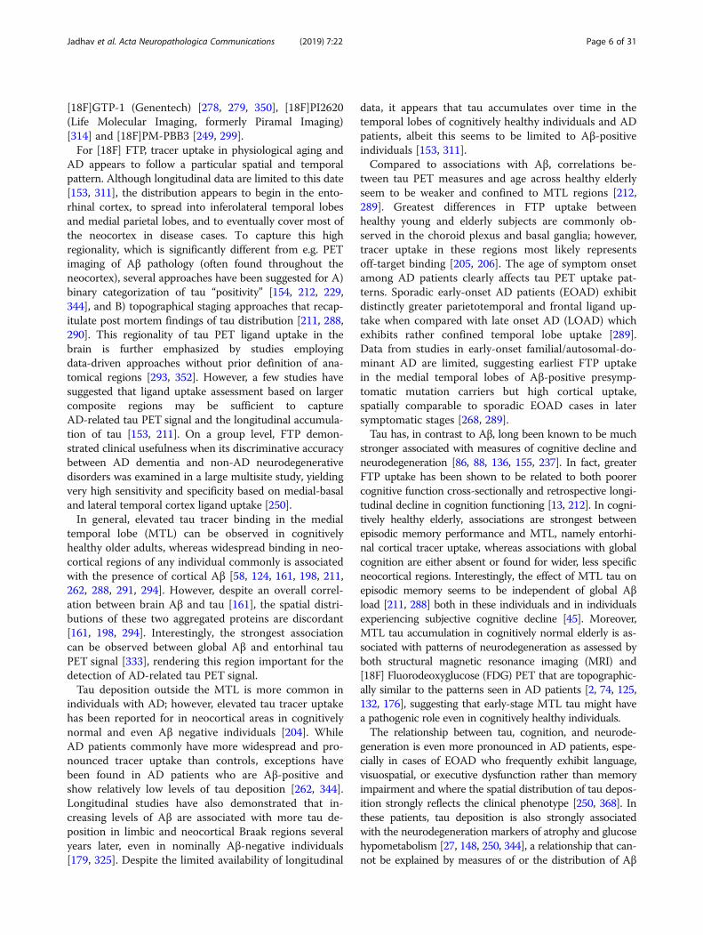
mild to moderate AD dementia demonstrated encour-aging results in both safety and immunogenicity. Twentynine of 30 patients developed an IgG response againstthe tau peptide component of AADvac1 and against re-combinant pathological tau (aa151-391/4R) [381]. Theserum antibodies showed a pronounced preference forpathological truncated tau over healthy full-length tauprotein [245]. Similarly, a 72-week open-label singlearm interventional follow-up trial (FUNDAMANT)displayed a benign safety profile of the vaccine. Nocases of meningoencephalitis or vasogenic oedemawere observed. There was a tendency towards sloweratrophy in MRI and less decline in cognitive assess-ment in patients with high titres [243]. Currently, aphase II clinical trial in AD and a phase I trial innon-fluent primary progressive aphasia are underway(alzforum.org) (Fig. 3).Much less is known about the ACI35 clinical trial.
ACI-35 is a liposome-based vaccine consisting of a syn-thetic peptide to mimic the phospho-epitope of tau at resi-dues pS396/pS404 anchored into a lipid bilayer. A phase 1bmulti-centre double-blind randomized placebo-controlledtrial in 24 patients with mild to moderate Alzheimer’s dis-ease compared low, medium, and high doses of the vaccineto placebo.

Fig. 3 Chemical structures of methylene blue derivatives
Jadhav et al. Acta Neuropathologica Communications (2019) 7:22 Page 14 of 31
Active immunization is long lasting because it inducesimmunological memory. Active vaccines are easy to ad-minister (different routes) and the production iscost-effective. Immunisation generates polyclonal re-sponse; antibodies can recognize multiple epitopes onthe target protein with different affinity and avidity. Onthe other hand, the immune response depends on thehost immune system, there is a variability in the anti-body response across patients [128, 353].
Antisense therapies for tauopathiesDirect targeting of tau gene (MAPT) expression is gain-ing currency as a therapeutic approach with an antisenseoligonucleotide (ASO) therapy already in Phase I clinicaltrials. Several in vivo and cell studies have demonstratedthe benefit of tau reduction in slowing pathological pro-gression and improving functional deficits in tauopathymodels both dependent and independent of ß-amyloidpathology. Tau reduction also results in significant im-provements in seizures associated with AD pathologyand in a model for Dravet syndrome [112].The fibrillar tau pathology in tauopathy brains consists of
abnormally hyperphosphorylated tau protein [169, 360].Normal phosphorylation and dephosphorylation of residueswithin and flanking the microtubule (MT)-binding repeatdomain (MTBR) mediates the dynamic binding and releaseof tau from MTs [303]. Hyperphosphorylation could causeor be the result of aberrant release of tau from MTs, withhyperphosphorylated tau unable to bind to MTs [41]. Theresulting surplus of unbound tau together with localisedconcentrations, could lead to the triggering of pathologicalconformational conversion of tau to a seed-competent form[228] and the initiation of the aggregation cascade thatleads to the fibrillar tau accumulations.The genetics of tau have informed us on the role of
tau defects as directly contributing to neurodegenera-tion. The early dominance of Aß and the amyloid hy-pothesis [292] subsumed tau to a consequence orbystander in the AD pathogenesis cascade. However, itwas clear that the spread and severity of tau pathology
better correlated with clinical progression of AD [40,116, 126]. The identification of mutations in the taugene (MAPT) that cause familial forms of FTLD withtau pathology (FTLD-tau) [147, 313] cemented the primaryrole of defective tau as a neurodegenerative agent. Fromthese genetic studies, the identification of common geneticvariation in MAPT emerged, defining the H1 haplotype,that is a strong risk factor for primary tauopathies withdominant 4R-tau pathology, progressive supranuclear palsy(PSP; OR = 5.46) [19, 139, 260] and corticobasal degener-ation (CBD; OR= 3.7) [139, 147, 171] and, more surpris-ingly, Parkinson’s disease (OR = 0.77) [306].The FTLD-tau mutations in MAPT fall into two broad
classes; missense mutations that chiefly affect residueswithin the MTBR that impair microtubule binding capacityand/or increase fibrillogenicity of tau, and splicing muta-tions in intronic sequences flanking the alternatively splicedexon 10 and in splicing regulatory motifs within exon 10[147]. The latter cause increased inclusion of exon 10 andensuing increased ratio of tau isoforms with four MTBRs(4R-tau) over those containing three MTBRs (3R-tau)[118]. The splicing of MAPT exons 2, 3 and 10 is develop-mentally regulated, and in the healthy adult brain, there areabout equal amounts of 3R- and 4R-tau [117, 170]. Thebasis of the increased risk conferred by the H1 haplotype ofMAPT and its defining common polymorphisms, spanningthe entire gene and beyond, could be the demonstratedallele-specific differences in transcription [233] and of spli-cing of exons 3 and 10 of the MAPT pre-mRNA [50, 233].The result is an overall increase in tau levels, particularlythe more fibrillogenic 4R-tau, leading to the 4R-tau domi-nated pathology seen in PSP and CBD [195]. Furthermore,it was shown that 17q21.31 duplication leads to early-onsetdementia with an AD clinical phenotype [178].
Therapeutic reduction of tauSurplus availability of unbound tau, particularly of themore fibrillogenic mutants or 4R-tau could, with abnor-mal hyperphosphorylation, lead to mislocalisation and ab-errant interaction with other cellular components and

Jadhav et al. Acta Neuropathologica Communications (2019) 7:22 Page 15 of 31
milieux. This leads to conformational conversion of taufrom its highly soluble, intrinsically disordered character-istic to the seed-competent aggregation-prone form [228].This has led to the notion that reduction of total tau (orsurplus 4R-tau) could be therapeutically beneficial. Al-though the recent stable of passive immunotherapy ap-proaches targeting tau could be blockading intercellulartransmission of pathological tau seeds, a plausible mech-anism could also be a reduction of pathological tau medi-ated by microglial or neuronal uptake and clearance ofextracellular tau-antibody complexes [107, 210, 223].Several published pre-clinical studies with cell and animal
models of AD and tauopathies have persuasively demon-strated the possible therapeutic benefit of tau reduction(Table 4). An ASO-based approach already having enteredPhase I of clinical trials [227]. In early work, SantaCruz andcolleagues demonstrated recovery of memory function andreduced neuronal loss after conditional repression of tauexpression in the rTg4510 mouse [282]. Reduction of en-dogenous tau levels in AD mouse models overexpressinghuman amyloid precursor protein (hAPP) with familial ADmutations dose-dependently ameliorated Aß-related learn-ing and memory deficits and protected the mice from earlymortality [152, 275]. The benefit of tau reduction occurredwithout influencing Aß burden suggesting that tau reduc-tion uncouples Aß from downstream pathogenic mecha-nisms [275] including the prevention of Aß-induced defectsin axonal transport [341]. Other mouse studies have alsoshown tau reduction-mediated mitigation of cognitive defi-cits as a consequence of mild repetitive brain injury [57], ortype-1 diabetes [1].With excitotoxicity implicated in AD, and increased
incidence of seizures in AD patients [11], tau reductionalso prevented increased susceptibility of hAPP mice toevoked seizures [275]. This protection extended to sei-zures independent of AD pathology with ASO-mediatedknockdown of endogenous tau in adult non-transgenicmice [81] and in mouse (Kcna1−/−) and Drosophila (kccand eas) models of hyperexcitability [141] as well as amouse model for Dravet syndrome [112].
Antisense therapiesThis is an exciting juncture in the hunt for therapies againstneurodegenerative disorders by directly targeting thosecausative genes. The efficacy and safety of ASO therapy hasbeen demonstrated in clinical trials for nusinersen (Spin-raza®; ClinicalTrials.gov Identifier: NCT02193074) for thetreatment of spinal muscular atrophy (SMA) and eteplirsen(Exondys51®; NCT00844597, NCT01396239/NCT01540409,NCT02255552) to treat Duchenne muscular dystrophy(DMD). More recently, IONIS-HTTRx (RG6042;NCT02519036) was tested for the treatment of Huntington’sdisease (HD) [317]. This specifically targets the mutant, ex-panded huntingtin gene (HTT) mRNA and supresses its
expression. A recent Phase 1/2a clinical trial with intrathecaldelivery of the ASO has had no adverse drug-related inci-dents and showed promising reduction of mutant HTTmRNA levels in CSF [317].ASOs are short, single-stranded oligonucleotides (8-50
nucleotides) that are designed to bind with completespecificity to complementary sense pre-messenger RNA(mRNA) or mature mRNA sequences. Depending on de-sign and binding site, they could mediate degradation ofthe target mRNA or prevent translation and thus attenu-ate protein production. Gene down-regulation by ASOsexploits cellular mechanisms either via RNA interference(RNAi) and the degradation of the target mRNA byRNA-induced silencing complex (RISC), or by recruit-ment RNase H1 to degrade mRNA at the site of theDNA-RNA duplex. Owing to their size and highlycharged nature, ASOs present challenges in terms of cel-lular uptake, stability and susceptibility to degradationby nucleases and, particularly with CNS targeted therap-ies, overcoming the blood-brain barrier (BBB). Thesecan in part be overcome by chemical modifications ofthe DNA or RNA phosphodiester backbone or ribosesugar [190] and the use of the likes of viral vectors, lipo-somes, polyplexes, or cell-penetrating peptides to en-hance delivery [96, 222, 367].Based on the striking success and safety profile of re-
cent ASO-based clinical trials and, and the recentin vivo ASO-based tau reduction work by de Vos andcolleagues [80], a clinical trial of IONIS-MAPTRx
(BIIB080, ISIS 814907), the first ASO targeting tau inmild AD patients, is currently under way [ClinicalTrials.-gov Identifier: NCT03186989]. Via repeated intrathecaldelivery, it appears that this ASO can overcome the BBBin non-human primates with about 75% reduction ofMAPT mRNA in both hippocampus and cortex and nodose-limiting side-effects [227].As shown with nusinersen in SMA and eteplirsen in
DMD, ASOs could also be used to target splice acceptoror donor sites or splicing enhancers or repressors toblock or enhance splicing of alternatively spliced exons[69, 190]. SMA is caused by survival motor neuron 1(SMN1) gene mutation causing loss of SMN1 protein,resulting in loss of motor neuron function [202]. Theintrathecally administered ASO targets the paralogousSMN2 pre-mRNA, promoting inclusion of exon 7 andproduction of active SMN in place of the depletedSMN1 product [307]. DMD is a fatal X-linked recessiveneuromuscular disorder characterised by progressivemuscle weakening and wasting caused by disruptive mu-tations throughout the large (79 exon) DMD gene [203].ASO approaches for DMD, including eteplirsen, are de-signed to induce exon skipping, thereby excluding dis-pensable downstream exons and avoiding exons withdisruptive loss-of-function frame-shift or splice site

Table 4 Studies on cell and animal models demonstrating therapeutic benefit of tau reduction
MODEL BENEFITS REFERENCES
Tet-repression of Tg-tau expression inrTg4510 mice
Reduced neuronal loss and improved memory function [282]
hAPP tau−/− crosses Blocks Aß and excitotoxin mediated neuronal dysfunction [275]
hAPP (APP23) Dtau or tau−/− crosses Prevention of Aß-mediated memory deficits and improved survival [152]
CSF delivered ASOs Reduces evoked seizures in adult nTg mice [81]
tau−/− Kcna−/− crosses Reduced network hyperexcitability in mouse and Drosophila epilepsy models [141]
Crossing tau−/− mice with nTg mice Reduces learning and memory deficits due to mild repetitive traumatic brain injury inmice
[57]
Streptozotocin-treated tau−/− and nTg mice Mitigates cognitive deficits in type-1 diabetes mouse model [1]
tau−/− Scn1a −/− R1407X loss-of-functiontruncation mice
Prevents seizure and improves survival in Dravet syndrome mouse model [112]
shRNA knockdown of Mapt in nTg mouseprimary neurons
Prevents Aß-induced axonal transport deficits [341]
ASO knockdown of Tg-tau overexpression inPS19 mice
Reduced tau pathology, reversal of existing tau pathology. Prevention of neuronal loss.Improved behavioural deficits
[82]
Inducible tau knockdown in APP/PS1 xrTg4510 mice
Prevents tau pathology and neuronal death in presence of Aß pathology [80]
Abbreviations: Tg transgenic, nTg non-transgenic (wild-type), Tet tetracycline, hAPP human amyloid precursor protein, shRNA short hairpin RNA
Jadhav et al. Acta Neuropathologica Communications (2019) 7:22 Page 16 of 31
mutations, while still producing an internally truncated,partially functional protein [190].Noting the pathogenic role of increased availability of
4R-tau due to exon 10 mutations in FTLD-tau and theMAPT H1 haplotype in PSP and CBD, rebalancing exon 10is also being tested [276, 287]. This includes ASO-basedtargeting of exon 10 splice motifs leading to exon-skippingand reduced 4R-tau [287], or reprogramming using aspliceosome-mediated trans-splicing (SMaRT) techniquethat acts by creating a hybrid mRNA through a trans-spli-cing reaction between the MAPT pre-mRNA and a pre--trans-splicing molecule, comprised of a binding domainthat hybridises with the 3′ end of intron 9 and exons 11–13, designed to exclude exon 10 [276].
MAPT-AS1 natural antisense transcript as a physiologicalrepressor of tau expressionIn addition to ASOs, we have seen recent upsurge in ourunderstanding of natural antisense transcripts (NATs).These are endogenous RNA molecules formed by anti-sense transcription at coding genes and playmulti-layered role(s) in regulation of expression of theirpaired coding gene [347]. The MAPT-AS1 longnon-coding RNA (lncRNA) gene partially overlapshead-to-head with the promoter and 5′ untranslated re-gion (5’-UTR) of MAPT and by alternative splicing anduse of alternate exons and splice sites, expresses multipleNATs (tau-NATs) [305]. Both in vitro and in vivo, someof the tau-NATs potently repress tau translation [305].This presents a novel, physiological repressor of tau pro-tein acting in the cytoplasm that, unlike synthetic ASOs,does not rely on RISC or RNAseH and is amenable toadeno-associated virus (AAV) vector-based delivery.
Several clinical trials using AAV vectors, including intra-cranial delivery, have been shown to be safe [137]. Wide-spread CNS distribution and persistence for up to 10years and no adverse effects [188] could imply treatmentlimited to a single delivery unlike ASOs where in on-going clinical trials, involve repeat intrathecal injectionof large doses, every few weeks over several months.
Consequences of tau reductionGiven the importance of tau in multiple facets of neur-onal function, mainly by its role in axonal MT assemblyand stabilisation and mediation of axonal transport, defi-cits in tau could have undesirable consequences. Micethat completely lack tau have normal learning and mem-ory and cognition [191, 230, 275], with a minor, variablemotor phenotype in later life [186, 191, 230, 330]. Onthe other hand, it is important to note, that tau deletionwas shown to be associated with brain iron accumula-tion, brain insulin resistance and deficits in synapticplasticity and cognition [6, 185, 218]. However, observa-tions in knockout models could be hampered by devel-opmental compensation by other MT-associatedproteins such as MAP1B [134, 318] and it is thus crucialto understand the consequences of tau knockdown,post-development, in the adult brain. In one recentstudy, bilateral hippocampal shRNA mediated knock-down of tau in adult mice caused significantly impairedmotor coordination and spatial memory accompanied byreduced synaptic markers and dendritic spine density.Behavioural deficits were restored once tau repressionwas removed [332]. However, in other studies, general-ised knockdown of CNS tau in adult mice caused no de-viations in normal sensory, motor or cognitive tasks

Jadhav et al. Acta Neuropathologica Communications (2019) 7:22 Page 17 of 31
[82]. Based on these mixed findings, it would be import-ant to ascertain the tolerability of different levels of tauknockdown – it is perceivable that partial knockdown oftau in the adult brain could be beneficial, and yet mini-mising any undesirable effects.
Anti-aggregation agentsHyperphosphorylated and truncated tau protein is sus-ceptible to aggregation and loss of cytoskeletalmicrotubule-stabilizing properties, leading to neuronaldamage and cell death. Compounds able to prevent ag-gregation may represent a promising strategy for effect-ive treatment of Alzheimer’s disease [162, 356]. Twomajor approaches focus on phosphorylation of tau andprevention of tau oligomerization. The former involvesthe search for inhibitors of kinases which phosphorylatetau or phosphatase activators which dephosphorylate theprotein [5, 189]. The latter seeks direct inhibitors of thetau aggregation process.
Regulation of tau phosphorylationPhosphorylation of tau is under tight control of variousprotein kinases and phosphatases [5, 189]. Among them,glycogen synthase kinase 3β (GSK-3β) and phosphatase2A (PP2A) are two key enzymes involved in regulation ofthe phosphorylation state of tau. GSK-3β is a multitaskingserine/threonine kinase largely expressed in CNS thatphosphorylates tau mainly at the Ser199, Ser396 andSer413 sites [16]. Furthermore, it has been shown that anincrease in GSK-3β activity induces Αβ formation and isalso implicated in other processes, including neuroinflam-mation and apoptosis [51]. Therefore, GSK-3β is validatedas a therapeutic target for AD, and several chemical clas-ses of GSK-3β inhibitors have been discovered and devel-oped in preclinical [217, 253] or even clinical trials.Tideglusib (NP031112, NP-12), is an ATP non-competitiveGSK-3β inhibitor demonstrated to reduce spatial memorydeficits in transgenic mice in preclinical studies [76]. Whileit has reached clinical trials, no satisfactory therapeutic re-sults were obtained during phase II.Tau phosphorylation is also regulated by
O-GlcNAcylation, a non-canonical glycosylation involvingthe attachment of single O-linked N-acetylglucosamine(O-GlcNAc) moieties to serine and threonine residues[376]. O-GlcNAcylation is regulated by two enzymes,O-GlcNAc transferase catalyzing the transfer of GlcNAc toproteins, and N-acetylglucosaminidase (OGA) catalyzingthe removal of GlcNAc from proteins [377]. Thiamet-G – apotent OGA inhibitor, that can influence O-GlcNAc levelsin the brain, reduced tau phosphorylation in the brain afterintraventricular administration [377]. This finding was suc-cessfully replicated in additional study, where Thiamet-Gprevented the progression of hyperactivity, slowed brain at-rophy, and reduced brain hyperphosphorylated tau in tau
transgenic model TG4510 [345]. Similarly, ASN120290 – asmall molecule that inhibits O-GlcNAcase reduced tauphosphorylation and the number of neurofibrillary path-ology in the brain of transgenic mice P301L. ASN120290which received Orphan Drug Designation from the Foodand Drug Administration (FDA) for PSP, has already initi-ated Phase I clinical trials.
Inhibition of tau aggregationThe most common direct inhibitor of tau protein aggre-gation is methylene blue (MB), which belongs to theclass of thiazine dyes. Methylene blue, also known asmethylthionine chloride (MTC), was originally synthe-sized at the end of the nineteenth century and used totreat malaria. It later found use as an antibacterial, anti-viral and anticancer agent, applied in the treatment ofvarious disorders. It is worth noting that its structurehas played an important role in the development ofphenothiazine-like compounds, including antipsychoticand antihistamine drugs. The anti-aggregating effect ofphenothiazines upon the tau protein, discovered byWischik and co-workers [355] over twenty years ago,paved the way for a new class of for potential anti-ADagents. However, during this time, most researchers fo-cused on β-amyloid targets, and over the next two de-cades few achievements concerning tau were reported.Nonetheless, several chemical classes of tau aggregationinhibitors have been synthesized and presented [47, 48].The tau aggregation inhibitor, methylene blue, occurs
in two main forms, which are in equilibrium, dependingon the redox potential of the solution. The first oxidizedcationic state is characterized by a dark blue color, whilethe second (reduced form) is colorless and also referredto as leucomethylene blue (leuco-methylthioninum,LMT). Structurally, MTC is an aromatic anthracenecompound (polyaromatic), whereas LMT is classified asa phenothiazine. It has been shown that anthracene-typecompounds inhibit the tau protein, while phenothia-zines, with nonaromatic tricyclic structures are inactivein this respect. MTC acts as a prodrug, and in acidic pHconverts into leuco-methylthioninium, which can pene-trate the BBB and reach brain tissues [17]. Many studieshave demonstrated that MTC has a broad spectrum ofpharmacological activity [251, 319]. The inhibition of tauaggregation by MTC has been confirmed by numerousin vitro tests as well as in in vivo models in transgenicmice. The dye has properties which inhibit microtubuleassembly, prevent tau interaction, inhibit β-amyloid ag-gregation as well as α-synuclein aggregation. MTC coun-teracts mitochondrial damage caused by oxidative stress;it also has a positive effect on regulation of autophagy,acetylcholine E (AchE) inhibition, monoamine oxidases,the glutamatergic system and inhibition of noradrenalineuptake. From the point of view of potential clinical

Jadhav et al. Acta Neuropathologica Communications (2019) 7:22 Page 18 of 31
applications, the most important properties of MTC in-clude: inhibition of microtubule formation, improvementof mitochondrial oxidation and inhibition of monoamineoxidase A [239].In clinical trials, MTC was introduced under the
name Rember™ (TauRx Therapeutics) as a potentialanti-AD drug candidate. Some improvements inAD-related symptoms have been reported, but thedrug failed phase II trials due to undesirable side ef-fects, including diarrhoea, urgency, painful urination,dizziness and others (Clinical Trial Identifier,NCT00515333 and NCT00684944). Results of thesestudies prompted researchers to develop a new gener-ation of MTC derivatives. These new compounds(LMTX) include leuco-methylthionium bis (hydro--methanesulfonate (LMTM) and leuco-methylthioniumdihydrobromide (LMTB) – stable, reduced forms thatpermit direct absorption of LMT without the needfor the aforementioned conversion step (Fig. 3).LMTM (TRx0237) has reached phase III trials, and
was better absorbed, with improved safety and tolerabil-ity compared to methylene blue (Rember™). Neverthe-less, results of Phase III clinical trials involving LMTMin the treatment of AD were disappointing as they didnot yield unambiguously positive data. The first phaseIII trial (NCT01689246) included 891 participants withmild to moderate AD, who received 125 mg of LMTMtwice a day, or 75 mg twice a day while the controlgroup received 4 mg twice a day. No significant differ-ence in cognitive faculties or the ability to perform dailyactivities was observed between the treatment and con-trol groups [110]. Due to the low number of participants(79) in this study, these results require further confirm-ation. Currently, TauRx has begun a new clinical trial(LUCIDUTY, NCT03446001) using FDG-PET imagingto examine the potential of LMTX in delaying the pro-gression of pathological changes in the brain in AD pa-tients who do not receive cholinesterase inhibitors ormemantine. This trial is aimed at patients with earlyAD, with treatment lasting for 9 months (at doses of8 mg/day and 16 mg/day). Thus, LMTM is being de-veloped as an anti-AD treatment option based on in-hibition of tau aggregation. Moreover, LMTC hasdemonstrated amelioration of α-synuclein pathologyin a transgenic mouse model of synucleinopathy, and
Fig. 4 Multifunctional derivatives of piperazine
may therefore find use as a potential disease modifi-cation therapy in Parkinson’s disease (PD) and othersynucleinopathies [290].Since the discovery of the tau aggregation inhibitory ac-
tivity of methylene blue, several chemical classes of com-pounds have been identified. These include derivatives ofphenothiazines, polyphenols, benzothiazoles and porphy-rins [319]. It has been observed that all these tested deriva-tives inhibited both tau filament formation and Aβ fibrilformation. Further research carried out by Bulic and E.Mandelkow [47, 48], based on screening of a random li-brary of 200,000 compounds, led to the identification ofnew chemical structures for potential tau inhibitors, includ-ing rhodamines, phenylthiazolyl-hydrazides, N-phenyla-mines, anthraquinones, benzothiazoles. Using quantitativehigh-throughput screening, Crowe and co-workers [70] dis-covered that aminothienopyrydazines (AZPZs) also inhibitof tau assembly.Another potential source of anti-aggregation agents is
provided by the multi-target-directed ligand approach.This strategy is suitable for complex diseases such asAlzheimer’s disease [18, 83, 264]. Therefore, many multi-functional compounds have been obtained by combiningvarious pharmacophores targeting neurodegenerativeprocesses into a single molecule. Among them multi-modal molecules have been discovered that are endowedwith tau aggregation inhibitory activity as well as otherdesirable properties. Selected examples of multifunc-tional agents are presented below.Compound AZP2006, an N,N′-disubstituted pipera-
zine [226, 297], reduces the release of Aβ species andtargets both amyloid and tau pathologies. It was demon-strated to improve cognitive faculties in various mousemodels of both amyloid and tau pathology [21].AZP2006 underwent phase I clinical trials on AD, andhas now been classed as an orphan drug for the treat-ment of progressive supranuclear palsy (PSP). Anothernew compound, named RPEL, is a piperazine derivativethat contains the pharmacophore fragment of tacrine[226] (Fig. 4). This dual-action compound showed in-hibitory potency against cholinesterase (IC50 hAChE =0.8 nM), reduced the phosphorylation of tau protein andinhibited the release of the Aβ peptide. Moreover, it dis-played in vivo potency in transgenic mouse models andreduced memory loss.

Fig. 6 Structure of multifunctional carbazole–basedcyanine compounds
Jadhav et al. Acta Neuropathologica Communications (2019) 7:22 Page 19 of 31
Japanese researchers [246, 247] presented a new tauinhibitor compound, PE859, based on the curcuminstructure (Fig. 5). Promising results were obtained inboth in vitro and in vivo studies – the compound wasshown to counteract tau aggregation and prevent theonset and progression of nerve dysfunction in an in vivomodel. Furthermore, it inhibits both tau and Aβ aggre-gation and alleviates cognitive dysfunction in vivo.Two carbazole-based cyanine compounds named SLM
and SLOH were described as strong inhibitors of Aβ aggre-gation in vitro and were able to alleviate the pathologicalsymptoms and memory deterioration in AD model mice[364–366] (Fig. 6). These multifunctional compounds alsoreduced tau hyperphosphorylation as well as significantlyattenuated neuroinflammation through inhibitiion ofGSK-3β activity. They showed a good pharmacokinetic pro-file, with high BBB permeability, which justifies their furtherdevelopment as AD drug candidates [379].Dual inhibitors acting against β-secretase (BACE1) and
glycogen synthase kinase 3β (GSK-3β), with well-balancedin vitro activity (in μM range), were synthesized in theclass of triazinone derivatives [265]. These compoundsdisplayed strong neuroprotective and neurogenic effects,and also showed good BBB permeability in a pharmacoki-netic evaluation in mice. A new multi-target strategy fordesigning anti-AD agents involves compounds whichcombine GSK-3β and tau aggregation inhibitors [109]. De-rivatives of 2,4-thiazolidinedione showed activity againstGSK-3β (at micromolar IC50 values) and were also foundto inhibit tau aggregation. Other examples of multifunc-tional compounds include rhein-huprine hybrids, whichshowed AChE and BACE1 inhibitory activity, as well asAβ1-42 and tau anti-aggregating properties [259]. A1-benzylamino-2-hydroxyalkyl derivative with a diphenyl-piperazine fragment, selected form a series of compounds,showed balanced inhibitory activity against bothdisease-modifying targets, inhibition of BACE1, inhibitionof Aβ, inhibition of tau aggregation, as well as inhibitionof BuChE as a symptomatic target [254]. Jiang andco-workers [158] described a new class of dual GSK-3βand AChE inhibitors. These multifunctional compoundswere designed by incorporating a tacrine fragment at thethiazolyl ring, as the pharmacophore responsible for
Fig. 5 Structure of curcumin derivative PE859 dual tau andβ-amyloid inhibitor
GSK-3β inhibition. The resulting derivatives were very po-tent inhibitors of both targets (in the nanomolar range).The most promising compound from this series signifi-cantly inhibited tau protein phosphorylation and counter-acted self-aggregation of Aβ1-42. In addition, it was nottoxic and proved effective in an in vivo assay in mice, bysignificantly improving memory.Most of the above-described direct tau inhibitors and
multifunctional compounds have shown activity inin vitro tests, but only some of them have been evalu-ated in vivo in extended pharmacological, preclinicalstudies. Moreover, it is difficult to predict further devel-opment of these compounds. Due to the complex natureof AD, it seems reasonable to pursue development ofcombination therapies, as well as new alternative ap-proaches which involve multi-target drugs. It is probablethat a molecule capable of acting on two recognized tar-gets, with one of them belonging to the tau cascade,might bring clinical benefits compared to drugs whichonly address only specific target.
Concluding remarks and future directionsTau is a multifaceted protein with a plethora of physio-logical functions. In the disease condition, tau proteindrives neurodegeneration and causes neurodegenerativedisorders such as Alzheimer’s disease. Pathologicallymodified tau has become an important therapeutic targetfor AD and related tauopathies. Although nodisease-modifying treatments are yet available, many newtherapeutic approaches targeting pathological forms of tauare being tested in clinical trials. Disease-modifying therapyis aimed at preventing, slowing or ameliorating the produc-tion, oligomerisation, aggregation and deposition of patho-logical tau protein. The most promising therapeuticstrategies include active tau vaccines and therapeuticmonoclonal antibodies. Besides immunotherapy, there aremany other therapies currently being explored in the treat-ment of tau neurodegeneration such as modulation of tauphosphorylation, inhibition of tau aggregation or regulationof its expression. While waiting for the results of ongoingclinical trials, we can continue to unravel the complexitiesof tau proteome and different biological functions of thispeculiar brain protein.

Jadhav et al. Acta Neuropathologica Communications (2019) 7:22 Page 20 of 31
Abbreviations1 N: First insert; 2 N: Second insert; 3R: Three repeat; 4R: Four repeat; Aβ: β-amyloid; aa: Amino acids; AAV: Adeno-associated virus; AChE: AcetylcholineE; AD: Alzheimer’s disease; AGD: Argyrophilic grain disease;ApoE4: Apolipoprotein E4; APP: Amyloid precursor protein; ARTAG: Age-related tau astrogliopathy; ASO: Antisense oligonucleotide; BBB: Blood-brainbarrier; BIN1: Myc box-dependent-interacting protein 1; CDB: Corticobasaldegeneration; CDRs: Complementarity determining regions; CNS: Centralnervous system; CSF: Cerebrospinal fluid; C-terminal: Carboxy-terminal;DMD: Duchenne muscular dystrophy; EOAD: Early-onset Alzheimer’s disease;FDA: Food and Drug Administration; FDG: Fluorodeoxyglucose; FGF-2: Fibroblast growth factor 2; FTD: Frontotemporal dementia;FTLD: Frontotemporal lobar degeneration; FTP: [18F] Flortaucipir;GGT: Globular glial tauopathy; GSK-3β: Glycogen synthase kinase 3β;HD: Huntington’s disease; HMW: High molecular weight tau; HTT: Huntingtingene; IRS-1: Insulin receptor substrate 1; Ka: Association constant;Kd: Dissociation constant; KLH: Keyhole limpet haemocyanin; LMT: Leuco-methylthioninum; LMTB: Leuco-methylthionium dihydrobromide;LMTM: Leuco-methylthionium bis(hydro-methanesulfonate; lncRNA: Longnon-coding RNA; LOAD: Late- onset Alzheimer’s disease; MAO-B: Monoaminoxidase B; MAP: Microtubule-associated protein; MB: Methyleneblue; MIR: Mammalian interspersed repeat; MRI: Magnetic resonance imaging;mRNA: Messenger RNA; MT: Microtubules; MTBR: Microtubule binding region;MTC: Methylthionine chloride; MTL: Medial temporal lobe; NATs: Naturalantisense transcripts; NFT: Neurofibrillary tangles; NPC: Nuclear pore complex;N-terminal: Amino-terminal; O-GlcNAc: O-linked N-acetylglucosamine;PART: Primary age-related tauopathy; PET: Positron emission tomography;PHF: Paired-helical filaments; PiD: Pick’s disease; PP2A: Phosphatase 2A;PSP: Progressive supranuclear palsy; PTEN: Phosphatase and tensin homolog;PTM: Post translational modification; RISC: RNA-induced silencing complex;RNAi: RNA interference; SMA: Spinal muscular atrophy; SMaRT: Spliceosome-mediated trans-splicing; SMN: Survival motor neuron; SPR: Surface plasmonresonance spectroscopy; SPs: Senile plaques; TRIM21: Tripartite motif-containing protein 21
AcknowledgementsWe would like to especially acknowledge the pioneering work of professorsHeiko Braak and Constantin Bouras. Without their outstanding contributionto the field, the current progress in the development of tau therapy wouldnot have been possible. The Eurotau2018 meeting was sponsored by UCBBioPharma, Janssen Pharmaceutical Companies, Sanofi, Axon Neuroscience,Servier, Quanterix, Proteome Sciences and Proteintech. It was also supportedby French and Spanish academic funds from University of Lille, SFR DN2M,LabEx DISTALZ, LiCEND, and Ciberned.
Authors’ contributionsAll authors participated in writing, read and approved the final manuscript.
Competing interestsSJ, RS, EK and NZ are Axon Neuroscience R&D Services employees. LBreceived research grants from UCB Biopharma. Other authors declare thatthey have no competing interests.
Publisher’s NoteSpringer Nature remains neutral with regard to jurisdictional claims inpublished maps and institutional affiliations.
Author details1Institute of Neuroimmunology, Slovak Academy of Sciences, Dubravska 9,845 10 Bratislava, Slovakia. 2Centro de Biologia Molecular “Severo Ochoa”,Consejo Superior de Investigaciones, Cientificas, Universidad Autonoma deMadrid, C/ Nicolas Cabrera, 1. Campus de Cantoblanco, 28049 Madrid, Spain.3Networking Research Center on Neurodegenerative, Diseases (CIBERNED),Instituto de Salud Carlos III, Madrid, Spain. 4Wallenberg Centre for Molecularand Translational Medicine, University of Gothenburg, Gothenburg, Sweden.5Department of, Psychiatry and Neurochemistry, University of Gothenburg,Gothenburg, Sweden. 6Clinical Memory Research Unit, Department of ClinicalSciences, Lund University, Malmö, Sweden. 7Dementia Research Centre,University College London, London, UK. 8Institute of Neurology, MedicalUniversity of Vienna, AKH 4J, Währinger Gürtel 18-20, 1097 Vienna, Austria.9Department of Mental Health and Psychiatry, University Hospitals of Geneva,
Geneva, Switzerland. 10AXON Neuroscience R&D Services SE, Dvorakovonabrezie 10, 811 02 Bratislava, Slovakia. 11Gurdon Institute and Departmentof Biochemistry, University of Cambridge, Cambridge CB2 1QN, UK.12Department of Physicochemical Drug Analysis, Faculty of Pharmacy,Jagiellonian University Medical College, Medyczna 9, 30-688 Cracow, Poland.13Reta Lila Weston Institute and Department of Clinical and MovementNeurosciences, UCL Queen Square Institute of Neurology, 1 Wakefield Street,London WC1N 1PJ, UK. 14Universite of Lille, Inserm, CHU-Lille, UMRS1172,Alzheimer & Tauopathies, Place de Verdun, 59045 Lille cedex, France.
Received: 30 November 2018 Accepted: 21 January 2019
References1. Abbondante S, Baglietto-Vargas D, Rodriguez-Ortiz CJ, Estrada-
Hernandez T, Medeiros R, Laferla FM (2014) Genetic ablation of taumitigates cognitive impairment induced by type 1 diabetes. Am JPathol 184:819–826. https://doi.org/10.1016/j.ajpath.2013.11.021
2. Adams JN, Lockhart SN, Li L, Jagust WJ (2018) Relationships between tauand glucose metabolism reflect Alzheimer’s disease pathology in cognitivelyNormal older adults. Cereb Cortex. https://doi.org/10.1093/cercor/bhy078
3. Agadjanyan MG, Petrovsky N, Ghochikyan A (2015) A fresh perspective fromimmunologists and vaccine researchers: active vaccination strategies toprevent and reverse Alzheimer's disease. Alzheimers Dement 11:1246–1259.https://doi.org/10.1016/j.jalz.2015.06.1884
4. Agadjanyan MG, Zagorski K, Petrushina I, Davtyan H, Kazarian K, AntonenkoM, Davis J, Bon C, Blurton-Jones M, Cribbs DH, Ghochikyan A (2017)Humanized monoclonal antibody armanezumab specific to N-terminus ofpathological tau: characterization and therapeutic potency. MolNeurodegener 12:33. https://doi.org/10.1186/s13024-017-0172-1
5. Ahmed T, Blum D, Burnouf S, Demeyer D, Buee-Scherrer V, D'Hooge R, BueeL, Balschun D (2015) Rescue of impaired late-phase long-term depression ina tau transgenic mouse model. Neurobiol Aging 36:730–739. https://doi.org/10.1016/j.neurobiolaging.2014.09.015
6. Ahmed T, Van der Jeugd A, Blum D, Galas MC, D'Hooge R, Buee L,Balschun D (2014) Cognition and hippocampal synaptic plasticity inmice with a homozygous tau deletion. Neurobiol Aging 35(11):2474-2478. https://doi.org/10.1016/j.neurobiolaging.2014.05.005.
7. Ahmed Z, Bigio EH, Budka H, Dickson DW, Ferrer I, Ghetti B, GiacconeG, Hatanpaa KJ, Holton JL, Josephs KA, Powers J, Spina S, Takahashi H,White CL 3rd, Revesz T, Kovacs GG (2013) Globular glial tauopathies(GGT): consensus recommendations. Acta Neuropathol 126:537–544.https://doi.org/10.1007/s00401-013-1171-0
8. Albert M, Mairet-Coello G, Danis C, Lieger S, Caillierez R, Carrier S, Skrobala E,Landrieu I, Michel A, Schmitt M, Citron M, Downey P, Courade JP, Buée L, ColinM (2018) Prevention of tau seeding and propagation by immunotherapy witha central tau epitope antibody. AAIC2018, Chicago, IL, USA.
9. Amadoro G, Corsetti V, Florenzano F, Atlante A, Ciotti MT, Mongiardi MP,Bussani R, Nicolin V, Nori SL, Campanella M, Calissano P (2014) AD-linked,toxic NH2 human tau affects the quality control of mitochondria inneurons. Neurobiol Dis 62:489–507. https://doi.org/10.1016/j.nbd.2013.10.018
10. Amadoro G, Corsetti V, Stringaro A, Colone M, D'Aguanno S, Meli G, CiottiM, Sancesario G, Cattaneo A, Bussani R, Mercanti D, Calissano P (2010) ANH2 tau fragment targets neuronal mitochondria at AD synapses: possibleimplications for neurodegeneration. J Alzheimers Dis 21:445–470. https://doi.org/10.3233/jad-2010-100120
11. Amatniek JC, Hauser WA, DelCastillo-Castaneda C, Jacobs DM, Marder K, BellK, Albert M, Brandt J, Stern Y (2006) Incidence and predictors of seizures inpatients with Alzheimer's disease. Epilepsia 47:867–872. https://doi.org/10.1111/j.1528-1167.2006.00554.x
12. Ando K, Kabova A, Stygelbout V, Leroy K, Heraud C, Frederick C, SuainV, Yilmaz Z, Authelet M, Dedecker R, Potier MC, Duyckaerts C, Brion JP(2014) Vaccination with Sarkosyl insoluble PHF-tau decreaseneurofibrillary tangles formation in aged tau transgenic mouse model: apilot study. J Alzheimers Dis 40(Suppl 1):S135–S145. https://doi.org/10.3233/jad-132237
13. Aschenbrenner AJ, Gordon BA, Benzinger TLS, Morris JC, Hassenstab JJ(2018) Influence of tau PET, amyloid PET, and hippocampal volume oncognition in Alzheimer disease. Neurology 91:e859–e866. https://doi.org/10.1212/wnl.0000000000006075

Jadhav et al. Acta Neuropathologica Communications (2019) 7:22 Page 21 of 31
14. Asuni AA, Boutajangout A, Quartermain D, Sigurdsson EM (2007)Immunotherapy targeting pathological tau conformers in a tangle mousemodel reduces brain pathology with associated functional improvements. JNeurosci 27:9115–9129. https://doi.org/10.1523/jneurosci.2361-07.2007
15. Augustinack JC, Schneider A, Mandelkow EM, Hyman BT (2002) Specific tauphosphorylation sites correlate with severity of neuronal cytopathology inAlzheimer's disease. Acta Neuropathol 103:26–35
16. Avila J, Wandosell F, Hernandez F (2010) Role of glycogen synthase kinase-3 inAlzheimer's disease pathogenesis and glycogen synthase kinase-3 inhibitors.Expert Rev Neurother 10:703–710. https://doi.org/10.1586/ern.10.40
17. Baddeley TC, McCaffrey J, Storey JM, Cheung JK, Melis V, Horsley D, HarringtonCR, Wischik CM (2015) Complex disposition of methylthioninium redox formsdetermines efficacy in tau aggregation inhibitor therapy for Alzheimer's disease. JPharmacol Exp Ther 352:110–118. https://doi.org/10.1124/jpet.114.219352
18. Bajda M, Guzior N, Ignasik M, Malawska B (2011) Multi-target-directedligands in Alzheimer's disease treatment. Curr Med Chem 18:4949–4975
19. Baker M, Litvan I, Houlden H, Adamson J, Dickson D, Perez-Tur J, Hardy J,Lynch T, Bigio E, Hutton M (1999) Association of an extended haplotype in thetau gene with progressive supranuclear palsy. Hum Mol Genet 8:711–715
20. Baker SL, Maass A, Jagust WJ (2017) Considerations and code for partialvolume correcting [(18)F]-AV-1451 tau PET data. Data Brief 15:648–657.https://doi.org/10.1016/j.dib.2017.10.024
21. Barrier MBL, Burlet S, Delacourte A, Estrella C, Melnyk P, Sergeant N,Verwaerde P (2014) Sulfate salts of N-(3-(4-(3-(diisobutylamino)propyl)piperazin-1-yl)propyl)-1H-benzo [d]imidazol-2amine, preparation thereof and use of the same
22. Batista FD, Neuberger MS (1998) Affinity dependence of the B cell response toantigen: a threshold, a ceiling, and the importance of off-rate. Immunity 8:751–759
23. Bejanin A, Schonhaut DR, La Joie R, Kramer JH, Baker SL, Sosa N, Ayakta N,Cantwell A, Janabi M, Lauriola M, O'Neil JP, Gorno-Tempini ML, Miller ZA,Rosen HJ, Miller BL, Jagust WJ, Rabinovici GD (2017) Tau pathology andneurodegeneration contribute to cognitive impairment in Alzheimer'sdisease. Brain 140:3286–3300. https://doi.org/10.1093/brain/awx243
24. Betthauser TJ, Cody KA, Zammit MD, Murali D, Converse AK, Barnhart TE,Stone CK, Rowley HA, Johnson SC, Christian BT (2018) In vivocharacterization and quantification of neurofibrillary tau PET radioligand[(18)F]MK-6240 in humans from Alzheimer's disease dementia to youngcontrols. J Nucl Med. https://doi.org/10.2967/jnumed.118.209650
25. Bi M, Ittner A, Ke YD, Gotz J, Ittner LM (2011) Tau-targeted immunization impedesprogression of neurofibrillary histopathology in aged P301L tau transgenic mice.PLoS One 6:e26860. https://doi.org/10.1371/journal.pone.0026860
26. Bischof GN, Endepols H, van Eimeren T, Drzezga A (2017) Tau-imaging inneurodegeneration. Methods 130:114–123. https://doi.org/10.1016/j.ymeth.2017.08.003
27. Bischof GN, Jessen F, Fliessbach K, Dronse J, Hammes J, Neumaier B, OnurO, Fink GR, Kukolja J, Drzezga A, van Eimeren T (2016) Impact of tau andamyloid burden on glucose metabolism in Alzheimer's disease. Ann ClinTransl Neurol 3:934–939. https://doi.org/10.1002/acn3.339
28. Blennow K, Wallin A, Agren H, Spenger C, Siegfried J, Vanmechelen E (1995)Tau protein in cerebrospinal fluid: a biochemical marker for axonaldegeneration in Alzheimer disease? Mol Chem Neuropathol 26:231–245.https://doi.org/10.1007/bf02815140
29. Blennow K, Zetterberg H (2018) Biomarkers for Alzheimer's disease: current statusand prospects for the future. J Intern Med. https://doi.org/10.1111/joim.12816
30. Boimel M, Grigoriadis N, Lourbopoulos A, Haber E, Abramsky O, RosenmannH (2010) Efficacy and safety of immunization with phosphorylated tauagainst neurofibrillary tangles in mice. Exp Neurol 224:472–485. https://doi.org/10.1016/j.expneurol.2010.05.010
31. Boluda S, Iba M, Zhang B, Raible KM, Lee VM, Trojanowski JQ (2015)Differential induction and spread of tau pathology in young PS19 tautransgenic mice following intracerebral injections of pathological tau fromAlzheimer's disease or corticobasal degeneration brains. Acta Neuropathol129:221–237. https://doi.org/10.1007/s00401-014-1373-0
32. Bondareff W, Wischik CM, Novak M, Amos WB, Klug A, Roth M (1990)Molecular analysis of neurofibrillary degeneration in Alzheimer's disease. Animmunohistochemical study. Am J Pathol 137:711–723
33. Borghgraef P, Menuet C, Theunis C, Louis JV, Devijver H, Maurin H, Smet-Nocca C,Lippens G, Hilaire G, Gijsen H, Moechars D, Van Leuven F (2013) Increasing brainprotein O-GlcNAc-ylation mitigates breathing defects and mortality of tau.P301Lmice. PLoS One 8:e84442. https://doi.org/10.1371/journal.pone.0084442
34. Borreca A, Latina V, Corsetti V, Middei S, Piccinin S, Della Valle F, Bussani R,Ammassari-Teule M, Nistico R, Calissano P, Amadoro G (2018) AD-related N-
terminal truncated tau is sufficient to recapitulate in vivo the earlyperturbations of human neuropathology: implications for immunotherapy.Mol Neurobiol 55:8124–8153. https://doi.org/10.1007/s12035-018-0974-3
35. Bou Samra E, Buhagiar-Labarchede G, Machon C, Guitton J, Onclercq-Delic R,Green MR, Alibert O, Gazin C, Veaute X, Amor-Gueret M (2017) A role for tauprotein in maintaining ribosomal DNA stability and cytidine deaminase-deficientcell survival. Nat Commun 8:693. https://doi.org/10.1038/s41467-017-00633-1
36. Boutajangout A, Ingadottir J, Davies P, Sigurdsson EM (2011) Passiveimmunization targeting pathological phospho-tau protein in a mousemodel reduces functional decline and clears tau aggregates from the brain.J Neurochem 118:658–667. https://doi.org/10.1111/j.1471-4159.2011.07337.x
37. Boutajangout A, Quartermain D, Sigurdsson EM (2010) Immunotherapy targetingpathological tau prevents cognitive decline in a new tangle mouse model. JNeurosci 30:16559–16566. https://doi.org/10.1523/jneurosci.4363-10.2010
38. Braak H, Alafuzoff I, Arzberger T, Kretzschmar H, Del Tredici K (2006) Stagingof Alzheimer disease-associated neurofibrillary pathology using paraffinsections and immunocytochemistry. Acta Neuropathol 112:389–404. https://doi.org/10.1007/s00401-006-0127-z
39. Braak H, Braak E (1991) Neuropathological stageing of Alzheimer-relatedchanges. Acta Neuropathol 82:239–259
40. Braak H, Braak E (1995) Staging of Alzheimer's disease-related neurofibrillarychanges. Neurobiol Aging 16:271–278 discussion 278-284
41. Bramblett GT, Goedert M, Jakes R, Merrick SE, Trojanowski JQ, Lee VM (1993)Abnormal tau phosphorylation at Ser396 in Alzheimer's disease recapitulatesdevelopment and contributes to reduced microtubule binding. Neuron 10:1089–1099
42. Brier MR, Gordon B, Friedrichsen K, McCarthy J, Stern A, Christensen J, OwenC, Aldea P, Su Y, Hassenstab J, Cairns NJ, Holtzman DM, Fagan AM, MorrisJC, Benzinger TL, Ances BM (2016) Tau and Abeta imaging, CSF measures,and cognition in Alzheimer’s disease. Sci Transl Med 8:338ra366. https://doi.org/10.1126/scitranslmed.aaf2362
43. Bright J, Hussain S, Dang V, Wright S, Cooper B, Byun T, Ramos C, Singh A,Parry G, Stagliano N, Griswold-Prenner I (2015) Human secreted tauincreases amyloid-beta production. Neurobiol Aging 36:693–709. https://doi.org/10.1016/j.neurobiolaging.2014.09.007
44. Brough D, Pelegrin P, Nickel W (2017) An emerging case for membrane poreformation as a common mechanism for the unconventional secretion of FGF2and IL-1beta. J Cell Sci 130:3197–3202. https://doi.org/10.1242/jcs.204206
45. Buckley RF, Hanseeuw B, Schultz AP, Vannini P, Aghjayan SL, Properzi MJ,Jackson JD, Mormino EC, Rentz DM, Sperling RA, Johnson KA, Amariglio RE(2017) Region-specific Association of Subjective Cognitive Decline withTauopathy Independent of global beta-amyloid burden. JAMA Neurol 74:1455–1463. https://doi.org/10.1001/jamaneurol.2017.2216
46. Buee L, Bussiere T, Buee-Scherrer V, Delacourte A, Hof PR (2000) Tau proteinisoforms, phosphorylation and role in neurodegenerative disorders. BrainRes Brain Res Rev 33:95–130
47. Bulic B, Pickhardt M, Mandelkow EM, Mandelkow E (2010) Tau protein andtau aggregation inhibitors. Neuropharmacology 59:276–289. https://doi.org/10.1016/j.neuropharm.2010.01.016
48. Bulic B, Pickhardt M, Schmidt B, Mandelkow EM, Waldmann H, Mandelkow E(2009) Development of tau aggregation inhibitors for Alzheimer's disease. AngewChem Int Ed Engl 48:1740–1752. https://doi.org/10.1002/anie.200802621
49. Burnouf S, Martire A, Derisbourg M, Laurent C, Belarbi K, Leboucher A,Fernandez-Gomez FJ, Troquier L, Eddarkaoui S, Grosjean ME, Demeyer D,Muhr-Tailleux A, Buisson A, Sergeant N, Hamdane M, Humez S, Popoli P,Buee L, Blum D (2013) NMDA receptor dysfunction contributes to impairedbrain-derived neurotrophic factor-induced facilitation of hippocampalsynaptic transmission in a tau transgenic model. Aging Cell 12:11–23.https://doi.org/10.1111/acel.12018
50. Caffrey TM, Joachim C, Wade-Martins R (2008) Haplotype-specific expressionof the N-terminal exons 2 and 3 at the human MAPT locus. NeurobiolAging 29:1923–1929. https://doi.org/10.1016/j.neurobiolaging.2007.05.002
51. Cai Z, Zhao Y, Zhao B (2012) Roles of glycogen synthase kinase 3 inAlzheimer's disease. Curr Alzheimer Res 9:864–879
52. Calafate S, Flavin W, Verstreken P, Moechars D (2016) Loss of Bin1 promotesthe propagation of tau pathology. Cell Rep 17:931–940. https://doi.org/10.1016/j.celrep.2016.09.063
53. Camero S, Benitez MJ, Barrantes A, Ayuso JM, Cuadros R, Avila J, Jimenez JS(2014) Tau protein provides DNA with thermodynamic and structuralfeatures which are similar to those found in histone-DNA complex. JAlzheimers Dis 39:649–660. https://doi.org/10.3233/jad-131415

Jadhav et al. Acta Neuropathologica Communications (2019) 7:22 Page 22 of 31
54. Castillo-Carranza DL, Sengupta U, Guerrero-Munoz MJ, Lasagna-Reeves CA,Gerson JE, Singh G, Estes DM, Barrett AD, Dineley KT, Jackson GR, Kayed R (2014)Passive immunization with tau oligomer monoclonal antibody reversestauopathy phenotypes without affecting hyperphosphorylated neurofibrillarytangles. J Neurosci 34:4260–4272. https://doi.org/10.1523/jneurosci.3192-13.2014
55. Chai X, Dage JL, Citron M (2012) Constitutive secretion of tau protein by anunconventional mechanism. Neurobiol Dis 48:356–366. https://doi.org/10.1016/j.nbd.2012.05.021
56. Chai X, Wu S, Murray TK, Kinley R, Cella CV, Sims H, Buckner N, Hanmer J,Davies P, O'Neill MJ, Hutton ML, Citron M (2011) Passive immunization withanti-tau antibodies in two transgenic models: reduction of tau pathologyand delay of disease progression. J Biol Chem 286:34457–34467. https://doi.org/10.1074/jbc.M111.229633
57. Cheng JS, Craft R, Yu GQ, Ho K, Wang X, Mohan G, Mangnitsky S,Ponnusamy R, Mucke L (2014) Tau reduction diminishes spatial learning andmemory deficits after mild repetitive traumatic brain injury in mice. PLoSOne 9:e115765. https://doi.org/10.1371/journal.pone.0115765
58. Cho H, Choi JY, Hwang MS, Kim YJ, Lee HM, Lee HS, Lee JH, Ryu YH, Lee MS, LyooCH (2016) In vivo cortical spreading pattern of tau and amyloid in the Alzheimerdisease spectrum. Ann Neurol 80:247–258. https://doi.org/10.1002/ana.24711
59. Choi JY, Cho H, Ahn SJ, Lee JH, Ryu YH, Lee MS, Lyoo CH (2018) Off-target (18)F-AV-1451 binding in the basal ganglia correlates with age-related Ironaccumulation. J Nucl Med 59:117–120. https://doi.org/10.2967/jnumed.117.195248
60. Clavaguera F, Akatsu H, Fraser G, Crowther RA, Frank S, Hench J, Probst A, WinklerDT, Reichwald J, Staufenbiel M, Ghetti B, Goedert M, Tolnay M (2013) Brainhomogenates from human tauopathies induce tau inclusions in mouse brain.Proc Natl Acad Sci U S A 110:9535–9540. https://doi.org/10.1073/pnas.1301175110
61. Collin L, Bohrmann B, Gopfert U, Oroszlan-Szovik K, Ozmen L, Gruninger F(2014) Neuronal uptake of tau/pS422 antibody and reduced progression oftau pathology in a mouse model of Alzheimer's disease. Brain 137:2834–2846. https://doi.org/10.1093/brain/awu213
62. Combs B, Hamel C, Kanaan NM (2016) Pathological conformations involvingthe amino terminus of tau occur early in Alzheimer's disease and aredifferentially detected by monoclonal antibodies. Neurobiol Dis 94:18–31.https://doi.org/10.1016/j.nbd.2016.05.016
63. Combs B, Kanaan NM (2017) Exposure of the amino terminus of tau is apathological event in multiple Tauopathies. Am J Pathol 187:1222–1229.https://doi.org/10.1016/j.ajpath.2017.01.019
64. Congdon EE, Lin Y, Rajamohamedsait HB, Shamir DB, Krishnaswamy S,Rajamohamedsait WJ, Rasool S, Gonzalez V, Levenga J, Gu J, Hoeffer C,Sigurdsson EM (2016) Affinity of tau antibodies for solubilized pathologicaltau species but not their immunogen or insoluble tau aggregates predictsin vivo and ex vivo efficacy. Mol Neurodegener 11:62. https://doi.org/10.1186/s13024-016-0126-z
65. Congdon EE, Sigurdsson EM (2018) Tau-targeting therapies for Alzheimerdisease. Nat Rev Neurol 14:399–415. https://doi.org/10.1038/s41582-018-0013-z
66. Cooper H M and Paterson Y. Determination of specific antibody titer.Current protocols in Molecular Biology. 2000, 11.17.1-11.17.13, John Wiley &Sons, Inc.
67. Courade J-P, Angers R, Mairet-Coello G, Pacico N, Tyson K, Lightwood D,Munro R, McMillan D, Griffin R, Baker T, Starkie D, Nan R, Westwood M,Mushikiwabo M-L, Jung S, Odede G, Sweeney B, Popplewell A, Burgess G,Downey P, Citron MJAN (2018) Epitope determines efficacy of therapeuticanti-tau antibodies in a functional assay with human Alzheimer tau. 136:729–745. https://doi.org/10.1007/s00401-018-1911-2
68. Crary JF, Trojanowski JQ, Schneider JA, Abisambra JF, Abner EL, Alafuzoff I,Arnold SE, Attems J, Beach TG, Bigio EH, Cairns NJ, Dickson DW, Gearing M,Grinberg LT, Hof PR, Hyman BT, Jellinger K, Jicha GA, Kovacs GG, Knopman DS,Kofler J, Kukull WA, Mackenzie IR, Masliah E, McKee A, Montine TJ, Murray ME,Neltner JH, Santa-Maria I, Seeley WW, Serrano-Pozo A, Shelanski ML, Stein T,Takao M, Thal DR, Toledo JB, Troncoso JC, Vonsattel JP, White CL 3rd,Wisniewski T, Woltjer RL, Yamada M, Nelson PT (2014) Primary age-relatedtauopathy (PART): a common pathology associated with human aging. ActaNeuropathol 128:755–766. https://doi.org/10.1007/s00401-014-1349-0
69. Crooke ST (2017) Molecular mechanisms of antisense oligonucleotides.Nucleic Acid Ther 27:70–77. https://doi.org/10.1089/nat.2016.0656
70. Crowe A, Huang W, Ballatore C, Johnson RL, Hogan AM, Huang R, WichtermanJ, McCoy J, Huryn D, Auld DS, Smith AB 3rd, Inglese J, Trojanowski JQ, AustinCP, Brunden KR, Lee VM (2009) Identification of aminothienopyridazineinhibitors of tau assembly by quantitative high-throughput screening.Biochemistry 48:7732–7745. https://doi.org/10.1021/bi9006435
71. Cummings J, Lee G, Ritter A, Zhong K (2018) Alzheimer's disease drugdevelopment pipeline: 2018. Alzheimers Dement (N Y) 4:195–214. https://doi.org/10.1016/j.trci.2018.03.009
72. d'Abramo C, Acker CM, Jimenez HT, Davies P (2013) Tau passiveimmunotherapy in mutant P301L mice: antibody affinity versus specificity.PLoS One 8:e62402. https://doi.org/10.1371/journal.pone.0062402
73. Dai CL, Chen X, Kazim SF, Liu F, Gong CX, Grundke-Iqbal I, Iqbal K (2015)Passive immunization targeting the N-terminal projection domain of taudecreases tau pathology and improves cognition in a transgenic mousemodel of Alzheimer disease and tauopathies. J Neural Transm (Vienna) 122:607–617. https://doi.org/10.1007/s00702-014-1315-y
74. Das SR, Xie L, Wisse LEM, Ittyerah R, Tustison NJ, Dickerson BC, YushkevichPA, Wolk DA (2018) Longitudinal and cross-sectional structural magneticresonance imaging correlates of AV-1451 uptake. Neurobiol Aging 66:49–58.https://doi.org/10.1016/j.neurobiolaging.2018.01.024
75. de Calignon A, Polydoro M, Suarez-Calvet M, William C, Adamowicz DH,Kopeikina KJ, Pitstick R, Sahara N, Ashe KH, Carlson GA, Spires-Jones TL, HymanBT (2012) Propagation of tau pathology in a model of early Alzheimer'sdisease. Neuron 73:685–697. https://doi.org/10.1016/j.neuron.2011.11.033
76. del Ser T, Steinwachs KC, Gertz HJ, Andres MV, Gomez-Carrillo B, Medina M,Vericat JA, Redondo P, Fleet D, Leon T (2013) Treatment of Alzheimer'sdisease with the GSK-3 inhibitor tideglusib: a pilot study. J Alzheimers Dis33:205–215. https://doi.org/10.3233/jad-2012-120805
77. Delaere P, He Y, Fayet G, Duyckaerts C, Hauw JJ (1993) Beta A4 deposits areconstant in the brain of the oldest old: an immunocytochemical study of 20French centenarians. Neurobiol Aging 14:191–194
78. Derisbourg M, Leghay C, Chiappetta G, Fernandez-Gomez FJ, Laurent C,Demeyer D, Carrier S, Buee-Scherrer V, Blum D, Vinh J, Sergeant N, Verdier Y,Buee L, Hamdane M (2015) Role of the tau N-terminal region inmicrotubule stabilization revealed by new endogenous truncated forms. SciRep 5:9659. https://doi.org/10.1038/srep09659
79. Derkinderen P, Scales TM, Hanger DP, Leung KY, Byers HL, Ward MA, Lenz C,Price C, Bird IN, Perera T, Kellie S, Williamson R, Noble W, Van Etten RA,Leroy K, Brion JP, Reynolds CH, Anderton BH (2005) Tyrosine 394 isphosphorylated in Alzheimer’s paired helical filament tau and in fetal tauwith c-Abl as the candidate tyrosine kinase. J Neurosci 25:6584–6593.https://doi.org/10.1523/jneurosci.1487-05.2005
80. DeVos SL, Corjuc BT, Commins C, Dujardin S, Bannon RN, Corjuc D, MooreBD, Bennett RE, Jorfi M, Gonzales JA, Dooley PM, Roe AD, Pitstick R, Irimia D,Frosch MP, Carlson GA, Hyman BT (2018) Tau reduction in the presence ofamyloid-beta prevents tau pathology and neuronal death in vivo. Brain 141:2194–2212. https://doi.org/10.1093/brain/awy117
81. DeVos SL, Goncharoff DK, Chen G, Kebodeaux CS, Yamada K, Stewart FR,Schuler DR, Maloney SE, Wozniak DF, Rigo F, Bennett CF, Cirrito JR,Holtzman DM, Miller TM (2013) Antisense reduction of tau in adult miceprotects against seizures. J Neurosci 33:12887–12897. https://doi.org/10.1523/jneurosci.2107-13.2013
82. DeVos SL, Miller RL, Schoch KM, Holmes BB, Kebodeaux CS, WegenerAJ, Chen G, Shen T, Tran H, Nichols B, Zanardi TA, Kordasiewicz HB,Swayze EE, Bennett CF, Diamond MI, Miller TM (2017) Tau reductionprevents neuronal loss and reverses pathological tau deposition andseeding in mice with tauopathy. Sci Transl Med 9. https://doi.org/10.1126/scitranslmed.aag0481
83. Dias KS, Viegas C Jr (2014) Multi-target directed drugs: a modern approach forDesign of new Drugs for the treatment of Alzheimer's disease. CurrNeuropharmacol 12:239–255. https://doi.org/10.2174/1570159x1203140511153200
84. Díaz-Hernández M, Gomez-Ramos A, Rubio A, Gomez-Villafuertes R, NaranjoJR, Miras-Portugal MT, Avila J (2010) Tissue-nonspecific alkaline phosphatasepromotes the neurotoxicity effect of extracellular tau. J Biol Chem 285:32539–32548. https://doi.org/10.1074/jbc.M110.145003
85. Dickson DW, Bergeron C, Chin SS, Duyckaerts C, Horoupian D, Ikeda K,Jellinger K, Lantos PL, Lippa CF, Mirra SS, Tabaton M, Vonsattel JP,Wakabayashi K, Litvan I (2002) Office of Rare Diseases neuropathologiccriteria for corticobasal degeneration. J Neuropathol Exp Neurol 61:935–946
86. Donohue MC, Sperling RA, Petersen R, Sun CK, Weiner MW, Aisen PS (2017)Association between elevated brain amyloid and subsequent cognitivedecline among cognitively Normal persons. Jama 317:2305–2316. https://doi.org/10.1001/jama.2017.6669
87. Dorval V, Fraser PE (2006) Small ubiquitin-like modifier (SUMO) modificationof natively unfolded proteins tau and alpha-synuclein. J Biol Chem 281:9919–9924. https://doi.org/10.1074/jbc.M510127200

Jadhav et al. Acta Neuropathologica Communications (2019) 7:22 Page 23 of 31
88. Dubois B, Epelbaum S, Nyasse F, Bakardjian H, Gagliardi G, Uspenskaya O,Houot M, Lista S, Cacciamani F, Potier MC, Bertrand A, Lamari F, Benali H,Mangin JF, Colliot O, Genthon R, Habert MO, Hampel H (2018) Cognitiveand neuroimaging features and brain beta-amyloidosis in individuals at riskof Alzheimer's disease (INSIGHT-preAD): a longitudinal observational study.Lancet Neurol 17:335–346. https://doi.org/10.1016/s1474-4422(18)30029-2
89. Dujardin S, Begard S, Caillierez R, Lachaud C, Delattre L, Carrier S, Loyens A,Galas MC, Bousset L, Melki R, Auregan G, Hantraye P, Brouillet E, Buee L, ColinM (2014) Ectosomes: a new mechanism for non-exosomal secretion of tauprotein. PLoS One 9:e100760. https://doi.org/10.1371/journal.pone.0100760
90. Dujardin S, Lachaud C, Caillierez R, Bégard S, Carrier S, Lieger S, Gonzales JA,Moore BD, Deramecourt V, Deglon N, Maurage CA, Frosch MP, Hyman BT,Colin M, Buée L (2018) Different tau species lead to heterogeneous taupathology propagation and misfolding. Acta Neuropathol Commun 6:132.
91. Duyckaerts C, Hauw JJ (1997) Diagnosis and staging of Alzheimer disease.Neurobiol Aging 18:S33–S42
92. Duyckaerts C, Hauw JJ (1997) Prevalence, incidence and duration of Braak'sstages in the general population: can we know? Neurobiol Aging 18:362–369 discussion 389-392
93. Eftekharzadeh B, Daigle JG, Kapinos LE, Coyne A, Schiantarelli J, CarlomagnoY, Cook C, Miller SJ, Dujardin S, Amaral AS, Grima JC, Bennett RE, Tepper K,DeTure M, Vanderburgh CR, Corjuc BT, DeVos SL, Gonzalez JA, Chew J,Vidensky S, Gage FH, Mertens J, Troncoso J, Mandelkow E, Salvatella X, LimRYH, Petrucelli L, Wegmann S, Rothstein JD, Hyman BT (2018) Tau proteindisrupts nucleocytoplasmic transport in Alzheimer's disease. Neuron 99:925–940.e927. https://doi.org/10.1016/j.neuron.2018.07.039
94. Elahi FM, Miller BL (2017) A clinicopathological approach to the diagnosis ofdementia. Nat Rev Neurol 13:457–476. https://doi.org/10.1038/nrneurol.2017.96
95. Evans LD, Wassmer T, Fraser G, Smith J, Perkinton M, Billinton A, Livesey FJ(2018) Extracellular monomeric and aggregated tau efficiently enter humanneurons through overlapping but distinct pathways. Cell Rep 22:3612–3624.https://doi.org/10.1016/j.celrep.2018.03.021
96. Evers MM, Toonen LJ, van Roon-Mom WM (2015) Antisenseoligonucleotides in therapy for neurodegenerative disorders. Adv DrugDeliv Rev 87:90–103. https://doi.org/10.1016/j.addr.2015.03.008
97. Ferrer I, Lopez-Gonzalez I, Carmona M, Arregui L, Dalfo E, Torrejon-EscribanoB, Diehl R, Kovacs GG (2014) Glial and neuronal tau pathology intauopathies: characterization of disease-specific phenotypes and taupathology progression. J Neuropathol Exp Neurol 73:81–97. https://doi.org/10.1097/nen.0000000000000030
98. Filipcik P, Zilka N, Bugos O, Kucerak J, Koson P, Novak P, Novak M (2012) Firsttransgenic rat model developing progressive cortical neurofibrillary tangles. NeurobiolAging 33:1448–1456. https://doi.org/10.1016/j.neurobiolaging.2010.10.015
99. Fitzpatrick AWP, Falcon B, He S, Murzin AG, Murshudov G, Garringer HJ,Crowther RA, Ghetti B, Goedert M, Scheres SHW (2017) Cryo-EM structuresof tau filaments from Alzheimer's disease. Nature 547:185–190. https://doi.org/10.1038/nature23002
100. Florenzano F, Veronica C, Ciasca G, Ciotti MT, Pittaluga A, Olivero G,Feligioni M, Iannuzzi F, Latina V, Maria Sciacca MF, Sinopoli A, Milardi D,Pappalardo G, Marco S, Papi M, Atlante A, Bobba A, Borreca A, Calissano P,Amadoro G (2017) Extracellular truncated tau causes early presynapticdysfunction associated with Alzheimer's disease and other tauopathies.Oncotarget 8:64745–64778. https://doi.org/10.18632/oncotarget.17371
101. Forrest SL, Kril JJ, Stevens CH, Kwok JB, Hallupp M, Kim WS, Huang Y,McGinley CV, Werka H, Kiernan MC, Gotz J, Spillantini MG, Hodges JR, IttnerLM, Halliday GM (2018) Retiring the term FTDP-17 as MAPT mutations aregenetic forms of sporadic frontotemporal tauopathies. Brain 141:521–534.https://doi.org/10.1093/brain/awx328
102. Frandemiche ML, De Seranno S, Rush T, Borel E, Elie A, Arnal I, Lante F,Buisson A (2014) Activity-dependent tau protein translocation to excitatorysynapse is disrupted by exposure to amyloid-beta oligomers. J Neurosci 34:6084–6097. https://doi.org/10.1523/jneurosci.4261-13.2014
103. Frost B, Bardai FH, Feany MB (2016) Lamin dysfunction mediatesneurodegeneration in Tauopathies. Curr Biol 26:129–136. https://doi.org/10.1016/j.cub.2015.11.039
104. Frost B, Hemberg M, Lewis J, Feany MB (2014) Tau promotesneurodegeneration through global chromatin relaxation. Nat Neurosci 17:357–366. https://doi.org/10.1038/nn.3639
105. Frost B, Jacks RL, Diamond MI (2009) Propagation of tau misfolding fromthe outside to the inside of a cell. J Biol Chem 284:12845–12852. https://doi.org/10.1074/jbc.M808759200
106. Fu H, Hussaini SA, Wegmann S, Profaci C, Daniels JD, Herman M, Emrani S,Figueroa HY, Hyman BT, Davies P, Duff KE (2016) 3D visualization of thetemporal and spatial spread of tau pathology reveals extensive sites of tauaccumulation associated with neuronal loss and recognition memory deficitin aged tau transgenic mice. PLoS One 11:e0159463. https://doi.org/10.1371/journal.pone.0159463
107. Funk KE, Mirbaha H, Jiang H, Holtzman DM, Diamond MI (2015) Distincttherapeutic mechanisms of tau antibodies: promoting microglial clearanceversus blocking neuronal uptake. J Biol Chem 290:21652–21662. https://doi.org/10.1074/jbc.M115.657924
108. Fuster-Matanzo A, Hernandez F, Avila J (2018) Tau spreading mechanisms;implications for dysfunctional Tauopathies. Int J Mol Sci 19. https://doi.org/10.3390/ijms19030645
109. Gandini A, Bartolini M, Tedesco D, Martinez-Gonzalez L, Roca C, Campillo NE,Zaldivar-Diez J, Perez C, Zuccheri G, Miti A, Feoli A, Castellano S, Petralla S,Monti B, Rossi M, Moda F, Legname G, Martinez A, Bolognesi ML (2018) Tau-centric multitarget approach for Alzheimer's disease: development of first-in-class dual glycogen synthase kinase 3beta and tau-aggregation inhibitors. JMed Chem 61:7640–7656. https://doi.org/10.1021/acs.jmedchem.8b00610
110. Gauthier S, Feldman HH, Schneider LS, Wilcock GK, Frisoni GB, Hardlund JH,Moebius HJ, Bentham P, Kook KA, Wischik DJ, Schelter BO, Davis CS, Staff RT,Bracoud L, Shamsi K, Storey JM, Harrington CR, Wischik CM (2016) Efficacy andsafety of tau-aggregation inhibitor therapy in patients with mild or moderateAlzheimer's disease: a randomised, controlled, double-blind, parallel-arm, phase3 trial. Lancet 388:2873–2884. https://doi.org/10.1016/s0140-6736(16)31275-2
111. Ghetti B, Oblak AL, Boeve BF, Johnson KA, Dickerson BC, Goedert M (2015) Invitedreview: frontotemporal dementia caused by microtubule-associated protein taugene (MAPT) mutations: a chameleon for neuropathology and neuroimaging.Neuropathol Appl Neurobiol 41:24–46. https://doi.org/10.1111/nan.12213
112. Gheyara AL, Ponnusamy R, Djukic B, Craft RJ, Ho K, Guo W, Finucane MM,Sanchez PE, Mucke L (2014) Tau reduction prevents disease in a mouse modelof Dravet syndrome. Ann Neurol 76:443–456. https://doi.org/10.1002/ana.24230
113. Giannakopoulos P, Duc M, Gold G, Hof PR, Michel JP, Bouras C (1998)Pathologic correlates of apraxia in Alzheimer disease. Arch Neurol 55:689–695
114. Giannakopoulos P, Gold G, Duc M, Michel JP, Hof PR, Bouras C (1999)Neuroanatomic correlates of visual agnosia in Alzheimer's disease: aclinicopathologic study. Neurology 52:71–77
115. Giannakopoulos P, Gold G, Duc M, Michel JP, Hof PR, Bouras C (2000) Neuralsubstrates of spatial and temporal disorientation in Alzheimer's disease. ActaNeuropathol 100:189–195
116. Giannakopoulos P, Herrmann FR, Bussiere T, Bouras C, Kovari E, Perl DP, MorrisonJH, Gold G, Hof PR (2003) Tangle and neuron numbers, but not amyloid load,predict cognitive status in Alzheimer's disease. Neurology 60:1495–1500
117. Goedert M, Jakes R (1990) Expression of separate isoforms of human tauprotein: correlation with the tau pattern in brain and effects on tubulinpolymerization. EMBO J 9:4225–4230
118. Goedert M, Spillantini MG (2001) Tau gene mutations andneurodegeneration. Biochem Soc Symp 67:59–71
119. Goedert M, Spillantini MG, Potier MC, Ulrich J, Crowther RA (1989) Cloningand sequencing of the cDNA encoding an isoform of microtubule-associated protein tau containing four tandem repeats: differentialexpression of tau protein mRNAs in human brain. EMBO J 8:393–399
120. Goedert M, Wischik CM, Crowther RA, Walker JE, Klug A (1988) Cloning andsequencing of the cDNA encoding a core protein of the paired helicalfilament of Alzheimer disease: identification as the microtubule-associatedprotein tau. Proc Natl Acad Sci U S A 85:4051–4055
121. Gold G, Bouras C, Kovari E, Canuto A, Glaria BG, Malky A, Hof PR, Michel JP,Giannakopoulos P (2000) Clinical validity of Braak neuropathological stagingin the oldest-old. Acta Neuropathol 99:579–582 discussion 583-574
122. Gomez-Ramos A, Diaz-Hernandez M, Cuadros R, Hernandez F, Avila J (2006)Extracellular tau is toxic to neuronal cells. FEBS Lett 580:4842–4850. https://doi.org/10.1016/j.febslet.2006.07.078
123. Gomez-Ramos A, Diaz-Hernandez M, Rubio A, Miras-Portugal MT, Avila J(2008) Extracellular tau promotes intracellular calcium increase through M1and M3 muscarinic receptors in neuronal cells. Mol Cell Neurosci 37:673–681. https://doi.org/10.1016/j.mcn.2007.12.010
124. Gordon BA, Friedrichsen K, Brier M, Blazey T, Su Y, Christensen J, Aldea P,McConathy J, Holtzman DM, Cairns NJ, Morris JC, Fagan AM, Ances BM,Benzinger TL (2016) The relationship between cerebrospinal fluid markers ofAlzheimer pathology and positron emission tomography tau imaging. Brain139:2249–2260. https://doi.org/10.1093/brain/aww139
Jadhav et al. Acta Neuropathologica Communications (2019) 7:22 Page 24 of 31
125. Gordon BA, McCullough A, Mishra S, Blazey TM, Su Y, Christensen J, Dincer A,Jackson K, Hornbeck RC, Morris JC, Ances BM, Benzinger TLS (2018) Cross-sectional and longitudinal atrophy is preferentially associated with tau ratherthan amyloid beta positron emission tomography pathology. AlzheimersDement (Amst) 10:245–252. https://doi.org/10.1016/j.dadm.2018.02.003
126. Grober E, Dickson D, Sliwinski MJ, Buschke H, Katz M, Crystal H, Lipton RB(1999) Memory and mental status correlates of modified Braak staging.Neurobiol Aging 20:573–579
127. Grundke-Iqbal I, Iqbal K, Tung YC, Quinlan M, Wisniewski HM, Binder LI(1986) Abnormal phosphorylation of the microtubule-associated proteintau (tau) in Alzheimer cytoskeletal pathology. Proc Natl Acad Sci U S A83:4913–4917
128. Gu H, Dodel R, Farlow M, Du Y (2014) Advances in the development ofantibody-based immunotherapy against prion disease. https://doi.org/10.2147/ANTI.S53336
129. Gu J, Congdon EE, Sigurdsson EM (2013) Two novel tau antibodiestargeting the 396/404 region are primarily taken up by neurons and reducetau protein pathology. J Biol Chem 288:33081–33095. https://doi.org/10.1074/jbc.M113.494922
130. Hamdane M, Bretteville A, Sambo AV, Schindowski K, Begard S, DelacourteA, Bertrand P, Buee L (2005) p25/Cdk5-mediated retinoblastomaphosphorylation is an early event in neuronal cell death. J Cell Sci 118:1291–1298. https://doi.org/10.1242/jcs.01724
131. Hampel H, Toschi N, Baldacci F, Zetterberg H, Blennow K, Kilimann I, Teipel SJ,Cavedo E, Melo Dos Santos A, Epelbaum S, Lamari F, Genthon R, Dubois B, FlorisR, Garaci F, Lista S (2018) Alzheimer's disease biomarker-guided diagnosticworkflow using the added value of six combined cerebrospinal fluid candidates:Abeta1-42, total-tau, phosphorylated-tau, NFL, neurogranin, and YKL-40.Alzheimers Dement 14:492–501. https://doi.org/10.1016/j.jalz.2017.11.015
132. Hanseeuw BJ, Betensky RA, Schultz AP, Papp KV, Mormino EC, Sepulcre J,Bark JS, Cosio DM, LaPoint M, Chhatwal JP, Rentz DM, Sperling RA, JohnsonKA (2017) Fluorodeoxyglucose metabolism associated with tau-amyloidinteraction predicts memory decline. Ann Neurol 81:583–596. https://doi.org/10.1002/ana.24910
133. Hansen AK, Brooks DJ, Borghammer P (2018) MAO-B inhibitors do not blockin vivo Flortaucipir([(18)F]-AV-1451) binding. Mol Imaging Biol 20:356–360.https://doi.org/10.1007/s11307-017-1143-1
134. Harada A, Oguchi K, Okabe S, Kuno J, Terada S, Ohshima T, Sato-YoshitakeR, Takei Y, Noda T, Hirokawa N (1994) Altered microtubule organization insmall-calibre axons of mice lacking tau protein. Nature 369:488–491. https://doi.org/10.1038/369488a0
135. Hayashi ML, Lu J, Driver D, Alvarado A. (2016) Antibodies to tau and usesthereof. Eli Lilly and Company
136. Hedden T, Oh H, Younger AP, Patel TA (2013) Meta-analysis of amyloid-cognition relations in cognitively normal older adults. Neurology 80:1341–1348. https://doi.org/10.1212/WNL.0b013e31828ab35d
137. Hocquemiller M, Giersch L, Audrain M, Parker S, Cartier N (2016) Adeno-associated virus-based gene therapy for CNS diseases. Hum Gene Ther 27:478–496. https://doi.org/10.1089/hum.2016.087
138. Hof PR, Vogt BA, Bouras C, Morrison JH (1997) Atypical form of Alzheimer'sdisease with prominent posterior cortical atrophy: a review of lesiondistribution and circuit disconnection in cortical visual pathways. Vis Res 37:3609–3625. https://doi.org/10.1016/s0042-6989(96)00240-4
139. Hoglinger GU, Melhem NM, Dickson DW, Sleiman PM, Wang LS, Klei L,Rademakers R, de Silva R, Litvan I, Riley DE, van Swieten JC, Heutink P,Wszolek ZK, Uitti RJ, Vandrovcova J, Hurtig HI, Gross RG, Maetzler W,Goldwurm S, Tolosa E, Borroni B, Pastor P, Cantwell LB, Han MR, Dillman A,van der Brug MP, Gibbs JR, Cookson MR, Hernandez DG, Singleton AB,Farrer MJ, Yu CE, Golbe LI, Revesz T, Hardy J, Lees AJ, Devlin B, HakonarsonH, Muller U, Schellenberg GD (2011) Identification of common variantsinfluencing risk of the tauopathy progressive supranuclear palsy. Nat Genet43:699–705. https://doi.org/10.1038/ng.859
140. Holmes BB, DeVos SL, Kfoury N, Li M, Jacks R, Yanamandra K, Ouidja MO,Brodsky FM, Marasa J, Bagchi DP, Kotzbauer PT, Miller TM, Papy-Garcia D,Diamond MI (2013) Heparan sulfate proteoglycans mediate internalizationand propagation of specific proteopathic seeds. Proc Natl Acad Sci U S A110:E3138–E3147. https://doi.org/10.1073/pnas.1301440110
141. Holth JK, Bomben VC, Reed JG, Inoue T, Younkin L, Younkin SG, Pautler RG,Botas J, Noebels JL (2013) Tau loss attenuates neuronal networkhyperexcitability in mouse and Drosophila genetic models of epilepsy. JNeurosci 33:1651–1659. https://doi.org/10.1523/jneurosci.3191-12.2013
142. Honer M, Gobbi L, Knust H, Kuwabara H, Muri D, Koerner M, Valentine H,Dannals RF, Wong DF, Borroni E (2018) Preclinical evaluation of (18)F-RO6958948, (11)C-RO6931643, and (11)C-RO6924963 as novel PETradiotracers for imaging tau aggregates in Alzheimer disease. J Nucl Med59:675–681. https://doi.org/10.2967/jnumed.117.196741
143. Hoover BR, Reed MN, Su J, Penrod RD, Kotilinek LA, Grant MK, Pitstick R, CarlsonGA, Lanier LM, Yuan LL, Ashe KH, Liao D (2010) Tau mislocalization to dendriticspines mediates synaptic dysfunction independently of neurodegeneration.Neuron 68:1067–1081. https://doi.org/10.1016/j.neuron.2010.11.030
144. Horiguchi T, Uryu K, Giasson BI, Ischiropoulos H, LightFoot R, Bellmann C etal (2003) Nitration of tau protein is linked to neurodegeneration intauopathies. Am J Pathol 163:1021–1031. https://doi.org/10.1016/s0002-9440(10)63462-1
145. Hu NW, Corbett GT, Moore S, Klyubin I, O'Malley TT, Walsh DM, Livesey FJ,Rowan MJ (2018) Extracellular forms of Abeta and tau from iPSC models ofAlzheimer's disease disrupt synaptic plasticity. Cell Rep 23:1932–1938.https://doi.org/10.1016/j.celrep.2018.04.040
146. Hua Q, He RQ (2003) Tau could protect DNA double helix structure.Biochim Biophys Acta 1645:205–211
147. Hutton M, Lendon CL, Rizzu P, Baker M, Froelich S, Houlden H, Pickering-Brown S, Chakraverty S, Isaacs A, Grover A, Hackett J, Adamson J, Lincoln S,Dickson D, Davies P, Petersen RC, Stevens M, de Graaff E, Wauters E, vanBaren J, Hillebrand M, Joosse M, Kwon JM, Nowotny P, Che LK, Norton J,Morris JC, Reed LA, Trojanowski J, Basun H, Lannfelt L, Neystat M, Fahn S,Dark F, Tannenberg T, Dodd PR, Hayward N, Kwok JB, Schofield PR,Andreadis A, Snowden J, Craufurd D, Neary D, Owen F, Oostra BA, Hardy J,Goate A, van Swieten J, Mann D, Lynch T, Heutink P (1998) Association ofmissense and 5′-splice-site mutations in tau with the inherited dementiaFTDP-17. Nature 393:702–705. https://doi.org/10.1038/31508
148. Iaccarino L, Tammewar G, Ayakta N, Baker SL, Bejanin A, Boxer AL, Gorno-Tempini ML, Janabi M, Kramer JH, Lazaris A, Lockhart SN, Miller BL, Miller ZA,O'Neil JP, Ossenkoppele R, Rosen HJ, Schonhaut DR, Jagust WJ, RabinoviciGD (2018) Local and distant relationships between amyloid, tau andneurodegeneration in Alzheimer's disease. Neuroimage Clin 17:452–464.https://doi.org/10.1016/j.nicl.2017.09.016
149. Iba M, Guo JL, McBride JD, Zhang B, Trojanowski JQ, Lee VM (2013)Synthetic tau fibrils mediate transmission of neurofibrillary tangles in atransgenic mouse model of Alzheimer's-like tauopathy. J Neurosci 33:1024–1037. https://doi.org/10.1523/jneurosci.2642-12.2013
150. Ikonomovic MD, Uryu K, Abrahamson EE, Ciallella JR, Trojanowski JQ, Lee VM,Clark RS, Marion DW, Wisniewski SR, DeKosky ST (2004) Alzheimer's pathologyin human temporal cortex surgically excised after severe brain injury. ExpNeurol 190:192–203. https://doi.org/10.1016/j.expneurol.2004.06.011
151. Ittner A, Bertz J, Suh LS, Stevens CH, Gotz J, Ittner LM (2015) Tau-targetingpassive immunization modulates aspects of pathology in tau transgenicmice. J Neurochem 132:135–145. https://doi.org/10.1111/jnc.12821
152. Ittner LM, Ke YD, Delerue F, Bi M, Gladbach A, van Eersel J, Wolfing H, ChiengBC, Christie MJ, Napier IA, Eckert A, Staufenbiel M, Hardeman E, Gotz J (2010)Dendritic function of tau mediates amyloid-beta toxicity in Alzheimer's diseasemouse models. Cell 142:387–397. https://doi.org/10.1016/j.cell.2010.06.036
153. Jack CR Jr, Wiste HJ, Schwarz CG, Lowe VJ, Senjem ML, Vemuri P, WeigandSD, Therneau TM, Knopman DS, Gunter JL, Jones DT, Graff-Radford J,Kantarci K, Roberts RO, Mielke MM, Machulda MM, Petersen RC (2018)Longitudinal tau PET in ageing and Alzheimer's disease. Brain 141:1517–1528. https://doi.org/10.1093/brain/awy059
154. Jack CR Jr, Wiste HJ, Weigand SD, Therneau TM, Lowe VJ, Knopman DS,Gunter JL, Senjem ML, Jones DT, Kantarci K, Machulda MM, Mielke MM,Roberts RO, Vemuri P, Reyes DA, Petersen RC (2017) Defining imagingbiomarker cut points for brain aging and Alzheimer's disease. AlzheimersDement 13:205–216. https://doi.org/10.1016/j.jalz.2016.08.005
155. Jansen WJ, Ossenkoppele R, Tijms BM, Fagan AM, Hansson O, Klunk WE, vander Flier WM, Villemagne VL, Frisoni GB, Fleisher AS, Lleo A, Mintun MA,Wallin A, Engelborghs S, Na DL, Chetelat G, Molinuevo JL, Landau SM,Mattsson N, Kornhuber J, Sabri O, Rowe CC, Parnetti L, Popp J, Fladby T,Jagust WJ, Aalten P, Lee DY, Vandenberghe R, Resende de Oliveira C, KapakiE, Froelich L, Ivanoiu A, Gabryelewicz T, Verbeek MM, Sanchez-Juan P,Hildebrandt H, Camus V, Zboch M, Brooks DJ, Drzezga A, Rinne JO,Newberg A, de Mendonca A, Sarazin M, Rabinovici GD, Madsen K,Kramberger MG, Nordberg A, Mok V, Mroczko B, Wolk DA, Meyer PT, TsolakiM, Scheltens P, Verhey FRJ, Visser PJ, Aarsland D, Alcolea D, Alexander M,Almdahl IS, Arnold SE, Baldeiras I, Barthel H, van Berckel BNM, Blennow K,

Jadhav et al. Acta Neuropathologica Communications (2019) 7:22 Page 25 of 31
van Buchem MA, Cavedo E, Chen K, Chipi E, Cohen AD, Forster S, Fortea J,Frederiksen KS, Freund-Levi Y, Gkatzima O, Gordon MF, Grimmer T, HampelH, Hausner L, Hellwig S, Herukka SK, Johannsen P, Klimkowicz-Mrowiec A,Kohler S, Koglin N, van Laere K, de Leon M, Lisetti V, Maier W, Marcusson J,Meulenbroek O, Mollergard HM, Morris JC, Nordlund A, Novak GP,Paraskevas GP, Perera G, Peters O, Ramakers I, Rami L, Rodriguez-RodriguezE, Roe CM, Rot U, Ruther E, Santana I, Schroder J, Seo SW, Soininen H, SpiruL, Stomrud E, Struyfs H, Teunissen CE, Vos SJB, van Waalwijk van Doorn LJC,Waldemar G, Wallin AK, Wiltfang J, Zetterberg H (2018) Association ofCerebral Amyloid-beta Aggregation with Cognitive Functioning in personswithout dementia. JAMA Psychiatry 75:84–95. https://doi.org/10.1001/jamapsychiatry.2017.3391
156. Jellinger KA, Attems J (2010) Prevalence of dementia disorders in theoldest-old: an autopsy study. Acta Neuropathol 119:421–433. https://doi.org/10.1007/s00401-010-0654-5
157. Ji M, Xie XX, Liu DQ, Yu XL, Zhang Y, Zhang LX, Wang SW, Huang YR, Liu RT(2018) Hepatitis B core VLP-based mis-disordered tau vaccine elicits strongimmune response and alleviates cognitive deficits and neuropathologyprogression in tau.P301S mouse model of Alzheimer's disease andfrontotemporal dementia. Alzheimers Res Ther 10:55. https://doi.org/10.1186/s13195-018-0378-7
158. Jiang XY, Chen TK, Zhou JT, He SY, Yang HY, Chen Y, Qu W, Feng F, Sun HP(2018) Dual GSK-3beta/AChE inhibitors as a new strategy for multitargetinganti-Alzheimer's disease drug discovery. ACS Med Chem Lett 9:171–176.https://doi.org/10.1021/acsmedchemlett.7b00463
159. Jicha GA, Bowser R, Kazam IG, Davies P (1997) Alz-50 and MC-1, a newmonoclonal antibody raised to paired helical filaments, recognizeconformational epitopes on recombinant tau. J Neurosci Res 48:128–132
160. Johnson GV, Seubert P, Cox TM, Motter R, Brown JP, Galasko D (1997) Thetau protein in human cerebrospinal fluid in Alzheimer's disease consists ofproteolytically derived fragments. J Neurochem 68:430–433
161. Johnson KA, Schultz A, Betensky RA, Becker JA, Sepulcre J, Rentz D,Mormino E, Chhatwal J, Amariglio R, Papp K, Marshall G, Albers M, Mauro S,Pepin L, Alverio J, Judge K, Philiossaint M, Shoup T, Yokell D, Dickerson B,Gomez-Isla T, Hyman B, Vasdev N, Sperling R (2016) Tau positron emissiontomographic imaging in aging and early Alzheimer disease. Ann Neurol 79:110–119. https://doi.org/10.1002/ana.24546
162. Jouanne M, Rault S, Voisin-Chiret AS (2017) Tau protein aggregation inAlzheimer's disease: an attractive target for the development of noveltherapeutic agents. Eur J Med Chem 139:153–167. https://doi.org/10.1016/j.ejmech.2017.07.070
163. Kanmert D, Cantlon A, Muratore CR, Jin M, O'Malley TT, Lee G, Young-PearseTL, Selkoe DJ, Walsh DM (2015) C-terminally truncated forms of tau, but notfull-length tau or its C-terminal fragments, are released from neuronsindependently of cell death. J Neurosci 35:10851–10865. https://doi.org/10.1523/jneurosci.0387-15.2015
164. Katsinelos T, Zeitler M, Dimou E, Karakatsani A, Muller HM, Nachman E,Steringer JP, Ruiz de Almodovar C, Nickel W, Jahn TR (2018) Unconventionalsecretion mediates the trans-cellular spreading of tau. Cell Rep 23:2039–2055. https://doi.org/10.1016/j.celrep.2018.04.056
165. Kfoury N, Holmes BB, Jiang H, Holtzman DM, Diamond MI (2012) Trans-cellular propagation of tau aggregation by fibrillar species. J Biol Chem 287:19440–19451. https://doi.org/10.1074/jbc.M112.346072
166. Kobayashi K, Nakano H, Hayashi M, Shimazaki M, Fukutani Y, Sasaki K, SugimoriK, Koshino Y (2003) Association of phosphorylation site of tau protein withneuronal apoptosis in Alzheimer's disease. J Neurol Sci 208:17–24
167. Kontsekova E, Zilka N, Kovacech B, Novak P, Novak M (2014) First-in-man tauvaccine targeting structural determinants essential for pathological tau-tauinteraction reduces tau oligomerisation and neurofibrillary degeneration inan Alzheimer's disease model. Alzheimers Res Ther 6:44. https://doi.org/10.1186/alzrt278
168. Kontsekova E, Zilka N, Kovacech B, Skrabana R, Novak M (2014)Identification of structural determinants on tau protein essential for itspathological function: novel therapeutic target for tau immunotherapyin Alzheimer's disease. Alzheimers Res Ther 6:45. https://doi.org/10.1186/alzrt277
169. Kosik KS, Joachim CL, Selkoe DJ (1986) Microtubule-associated protein tau(tau) is a major antigenic component of paired helical filaments inAlzheimer disease. Proc Natl Acad Sci U S A 83:4044–4048
170. Kosik KS, Orecchio LD, Bakalis S, Neve RL (1989) Developmentally regulatedexpression of specific tau sequences. Neuron 2:1389–1397
171. Kouri N, Ross OA, Dombroski B, Younkin CS, Serie DJ, Soto-Ortolaza A, Baker M,Finch NC, Yoon H, Kim J, Fujioka S, McLean CA, Ghetti B, Spina S, Cantwell LB,Farlow MR, Grafman J, Huey ED, Ryung Han M, Beecher S, Geller ET,Kretzschmar HA, Roeber S, Gearing M, Juncos JL, Vonsattel JP, Van Deerlin VM,Grossman M, Hurtig HI, Gross RG, Arnold SE, Trojanowski JQ, Lee VM, WenningGK, White CL, Hoglinger GU, Muller U, Devlin B, Golbe LI, Crook J, Parisi JE,Boeve BF, Josephs KA, Wszolek ZK, Uitti RJ, Graff-Radford NR, Litvan I, YounkinSG, Wang LS, Ertekin-Taner N, Rademakers R, Hakonarsen H, Schellenberg GD,Dickson DW (2015) Genome-wide association study of corticobasaldegeneration identifies risk variants shared with progressive supranuclear palsy.Nat Commun 6:7247. https://doi.org/10.1038/ncomms8247
172. Kovacs GG (2015) Invited review: neuropathology of tauopathies: principles andpractice. Neuropathol Appl Neurobiol 41:3–23. https://doi.org/10.1111/nan.12208
173. Kovacs GG (2016) Molecular pathological classification of neurodegenerativediseases: turning towards precision medicine. Int J Mol Sci 17. https://doi.org/10.3390/ijms17020189
174. Kovacs GG, Lee VM, Trojanowski JQ (2017) Protein astrogliopathies inhuman neurodegenerative diseases and aging. Brain pathol 27:675–690.https://doi.org/10.1111/bpa.12536
175. Kovacs GG, Xie SX, Robinson JL, Lee EB, Smith DH, Schuck T, Lee VM,Trojanowski JQ (2018) Sequential stages and distribution patterns of aging-related tau astrogliopathy (ARTAG) in the human brain. Acta NeuropatholCommun 6:50. https://doi.org/10.1186/s40478-018-0552-y
176. LaPoint MR, Chhatwal JP, Sepulcre J, Johnson KA, Sperling RA, Schultz AP(2017) The association between tau PET and retrospective cortical thinningin clinically normal elderly. Neuroimage 157:612–622. https://doi.org/10.1016/j.neuroimage.2017.05.049
177. Lathuiliere A, Valdes P, Papin S, Cacquevel M, Maclachlan C, Knott GW,Muhs A, Paganetti P, Schneider BL (2017) Motifs in the tau protein thatcontrol binding to microtubules and aggregation determine pathologicaleffects. Sci Rep 7:13556. https://doi.org/10.1038/s41598-017-13786-2
178. Le Guennec K, Quenez O, Nicolas G, Wallon D, Rousseau S, Richard AC,Alexander J, Paschou P, Charbonnier C, Bellenguez C, Grenier-Boley B,Lechner D, Bihoreau MT, Olaso R, Boland A, Meyer V, Deleuze JF, AmouyelP, Munter HM, Bourque G, Lathrop M, Frebourg T, Redon R, Letenneur L,Dartigues JF, Martinaud O, Kalev O, Mehrabian S, Traykov L, Strobel T, Le BerI, Caroppo P, Epelbaum S, Jonveaux T, Pasquier F, Rollin-Sillaire A, Genin E,Guyant-Marechal L, Kovacs GG, Lambert JC, Hannequin D, Campion D,Rovelet-Lecrux A (2017) 17q21.31 duplication causes prominent tau-relateddementia with increased MAPT expression. Mol Psychiatry 22:1119–1125.https://doi.org/10.1038/mp.2016.226
179. Leal SL, Lockhart SN, Maass A, Bell RK, Jagust WJ (2018) Subthresholdamyloid predicts tau deposition in aging. J Neurosci 38:4482–4489. https://doi.org/10.1523/jneurosci.0485-18.2018
180. Lee CM, Jacobs HIL, Marquie M, Becker JA, Andrea NV, Jin DS, Schultz AP,Frosch MP, Gomez-Isla T, Sperling RA, Johnson KA (2018) 18F-Flortaucipirbinding in choroid plexus: related to race and Hippocampus signal. JAlzheimers Dis 62:1691–1702. https://doi.org/10.3233/jad-170840
181. Lee MJ, Lee JH, Rubinsztein DC (2013) Tau degradation: the ubiquitin-proteasome system versus the autophagy-lysosome system. Prog Neurobiol105:49–59. https://doi.org/10.1016/j.pneurobio.2013.03.001
182. Lee SH, Le Pichon CE, Adolfsson O, Gafner V, Pihlgren M, Lin H, Solanoy H,Brendza R, Ngu H, Foreman O, Chan R, Ernst JA, DiCara D, Hotzel I,Srinivasan K, Hansen DV, Atwal J, Lu Y, Bumbaca D, Pfeifer A, Watts RJ, MuhsA, Scearce-Levie K, Ayalon G (2016) Antibody-mediated targeting of tauin vivo does not require effector function and microglial engagement. CellRep 16:1690–1700. https://doi.org/10.1016/j.celrep.2016.06.099
183. Lee VM, Balin BJ, Otvos L Jr, Trojanowski JQ (1991) A68: a major subunit of pairedhelical filaments and derivatized forms of normal tau. Science 251:675–678
184. Lee VM, Goedert M, Trojanowski JQ (2001) Neurodegenerative tauopathies. AnnuRev Neurosci 24:1121–1159. https://doi.org/10.1146/annurev.neuro.24.1.1121
185. Lei P, Ayton S, Finkelstein DI, Spoerri L, Ciccotosto GD, Wright DK et al (2012)Tau deficiency induces parkinsonism with dementia by impairing APP-mediated iron export. Nat Med 18:291–295. https://doi.org/10.1038/nm.2613
186. Lei P, Ayton S, Moon S, Zhang Q, Volitakis I, Finkelstein DI, Bush AI (2014)Motor and cognitive deficits in aged tau knockout mice in two backgroundstrains. Mol Neurodegener 9:29. https://doi.org/10.1186/1750-1326-9-29
187. Lemoine L, Leuzy A, Chiotis K, Rodriguez-Vieitez E, Nordberg A (2018) Taupositron emission tomography imaging in tauopathies: the added hurdle ofoff-target binding. Alzheimers Dement (Amst) 10:232–236. https://doi.org/10.1016/j.dadm.2018.01.007

Jadhav et al. Acta Neuropathologica Communications (2019) 7:22 Page 26 of 31
188. Leone P, Shera D, McPhee SW, Francis JS, Kolodny EH, Bilaniuk LT, Wang DJ,Assadi M, Goldfarb O, Goldman HW, Freese A, Young D, During MJ, Samulski RJ,Janson CG (2012) Long-term follow-up after gene therapy for canavan disease.Sci Transl Med 4:165ra163. https://doi.org/10.1126/scitranslmed.3003454
189. Li C, Gotz J (2017) Tau-based therapies in neurodegeneration: opportunities andchallenges. Nat Rev Drug Discov 16:863–883. https://doi.org/10.1038/nrd.2017.155
190. Li D, Mastaglia FL, Fletcher S, Wilton SD (2018) Precision medicine throughantisense oligonucleotide-mediated exon skipping. Trends Pharmacol Sci39:982–994. https://doi.org/10.1016/j.tips.2018.09.001
191. Li Z, Hall AM, Kelinske M, Roberson ED (2014) Seizure resistance withoutparkinsonism in aged mice after tau reduction. Neurobiol Aging 35:2617–2624. https://doi.org/10.1016/j.neurobiolaging.2014.05.001
192. Lindwall G, Cole RD (1984) The purification of tau protein and the occurrenceof two phosphorylation states of tau in brain. J Biol Chem 259:12241–12245
193. Ling H, Kovacs GG, Vonsattel JP, Davey K, Mok KY, Hardy J, Morris HR,Warner TT, Holton JL, Revesz T (2016) Astrogliopathy predominates theearliest stage of corticobasal degeneration pathology. Brain 139:3237–3252.https://doi.org/10.1093/brain/aww256
194. Liu C, Song X, Nisbet R, Gotz J (2016) Co-immunoprecipitation with tauisoform-specific antibodies reveals distinct protein interactions andhighlights a putative role for 2N tau in disease. J Biol Chem 291:8173–8188.https://doi.org/10.1074/jbc.M115.641902
195. Liu F, Gong CX (2008) Tau exon 10 alternative splicing and tauopathies. MolNeurodegener 3:8. https://doi.org/10.1186/1750-1326-3-8
196. Liu F, Zaidi T, Iqbal K, Grundke-Iqbal I, Merkle RK, Gong CX (2002) Role ofglycosylation in hyperphosphorylation of tau in Alzheimer's disease. FEBSLett 512:101–106
197. Liu L, Drouet V, Wu JW, Witter MP, Small SA, Clelland C, Duff K (2012) Trans-synaptic spread of tau pathology in vivo. PLoS One 7:e31302. https://doi.org/10.1371/journal.pone.0031302
198. Lockhart SN, Schöll M, Baker SL, Ayakta N, Swinnerton KN, Bell RK, MellingerTJ, Shah VD, O'Neil JP, Janabi M, Jagust WJ (2017) Amyloid and tau PETdemonstrate region-specific associations in normal older people.Neuroimage 150:191–199. https://doi.org/10.1016/j.neuroimage.2017.02.051
199. Lohith TG, Bennacef I, Vandenberghe R, Vandenbulcke M, Salinas-Valenzuela C,Declercq R, Reynders T, Telan-Choing F, Riffel K, Celen S, Serdons K, Bormans G,Tsai K, Walji A, Hostetler ED, Evelhoch JL, Van Laere K, Forman M, Stoch A, Sur C,Struyk A (2018) First-in-human brain imaging of Alzheimer dementia patients andelderly controls with (18)F-MK-6240, a PET tracer targeting neurofibrillary tanglepathology. J Nucl Med. https://doi.org/10.2967/jnumed.118.208215
200. Lois C, Gonzalez I, Johnson KA, Price JC (2018) PET imaging of tau proteintargets: a methodology perspective. Brain Imaging Behav. https://doi.org/10.1007/s11682-018-9847-7
201. Loomis PA, Howard TH, Castleberry RP, Binder LI (1990) Identification ofnuclear tau isoforms in human neuroblastoma cells. Proc Natl Acad Sci U S A87:8422–8426
202. Lorson CL, Rindt H, Shababi M (2010) Spinal muscular atrophy: mechanismsand therapeutic strategies. Hum Mol Genet 19:R111–R118. https://doi.org/10.1093/hmg/ddq147
203. Love DR, Davies KE (1989) Duchenne muscular dystrophy: the gene and theprotein. Mol Biol Med 6:7–17
204. Lowe VJ, Bruinsma TJ, Min HK, Lundt ES, Fang P, Senjem ML, Boeve BF,Josephs KA, Pandey MK, Murray ME, Kantarci K, Jones DT, Schwarz CG,Knopman DS, Petersen RC, Jack CR Jr (2018) Elevated medial temporal lobeand pervasive brain tau-PET signal in normal participants. AlzheimersDement (Amst) 10:210–216. https://doi.org/10.1016/j.dadm.2018.01.005
205. Lowe VJ, Curran G, Fang P, Liesinger AM, Josephs KA, Parisi JE, Kantarci K,Boeve BF, Pandey MK, Bruinsma T, Knopman DS, Jones DT, Petrucelli L,Cook CN, Graff-Radford NR, Dickson DW, Petersen RC, Jack CR Jr, Murray ME(2016) An autoradiographic evaluation of AV-1451 tau PET in dementia.Acta Neuropathol Commun 4:58. https://doi.org/10.1186/s40478-016-0315-6
206. Lowe VJ, Wiste HJ, Senjem ML, Weigand SD, Therneau TM, Boeve BF,Josephs KA, Fang P, Pandey MK, Murray ME, Kantarci K, Jones DT, Vemuri P,Graff-Radford J, Schwarz CG, Machulda MM, Mielke MM, Roberts RO,Knopman DS, Petersen RC, Jack CR Jr (2018) Widespread brain tau and itsassociation with ageing, Braak stage and Alzheimer's dementia. Brain 141:271–287. https://doi.org/10.1093/brain/awx320
207. Luna-Munoz J, Chavez-Macias L, Garcia-Sierra F, Mena R (2007) Earlieststages of tau conformational changes are related to the appearance of asequence of specific phospho-dependent tau epitopes in Alzheimer'sdisease. J Alzheimers Dis 12:365–375
208. Luna-Munoz J, Garcia-Sierra F, Falcon V, Menendez I, Chavez-Macias L, MenaR (2005) Regional conformational change involving phosphorylation of tauprotein at the Thr231, precedes the structural change detected by Alz-50antibody in Alzheimer's disease. J Alzheimers Dis 8:29–41
209. Luo HB, Xia YY, Shu XJ, Liu ZC, Feng Y, Liu XH et al (2014) SUMOylation atK340 inhibits tau degradation through deregulating its phosphorylation andubiquitination. Proc Natl Acad Sci U S A 111:16586–16591. https://doi.org/10.1073/pnas.1417548111
210. Luo W, Liu W, Hu X, Hanna M, Caravaca A, Paul SM (2015) Microglialinternalization and degradation of pathological tau is enhanced by an anti-taumonoclonal antibody. Sci Rep 5:11161. https://doi.org/10.1038/srep11161
211. Maass A, Landau S, Baker SL, Horng A, Lockhart SN, La Joie R, Rabinovici GD,Jagust WJ (2017) Comparison of multiple tau-PET measures as biomarkers inaging and Alzheimer's disease. Neuroimage 157:448–463. https://doi.org/10.1016/j.neuroimage.2017.05.058
212. Maass A, Lockhart SN, Harrison TM, Bell RK, Mellinger T, Swinnerton K, BakerSL, Rabinovici GD, Jagust WJ (2018) Entorhinal tau pathology, episodicmemory decline, and neurodegeneration in aging. J Neurosci 38:530–543.https://doi.org/10.1523/jneurosci.2028-17.2017
213. Magnani E, Fan J, Gasparini L, Golding M, Williams M, Schiavo G, Goedert M,Amos LA, Spillantini MG (2007) Interaction of tau protein with the dynactincomplex. EMBO J 26:4546–4554. https://doi.org/10.1038/sj.emboj.7601878
214. Mailliot C, Sergeant N, Bussiere T, Caillet-Boudin ML, Delacourte A, Buee L(1998) Phosphorylation of specific sets of tau isoforms reflects differentneurofibrillary degeneration processes. FEBS Lett 433:201–204
215. Mallery DL, McEwan WA, Bidgood SR, Towers GJ, Johnson CM, James LC(2010) Antibodies mediate intracellular immunity through tripartite motif-containing 21 (TRIM21). Proc Natl Acad Sci U S A 107:19985–19990. https://doi.org/10.1073/pnas.1014074107
216. Maphis N, Xu G, Kokiko-Cochran ON, Jiang S, Cardona A, Ransohoff RM,Lamb BT, Bhaskar K (2015) Reactive microglia drive tau pathology andcontribute to the spreading of pathological tau in the brain. Brain 138:1738–1755. https://doi.org/10.1093/brain/awv081
217. Maqbool M, Mobashir M, Hoda N (2016) Pivotal role of glycogen synthasekinase-3: a therapeutic target for Alzheimer's disease. Eur J Med Chem 107:63–81. https://doi.org/10.1016/j.ejmech.2015.10.018
218. Marciniak E, Leboucher A, Caron E, Ahmed T, Tailleux A, Dumont J, Issad T,Gerhardt E, Pagesy P, Vileno M, Bournonville C, Hamdane M, Bantubungi K,Lancel S, Demeyer D, Eddarkaoui S, Vallez E, Vieau D, Humez S, Faivre E,Grenier-Boley B, Outeiro TF, Staels B, Amouyel P, Balschun D, Buee L, BlumD (2017) Tau deletion promotes brain insulin resistance. J Exp Med 214:2257–2269. https://doi.org/10.1084/jem.20161731
219. Marquie M, Normandin MD, Vanderburg CR, Costantino IM, Bien EA, RycynaLG, Klunk WE, Mathis CA, Ikonomovic MD, Debnath ML, Vasdev N, DickersonBC, Gomperts SN, Growdon JH, Johnson KA, Frosch MP, Hyman BT, Gomez-Isla T (2015) Validating novel tau positron emission tomography tracer [F-18]-AV-1451 (T807) on postmortem brain tissue. Ann Neurol 78:787–800.https://doi.org/10.1002/ana.24517
220. Marquie M, Siao Tick Chong M, Anton-Fernandez A, Verwer EE, Saez-Calveras N, Meltzer AC, Ramanan P, Amaral AC, Gonzalez J, Normandin MD,Frosch MP, Gomez-Isla T (2017) [F-18]-AV-1451 binding correlates withpostmortem neurofibrillary tangle Braak staging. Acta Neuropathol 134:619–628. https://doi.org/10.1007/s00401-017-1740-8
221. Marquie M, Verwer EE, Meltzer AC, Kim SJW, Aguero C, Gonzalez J, MakaretzSJ, Siao Tick Chong M, Ramanan P, Amaral AC, Normandin MD, VanderburgCR, Gomperts SN, Johnson KA, Frosch MP, Gomez-Isla T (2017) Lessonslearned about [F-18]-AV-1451 off-target binding from an autopsy-confirmedParkinson's case. Acta Neuropathol Commun 5:75. https://doi.org/10.1186/s40478-017-0482-0
222. McClorey G, Banerjee S (2018) Cell-Penetrating Peptides to EnhanceDelivery of Oligonucleotide-Based Therapeutics. Biomedicines 6. https://doi.org/10.3390/biomedicines6020051
223. McEwan WA, Falcon B, Vaysburd M, Clift D, Oblak AL, Ghetti B, Goedert M,James LC (2017) Cytosolic fc receptor TRIM21 inhibits seeded tauaggregation. Proc Natl Acad Sci U S A 114:574–579. https://doi.org/10.1073/pnas.1607215114
224. McInnes J, Wierda K, Snellinx A, Bounti L, Wang YC, Stancu IC, Apostolo N,Gevaert K, Dewachter I, Spires-Jones TL, De Strooper B, De Wit J, Zhou L,Verstreken P (2018) Synaptogyrin-3 mediates presynaptic dysfunctioninduced by tau. Neuron 97:823–835.e828. https://doi.org/10.1016/j.neuron.2018.01.022

Jadhav et al. Acta Neuropathologica Communications (2019) 7:22 Page 27 of 31
225. Meier S, Bell M, Lyons DN, Rodriguez-Rivera J, Ingram A, Fontaine SN,Mechas E, Chen J, Wolozin B, LeVine H 3rd, Zhu H, Abisambra JF (2016)Pathological tau promotes neuronal damage by impairing ribosomalfunction and decreasing protein synthesis. J Neurosci 36:1001–1007. https://doi.org/10.1523/jneurosci.3029-15.2016
226. Melnyk P, Vingtdeux V, Burlet S, Eddarkaoui S, Grosjean ME, Larchanche PE,Hochart G, Sergheraert C, Estrella C, Barrier M, Poix V, Plancq P, Lannoo C,Hamdane M, Delacourte A, Verwaerde P, Buee L, Sergeant N (2015)Chloroquine and chloroquinoline derivatives as models for the design ofmodulators of amyloid peptide precursor metabolism. ACS Chem Neurosci6:559–569. https://doi.org/10.1021/cn5003013
227. Mignon L, Kordasiewicz H, Lane R, Smith A, Miller T, Narayanan P, SwayzeEN, D., Fitzsimmons B, Bennett F (2018) Design of the First-in-Human Studyof IONIS-MAPTRx, a Tau-lowering Antisense Oligonucleotide, in PatientsWith Alzheimer Disease (S2.006). Neurology 90; S2.006.
228. Mirbaha H, Chen D, Morazova OA, Ruff KM, Sharma AM, Liu X, Goodarzi M,Pappu RV, Colby DW, Mirzaei H, Joachimiak LA, Diamond MI (2018) Inertand seed-competent tau monomers suggest structural origins ofaggregation. Elife 7. https://doi.org/10.7554/eLife.36584
229. Mishra S, Gordon BA, Su Y, Christensen J, Friedrichsen K, Jackson K,Hornbeck R, Balota DA, Cairns NJ, Morris JC, Ances BM, Benzinger TLS (2017)AV-1451 PET imaging of tau pathology in preclinical Alzheimer disease:defining a summary measure. NeuroImage 161:171–178. https://doi.org/10.1016/j.neuroimage.2017.07.050
230. Morris M, Hamto P, Adame A, Devidze N, Masliah E, Mucke L (2013) Age-appropriate cognition and subtle dopamine-independent motor deficits inaged tau knockout mice. Neurobiol Aging 34:1523–1529. https://doi.org/10.1016/j.neurobiolaging.2012.12.003
231. Mudher A, Colin M, Dujardin S, Medina M, Dewachter I, Alavi Naini SM,Mandelkow EM, Mandelkow E, Buee L, Goedert M, Brion JP (2017) What isthe evidence that tau pathology spreads through prion-like propagation?Acta Neuropathol Commun 5:99. https://doi.org/10.1186/s40478-017-0488-7
232. Mukrasch MD, von Bergen M, Biernat J, Fischer D, Griesinger C, MandelkowE, Zweckstetter M (2007) The “jaws” of the tau-microtubule interaction. JBiol Chem 282:12230–12239. https://doi.org/10.1074/jbc.M607159200
233. Myers AJ, Pittman AM, Zhao AS, Rohrer K, Kaleem M, Marlowe L, Lees A,Leung D, McKeith IG, Perry RH, Morris CM, Trojanowski JQ, Clark C, KarlawishJ, Arnold S, Forman MS, Van Deerlin V, de Silva R, Hardy J (2007) The MAPTH1c risk haplotype is associated with increased expression of tau andespecially of 4 repeat containing transcripts. Neurobiol Dis 25:561–570.https://doi.org/10.1016/j.nbd.2006.10.018
234. Myszka DG (1999) Improving biosensor analysis. J Mol Recognit 12:279–284.https://doi.org/10.1002/(sici)1099-1352(199909/10)12:5<279::Aid-jmr473>3.0.Co;2-3
235. Myszka DG, He X, Dembo M, Morton TA, Goldstein B (1998) Extending therange of rate constants available from BIACORE: interpreting mass transport-influenced binding data. Biophys J 75:583–594. https://doi.org/10.1016/s0006-3495(98)77549-6
236. Narasimhan S, Guo JL, Changolkar L, Stieber A, McBride JD, Silva LV, He Z,Zhang B, Gathagan RJ, Trojanowski JQ, Lee VMY (2017) Pathological taustrains from human brains recapitulate the diversity of Tauopathies innontransgenic mouse brain. J Neurosci 37:11406–11423. https://doi.org/10.1523/jneurosci.1230-17.2017
237. Nelson PT, Alafuzoff I, Bigio EH, Bouras C, Braak H, Cairns NJ, Castellani RJ,Crain BJ, Davies P, Del Tredici K, Duyckaerts C, Frosch MP, Haroutunian V,Hof PR, Hulette CM, Hyman BT, Iwatsubo T, Jellinger KA, Jicha GA, Kovari E,Kukull WA, Leverenz JB, Love S, Mackenzie IR, Mann DM, Masliah E, McKeeAC, Montine TJ, Morris JC, Schneider JA, Sonnen JA, Thal DR, TrojanowskiJQ, Troncoso JC, Wisniewski T, Woltjer RL, Beach TG (2012) Correlation ofAlzheimer disease neuropathologic changes with cognitive status: a reviewof the literature. J Neuropathol Exp Neurol 71:362–381. https://doi.org/10.1097/NEN.0b013e31825018f7
238. Neve RL, Harris P, Kosik KS, Kurnit DM, Donlon TA (1986) Identification ofcDNA clones for the human microtubule-associated protein tau andchromosomal localization of the genes for tau and microtubule-associatedprotein 2. Brain Res 387:271–280
239. Ng KP, Pascoal TA, Mathotaarachchi S, Therriault J, Kang MS, Shin M, GuiotMC, Guo Q, Harada R, Comley RA, Massarweh G, Soucy JP, Okamura N,Gauthier S, Rosa-Neto P (2017) Monoamine oxidase B inhibitor, selegiline,reduces (18)F-THK5351 uptake in the human brain. Alzheimers Res Ther 9:25. https://doi.org/10.1186/s13195-017-0253-y
240. Nobuhara CK, DeVos SL, Commins C, Wegmann S, Moore BD, Roe AD,Costantino I, Frosch MP, Pitstick R, Carlson GA, Hock C, Nitsch RM, MontrasioF, Grimm J, Cheung AE, Dunah AW, Wittmann M, Bussiere T, Weinreb PH,Hyman BT, Takeda S (2017) Tau antibody targeting pathological speciesblocks neuronal uptake and interneuron propagation of tau in vitro. Am JPathol 187:1399–1412. https://doi.org/10.1016/j.ajpath.2017.01.022
241. Novak M (1994) Truncated tau protein as a new marker for Alzheimer'sdisease. Acta Virol 38:173–189
242. Novak M, Kabat J, Wischik CM (1993) Molecular characterization of theminimal protease resistant tau unit of the Alzheimer's disease paired helicalfilament. EMBO J 12:365–370
243. Novak P, Cehlar O, Skrabana R, Novak M (2018) Tau conformation as atarget for disease-modifying therapy: the role of truncation. J Alzheimers Dis64:S535–s546. https://doi.org/10.3233/jad-179942
244. Novak P, Kontsekova E, Zilka N, Novak M (2018) Ten years of tau-targetedimmunotherapy: the path walked and the roads ahead. Front Neurosci 12:798. https://doi.org/10.3389/fnins.2018.00798
245. Novak P, Schmidt R, Kontsekova E, Zilka N, Kovacech B, Skrabana R, Vince-Kazmerova Z, Katina S, Fialova L, Prcina M, Parrak V, Dal-Bianco P, BrunnerM, Staffen W, Rainer M, Ondrus M, Ropele S, Smisek M, Sivak R, Winblad B,Novak M (2017) Safety and immunogenicity of the tau vaccine AADvac1 inpatients with Alzheimer's disease: a randomised, double-blind, placebo-controlled, phase 1 trial. Lancet Neurol 16:123–134. https://doi.org/10.1016/s1474-4422(16)30331-3
246. Okuda M, Hijikuro I, Fujita Y, Teruya T, Kawakami H, Takahashi T, Sugimoto H(2016) Design and synthesis of curcumin derivatives as tau and amyloidbeta dual aggregation inhibitors. Bioorg Med Chem Lett 26:5024–5028.https://doi.org/10.1016/j.bmcl.2016.08.092
247. Okuda M, Hijikuro I, Fujita Y, Wu X, Nakayama S, Sakata Y, Noguchi Y, OgoM, Akasofu S, Ito Y, Soeda Y, Tsuchiya N, Tanaka N, Takahashi T, Sugimoto H(2015) PE859, a novel tau aggregation inhibitor, reduces aggregated tauand prevents onset and progression of neural dysfunction in vivo. PLoSOne 10:e0117511. https://doi.org/10.1371/journal.pone.0117511
248. Olsson A, Vanderstichele H, Andreasen N, De Meyer G, Wallin A, HolmbergB, Rosengren L, Vanmechelen E, Blennow K (2005) Simultaneousmeasurement of beta-amyloid(1-42), total tau, and phosphorylated tau(Thr181) in cerebrospinal fluid by the xMAP technology. Clin Chem 51:336–345. https://doi.org/10.1373/clinchem.2004.039347
249. Ono Mea., et al. Development of novel tau PET tracers, [18F]AM-PPB3 and[18F]PM-PPB3. In: Human Amyloid Imaging Meeting, 2017. p 34
250. Ossenkoppele R, Schonhaut DR, Schöll M, Lockhart SN, Ayakta N, Baker SL, O'NeilJP, Janabi M, Lazaris A, Cantwell A, Vogel J, Santos M, Miller ZA, Bettcher BM,Vossel KA, Kramer JH, Gorno-Tempini ML, Miller BL, Jagust WJ, Rabinovici GD(2016) Tau PET patterns mirror clinical and neuroanatomical variability inAlzheimer's disease. Brain 139:1551–1567. https://doi.org/10.1093/brain/aww027
251. Oz M, Lorke DE, Petroianu GA (2009) Methylene blue and Alzheimer's disease.Biochem Pharmacol 78:927–932. https://doi.org/10.1016/j.bcp.2009.04.034
252. Padmaraju V, Indi SS, Rao KS (2010) New evidences on tau-DNA interactionsand relevance to neurodegeneration. Neurochem Int 57:51–57. https://doi.org/10.1016/j.neuint.2010.04.013
253. Palomo V, Perez DI, Roca C, Anderson C, Rodriguez-Muela N, Perez C,Morales-Garcia JA, Reyes JA, Campillo NE, Perez-Castillo AM, Rubin LL,Timchenko L, Gil C, Martinez A (2017) Subtly modulating glycogen synthasekinase 3 beta: allosteric inhibitor development and their potential for thetreatment of chronic diseases. J Med Chem 60:4983–5001. https://doi.org/10.1021/acs.jmedchem.7b00395
254. Panek D, Wieckowska A, Jonczyk J, Godyn J, Bajda M, Wichur T, Pasieka A,Knez D, Pislar A, Korabecny J, Soukup O, Sepsova V, Sabate R, Kos J, GobecS, Malawska B (2018) Design, synthesis, and biological evaluation of 1-Benzylamino-2-hydroxyalkyl derivatives as new potential disease-modifyingmultifunctional anti-Alzheimer's agents. ACS Chem Neurosci 9:1074–1094.https://doi.org/10.1021/acschemneuro.7b00461
255. Pascoal TA, Shin M, Kang MS, Chamoun M, Chartrand D, Mathotaarachchi S,Bennacef I, Therriault J, Ng KP, Hopewell R, Bouhachi R, Hsiao HH, BenedetAL, Soucy JP, Massarweh G, Gauthier S, Rosa-Neto P (2018) In vivoquantification of neurofibrillary tangles with [(18)F]MK-6240. Alzheimers ResTher 10:74. https://doi.org/10.1186/s13195-018-0402-y
256. Patterson KR, Remmers C, Fu Y, Brooker S, Kanaan NM, Vana L, Ward S,Reyes JF, Philibert K, Glucksman MJ, Binder LI (2011) Characterization ofprefibrillar tau oligomers in vitro and in Alzheimer disease. J Biol Chem 286:23063–23076. https://doi.org/10.1074/jbc.M111.237974
Jadhav et al. Acta Neuropathologica Communications (2019) 7:22 Page 28 of 31
257. Pérez M, Arrasate M, Montejo De Garcini E, Munoz V, Avila J (2001) In vitroassembly of tau protein: mapping the regions involved in filamentformation. Biochemistry 40:5983–5991
258. Pérez M, Valpuesta JM, Medina M, Montejo de Garcini E, Avila J (1996)Polymerization of tau into filaments in the presence of heparin: the minimalsequence required for tau-tau interaction. J Neurochem 67:1183–1190
259. Perez-Areales FJ, Betari N, Viayna A, Pont C, Espargaro A, Bartolini M, De SimoneA, Rinaldi Alvarenga JF, Perez B, Sabate R, Lamuela-Raventos RM, Andrisano V,Luque FJ, Munoz-Torrero D (2017) Design, synthesis and multitarget biologicalprofiling of second-generation anti-Alzheimer rhein-huprine hybrids. Future MedChem 9:965–981. https://doi.org/10.4155/fmc-2017-0049
260. Pittman AM, Myers AJ, Abou-Sleiman P, Fung HC, Kaleem M, Marlowe L,Duckworth J, Leung D, Williams D, Kilford L, Thomas N, Morris CM, DicksonD, Wood NW, Hardy J, Lees AJ, de Silva R (2005) Linkage disequilibrium finemapping and haplotype association analysis of the tau gene in progressivesupranuclear palsy and corticobasal degeneration. J Med Genet 42:837–846.https://doi.org/10.1136/jmg.2005.031377
261. Plouffe V, Mohamed NV, Rivest-McGraw J, Bertrand J, Lauzon M, Leclerc N(2012) Hyperphosphorylation and cleavage at D421 enhance tau secretion.PLoS One 7:e36873. https://doi.org/10.1371/journal.pone.0036873
262. Pontecorvo MJ, Devous MD Sr, Navitsky M, Lu M, Salloway S, Schaerf FW,Jennings D, Arora AK, McGeehan A, Lim NC, Xiong H, Joshi AD, Siderowf A,Mintun MA (2017) Relationships between flortaucipir PET tau binding andamyloid burden, clinical diagnosis, age and cognition. Brain 140:748–763.https://doi.org/10.1093/brain/aww334
263. Pooler AM, Phillips EC, Lau DH, Noble W, Hanger DP (2013) Physiologicalrelease of endogenous tau is stimulated by neuronal activity. EMBO Rep 14:389–394. https://doi.org/10.1038/embor.2013.15
264. Prati F, Cavalli A, Bolognesi ML (2016) Navigating the chemical space ofmultitarget-directed ligands: from hybrids to fragments in Alzheimer'sdisease. Molecules 21:466. https://doi.org/10.3390/molecules21040466
265. Prati F, De Simone A, Bisignano P, Armirotti A, Summa M, Pizzirani D,Scarpelli R, Perez DI, Andrisano V, Perez-Castillo A, Monti B, Massenzio F,Polito L, Racchi M, Favia AD, Bottegoni G, Martinez A, Bolognesi ML, CavalliA (2015) Multitarget drug discovery for Alzheimer's disease: triazinones asBACE-1 and GSK-3beta inhibitors. Angew Chem Int Ed Engl 54:1578–1582.https://doi.org/10.1002/anie.201410456
266. Qi H, Cantrelle FX, Benhelli-Mokrani H, Smet-Nocca C, Buee L, Lippens G,Bonnefoy E, Galas MC, Landrieu I (2015) Nuclear magnetic resonancespectroscopy characterization of interaction of tau with DNA and itsregulation by phosphorylation. Biochemistry 54:1525–1533. https://doi.org/10.1021/bi5014613
267. Qu MH, Li H, Tian R, Nie CL, Liu Y, Han BS, He RQ (2004) Neuronal tauinduces DNA conformational changes observed by atomic forcemicroscopy. Neuroreport 15:2723–2727
268. Quiroz YT, Sperling RA, Norton DJ, Baena A, Arboleda-Velasquez JF,Cosio D, Schultz A, Lapoint M, Guzman-Velez E, Miller JB, Kim LA, ChenK, Tariot PN, Lopera F, Reiman EM, Johnson KA (2018) Associationbetween amyloid and tau accumulation in Young adults withautosomal dominant Alzheimer disease. JAMA Neurol 75:548–556.https://doi.org/10.1001/jamaneurol.2017.4907
269. Rabinovici GD, Furst AJ, Alkalay A, Racine CA, O'Neil JP, Janabi M, Baker SL,Agarwal N, Bonasera SJ, Mormino EC, Weiner MW, Gorno-Tempini ML,Rosen HJ, Miller BL, Jagust WJ (2010) Increased metabolic vulnerability inearly-onset Alzheimer's disease is not related to amyloid burden. Brain 133:512–528. https://doi.org/10.1093/brain/awp326
270. Rajamohamedsait H, Rasool S, Rajamohamedsait W, Lin Y, Sigurdsson EM(2017) Prophylactic active tau immunization leads to sustained reduction inboth tau and amyloid-beta pathologies in 3xTg mice. Sci Rep 7:17034.https://doi.org/10.1038/s41598-017-17313-1
271. Reyes JF, Fu Y, Vana L, Kanaan NM, Binder LI (2011) Tyrosine nitration withinthe proline-rich region of tau in Alzheimer's disease. Am J Pathol 178:2275–2285. https://doi.org/10.1016/j.ajpath.2011.01.030
272. Reynolds MR, Reyes JF, Fu Y, Bigio EH, Guillozet-Bongaarts AL, Berry RW,Binder LI (2006) Tau nitration occurs at tyrosine 29 in the fibrillar lesions ofAlzheimer's disease and other tauopathies. J Neurosci 26:10636–10645.https://doi.org/10.1523/jneurosci.2143-06.2006
273. Richter M, Hoffmann R, Singer D (2013) T-cell epitope-dependentimmune response in inbred (C57BL/6J, SJL/J, and C3H/HeN) andtransgenic P301S and Tg2576 mice. J Pept Sci 19:441–451. https://doi.org/10.1002/psc.2518
274. Richter M, Mewes A, Fritsch M, Krugel U, Hoffmann R, Singer D (2014)Doubly phosphorylated peptide vaccines to protect transgenic P301S miceagainst Alzheimer's disease like tau aggregation. Vaccines 2:601–623.https://doi.org/10.3390/vaccines2030601
275. Roberson ED, Scearce-Levie K, Palop JJ, Yan F, Cheng IH, Wu T, Gerstein H,Yu GQ, Mucke L (2007) Reducing endogenous tau ameliorates amyloidbeta-induced deficits in an Alzheimer's disease mouse model. Science 316:750–754. https://doi.org/10.1126/science.1141736
276. Rodriguez-Martin T, Anthony K, Garcia-Blanco MA, Mansfield SG, AndertonBH, Gallo JM (2009) Correction of tau mis-splicing caused by FTDP-17 MAPTmutations by spliceosome-mediated RNA trans-splicing. Hum Mol Genet 18:3266–3273. https://doi.org/10.1093/hmg/ddp264
277. Saito Y, Ruberu NN, Sawabe M, Arai T, Tanaka N, Kakuta Y, Yamanouchi H,Murayama S (2004) Staging of argyrophilic grains: an age-associatedtauopathy. J Neuropathol Exp Neurol 63:911–918
278. Sanabria Bohorquez S, Barret O, Tamagnan G, Alagille D, Marik J, Ayalon G,Bengtsson T, de Crespigny A, Jennings D, Seibyl JP, Marek K, Weimer R,Kerchner GA (2016) Evaluation of tau burden in a cross-sectional cohort ofAlzheimer’s disease subjects using [18F]GTP1 (Genentech tau probe 1).Alzheimers Dement 12:P1172. https://doi.org/10.1016/j.jalz.2016.07.096
279. Sanabria Bohorquez, S., et al., Kinetics of [18F]GTP (Genentech tau probe 1)in the basal ganglia of Alzheimer's disease patients and healthy controls, inHuman Amyloid Imaging Meeting. 2017. p. 32
280. Sanders DW, Kaufman SK, DeVos SL, Sharma AM, Mirbaha H, Li A, Barker SJ, FoleyAC, Thorpe JR, Serpell LC, Miller TM, Grinberg LT, Seeley WW, Diamond MI (2014)Distinct tau prion strains propagate in cells and mice and define differenttauopathies. Neuron 82:1271–1288. https://doi.org/10.1016/j.neuron.2014.04.047
281. Sankaranarayanan S, Barten DM, Vana L, Devidze N, Yang L, Cadelina G,Hoque N, DeCarr L, Keenan S, Lin A, Cao Y, Snyder B, Zhang B, Nitla M,Hirschfeld G, Barrezueta N, Polson C, Wes P, Rangan VS, Cacace A, AlbrightCF, Meredith J Jr, Trojanowski JQ, Lee VM, Brunden KR, Ahlijanian M (2015)Passive immunization with phospho-tau antibodies reduces tau pathologyand functional deficits in two distinct mouse tauopathy models. PLoS One10:e0125614. https://doi.org/10.1371/journal.pone.0125614
282. Santacruz K, Lewis J, Spires T, Paulson J, Kotilinek L, Ingelsson M, GuimaraesA, DeTure M, Ramsden M, McGowan E, Forster C, Yue M, Orne J, Janus C,Mariash A, Kuskowski M, Hyman B, Hutton M, Ashe KH (2005) Tausuppression in a neurodegenerative mouse model improves memoryfunction. Science 309:476–481. https://doi.org/10.1126/science.1113694
283. Sasaki N, Fukatsu R, Tsuzuki K, Hayashi Y, Yoshida T, Fujii N et al (1998)Advanced glycation end products in Alzheimer's disease and otherneurodegenerative diseases. Am J Pathol 153:1149–1155. https://doi.org/10.1016/s0002-9440(10)65659-3
284. Sato C, Barthelemy NR, Mawuenyega KG, Patterson BW, Gordon BA, Jockel-Balsarotti J, Sullivan M, Crisp MJ, Kasten T, Kirmess KM, Kanaan NM, YarasheskiKE, Baker-Nigh A, Benzinger TLS, Miller TM, Karch CM, Bateman RJ (2018) Taukinetics in neurons and the human central nervous system. Neuron 98:861–864. https://doi.org/10.1016/j.neuron.2018.04.035
285. Saxena S, Caroni P (2011) Selective neuronal vulnerability inneurodegenerative diseases: from stressor thresholds to degeneration.Neuron 71:35–48. https://doi.org/10.1016/j.neuron.2011.06.031
286. Scheltens P, Blennow K, Breteler MM, de Strooper B, Frisoni GB, Salloway S,Van der Flier WM (2016) Alzheimer's disease. Lancet 388:505–517. https://doi.org/10.1016/s0140-6736(15)01124-1
287. Schoch KM, DeVos SL, Miller RL, Chun SJ, Norrbom M, Wozniak DF, DawsonHN, Bennett CF, Rigo F, Miller TM (2016) Increased 4R-tau inducespathological changes in a human-tau mouse model. Neuron 90:941–947.https://doi.org/10.1016/j.neuron.2016.04.042
288. Schöll M, Lockhart SN, Schonhaut DR, O'Neil JP, Janabi M, Ossenkoppele R,Baker SL, Vogel JW, Faria J, Schwimmer HD, Rabinovici GD, Jagust WJ (2016)PET imaging of tau deposition in the aging human brain. Neuron 89:971–982. https://doi.org/10.1016/j.neuron.2016.01.028
289. Schöll M, Ossenkoppele R, Strandberg O, Palmqvist S, Jogi J, Ohlsson T,Smith R, Hansson O (2017) Distinct 18F-AV-1451 tau PET retention patternsin early- and late-onset Alzheimer's disease. Brain 140:2286–2294. https://doi.org/10.1093/brain/awx171
290. Schwab K, Frahm S, Horsley D, Rickard JE, Melis V, Goatman EA,Magbagbeolu M, Douglas M, Leith MG, Baddeley TC, Storey JMD, Riedel G,Wischik CM, Harrington CR, Theuring F (2017) A protein aggregationinhibitor, Leuco-Methylthioninium Bis (Hydromethanesulfonate), decreasesalpha-Synuclein inclusions in a transgenic mouse model of
Jadhav et al. Acta Neuropathologica Communications (2019) 7:22 Page 29 of 31
Synucleinopathy. Front Mol Neurosci 10:447. https://doi.org/10.3389/fnmol.2017.00447
291. Schwarz AJ, Yu P, Miller BB, Shcherbinin S, Dickson J, Navitsky M, Joshi AD,Devous MD Sr, Mintun MS (2016) Regional profiles of the candidate tau PETligand 18F-AV-1451 recapitulate key features of Braak histopathologicalstages. Brain 139:1539–1550. https://doi.org/10.1093/brain/aww023
292. Selkoe DJ, Hardy J (2016) The amyloid hypothesis of Alzheimer's disease at 25years. EMBO Mol Med 8:595–608. https://doi.org/10.15252/emmm.201606210
293. Sepulcre J, Grothe MJ, Sabuncu M, Chhatwal J, Schultz AP, Hanseeuw B, ElFakhri G, Sperling R, Johnson KA (2017) Hierarchical Organization of tau andAmyloid Deposits in the cerebral cortex. JAMA Neurol 74:813–820. https://doi.org/10.1001/jamaneurol.2017.0263
294. Sepulcre J, Schultz AP, Sabuncu M, Gomez-Isla T, Chhatwal J, Becker A,Sperling R, Johnson KA (2016) In vivo tau, amyloid, and gray matter profilesin the aging brain. J Neurosci 36:7364–7374. https://doi.org/10.1523/jneurosci.0639-16.2016
295. Sergeant N, David JP, Goedert M, Jakes R, Vermersch P, Buee L, Lefranc D,Wattez A, Delacourte A (1997) Two-dimensional characterization of pairedhelical filament-tau from Alzheimer's disease: demonstration of anadditional 74-kDa component and age-related biochemical modifications. JNeurochem 69:834–844
296. Sergeant N, Delacourte A, Buee L (2005) Tau protein as a differentialbiomarker of tauopathies. Biochim Biophys Acta 1739:179–197. https://doi.org/10.1016/j.bbadis.2004.06.020
297. Sergeant N, Delacourte A, Melnyk P, Buée L (2006) Use of 1,4-bis (3-aminoalkyl)piperazine derivatives in the treatment of neurodegenerative diseases
298. Shammas SL, Garcia GA, Kumar S, Kjaergaard M, Horrocks MH, Shivji N,Mandelkow E, Knowles TP, Mandelkow E, Klenerman D (2015) Amechanistic model of tau amyloid aggregation based on directobservation of oligomers. Nat Commun 6:7025. https://doi.org/10.1038/ncomms8025
299. Shimada H, Kitamura S, Ono M, Kimura Y, Ichise M, Takahata K, Moriguchi S,Kubota M, Ishii T, Takado Y, Seki C, Hirano S, Shinotoh H, Sahara N, TempestP, Tamagnan G, Seibyl J, Barret O, Alagille D, Zhang M-R, Kuwabara S, JangM-K, Marek K, Suhara T, Higuchi M (2017) FIRST-IN-HUMAN PET STUDY WITH<sup>18</sup>F-AM-PBB3 AND <sup>18</sup>F-PM-PBB3. AlzheimersDement 13:P146. https://doi.org/10.1016/j.jalz.2017.06.2573
300. Shiurba RA, Ishiguro K, Takahashi M, Sato K, Spooner ET, Mercken M,Yoshida R, Wheelock TR, Yanagawa H, Imahori K, Nixon RA (1996)Immunocytochemistry of tau phosphoserine 413 and tau protein kinase I inAlzheimer pathology. Brain Res 737:119–132
301. Sigurdsson EM (2016) Tau immunotherapy. Neurodegener Dis 16:34–38.https://doi.org/10.1159/000440842
302. Sigurdsson EM (2018) Tau immunotherapies for Alzheimer's disease andrelated Tauopathies: Progress and potential pitfalls. J Alzheimers Dis 64:S555–s565. https://doi.org/10.3233/jad-179937
303. Simic G, Babic Leko M, Wray S, Harrington C, Delalle I, Jovanov-Milosevic N,Bazadona D, Buee L, de Silva R, Di Giovanni G, Wischik C, Hof PR (2016) Tauprotein hyperphosphorylation and aggregation in Alzheimer's disease andother Tauopathies, and possible neuroprotective strategies. Biomolecules 6:6. https://doi.org/10.3390/biom6010006
304. Simon D, Garcia-Garcia E, Gomez-Ramos A, Falcon-Perez JM, Diaz-Hernandez M, Hernandez F, Avila J (2012) Tau overexpression results in itssecretion via membrane vesicles. Neurodegener Dis 10:73–75. https://doi.org/10.1159/000334915
305. Simone R (2017) Antisense long non-coding RNA represses MAPTtranslation through an embedded MIR repeat. Alzheimers Dement 13:918
306. Simon-Sanchez J, Schulte C, Bras JM, Sharma M, Gibbs JR, Berg D,Paisan-Ruiz C, Lichtner P, Scholz SW, Hernandez DG, Kruger R, FederoffM, Klein C, Goate A, Perlmutter J, Bonin M, Nalls MA, Illig T, Gieger C,Houlden H, Steffens M, Okun MS, Racette BA, Cookson MR, Foote KD,Fernandez HH, Traynor BJ, Schreiber S, Arepalli S, Zonozi R, Gwinn K,van der Brug M, Lopez G, Chanock SJ, Schatzkin A, Park Y, HollenbeckA, Gao J, Huang X, Wood NW, Lorenz D, Deuschl G, Chen H, Riess O,Hardy JA, Singleton AB, Gasser T (2009) Genome-wide association studyreveals genetic risk underlying Parkinson's disease. Nat Genet 41:1308–1312. https://doi.org/10.1038/ng.487
307. Singh NK, Singh NN, Androphy EJ, Singh RN (2006) Splicing of a criticalexon of human survival motor neuron is regulated by a unique silencerelement located in the last intron. Mol Cell Biol 26:1333–1346. https://doi.org/10.1128/mcb.26.4.1333-1346.2006
308. Skrabana R, Sevcik J, Novak M (2006) Intrinsically disordered proteins in theneurodegenerative processes: formation of tau protein paired helicalfilaments and their analysis. Cell Mol Neurobiol 26:1085–1097. https://doi.org/10.1007/s10571-006-9083-3
309. Smith R, Puschmann A, Schöll M, Ohlsson T, van Swieten J, Honer M,Englund E, Hansson O (2016) 18F-AV-1451 tau PET imaging correlatesstrongly with tau neuropathology in MAPT mutation carriers. Brain 139:2372–2379. https://doi.org/10.1093/brain/aww163
310. Sotiropoulos I, Galas MC, Silva JM, Skoulakis E, Wegmann S, Maina MB, BlumD, Sayas CL, Mandelkow EM, Mandelkow E, Spillantini MG, Sousa N, Avila J,Medina M, Mudher A, Buee L (2017) Atypical, non-standard functions of themicrotubule associated tau protein. Acta Neuropathol Commun 5:91.https://doi.org/10.1186/s40478-017-0489-6
311. Southekal S, Devous MD Sr, Kennedy I, Navitsky M, Lu M, Joshi AD,Pontecorvo MJ, Mintun MA (2018) Flortaucipir F 18 quantitation usingparametric estimation of Reference signal intensity. J Nucl Med 59:944–951.https://doi.org/10.2967/jnumed.117.200006
312. Spillantini MG, Goedert M (2013) Tau pathology and neurodegeneration.Lancet Neurol 12:609–622. https://doi.org/10.1016/s1474-4422(13)70090-5
313. Spillantini MG, Murrell JR, Goedert M, Farlow MR, Klug A, Ghetti B (1998)Mutation in the tau gene in familial multiple system tauopathy withpresenile dementia. Proc Natl Acad Sci U S A 95:7737–7741
314. Stephens A, Seibyl JP, Mueller A, Barret O, Berndt M, Madonia J, et al.Clinical Update: [18F]PI-2620, a next generation Tau PET agent evaluated insubjects with Alzheimer's disease and progressive supranuclear palsy.Alzheimer's Association International Conference (2018), p. 24827
315. Stöhr J, Wu H, Nick M, Wu Y, Bhate M, Condello C, Johnson N, Rodgers J,Lemmin T, Acharya S, Becker J, Robinson K, Kelly MJS, Gai F, Stubbs G,Prusiner SB, DeGrado WF (2017) A 31-residue peptide induces aggregationof tau's microtubule-binding region in cells. Nat Chem 9:874–881. https://doi.org/10.1038/nchem.2754
316. Sultan A, Nesslany F, Violet M, Begard S, Loyens A, Talahari S, Mansuroglu Z,Marzin D, Sergeant N, Humez S, Colin M, Bonnefoy E, Buee L, Galas MC(2011) Nuclear tau, a key player in neuronal DNA protection. J Biol Chem286:4566–4575. https://doi.org/10.1074/jbc.M110.199976
317. Tabrizi S, Leavitt B, Kordasiewicz H, Czech C, Swayze EE, Norris DA, BaumannT, Gerlach I, Schobel S, Smith A et al (2018) Effects of IONIS-HTTRx inPatients with Early Huntington’s Disease, Results of the First HTT-LoweringDrug Trial (CT.002). Neurology 90
318. Takei Y, Teng J, Harada A, Hirokawa N (2000) Defects in axonal elongationand neuronal migration in mice with disrupted tau and map 1b genes. JCell Biol 150:989–1000
319. Taniguchi S, Suzuki N, Masuda M, Hisanaga S, Iwatsubo T, Goedert M,Hasegawa M (2005) Inhibition of heparin-induced tau filament formation byphenothiazines, polyphenols, and porphyrins. J Biol Chem 280:7614–7623.https://doi.org/10.1074/jbc.M408714200
320. Thal DR, Rub U, Orantes M, Braak H (2002) Phases of a beta-deposition inthe human brain and its relevance for the development of AD. Neurology58:1791–1800
321. Theunis C, Adolfsson O, Crespo-Biel N, Piorkowska K, Pihlgren M,Hickman DT, Gafner V, Borghgraef P, Devijver H, Pfeifer A, Van LeuvenF, Muhs A (2017) Novel Phospho-tau monoclonal antibody generatedusing a liposomal vaccine, with enhanced recognition of aconformational Tauopathy epitope. J Alzheimers Dis 56:585–599.https://doi.org/10.3233/JAD-160695
322. Theunis C, Crespo-Biel N, Gafner V, Pihlgren M, Lopez-Deber MP, Reis P,Hickman DT, Adolfsson O, Chuard N, Ndao DM, Borghgraef P, Devijver H,Van Leuven F, Pfeifer A, Muhs A (2013) Efficacy and safety of a liposome-based vaccine against protein tau, assessed in tau. P301L mice that modeltauopathy. PLoS One 8:e72301. https://doi.org/10.1371/journal.pone.0072301
323. Thies E, Mandelkow EM (2007) Missorting of tau in neurons causesdegeneration of synapses that can be rescued by the kinase MARK2/Par-1. JNeurosci 27:2896–2907. https://doi.org/10.1523/jneurosci.4674-06.2007
324. Tolnay M, Probst A (2008) Argyrophilic grain disease. Handb Clin Neurol 89:553–563. https://doi.org/10.1016/s0072-9752(07)01251-1
325. Tosun D, Landau S, Aisen PS, Petersen RC, Mintun M, Jagust W, Weiner MW(2017) Association between tau deposition and antecedent amyloid-betaaccumulation rates in normal and early symptomatic individuals. Brain 140:1499–1512. https://doi.org/10.1093/brain/awx046
326. Troquier L, Caillierez R, Burnouf S, Fernandez-Gomez FJ, Grosjean ME, ZommerN, Sergeant N, Schraen-Maschke S, Blum D, Buee L (2012) Targeting phospho-

Jadhav et al. Acta Neuropathologica Communications (2019) 7:22 Page 30 of 31
Ser422 by active tau immunotherapy in the THYTau22 mouse model: asuitable therapeutic approach. Curr Alzheimer Res 9:397–405
327. Tsartsalis S, Xekardaki A, Hof PR, Kovari E, Bouras C (2018) Early Alzheimer-type lesions in cognitively normal subjects. Neurobiol Aging 62:34–44.https://doi.org/10.1016/j.neurobiolaging.2017.10.002
328. Umeda T, Eguchi H, Kunori Y, Matsumoto Y, Taniguchi T, Mori H,Tomiyama T (2015) Passive immunotherapy of tauopathy targetingpSer413-tau: a pilot study in mice. Ann Clin Transl Neurol 2:241–255.https://doi.org/10.1002/acn3.171
329. Uversky VN (2002) Natively unfolded proteins: a point where biology waitsfor physics. Protein Sci 11:739–756. https://doi.org/10.1110/ps.4210102
330. van Hummel A, Bi M, Ippati S, van der Hoven J, Volkerling A, Lee WS, TanDC, Bongers A, Ittner A, Ke YD, Ittner LM (2016) No overt deficits in agedtau-deficient C57Bl/6.Mapttm1(EGFP) kit GFP Knockin mice. PLoS One 11:e0163236. https://doi.org/10.1371/journal.pone.0163236
331. Vandermeeren M, Borgers M, Van Kolen K, Theunis C, Vasconcelos B,Bottelbergs A, Wintmolders C, Daneels G, Willems R, Dockx K, Delbroek L,Marreiro A, Ver Donck L, Sousa C, Nanjunda R, Lacy E, Van De Casteele T,Van Dam D, De Deyn PP, Kemp JA, Malia TJ, Mercken MH (2018) Anti-taumonoclonal antibodies derived from soluble and filamentous tau showdiverse functional properties in vitro and in vivo. J Alzheimers Dis 65:265–281. https://doi.org/10.3233/jad-180404
332. Velazquez R, Ferreira E, Tran A, Turner EC, Belfiore R, Branca C, Oddo S(2018) Acute tau knockdown in the hippocampus of adult mice causeslearning and memory deficits. Aging Cell:e12775. https://doi.org/10.1111/acel.12775
333. Vemuri P, Lowe VJ, Knopman DS, Senjem ML, Kemp BJ, Schwarz CG, PrzybelskiSA, Machulda MM, Petersen RC, Jack CR Jr (2017) Tau-PET uptake: regionalvariation in average SUVR and impact of amyloid deposition. AlzheimersDement (Amst) 6:21–30. https://doi.org/10.1016/j.dadm.2016.12.010
334. Verma S, Kumar A, Tripathi T, Kumar A (2018) Muscarinic and nicotinicacetylcholine receptor agonists: current scenario in Alzheimer's diseasetherapy. J Pharm Pharmacol 70:985–993. https://doi.org/10.1111/jphp.12919
335. Vermeiren C, Motte P, Viot D, Mairet-Coello G, Courade JP, Citron M, MercierJ, Hannestad J, Gillard M (2018) The tau positron-emission tomographytracer AV-1451 binds with similar affinities to tau fibrils and monoamineoxidases. Mov Disord 33:273–281. https://doi.org/10.1002/mds.27271
336. Vijayraghavan S, Major AJ, Everling S (2018) Muscarinic M1 receptoroverstimulation disrupts working memory activity for rules in primateprefrontal cortex. Neuron 98:1256–1268.e1254. https://doi.org/10.1016/j.neuron.2018.05.027
337. Violet M, Delattre L, Tardivel M, Sultan A, Chauderlier A, Caillierez R, TalahariS, Nesslany F, Lefebvre B, Bonnefoy E, Buee L, Galas MC (2014) A major rolefor tau in neuronal DNA and RNA protection in vivo under physiologicaland hyperthermic conditions. Front Cell Neurosci 8:84. https://doi.org/10.3389/fncel.2014.00084
338. von Bergen M, Barghorn S, Biernat J, Mandelkow EM, Mandelkow E (2005)Tau aggregation is driven by a transition from random coil to beta sheetstructure. Biochim Biophys Acta 1739:158–166. https://doi.org/10.1016/j.bbadis.2004.09.010
339. von Bergen M, Friedhoff P, Biernat J, Heberle J, Mandelkow EM, MandelkowE (2000) Assembly of tau protein into Alzheimer paired helical filamentsdepends on a local sequence motif ((306)VQIVYK(311)) forming betastructure. Proc Natl Acad Sci U S A 97:5129–5134
340. von Gunten A, Miklossy J, Suva ML, Hof PR, Glannakopoulos P (2005)Environmental reduplicative paramnesia in a case of atypical Alzheimer'sdisease. Neurocase 11:216–226. https://doi.org/10.1080/13554790590944825
341. Vossel KA, Xu JC, Fomenko V, Miyamoto T, Suberbielle E, Knox JA, Ho K, KimDH, Yu GQ, Mucke L (2015) Tau reduction prevents Abeta-induced axonaltransport deficits by blocking activation of GSK3beta. J Cell Biol 209:419–433. https://doi.org/10.1083/jcb.201407065
342. Walls KC, Ager RR, Vasilevko V, Cheng D, Medeiros R, LaFerla FM (2014) P-tau immunotherapy reduces soluble and insoluble tau in aged 3xTg-ADmice. Neurosci Lett 575:96–100. https://doi.org/10.1016/j.neulet.2014.05.047
343. Wang JZ, Grundke-Iqbal I, Iqbal K (1996) Glycosylation of microtubule-associated protein tau: an abnormal posttranslational modification inAlzheimer's disease. Nat Med 2:871–875
344. Wang L, Benzinger TL, Su Y, Christensen J, Friedrichsen K, Aldea P,McConathy J, Cairns NJ, Fagan AM, Morris JC, Ances BM (2016)Evaluation of tau imaging in staging Alzheimer disease and revealing
interactions between beta-amyloid and Tauopathy. JAMA Neurol 73:1070–1077. https://doi.org/10.1001/jamaneurol.2016.2078
345. Wang X, Smith K, Pearson M, Hughes A, Cosden ML, Marcus J, Hess JF,Savage MJ, Rosahl T, Smith SM, Schachter JB, Uslaner JM (2018) Earlyintervention of tau pathology prevents behavioral changes in the rTg4510mouse model of tauopathy. PLoS One 13:e0195486. https://doi.org/10.1371/journal.pone.0195486
346. Wang Y, Balaji V, Kaniyappan S, Kruger L, Irsen S, Tepper K, Chandupatla R,Maetzler W, Schneider A, Mandelkow E, Mandelkow EM (2017) The releaseand trans-synaptic transmission of tau via exosomes. Mol Neurodegener 12:5. https://doi.org/10.1186/s13024-016-0143-y
347. Wanowska E, Kubiak MR, Rosikiewicz W, Makalowska I, Szczesniak MW (2018)Natural antisense transcripts in diseases: from modes of action to targetedtherapies. Wiley Interdiscip Rev RNA 9. https://doi.org/10.1002/wrna.1461
348. Weaver CL, Espinoza M, Kress Y, Davies P (2000) Conformational change asone of the earliest alterations of tau in Alzheimer's disease. Neurobiol Aging21:719–727
349. Wei Y, Qu MH, Wang XS, Chen L, Wang DL, Liu Y, Hua Q, He RQ (2008)Binding to the minor groove of the double-strand, tau protein preventsDNA from damage by peroxidation. PLoS One 3:e2600. https://doi.org/10.1371/journal.pone.0002600
350. Weimer R et al (2017) Evaluation of baseline and longitudinal tau burden inAlzheimer's disease using [18F]GTP1 (Genentech Tau Probe 1) PET imaging.In: Human Amyloid Imaging Meeting, p 91
351. Weingarten MD, Lockwood AH, Hwo SY, Kirschner MW (1975) A protein factoressential for microtubule assembly. Proc Natl Acad Sci U S A 72:1858–1862
352. Whitwell JL, Graff-Radford J, Tosakulwong N, Weigand SD, Machulda M,Senjem ML, Schwarz CG, Spychalla AJ, Jones DT, Drubach DA, Knopman DS,Boeve BF, Ertekin-Taner N, Petersen RC, Lowe VJ, Jack CR Jr, Josephs KA (2018)[(18) F]AV-1451 clustering of entorhinal and cortical uptake in Alzheimer'sdisease. Ann Neurol 83:248–257. https://doi.org/10.1002/ana.25142
353. Winblad B, Graf A, Riviere ME, Andreasen N, Ryan JM (2014) Activeimmunotherapy options for Alzheimer's disease. Alzheimers Res Ther 6:7.https://doi.org/10.1186/alzrt237
354. Winer JR, Maass A, Pressman P, Stiver J, Schonhaut DR, Baker SL, Kramer J,Rabinovici GD, Jagust WJ (2018) Associations between tau, beta-amyloid,and cognition in Parkinson disease. JAMA Neurol 75:227–235. https://doi.org/10.1001/jamaneurol.2017.3713
355. Wischik CM, Edwards PC, Lai RY, Roth M, Harrington CR (1996) Selectiveinhibition of Alzheimer disease-like tau aggregation by phenothiazines. ProcNatl Acad Sci U S A 93:11213–11218
356. Wischik CM, Harrington CR, Storey JM (2014) Tau-aggregation inhibitortherapy for Alzheimer's disease. Biochem Pharmacol 88:529–539. https://doi.org/10.1016/j.bcp.2013.12.008
357. Wischik CM, Novak M, Edwards PC, Klug A, Tichelaar W, Crowther RA (1988)Structural characterization of the core of the paired helical filament ofAlzheimer disease. Proc Natl Acad Sci U S A 85:4884–4888
358. Wischik CM, Novak M, Thogersen HC, Edwards PC, Runswick MJ, Jakes Ret al (1988) Isolation of a fragment of tau derived from the core of thepaired helical filament of Alzheimer disease. Proc Natl Acad Sci U S A85:4506–4510
359. Wong DF, Comley R, Kuwabara H, Rosenberg PB, Resnick SM, Ostrowitzki S,Vozzi C, Boess F, Oh E, Lyketsos CG, Honer M, Gobbi L, Klein G, George N,Gapasin L, Kitzmiller K, Roberts J, Sevigny J, Nandi A, Brasic JR, Mishra C,Thambisetty M, Moghekar A, Mathur A, Albert M, Dannals RF, Borroni E(2018) First in-human PET study of 3 novel tau radiopharmaceuticals:[(11)C]RO6924963, [(11)C]RO6931643, and [(18)F]RO6958948. J Nucl Med.https://doi.org/10.2967/jnumed.118.209916
360. Wood JG, Mirra SS, Pollock NJ, Binder LI (1986) Neurofibrillary tangles ofAlzheimer disease share antigenic determinants with the axonalmicrotubule-associated protein tau (tau). Proc Natl Acad Sci U S A 83:4040–4043
361. Wren MC, Lashley T, Arstad E, Sander K (2018) Large inter- and intra-casevariability of first generation tau PET ligand binding in neurodegenerativedementias. Acta Neuropathol Commun 6:34. https://doi.org/10.1186/s40478-018-0535-z
362. Wu JW, Herman M, Liu L, Simoes S, Acker CM, Figueroa H, Steinberg JI,Margittai M, Kayed R, Zurzolo C, Di Paolo G, Duff KE (2013) Small misfoldedtau species are internalized via bulk endocytosis and anterogradely andretrogradely transported in neurons. J Biol Chem 288:1856–1870. https://doi.org/10.1074/jbc.M112.394528

Jadhav et al. Acta Neuropathologica Communications (2019) 7:22 Page 31 of 31
363. Wu JW, Hussaini SA, Bastille IM, Rodriguez GA, Mrejeru A, Rilett K, SandersDW, Cook C, Fu H, Boonen RA, Herman M, Nahmani E, Emrani S, FigueroaYH, Diamond MI, Clelland CL, Wray S, Duff KE (2016) Neuronal activityenhances tau propagation and tau pathology in vivo. Nat Neurosci 19:1085–1092. https://doi.org/10.1038/nn.4328
364. Wu X, Kosaraju J, Tam KY (2017) SLM, a novel carbazole-based fluorophoreattenuates okadaic acid-induced tau hyperphosphorylation via down-regulating GSK-3beta activity in SH-SY5Y cells. Eur J Pharm Sci 110:101–108.https://doi.org/10.1016/j.ejps.2017.03.037
365. Wu X, Kosaraju J, Tam KY (2018) Anti-neuroinflammatory effects of SLOH inAbeta-induced BV-2 microglial cells and 3xTg-AD mice involve theinhibition of GSK-3beta. Neurosci Lett 687:207–215. https://doi.org/10.1016/j.neulet.2018.09.056
366. Wu X, Kosaraju J, Zhou W, Tam KY (2018) SLOH, a carbazole-basedfluorophore, mitigates neuropathology and behavioral impairment in thetriple-transgenic mouse model of Alzheimer's disease. Neuropharmacology131:351–363. https://doi.org/10.1016/j.neuropharm.2018.01.003
367. Wurster CD, Ludolph AC (2018) Antisense oligonucleotides in neurologicaldisorders. Ther Adv Neurol Disord 11:1756286418776932. https://doi.org/10.1177/1756286418776932
368. Xia C, Makaretz SJ, Caso C, McGinnis S, Gomperts SN, Sepulcre J, Gomez-IslaT, Hyman BT, Schultz A, Vasdev N, Johnson KA, Dickerson BC (2017)Association of in Vivo [18F]AV-1451 tau PET imaging results with corticalatrophy and symptoms in typical and atypical Alzheimer disease. JAMANeurol 74:427–436. https://doi.org/10.1001/jamaneurol.2016.5755
369. Yagi M, Palacpac NM, Ito K, Oishi Y, Itagaki S, Balikagala B, Ntege EH, Yeka A,Kanoi BN, Katuro O, Shirai H, Fukushima W, Hirota Y, Egwang TG, Horii T(2016) Antibody titres and boosting after natural malaria infection in BK-SE36 vaccine responders during a follow-up study in Uganda. Sci Rep 6:34363. https://doi.org/10.1038/srep34363
370. Yamada K, Cirrito JR, Stewart FR, Jiang H, Finn MB, Holmes BB, Binder LI,Mandelkow EM, Diamond MI, Lee VM, Holtzman DM (2011) In vivomicrodialysis reveals age-dependent decrease of brain interstitial fluid taulevels in P301S human tau transgenic mice. J Neurosci 31:13110–13117.https://doi.org/10.1523/jneurosci.2569-11.2011
371. Yamada K, Holth JK, Liao F, Stewart FR, Mahan TE, Jiang H, Cirrito JR, PatelTK, Hochgrafe K, Mandelkow EM, Holtzman DM (2014) Neuronal activityregulates extracellular tau in vivo. J Exp Med 211:387–393. https://doi.org/10.1084/jem.20131685
372. Yamada M, Hamaguchi T (2018) The sulfation code for propagation ofneurodegeneration. J Biol Chem 293:10841–10842. https://doi.org/10.1074/jbc.H118.003970
373. Yan SD, Chen X, Schmidt AM, Brett J, Godman G, Zou YS et al (1994)Glycated tau protein in Alzheimer disease: a mechanism for induction ofoxidant stress. Proc Natl Acad Sci U S A 91:7787–7791
374. Yanamandra K, Jiang H, Mahan TE, Maloney SE, Wozniak DF, Diamond MI,Holtzman DM (2015) Anti-tau antibody reduces insoluble tau and decreasesbrain atrophy. Ann Clin Transl Neurol 2:278–288. https://doi.org/10.1002/acn3.176
375. Yanamandra K, Kfoury N, Jiang H, Mahan TE, Ma S, Maloney SE,Wozniak DF, Diamond MI, Holtzman DM (2013) Anti-tau antibodies thatblock tau aggregate seeding in vitro markedly decrease pathology andimprove cognition in vivo. Neuron 80:402–414. https://doi.org/10.1016/j.neuron.2013.07.046
376. Yang X, Qian K (2017) Protein O-GlcNAcylation: emerging mechanisms andfunctions. Nat Rev Mol Cell Biol 18:452–465. https://doi.org/10.1038/nrm.2017.22
377. Yu Y, Zhang L, Li X, Run X, Liang Z, Li Y et al (2012) Differential effects of anO-GlcNAcase inhibitor on tau phosphorylation. PLoS One 7:e35277. https://doi.org/10.1371/journal.pone.0035277
378. Zhang X, Lin Y, Eschmann NA, Zhou H, Rauch JN, Hernandez I et al (2017)RNA stores tau reversibly in complex coacervates. PLoS Biol 15:e2002183.https://doi.org/10.1371/journal.pbio.2002183
379. Zhou W, Hu X, Tam KY (2017) Systemic clearance and brain distribution ofcarbazole-based cyanine compounds as Alzheimer's disease drugcandidates. Sci Rep 7:16368. https://doi.org/10.1038/s41598-017-16635-4
380. Zhou Y, Shi J, Chu D, Hu W, Guan Z, Gong CX et al (2018) Relevance ofphosphorylation and truncation of tau to the Etiopathogenesis of Alzheimer'sdisease. Front Aging Neurosci 10:27. https://doi.org/10.3389/fnagi.2018.00027
381. Zilka N, Filipcik P, Koson P, Fialova L, Skrabana R, Zilkova M, Rolkova G,Kontsekova E, Novak M (2006) Truncated tau from sporadic Alzheimer'sdisease suffices to drive neurofibrillary degeneration in vivo. FEBS Lett 580:3582–3588. https://doi.org/10.1016/j.febslet.2006.05.029
382. Zilka N, Kazmerova Z, Jadhav S, Neradil P, Madari A, Obetkova D, Bugos O,Novak M. Who fans the flames of Alzheimer's disease brains? Misfolded tauon the crossroad of neurodegenerative and inflammatory pathways. JNeuroinflammation. 2012 Mar 7;9:47. https://doi.org/10.1186/1742-2094-9-47
383. Zilka N, Korenova M, Novak M (2009) Misfolded tau protein and diseasemodifying pathways in transgenic rodent models of human tauopathies.Acta Neuropathol 118:71–86. https://doi.org/10.1007/s00401-009-0499-y
384. Zilka N, Kovacech B, Barath P, Kontsekova E, Novak M (2012) The self-perpetuating tau truncation circle. Biochem Soc Trans 40:681–686. https://doi.org/10.1042/bst20120015
385. Zimova I, Brezovakova V, Hromadka T, Weisova P, Cubinkova V, Valachova Bet al (2016) Human truncated tau induces mature neurofibrillary pathologyin a mouse model of human Tauopathy. J Alzheimers Dis 54:831–843.https://doi.org/10.3233/jad-160347