a two-active site one-polypeptide enzyme: the lsornaltase from
TRANSCRIPT

THE JOURNAL OF BIOLOGICAL CHEMISTRY 0 1984 by The American Society of Biological Chemists, Inc. Vol. 259, No. 8, Issue of April 25, pp. 48’7&48&L, 1984
Printed in U.S.A.
A Two-active Site One-Polypeptide Enzyme: The Lsornaltase from Sea Lion Small Intestinal Brush-Border Membrane ITS POSSIBLE PHYLOGENETIC RELATIONSHIP WITH SUCRASE-ISOMALTASE*
(Received for publication, November 7, 1983)
The enzyme responsible for all of the isomaltase ac- tivity and much of the maltase activity in the small intestine of the Californian sea lion (Zalophw califor- nianus) was isolated by detergent solubilization of the brush-border membrane, followed by immunoadsorp- tion chromatography using antibodies directed against rabbit sucrase-isomaltase. In 0.1% Triton X-100, sea lion isomaltase occurs as a monomer of M, = 245,000 and is composed of a single polypeptide chain. As judged from the stoichiometry of the covalent binding of the affinity label, conduritol-B-epoxide, this poly- peptide chain carries two enzymatically active sites; they are apparently identical and do not show either positive or negative cooperativity.
In addition to cross-reacting immunologically with rabbit sucrase-isomaltase, sea lion isomaltase has sim- ilar overall enzymatic properties, with the exception of not hydrolyzing sucrose.
The Alaskan fur seal (Collarhinus ursinus) has a two-active site isomaltase; however, in contrast to the sea lion, this animal is endowed with a small but sig- nificant sucrase activity.
Along with (fully active) pro-sucrase-isomaltase, sea lion isomaltase is one of the very few examples of enzymes with more than one active site on a single polypeptide chain acting “in parallel” (rather than “in series”). Furthermore, this enzyme triggers some in- teresting questions on the phylogenetical pedigree of small intestinal sucrase-isomaltase.
The sucrase-isomaltase complex, which occurs in the small intestinal brush-border membrane of most mammals (re- viewed in Ref. I), is composed of two subunits (a maltase- sucrase and an isomaltase-sucrase, respectively) (1-5). It is synthesized and inserted in the membrane as a single gigantic polypeptide chain (pro-sucrase-isomaltase, approximately 275 kDa), which is then split into the two “final” subunits by a pancreatic protease(s) (6-10).
It has been proposed (3-5) that this single chain precursor might have arisen by (partial) gene duplication of an ancestor
* This work was partially supported by the Swiss National Science Foundation, Berne and by Hoffmann-LaRoche, Basle. The costs of publication of this article were defrayed in part by the payment of page charges. This article must therefore be hereby marked “aduer- tbement” in accordance with 18 U.S.C. Section 1734 solely to indicate this fact.
ll The Research Institute of Environmental Medicine, Nagoya Uni- versity, Furo-cho, Chikusa-Ku, Nagoya, Japan.
)I To whom correspondence should be addressed.
-
gene coding for a single-site enzyme, splitting both maltose and isomaltose. That is, in the phylogeny of pro-sucrase- isomaltase a similarly gigantic polypeptide chain would occur carrying two identical (rather than different, as in pro-su- crase-isomaltase) active sites, each splitting both maltose and isomaltose.
The Californian sea lion (Zalophus calijotnianus) has no detectable intestinal sucrase activity (11). Although this mam- mal is obviously not in the direct phylogenetic line which leads to the other mammals who have both sucrase and isomaltase activities, we have investigated the isomaltase activity of the small intestinal brush-borders of a pup of this species and found it to be associated with a large molecular weight polypeptide carrying two apparently identical active sites, each splitting maltose and isomaltose (a “double iso- maltase”).
MATERIALS AND METHODS
Isolation of Sea Lion Zsomultase-Macroscopically normal small intestines were obtained from a newborn sea lion, everted, and rinsed with saline. Brush-border membranes were prepared by the Caz+ precipitation method (12) as modified in Ref. 13, frozen, and kept a t -20 “C.
Isomaltase activity was solubilized with Triton X-100 essentially as described for rabbit sucrase-isomaltase (141, but in the presence of phenylmethyIsulfony1 fluoride. Brush-border membranes (28.6 mg of protein/ml), after thawing, were suspended in 10 mM sodium phos- phate buffer, pH 7.0, 100 mM NaCI, 0.02% NaN3, 0.05% Triton X- 100, containing 7.5 NM phenylmethylsulfonyl fluoride at 4 “C. After stirring for 2 h at 4 “C, the solution was centrifuged at 120,000 X g for 40 min. For final purification the Triton extract was applied to a column of Sepharose 4B to which antibodies against rabbit sucrase- isomaltase raised in guinea pigs (15) had been coupled using the cyanogen bromide method (16); the capacity of the immunosorbents was -0.3 mg of rabbit sucrase-isomaltase/ml of settled bed volume. The column (1.6 X 9 cm) had been equilibrated with 10 mM phosphate buffer, pH 7.0, 100 mM NaCI, 0.02% NaN3, 0.1% Triton. After application of the sample, the column was washed with the same buffer. As the capacity of the column was rather low (0.7-0.8 unit or 0.5-0.6 mg), only a fraction of isomaltase activity was retained. The isomaltase adsorbed to the column was eluted with buffer of low ionic strength (1 mM sodium phosphate, pH 7.0,0.1% Triton), as described for the purification by immunoadsorption of other brush-border en- zymes (17-19). The column was then re-equilibrated with the starting buffer; the isomaltase which had not been retained in the previous step was reapplied and the column was again washed and eluted as before. The procedure was repeated a total of eight times.
The isomaltase fractions were concentrated by ultrafiltration (Amicon, PM IO), excess Triton was removed with Bio-Beads SM-2 (Bio-Rad) using 1 g of moist beads for 70 mg of Triton (20). and dialyzed against starting buffer. The preparation finally obtained was essentially homogeneous as judged by SDS’-PAGE. Sucrase-iso-
The abbreviations used are: SDS, sodium dodecyl sulfate; PAGE, polyacrylamide gel electrophoresis.
4878
by guest on February 17, 2018http://w
ww
.jbc.org/D
ownloaded from
Sea Lion Small Intestinal Isomaltase 4879
maltase complex was isolated from rabbit small intestinal brush border membrane after solubilization with Triton X-100 (14) or controlled papain digestion (21).
Polyacrylamide Gel Electrophoresis in SDS-This was accom- plished in tubes or on slabs using a discontinuous sulfate-borate system modified from Ref. 22, and consisting of a polyacrylamide stacking gel (3.6 X 2.6)’ buffered in 0.055 M Tris/H’S04, pH 6.14, 0.1% SDS, and a separation gel (6.2 X 2.6) buffered in 0.74 M Tris/ HCI, pH 9.18, 0.1% SDS. Cathode (upper) buffer was 0.044 M boric acid, 0.065 M Tris, pH 8.5, 0.1% SDS, anode (lower) buffer was 0.43 Tris/HCI, pH 9.28 (8).
Protein samples were denatured by boiling for 5 min in 0.05 M Tris/H2S04, pH 6.1, 4% SDS, 10% mercaptoethanol, 25% glycerol. This treatment fully dissociates the subunits of sucrase-isomaltase and of aminopeptidase, produces no protein aggregates when applied to unfractionated brush-border membranes, and yields a single, high molecular weight band for “pro-sucrase-isomaltase” from hog (7) and rat (9). Denaturation of the samples prior to SDS-PAGE in 6 M guanidinium HCI, using dithioerythritol as the reducing agent fol- lowed by alkylation with iodoacetamide, yielded identical results. Reference proteins were hog (7) and rat (9) pro-sucrase-isomaltase (M, = 260,000) and the isomaltase (M, = 150,000) and sucrase (M, = 130,000) subunits of rabbit sucrase-isomaltase complex (M, = 275,000) (24).
Gel Filtration-The apparent molecular weight of the native en- zyme was estimated by gel filtration using a Bio-Gel A-1.5m (Bio- Rad) column (2.0 X 40 cm) equilibrated with 10 mM sodium phosphate buffer, pH 7.0, 100 mM NaCI, 0.02% NaN3, 0.1% Triton. The marker proteins were Triton-solubilized and papain-solubilized sucrase-iso- maltase complex from rabbit small intestine ( M , = 275,000 and 235,000, respectively (24)), aldolase from rabbit muscle (M, = 158,000 (25)), and hemoglobin (human, M, = 65,000 (26)).
Affinity Labeling of Sea Lion Isomaltase-Affinity labeling with [3H]conduritol-B-epoxide (27) was done under the same conditions as those worked out by Quaroni et al. (28) to label sucrase-isomaltase.
Enzyme Assays-The glucose, liberated at 37 “C in 33 mM sodium maleate buffer, pH 6.8, from the respective substrates, palatinose (Fluka, Switzerland), isomaltose (BDH, Great Britain,) maltose (Merck, Federal Republic of Germany), and amylose (grade I, Sigma), was determined using a glucose dehydrogenase kit (29) (Merck).
Protein-Protein was determined according to a modified Lowry procedure (30) using bovine serum albumin as standard.
RESULTS
Isomaltase from the Small Intestine of Californian Sea Lion (2. californinnus)
Isolation Procedure, Apparent Molecular Weight, and Sub- unit Composition-The partial purification of brush-border membranes from small intestine of sea lion and the solubili- zation of isomaltase using Triton X-100 were accomplished essentially as previously described (14) in connection with the isolation of the sucrase-isomaltase complex from rabbit small intestine.
Sea lion isomaltase is eluted with the void volume in Seph- adex G-200 chromatography (in contrast, rabbit sucrase-iso- maltase (31) and its isomaltase subunit (32) are retarded by a specific interaction with the dextran matrix). The soluble material was subjected to immunochromatography on Seph- arose 4B to which antibodies against rabbit sucrase-iso- maltase had been covalently bound. Due to the very low capacity of the immunosorbent (0.7-0.8 unit, or 0.5-0.6 mg) this step had to be repeated a number of times (e.g. 8 times in the preparation of Table I). The overall yield was approx- imately 35%; the final, essentially homogeneous preparation (as judged by SDS-PAGE) (Fig. 1) had a palatinase activity (at saturating substrate concentration) of 1.5 units. mg” pro-
’ The first numeral denotes the total weight of monomer (acryl- amide plus N,N‘-methylenebisacrylamide, gram/100 ml (w/v)) and the second numeral denotes the amount of N,N’-methylenebisac- rylamide expressed as a percentage (w/w) of the total amount of monomer (23).
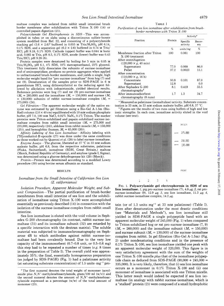
TABLE I Purification of sea lion isomaltase after solubilization from brush-
border membranes with Triton X-100 Activity
Fraction Protein specific ~ e c o v - activitv erv
Membrane fraction after Triton X-100 treatment
After centrifugation (120,000 X g, 40 min)
Supernatant Pellet
After concentration (110,000 x g, 16 h)
Concentrate Supernatant
After Sephadex G-200 chromatograph$
After immunoadsorbent chromatography (8 runs)
?& 100
77.0 37.2
50.8 12.8 8.1
1.7
unitslmg 76 0.061 100
0.068 86.0 0.0023 13.1
0.101 87.0 0.0
0.419 55.5
1.3 34.7
a Measured as palatinase (isomaltulase) activity. Substrate concen- tration is 33 mM, in 33 mM sodium maleate buffer, pH 6.8, 37 “C.
Figures are the results of two runs using buffers of high and low ionic strengths. In each case, isomaltase activity eluted in the void volume (see text).
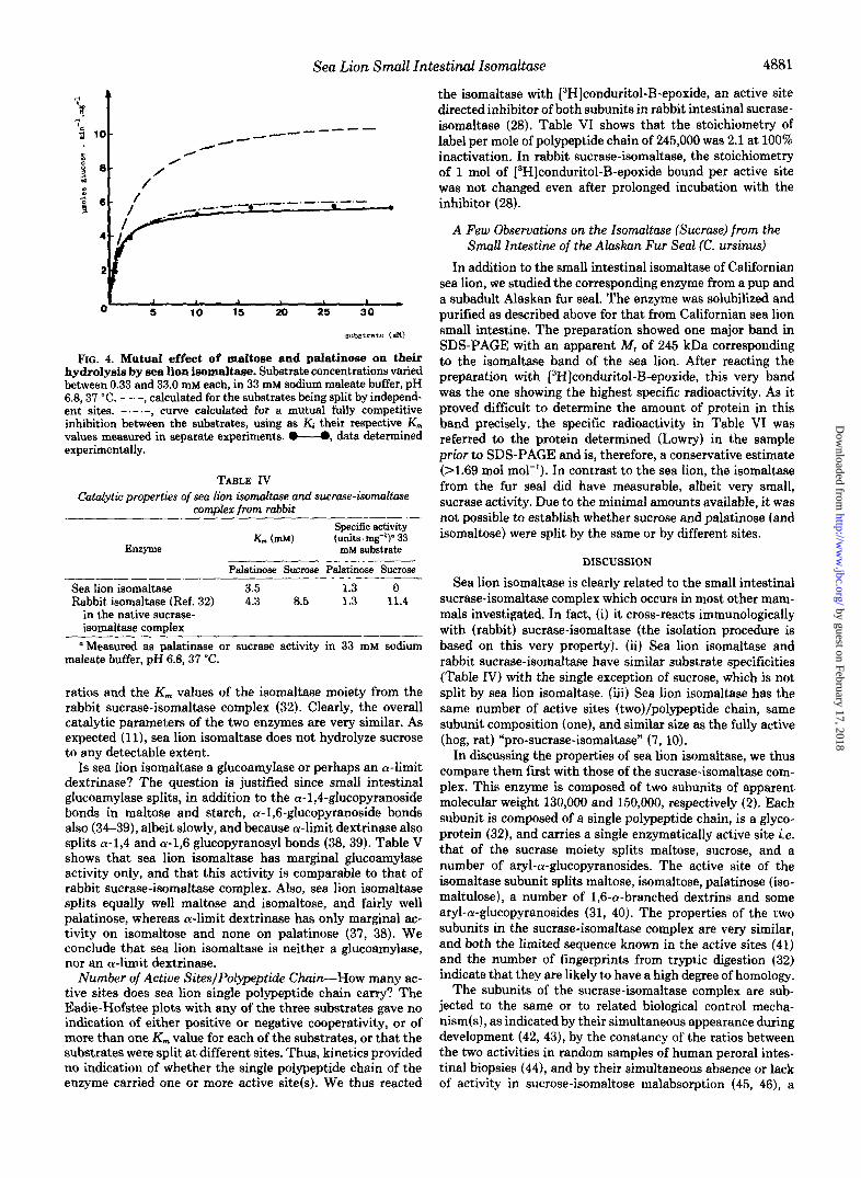
I ORIGIN
- 275.003 - 150.003
m.003 -
FRONT
1 2 3 4
FIG. 1. Polyacrylamide gel electrophoresis in SDS of sea lion isomaltase. 1 , pig pro-sucrase-isomaltase (7), 4.8 pg; 2, rat pro- sucrase-isomaltase (9), 12.9 pg; 3, sea lion isomaltase, 11.3 pg; 4, rabbit sucrase-isomaltase complex, 14.2 pg.
tein (or of 1.3 units.mg” at 33 mM palatinose) (Table I). Even after denaturation under the most drastic conditions (see “Materials and Methods”), sea lion isomaltase still yielded in SDS-PAGE a single polypeptide band with an apparent molecular weight of 245,000 (Fig. 1) when compared to Triton-solubilized hog or rat pro-sucrase-isomaltase (7, 9) ( M r = 260,000) and the isomaltase subunit ( M , = 150,000) and sucrase subunit ( M , = 130,000) of the sucrase-isomaltase complex from rabbit. In gel filtration (Bio-Gel A-1.5m) (Fig. 2) under nondenaturing conditions and in the presence of 0.1% Triton X-100, sea lion isomaltase yielded one peak with an apparent molecular weight of 320,000. This figure is in very satisfactory agreement with the sum of the weights of one Triton X-100 micelle plus that of the isomaltase polypep- tide chain as deduced from SDS-PAGE (90,000 + 245,000 = 335,000). It is very likely, therefore, that (i) sea lion isomaltase occurs as a monomer in 0.1% Triton X-100 and (ii) one monomer of isomaltase is associated with one Triton micelle. This latter result would indeed be expected if sea lion iso- maltase (in analogy with rabbit sucrase-isomaltase, which is a “stalked” protein (2)) were composed of a small hydrophobic
by guest on February 17, 2018http://w
ww
.jbc.org/D
ownloaded from

4880 Sea Lion Small Intestinal Isomaltase
4 9 i
1 1 a D
, 1s a D b
FIG. 2. Estimation of the apparent molecular weight of sea lion iaomaltase by gel filtration. A , rabbit Triton-solubilized sucrase-isomaltase complex (M, = 275,000 (U), or plus one Triton micelle 275,000 + 90,000 = 365,000 (0)); B, rabbit papain-solubilized sucrase-isomakase (M, = 235,000); C, aldolase from rabbit muscle (M, = 158,000); D, hemoglobin (M, = 65,000); E, Triton micelle (M, = 90,000, Ref. 58).
TABLE I1 Relative electrophoretic mobilities of sea lion isomaltase and rabbit
sucrase-isomaltase complex (detergent- and papain-solubilized). (Charge-shift ekctrophoresis)
Electrophoresis was carried out on 1% agarose gels in 50 mM glycine NaOH, 100 mM NaCl at pH 9.0 containing 0.5% Triton X- 100 alone, or with the addition of a negatively charged (deoxycholate, 0.25%) or of a positively charged (cetyltrimethylammonium bromide, 0.05%) detergent. The mobility of papain-solubilized rabbit sucrase- isomaltase did not change detectably with the charge of the detergent present and was thus used as the reference (100%).
Triton X-100 Triton and cetyltri- Triton X-100
deoxycholate alone ammonium
%
Enzyme and X-100 methyl-
bromide
Rabbit sucrase-isomaltase 100 100 100 (papain-solubilized) (reference)
(Triton-solubilized)
(Triton-solubilized)
Rabbit sucrase-isomaltase 100 64 41
Sea lion isomaltase 64 38 23
part and a much larger hydrophilic “body.” Hydrophobic Region(s) in Sea Lion Isomaltase-As this en-
zyme could not be detached from the brush-border membrane by aqueous solutions unless they contained a detergent, it is, by definition, an integral membrane protein, and was expected to possess one or more hydrophobic regions. This was tested by Helenius‘ and Simons’ (33) procedure of charge shift electrophoresis (Table 11). Clearly, sea lion isomaltase, much as Triton-solubilized rabbit sucrase-isomaltase, has the high- est electrophoretic mobility in the presence of the anionic detergent and the lowest in the presence of the cationic detergent. In contrast, the electrophoretic mobility of papain- solubilized rabbit sucrase-isomaltase, whose hydrophobic “an- chor” had been split off by the proteolytic treatment (2), was not detectably affected by the charge of the detergent present. We conclude that sea lion isomaltase, much as Triton-solu- bilized rabbit sucrase-isomaltase, has at least one hydrophobic domain.
Catalytic Properties-Sea lion isomaltase had a pH opti- mum in the range of 5-6.4 and was fairly stable in the pH range 5.3-7.8 (Fig. 3). It had maltase, isomaltase, and palati- nase activities in ratios approximately 1:0.92:0.31 (at saturat-
I -\
I ’ I
o-6 t I t i 1
FIG. 3. Effect of pH on sea lion isomaltase activity and stability after 1 h of incubation at 37 O C . A, o”-o, activity; B, 0- - 4, stability. The buffers used were Na+ acetate, pH 4.0 through 6.0, and Na+ phosphate, pH 6.0 through 12.0. For stability measure-
pH 2.1 through 12.3, were preincubated at 37 “C for 1 h. Before ments, samples containing 0.5 wg of protein in 50 pl of 10 mM buffer,
addition of the substrate, the pH was brought to 6.5 by adding 0.1 ml of 100 mM Na+ phosphate buffer, pH 6.5. Activity measurements were carried out in 33 mM buffer, pH 4.0 through 9.1. Above pH 7.7, the mixture had to be neutralized with phosphoric acid (100 mM) before the heating step to avoid nonenzymatic release of glucose.
TABLE I11 Catalytic properties of sea lion isomaltase
K,,, and V,. values for palatinose, maltose, and isomaltose were determined according to Lineweaver-Burk, Eadie-Hofstee, and Hill. Curves were fitted by linear regression. Assays were performed at 37 ‘C in 33 mM sodium maleate buffer (pH 6.8) and in 33 mM sodium phosphate (pH 7.6), respectively.
Substrate V-
split pH KO7 substrate
mM r ~ l m g ” . min”
Palatinose 6.8 3.5 1.5 Maltose 6.8 2.4 4.9 Isomaltose 6.8 2.5” 4.0”
3.8b 5.0’ 7.6 4.3 2.4
a Derived from an Eadie-Hofstee plot assuming Michaelian kinet- ics (correlation factor for linear regression r = 0.973). ’ Derived from a Hill plot; Hill coefficient is 0.786.
Determined using a plot u uersus v/s.
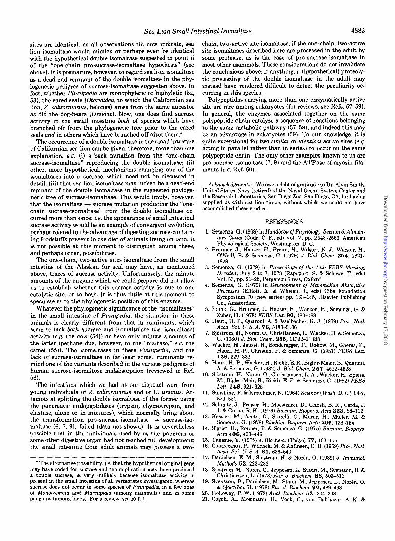
ing substrate concentrations and pH 6.8). The K , values were 2.4, 3.5, and 3.5 mM, respectively. The Eadie-Hofstee plots were linear with Hill coefficients between 0.97 and 1.03 (Table 111). In order to test whether these substrates were split by the same or by independent active site(s), mixed substrate incubations were carried out (Fig. 4). Clearly, the amount of glucose liberated from mixed substrate incubations corre- sponded closely to that calculated from a mutual fully com- petitive inhibition between the substrates, using as Ki their respective K,,, values as measured in separate experiments. Thus, maltose, palatinose, and isomaltose were split at the same site(s). Table IV reports for comparison the activity
by guest on February 17, 2018http://w
ww
.jbc.org/D
ownloaded from
Sea Lion Small Intestinal Isomaltase 4881
FIG. 4. Mutua1 effezt of maltose and palatinose on their hydrolysis by sea lion isomaltase. Substrate concentrations varied between 0.33 and 33.0 mM each, in 33 mM sodium maleate buffer, pH 6 4 3 7 "C. - - -, calculated for the substrates being split by independ- ent sites. curve calculated for a mutual fully competitive inhibition between the substrates, using as Kj their respective K,,, values measured in separate experiments. @"O, data determined experimentally.
TABLE IV Catalytic properties of sea lion iomultase and sucrase-iomnltase
comvlex from rabbit
Km (mM) (units. rng")= 33 Specifc activity
Enzyme mM substrate
Palatinose Sucrose Palatinose Sucrose
Sea lion isomaltase 3.5 1.3 0 Rabbit isomaltase (Ref. 32) 4.3 8.5 1.3 11.4
in the native sucrase- isomaltase complex
"Measured as palatinase or sucrase activity in 33 mM sodium maleate buffer, pH 6.8, 37 'C.
ratios and the K,,, values of the isomaltase moiety from the rabbit sucrase-isomaltase complex (32). Clearly, the overall catalytic parameters of the two enzymes are very similar. As expected (]I), sea lion isomaltase does not hydrolyze sucrose to any detectable extent.
Is sea lion isomaltase a glucoamylase or perhaps an a-limit dextrinase? The question is justified since small intestinal glucoamylase splits, in addition to the a-1,4-glucopyranoside bonds in maltose and starch, a-1,6-glucopyranoside bonds also (34-39), albeit slowly, and because a-limit dextrinase also splits a-1,4 and (~-1,6 glucopyranosyl bonds (38, 39). Table V shows that sea lion isomaltase has marginal glucoamylase activity only, and that this activity is comparable to that of rabbit sucrase-isomaltase complex. Also, sea lion isomaltase splits equally well maltose and isomaltose, and fairly well palatinose, whereas a-limit dextrinase has only marginal ac- tivity on isomaltose and none on palatinose (37, 38). We conclude that sea lion isomaltase is neither a glucoamylase, nor an a-limit dextrinase.
Number of Active SiteslPolypeptide Chain-How many ac- tive sites does sea lion single polypeptide chain carry? The Eadie-Hofstee plots with any of the three substrates gave no indication of either positive or negative cooperativity, or of more than one K, value for each of the substrates, or that the substrates were split at different sites. Thus, kinetics provided no indication of whether the single polypeptide chain of the enzyme carried one or more active site(s). We thus reacted
the isomaltase with [3H]c~nduritol-B-ep~xide, an active site directed inhibitor of both subunits in rabbit intestinal sucrase- isomaltase (28). Table VI shows that the stoichiometry of label per mole of polypeptide chain of 245,000 was 2.1 at 100% inactivation. In rabbit sucrase-isomaltase, the stoichiometry of 1 mol of [3H]conduritol-B-epoxide bound per active site was not changed even after prolonged incubation with the inhibitor (28).
A Few Observations on the Isomaltase (Sucrase) from the Small Intestine of the Alaskan Fur Seal (C. ursinus)
In addition to the small intestinal isomaltase of Californian sea lion, we studied the corresponding enzyme from a pup and a subadult Alaskan fur seal. The enzyme was solubilized and purified as described above for that from Californian sea lion small intestine. The preparation showed one major band in SDS-PAGE with an apparent M, of 245 kDa corresponding to the isomaltase band of the sea lion. After reacting the preparation with [3H]conduritol-B-epoxide, this very band was the one showing the highest specific radioactivity. As it proved difficult to determine the amount of protein in this band precisely, the specific radioactivity in Table VI was referred to the protein determined (Lowry) in the sample prior to SDS-PAGE and is, therefore, a conservative estimate (r1.69 mol mol"). In contrast to the sea lion, the isomaltase from the fur seal did have measurable, albeit very small, sucrase activity. Due to the minimal amounts available, it was not possible to establish whether sucrose and palatinose (and isomaltose) were split by the same or by different sites.
DISCUSSION
Sea lion isomaltase is clearly related to the small intestinal sucrase-isomaltase complex which occurs in most other mam- mals investigated. In fact, (i) it cross-reacts immunologically with (rabbit) sucrase-isomaltase (the isolation procedure is based on this very property). (ii) Sea lion isomaltase and rabbit sucrase-isomaltase have similar substrate specificities (Table IV) with the single exception of sucrose, which is not split by sea lion isomaltase. (iii) Sea lion isomaltase has the same number of active sites (two)/polypeptide chain, same subunit composition (one), and similar size as the fully active (hog, rat) "pro-sucrase-isomaltase" (7,lO).
In discussing the properties of sea lion isomaltase, we thus compare them first with those of the sucrase-isomaltase com- plex. This enzyme is composed of two subunits of apparent molecular weight 130,000 and 150,000, respectively (2). Each subunit is composed of a single polypeptide chain, is a glyco- protein (32), and carries a single enzymatically active site i.e. that of the sucrase moiety splits maltose, sucrose, and a number of aryl-a-glucopyranosides. The active site of the isomaltase subunit splits maltose, isomaltose, palatinose (iso- maltulose), a number of 1,6-a-branched dextrins and some aryl-a-giucopyranosides (31, 40). The properties of the two subunits in the sucrase-isomaltase complex are very similar, and both the limited sequence known in the active sites (41) and the number of fingerprints from tryptic digestion (32) indicate that they are likely to have a high degree of homology.
The subunits of the sucrase-isomaltase complex are sub- jected to the same or to related biological control mecha- nism(s), as indicated by their simultaneous appearance during development (42,43), by the constancy of the ratios between the two activities in random samples of human peroral intes- tinal biopsies (441, and by their simultaneous absence or lack of activity in sucrose-isomaltose malabsorption (45, 46), a
by guest on February 17, 2018http://w
ww
.jbc.org/D
ownloaded from

4882 Sea Lion Small Intestinal Isomaltase TABLE V
Glucoamyhe and isomaltase activities of some intestinal a-glucosidases Sources of amylose were as follows: A, amylose according to Zulkowsky (Merck); B, amylose from potato starch
(BDH); C, amylose from potato (type I, Sigma); D, amylose (Koch-Light); E, soluble starch (Baker); F, starch of unspecified source.
Isomaltase Glucoamylase activity (pmol glucose liberated-min”.mg” protein) activity (V-,
Enzyme pmol glucosidic bonds split at
optimum pH. min” A B C D E F mg” protein)
Sea lion isomaltase (present paper) -4.5 1.13 0.02 0.056 Rabbit sucrase-isomaltase complex -10 0.735 0.04 0.064
Rat glucoamylase (from Ref. 36) 7.6 Rat glucoamylase (from Ref. 37) NDb Hog glucoamylase (from Ref. 38) Hog a-limit dextrinase
(present paper) 28.4
-36 1.05 0.38
-0.65 -1.6
(from Ref. 39) 1.4
Glucoamylase activity was assayed in the present paper at 37 “C in 50 mM Na maleate buffer, pH 6.0, 2.5 mM EDTA, containing 10 mg of amylose/ml. The conditions in Ref. 36 were the same; those in other references were similar, although not necessarily identical.
* Not determined.
TABLE VI Stoichiometry of labeling of sea lion bomaltase with [3H]conduritol-
B-epoxide The enzyme (0.62 mg) was incubated for 7 h in 1 ml of 92 mM
sodium maleate buffer, with 23.9 pmol of [3H]conduritol-B-epoxide (specific radioactivity 1.25 X lo6 cpmlpmol). Excess reagent was removed by exhaustive dialysis (3 days) against 50 mM sodium maleate buffer pH 6.8, followed by vacuum dialysis against distilled water. The concentrate was percolated (13.2 ml/h) through a Bio-Gel P-100 column (2 X 26 cm) equilibrated in 10 mM ammonium carba- minate, 0.05% Triton X-100. The fractions eluted with the void volume were pooled, protein concentration was determined by a modified Lowry procedure (30), and the radioactivity of the fractions was counted.
Enzyme Inacti- Radio- Ratio of epoxide vation activity to enzyme
% TrT;2 mol/mol
Sea lion isomaltase 100 1055 2.07 Alaskan fur seal 100 6067” >1.69’ Rabbit sucrase-iso- 100 1709‘ 1.96
maltase complex (from Ref. 28)
‘ Specific radioactivity of [3H]conduritol-B-epoxide 8.80 X 10’ cpm/pmol (0.99 pCi/pmol).
See text. Specific radioactivity of [3H]conduritol-B-epoxide, 1.92 X lo5
cpm/pmol (0.197 pCi/pmol).
monofactorial genetic disease (47).3 One more point worth mentioning is the unusual mode of
anchoring of the sucrase-isomaltase in the brush-border mem- brane, i.e. the COOH-terminal regions of both subunits and the NH2-terminal regions of the sucrase subunit are not involved in the anchoring to the membrane fabric and are exposed to the outer, luminal face of the membrane (2). The isomaltase subunit is anchored in the membrane fabric via a highly hydrophobic segment which is located not far from the NH, terminus of the polypeptide chain (2, 5 ) ; the sucrase subunit has a peripheral position and seems to interact with the membrane fabric via the isomaltase subunit only (2, 48, 49). (For a very recent review on sucrase-isomaltase, see Ref. 50).
In some pedigrees of this pathological condition, the pattern of sucrase and isomaltase activities left indicates a fairly complicated situation (for a review, see Ref. 56).
In order to explain these three groups of characteristics of the small intestinal sucrase-isomaltase complex (i.e. the anal- ogy, if not homology, between the two subunits, their common or related biological control mechanism(s), and the unusual mode of anchoring to the membrane), the following has been suggested (3-5). (i) Sucrase and isomaltase have arisen phy- logenetically by (partial) duplication of an original isomaltase- maltase gene. (ii) This would have led first to a gene coding for a single polypeptide chain carrying two identical domains, each endowed with enzymatic activity i.e. a double isomaltase. (iii) Subsequent mutation would have transformed one of these domains from an isomaltase-maltase into a sucrase- maltase with the appearance of a single chain two-active site precursor (pro-sucrase-isomaltase). (iv) This single chain pro- sucrase-isomaltase would be synthesized, glycosylated, and inserted in the membrane of the endoplasmic reticulum and transferred, along with other plasma membrane proteins, to the brush-border membrane. (v) Post-translational proteoly- tic modification of this single chain (perhaps by one or more pancreatic proteases), would lead to the two subunits of the “ripe” sucrase-isomaltase complex; they would still remain associated via interactions formed during the folding of single chain pro-sucrase-isomaltase.
This “one-chain pro-sucrase-isomaltase hypothesis” seems now well established a single chain enzymatically active pro- sucrase-isomaltase has indeed been isolated from the small intestines of hogs whose pancreases had been disconnected from the duodena three days prior to death (7); a fast fucose- labeled, high molecular weight band could be isolated from the rat intestine (6) and from subcutaneous transplants of small intestine from fetal rats (9); and finally, in vitro trans- lation of RNA from rabbit small intestine yielded a high molecular weight band (8), which was identified as pro-su- crase-isomaltase (or pre-pro-sucrase-isomaltase). Both hog and rat pro-sucrase-isomaltases are converted into “normal” sucrase-isomaltase bands by treatment with pancreatic en- doproteases. In addition, recent work on glucoamylase (51) has shown that this enzyme also is synthesized as a large single chain polypeptide which is split post-translationally. Thus, points iv and v above seem to be secured. Points i and ii are of course made likely by the very probable homology between sucrase and isomaltase.
The observations of the present paper show that sea lion isomaltase carries two active sites/polypeptide chain. If the
by guest on February 17, 2018http://w
ww
.jbc.org/D
ownloaded from
Sea Lion Small Intestinal Isomattase 4883
sites are identical, as all observations till now indicate, sea lion isomaltase would mimick or perhaps even be identical with the hypothetical double isomaltase suggested in point ii of the “one-chain pro-sucrase-isomaltase hypothesis” (see above). It is premature, however, to regard sea lion isomaltase as a dead end remnant of the double isomaltase in the phy- logenetic pedigree of sucrase-isomaltase suggested above. In fact, whether Pinnipedia are monophyletic or biphyletic (52, 53), the eared seals (Otorioideu, to which the Californian sea lion, Z. califurnianus, belongs) arose from the same ancestor as did the dog-bears (Ursidue). Now, one does find sucrase activity in the small intestine both of species which have branched off from the phylogenetic tree prior to the eared seals and in others which have branched off after them:
The occurrence of a double isomaltase in the small intestine of Californian sea lion can be given, therefore, more than one explanation, e.g. (i) a back mutation from the “one-chain sucrase-isomaltase” reproducing the double isomaltase; (ii) other, more hypothetical, mechanisms changing one of the isomaltases into a sucrase, which need not be discussed in detail; (iii) that sea lion isomaltase may indeed be a dead-end remnant of the double isomaltase in the suggested phyloge- netic tree of sucrase-isomaltase. This would imply, however, that the isomaltase -+ sucrase mutation producing the “one- chain sucrase-isomaltase” from the double isomaltase oc- curred more than once; i.e. the appearance of small intestinal sucrase activity would be an example of convergent evolution, perhaps related to the advantage of digesting sucrose-contain- ing foodstuffs present in the diet of animals living on land. It is not possible at this moment to distinguish among these, and perhaps other, possibilities.
The one-chain, two-active sites isomaltase from the small intestine of the Alaskan fur seal may have, as mentioned above, traces of sucrase activity. Unfortunately, the minute amounts of the enzyme which we could prepare did not allow us to establish whether this sucrase activity is due to one catalytic site, or to both. It is thus futile at this moment to speculate as to the phylogenetic position of this enzyme.
Whatever the phylogenetic significance of the “isomaltases” in the small intestine of Pinnipediu, the situation in these animals is clearly different from that in ruminants, which seem to lack both sucrase and isomaltulase (i.e. isomaltase) activity (e.g. the cow (54)) or have only minute amounts of the latter (perhaps due, however, to the “maltase,” e.g. the camel ( 5 5 ) ) . The isomaltases in these Pinnipediu, and the lack of sucrase-isomaltase in (at least some) ruminants re- mind one of the variants described in the various pedigrees of human sucrose-isomaltose malabsorption (reviewed in Ref. 56).
The intestines which we had at our disposal were from young individuals of 2. californianus and of C. ursinus. At- tempts at splitting the double isomaltase of the former using the pancreatic endopeptidases (trypsin, chymotrypsin, and elastase, alone or in mixtures), which normally bring about the transformation pro-sucrase-isomaltase -+ sucrase-iso- maltase (6, 7, 9), failed (data not shown). It is nevertheless possible that in the individuals used by us the pancreas or some other digestive organ had not reached full development; the small intestine from adult animals may possess a two-
‘ The alternative possibility, i.e. that the hypothetical original gene may have coded for sucrase and the duplication may have produced a double sucrase, is very unlikely because isomaltase activity is present in the small intestine of all vertebrates investigated, whereas sucrase does not occur in some species of Pinnipedia, in a few ones of Monotremata and Marsupialn (among mammals) and in some penguins (among birds). For a review, see Ref. 1.
chain, two-active site isomaltase, if the one-chain, two-active site isomaltases described here are processed in the adult by some protease, as is the case of pro-sucrase-isomaltase in most other mammals. These considerations do not invalidate the conclusions above; if anything, a (hypothetical) proteoly- tic processing of the double isomaltase in the adult may instead have rendered difficult to detect the peculiarity oc- curring in this species.
Polypeptides carrying more than one enzymatically active site are rare among eukaryotes (for reviews, see Refs. 57-59). In general, the enzymes associated together on the same polypeptide chain catalyze a sequence of reactions belonging to the same metabolic pathway (57-59), and indeed this may be an advantage in eukaryotes (59). To our knowledge, it is quite exceptional for two similar or identical active sites (e.g. acting in parallel rather than in series) to occur on the same polypeptide chain. The only other examples known to us are pro-sucrase-isomaltase (7,9) and the ATPase of myosin fila- ments (e.g. Ref. 60).
Acknowledgments-We owe a debt of gratitude to Dr. Alvin Smith, United States Navy (retired) of the Naval Ocean System Center and the Research Laboratories, San Diego Zoo, San Diego, CA, for having supplied us with sea lion tissue, without which we could not have accomplished these studies.
REFERENCES
1. Semenza, G . (1968) in Handbook of Physiology, Section 6: Alimen- tary Canal (Code, C. F., ed) Vol. V, pp. 2543-2566, American Physiological Society, Washington, D. C.
2. Brunner, J., Hauser, H., Braun, H., Wilson, K . J., Wacker, H., O’Neill, B. & Semenza, G. (1979) J. Bwl. Chem. 254, 1821- 1828
3. Semenza, G. (1979) in Proceedings of the 12th FEBS Meeting, Dresden, July 2 to 7, 1978 (Rapoport, S. & Schewe, T., eds) Vol. 53, pp. 21-28, Pergamon Press, Oxford
4. Semenza, G. (1979) in Development of Mammalian Absorptive Processes (Elliott, K. 8c Whelan, J., eds) Ciba Foundation Symposium 70 (new series) pp. 133-145, Elsevier Publishing Co., Amsterdam
5. Frank, G., Brunner, J., Hauser, H., Wacker, H., Semenza, G. & Zuber, H. (1978) FEBS Lett. 96,183-188
6. Hauri, H. P., Quaroni, A. & Isselbacher, K. J. (1979) Proc. Natl.
7. Sjostrom, H., Norin, O., Christiansen, L., Wacker, H. & Semenza, G. (1980) J. Biol. Chem. 255 , 11332-11338
8. Wacker, H., Jaussi, R., Sonderegger, P., Dokow, M., Ghersa, P., Hauri, H.-P., Christen, P. & Semenza, G. (1981) FEBS Lett.
9. Hauri, H.-P., Wacker, H., Rickli, E. E., Bigler-Meier, B., Quaroni, A. & Semenza, G. (1982) J. Bwl. Chem. 257, 4522-4528
10. Sjostrom, H., Noren, O., Christiansen, L. A., Wacker, H., Spiess, M., Bigler-Meir, B., Rickli, E. E. & Semenza, G. (1982) FEBS
11. Sunshine, P. & Kretchmer, N. (1964) Science (Wash. D. C.) 144 ,
12. Schmitz, J., Preiser, H., Maestracci, D., Ghosh, B. K., Cerda, J. J. & Crane, R. K . (1973) Biochim. Biophys. Acta 323,98-112
13. Kessler, M., Acuto, O., Storelli, C., Murer, H., Muller, M. & Semenza, G. (1978) Biochim. Biophys. Acta 506,136-154
14. Sigrist, H., Ronner, P. & Semenza, G. (1975) Biochim. Biophys. Acta 406,433-446
15. Takesue, Y. (1975) J. Biochem. (Tokyo) 77, 103-115 16. Cuatrecasas, P., Wilchek, M. & Anfinsen, C. B. (1969) Proc. Natl.
17. Danielsen, E. M., Sjostrom, H. & Nor&, 0. (1982) J. Irnmunol.
18. Sjostrom, H., North, O., Jeppesen, L., Staun, M., Svensson, B. &
19. Svensson, B., Danielsen, M., Staun, M., Jeppesen, L., Norin, 0.
20. Holloway, P. W. (1973) Anal. Biochem. 5 3 , 304-308 21. Cogoli, A., Mosimann, H., Vock, C., von Balthazar, A.-K. &
A c d . Sci. U. S. A. 76,5183-5186
136,329-332
Lett. 148,321-325
850-851
Acad. Sci. U. S. A. 61,636-643
Methods 52,223-232
Christiansen, L. (1978) Eur. J. Biochem. 88,503-511
& Sjostrom, H. (1978) Eur. J . Biochen. 90,489-498
by guest on February 17, 2018http://w
ww
.jbc.org/D
ownloaded from
4884 Sea Lion Small Intestinal Isomaltase
22. 23. 24.
25.
26.
27.
28.
29.
30. 31.
32.
33.
34.
35. 36.
37.
38.
39.
Semenza, G. (1972) Eur. J. Biochem. 30, 7-14 Neville, D. M., Jr. (1971) J. Biot. Chem. 246,6328-6334 Hjerten, S. (1962) Arch. Biochem. Biophys. Suppl. 1, 147-151 Spiess, M., Hauser, H., Rosenbusch, J. P. & Semenza, G. (1981)
Sia, S. L. & Horecker, B. L. (1968) Arch. Biochem. Biophys. 123,
Braunitzer, G., Gehring-Miiller, R., Hilschmann, H., Hilse, K., Hobom, G., Rudloff, V. & Wittmann-Liebold, B. (1961) 2. Physiol. Chem. 325,283-287
Braun, H., Legler, G., Deshusses, J. & Semenza, G. (1977) Biochim. Biophys. Acta 483,135-140
Quaroni, A., Gershon, E. & Semenza, G. (1974) J. Biol. Chem.
Banauch, D., Briimmer, W., Ebeling, W., Metz, H., Rindfrey, H., Lang, H., Leybold, K. & Rick, W. (1975) 2. Klin. Chem. Klin. Biochem. 13, 101-107
J. Biol. Chem. 256,8977-8982
186-194
249,6424-6433
Wang, C.4. & Smith, R. L. (1975) Anal. Biochem. 414-417 Kolinska, J. & Semenza, G. (1967) Biochim. Biophys. Acta 146,
Cogoli, A., Eberle, A., Sigrist, H., Joss, C., Robinson, E., Mosi- mann, H. & Semenza, G. (1973) Eur. J . Biochem. 33,40-48
Helenius, A. & Simons, K. (1977) Proc. Natl. Acad. Sci. U. S. A.
Auricchio, S., Semenza, G. & Ruhino, A. (1965) Biochim. Biophys.
Eggermont, E. (1969) Eur. J. Biochem. 9,483-487 Kolinska, J. & Kraml, J. (1972) Biochim. Biophys. Acta 284,
Schlegel-Haueter, S., Hore, P., Kerry, K. R. & Semenza, G. (1972)
S~rensen, S. H., NorCn, O., Sjostrom, H. & Danielsen, E. M.
Taravel, F. R., Datema, R., Woloszczuk, W., Marshall, J. J. &
181-195
74,529-532
Acta 96,498-507
235-247
Biochim. Biophys. Acta 258, 506-519
(1982) Eur. J . Biochem. 130,147-153
Whelan, W. J. (1983) Eur. J. Biochem. 130, 147-153
40. Semenza, G. & von Balthazar, A.-K. (1974) Eur. J. Biochem. 41,
41. Quaroni, A. & Semenza, G. (1976) J. Biol. Chem. 251,3250-3253 42. Rubino. A.. Zimbalatti. F. & Auricchio. S. (1964) Biochim. Bio-
149-162
I . I~
phys. Acta 92,305-311
528
~
43. Dahlqvist, A. & Lindberg, T. (1966) Clin. Sci. (Lond. ) 30, 517-
44. Auricchio, S., Rubino, A., Tosi, R., Semenza, G., Landolt, M., Kistler, H. & Prader, A. (1963) Enzyml . Biol. Clin. 3, 193-208
45. Semenza, G., Auricchio, S., Rubino, A,, Prader, A. & Welsh, J. D. (1965) Biochim. Biophys. Acta 195,386-389
46. Auricchio, S., Rubino, A., Prader, A., Rey, J., Jos, J., Frezal, J. & Davidson, M. (1965) J. Pediatr. 66,555-564
47. Kerry, K. R. & Townley, R. R. W. (1965) Aust. Paecfiatr. J. 1,
48. Spiess, M., Brunner, J. & Semenza, G. (1982) J. Biol. Chem. 257,
49. Biirgi, R., Brunner, J. & Semenza, G. (1983) J. Biol. Chem. 258,
50. Hauser, H. & Semenza, G. (1983) CRC Crit. Rev. Biochem. 14,
51. Danielsen, E. M., Sjostrom, H. & Norkn, 0. (1983) Biochem. J.
52. McLaren, I. A. (1960) Syst. Zool. 9, 18-28 53. Kretchmer, N. & Sunshine, P. (1967) Gastroenterology 53, 123-
54. Siddons, R. C. (1968) Biochem. J. 108,839-844 55. Sir El Khatim, M. M. & Osman, A. M. (1982) Comp. Biochem.
Physiol. A. Comp. Physiol. 71, 199-204 56. Semenza, G. (1981) Clin. Gatroenterol. 10,691-706 57. Kirschner, K. & Bisswanger, H. (1976) Annu. Reu. Biochem. 45,
58. Stark, G. R. (1977) Trends Biochem. Sci. 2, 64-66 59. Bisswanger, H. & Schmincke-Ott, E. (eds) (1980) Multifunctional
Proteins, John Wiley & Sons, New York 60. Margossian, S. S. & Lowey, S. (1975) J. Mol. Biol. 74,301-330 61. Kushner, L. M. & Hubbard, W. D. (1954) J. Phys. Chem. 58,
- "
223-235
2370-2377
15114-15119
319-345
210,389-393
129
143-166
1163-1167
by guest on February 17, 2018http://w
ww
.jbc.org/D
ownloaded from

H Wacker, R Aggeler, N Kretchmer, B O'Neill, Y Takesue and G Semenzasucrase-isomaltase.
intestinal brush-border membrane. Its possible phylogenetic relationship with A two-active site one-polypeptide enzyme: the isomaltase from sea lion small
1984, 259:4878-4884.J. Biol. Chem.
http://www.jbc.org/content/259/8/4878Access the most updated version of this article at
Alerts:
When a correction for this article is posted•
When this article is cited•
to choose from all of JBC's e-mail alertsClick here
http://www.jbc.org/content/259/8/4878.full.html#ref-list-1
This article cites 0 references, 0 of which can be accessed free at
by guest on February 17, 2018http://w
ww
.jbc.org/D
ownloaded from