a tool for the quantification of radial neo-vessels in ... · a tool for the quantication of radial...
TRANSCRIPT

A tool for the quantification of radial neo-vessels in chickchorioallantoic membrane angiogenic assays
Alessandro Gnutti1, Alberto Signoroni1, Riccardo Leonardi1,Michela Corsini2, Marco Presta2, and Stefania Mitola2
Abstract— Angiogenesis, the process of new blood vesselsformation, plays a key role in different physiological andpathological conditions and it is considered a promising targetfor the development of new anti-inflammatory and anti-tumortherapies. Several assays have been developed to mimic theangiogenic process in vitro and in vivo. Here we proposea technique for the quantification of the pro-angiogenic oranti-angiogenic responses induced by different molecules whenimplanted in vivo on the chick embryo chorioallantoic mem-brane (CAM). At day 11 of development CAM is completelyvascularized and neo-vessels induced by exogenous moleculesconverge radially to the implant. Our algorithm is an effectiveand rapid tool to characterize molecules endowed with pro-or anti-angiogenic effects by means of the quantification ofthe vessels present in the CAM macroscopic images. Basedon conventional and dedicated image morphology tools, theproposed technique is able to discriminate radial from non-radial vessels, excluding the last ones from the count.
I. INTRODUCTION
Blood vessels are a prerequisite for normal development,tissue growth and repair as they provide nutrients, removewaste products and are means for conveying cells to distantsites. Blood vessels arise through one of two processes:vasculogenesis, where the blood vessels develop de novovia the assembly of endothelial cell (EC) precursors (an-gioblasts), or angiogenesis, when new blood vessels ariseby sprouting from pre-existing ones. Angiogenesis plays akey role in many physiological and pathological conditions,including tumor growth, progression and metastasis [1]. Thusit is considered a promising target for the development ofnew anti-tumor strategies, as demonstrated by a logarithmicincrease in the number of reports dealing with angiogenesisin the last few years. Angiogenesis processes can be dividedin several steps including EC survival, proliferation, migra-tion and differentiation leading lumen formation. Several invitro and in vivo assays have been developed to mimic thedifferent steps of the angiogenesis process [2]. In vivo testsare more difficult and time-consuming to perform, therebylimiting the number of tests that can run at any one time.One of the most critical technical problems in the fieldof angiogenesis is the accurate interpretation of the highlyvaried results obtained from the many assays currently in use.Among the in vivo assays the chick embryo chorionallantoicmembrane (CAM) is rapid and easy to perform [3]. The
1Department of Information Engineering, University of Brescia, viaBranze 38, 25123 Brescia (Italy) {name.surname}@unibs.it
2Department of Molecular and Translational Medicine,University of Brescia, Viale Europa 11, 25123 Brescia (Italy){name.surname}@unibs.it
chorionallantoic membrane is an extraembryonic membraneconstituted by an extensive capillary network. CAM mediatesgas and nutrient exchanges until hatching. Its developmentstarts at day 4 post fertilization. Blood vessels grow veryrapidly until day 11 thereafter EC mitotic index declines andthe vasculature system attains its final arrangement at day 18.In its original form, the pro- or anti-angiogenic effects havebeen limited to ranking the vascularization on a semiquanti-tative scale. Actually it is possible to follow the vasculaturedevelopment in vivo by time-lapse imaging techniques, andsomeone suggests the measurement of bifurcation points inthe area around the stimulus. In the quantification of pro-angiogenic response the ability of researcher may alter theresponses. Although there are a lot of CAM-related bloodvessel extraction tools, many and recent works continue tobe realized in order to increase the accuracy and to allow aspecific selection of a particular set of vessels [4] [5].
In this paper, we present a novel tool for the quantifi-cation of neo-formed vessels. In literature different ves-sel segmentation approaches are employed [6]. Here weadopt a morphological image processing approach whichis often exploited also in retinal image analysis [7] [8][9], an application domain that shares some aspects withCAM image analysis. However, there are peculiar aspects ofthe considered experimental assays which require dedicatedsolutions. In fact, since neo-vessels inducted by the drug areconvergent and radial to the stimulus, there is the need toprocess CAM images so as to be able to distinguish radialand non radial vessels, and then to discard the latter ones.
The paper is structured as follows: a brief illustration aboutthe procedure of the medical tests is enunciated in Section II.Section III describes the software, comprising a first modulewhere the complete vessels segmentation is implementedand a second module where specifically neo-formed radialvessels are filtered. The performance evaluation is presentedin section IV, and finally the paper is concluded in sectionV in which main points of the work and possible futuredevelopments are highlighted.
II. MATERIALS
Chick embryo chorioallantoic membrane (CAM) assayCAM of day 7-9 chick embryos has been exposed by makinga window in the egg shell. Alginate beads (5 µl) containingFGF2 (both at 100 ng per embryo) in the absence or inthe presence of combretastatin have been placed on theCAM of fertilized White Leghorn chicken eggs at day 11of incubation. After 72 hours, newly formed blood vessels
978-1-4244-9270-1/15/$31.00 ©2015 IEEE 763
Pre-processing operations
Large vessels removal (insignificant information)
Region of interest definition
Topological skeleton
Broken vessels reunification
Branches separation
Radial vessels maintenance
Vessels quantification and data analysis
Specific region Removal (stimulus)
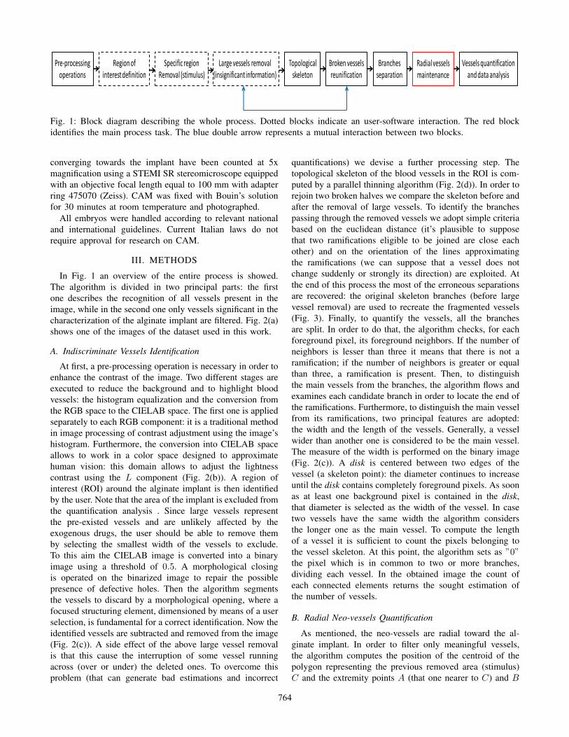
Fig. 1: Block diagram describing the whole process. Dotted blocks indicate an user-software interaction. The red blockidentifies the main process task. The blue double arrow represents a mutual interaction between two blocks.
converging towards the implant have been counted at 5xmagnification using a STEMI SR stereomicroscope equippedwith an objective focal length equal to 100 mm with adapterring 475070 (Zeiss). CAM was fixed with Bouin’s solutionfor 30 minutes at room temperature and photographed.
All embryos were handled according to relevant nationaland international guidelines. Current Italian laws do notrequire approval for research on CAM.
III. METHODS
In Fig. 1 an overview of the entire process is showed.The algorithm is divided in two principal parts: the firstone describes the recognition of all vessels present in theimage, while in the second one only vessels significant in thecharacterization of the alginate implant are filtered. Fig. 2(a)shows one of the images of the dataset used in this work.
A. Indiscriminate Vessels Identification
At first, a pre-processing operation is necessary in order toenhance the contrast of the image. Two different stages areexecuted to reduce the background and to highlight bloodvessels: the histogram equalization and the conversion fromthe RGB space to the CIELAB space. The first one is appliedseparately to each RGB component: it is a traditional methodin image processing of contrast adjustment using the image’shistogram. Furthermore, the conversion into CIELAB spaceallows to work in a color space designed to approximatehuman vision: this domain allows to adjust the lightnesscontrast using the L component (Fig. 2(b)). A region ofinterest (ROI) around the alginate implant is then identifiedby the user. Note that the area of the implant is excluded fromthe quantification analysis . Since large vessels representthe pre-existed vessels and are unlikely affected by theexogenous drugs, the user should be able to remove themby selecting the smallest width of the vessels to exclude.To this aim the CIELAB image is converted into a binaryimage using a threshold of 0.5. A morphological closingis operated on the binarized image to repair the possiblepresence of defective holes. Then the algorithm segmentsthe vessels to discard by a morphological opening, where afocused structuring element, dimensioned by means of a userselection, is fundamental for a correct identification. Now theidentified vessels are subtracted and removed from the image(Fig. 2(c)). A side effect of the above large vessel removalis that this cause the interruption of some vessel runningacross (over or under) the deleted ones. To overcome thisproblem (that can generate bad estimations and incorrect
quantifications) we devise a further processing step. Thetopological skeleton of the blood vessels in the ROI is com-puted by a parallel thinning algorithm (Fig. 2(d)). In order torejoin two broken halves we compare the skeleton before andafter the removal of large vessels. To identify the branchespassing through the removed vessels we adopt simple criteriabased on the euclidean distance (it’s plausible to supposethat two ramifications eligible to be joined are close eachother) and on the orientation of the lines approximatingthe ramifications (we can suppose that a vessel does notchange suddenly or strongly its direction) are exploited. Atthe end of this process the most of the erroneous separationsare recovered: the original skeleton branches (before largevessel removal) are used to recreate the fragmented vessels(Fig. 3). Finally, to quantify the vessels, all the branchesare split. In order to do that, the algorithm checks, for eachforeground pixel, its foreground neighbors. If the number ofneighbors is lesser than three it means that there is not aramification; if the number of neighbors is greater or equalthan three, a ramification is present. Then, to distinguishthe main vessels from the branches, the algorithm flows andexamines each candidate branch in order to locate the end ofthe ramifications. Furthermore, to distinguish the main vesselfrom its ramifications, two principal features are adopted:the width and the length of the vessels. Generally, a vesselwider than another one is considered to be the main vessel.The measure of the width is performed on the binary image(Fig. 2(c)). A disk is centered between two edges of thevessel (a skeleton point): the diameter continues to increaseuntil the disk contains completely foreground pixels. As soonas at least one background pixel is contained in the disk,that diameter is selected as the width of the vessel. In casetwo vessels have the same width the algorithm considersthe longer one as the main vessel. To compute the lengthof a vessel it is sufficient to count the pixels belonging tothe vessel skeleton. At this point, the algorithm sets as ”0”the pixel which is in common to two or more branches,dividing each vessel. In the obtained image the count ofeach connected elements returns the sought estimation ofthe number of vessels.
B. Radial Neo-vessels Quantification
As mentioned, the neo-vessels are radial toward the al-ginate implant. In order to filter only meaningful vessels,the algorithm computes the position of the centroid of thepolygon representing the previous removed area (stimulus)C and the extremity points A (that one nearer to C) and B
764

(a) Original image. (b) Pre-processed image. (c) ROI definition and non significant informa-tion removal.
(d) Topological skeleton. (e) Radial vessels filtering. (f) Presented result.
Fig. 2: Principal images at the end of each stage. In (f) we show a complete representation of the result. We highlight thequantified vessels overlapped to the original image. Colors represent the width of the vessels: a color scale from red to blueis used to indicate the width scale from the greatest one and the smallest one.
(that one more distant to C) of each connected element. Nowlet be α, β and γ the angles between the 0 angular coefficientline and the CA, CB and AB lines, respectively (Fig. 4).Then only the elements that have similar angle values aremaintained, while the other ones are removed. Furthermore,a check on the distance between stimulus and vessel isimplemented. Indeed only vessels near to the alginate implanthave to be considered neo-vessels. The above requisitesare formulated in the following test which each element issubjected:
|α− β| < Th∠
|α− γ| < Th∠
|β − γ| < Th∠
d(C,A) < Thd
(1)
The second and the third row of the equation are necessaryin the event of short vessels. Indeed, in that case the anglesα and β can assume similar values despite the presence ofa non radial vessel. In this case however the angle γ willbe different to them. Experimental results show that a goodthresholds selection consists in Th∠ = 20◦ and Thd = 50pixel (Fig. 2(e)).
IV. RESULTS
To set a rapidly and accurate quantification of CAM assaywe developed an algorithm for the quantification of radialvessels induced by the xenograft compound. CAMs havebeen treated at day 11 of development, when EC mitotic
index declined. CAMs have been grafted with 100 ng ofFGF2 in the absence or in the presence of 1-100 pmol/eggof combretastatin 4A (CA-4) drug and photographed after 72h. CA-4 is an antimitotic molecule with antivascular effects.
(a) Fragmented vessels.
(b) Repaired vessel network.
Fig. 3: Connection between interrupted vessels.
765

C
A
B
β
α
γ
Fig. 4: Model visualization of the polygonal mask represent-ing the implant. A radial neo-vessel is showed.
The implemented algorithm allows to identify all vesselspresent in the image and to extract the length and the widthof each of them. In this kind of experiment, in order tocharacterize the inhibitor compound, it has been necessaryto quantify only neo-vessels induced by the implant: sinceneo-vessels are convergent and radial to the stimulus, thealgorithm computes a directional analysis of the vessel. Inaccordance with previous results [10], FGF2 recruits radialneo-vessels from non radial pre-existing ones while CA-4inhibits the pro-angiogenic activities of FGF2. In Fig. 5 acomparison between manually and automatically computedquantification tests is illustrated. The measurements on 32CAMs where the inductor compound is combined with theinhibitor one are reported (red lines). To assess the efficiencyof the tested inhibitor molecule, a dedicated series of CAMshas been used to evaluate both manual and automated aver-age levels of reference, namely for the case where only theinductor compound is present (blue lines). Both manual andautomatic quantifications are able to discern pro-angiogenicand anti-angiogenic molecules. Software results are coherentand consistent to the manual quantification of the researchers,with an observed 20% average overestimation due to thefact that our software is more sensitive since it is also ableto detect blurred and hard visible vessels. This has beenjudged by experts a positive fact because this accuracy mayallow to better distinguish the biological activity of similardrugs. Dose dependent analysis is in progress to characterizedifferent CA-4. Fig. 2(f) shows a particular kind of the resultpresentation where the quantified vessels are overlapped tothe original image: the different colors indicate differentvessels widths. Specifically a color scale from red to blueis used to indicate a width scale from the thicker to thethinner one. These additional quantitative data have not beenused here but their exploitation is under evaluation for amore general quantitative imaging approach to the study ofangiogenesis.
V. CONCLUSION
In this paper, we presented a new tool able to segment allvessels present in CAM macroscopic images and to select
CAMs5 10 15 20 25 30
Nu
mb
er o
f n
eo-v
esse
ls
20
40
60
80
100
120 Inductor compound automatic quantification (average)Inductor compound manual quantification (average)Inductor and inhibitor compounds automatic quantificationInductor and inhibitor compounds manual quantification
Fig. 5: Comparison between manual and automatic quantifi-cation.
only neo-formed vessels radial to the implant. The softwarereturns the number of sought vessels, and furthermore itproduces other kinds of data as vessels length, vessels widthand area. Experimental results demonstrate the suitabilityof the method for automated quantification studies of theangiogenic effects of target compounds. Additional dataproduced by the technique could be further exploited toimprove its capability to characterize and to distinguishamong similar molecules.
REFERENCES
[1] Carmeliet P., Jain RK. Molecular mechanisms and clinical applica-tions of angiogenesis, in Nature, 2011 May.
[2] Corsini M., Moroni E., Ravelli C., Andrs G., Grillo E., Ali I.H., BrazilD.P., Presta M., Mitola S. Cyclic adenosine monophosphate-responseelement-binding protein mediates the proangiogenic or proinflamma-tory activity of gremlin, in Arteriosclerosis, Thrombosis, and VascularBiology, 34 (1), pp. 136-145, 2014 January.
[3] Ribatti D., Chick embryo chorioallantoic membrane as a useful toolto study angiogenesis, in Int Rev Cell Mol Biol, 270, pp. 181-224,2008.
[4] Peng S., Jinsheng H., Yue H., Mei Z., Lurong Z., Automated compu-tational framework of blood vessel quantification in chick chorioal-lantoic membrane angiogenesis, in Journal of Biomedical Optics, 19(10), 2014 October.
[5] Yongfeng H., Zhihan Z., Cairong Y., Qi L., Research on evaluationof CAM image segmentation algorithms on a new database, inInternational Symposium on Computers & Informatics, 2015.
[6] Fraz M.M., Remagnino P., Hoppe A., Uyyanonvara B., Rudnicka A.R.,Owen C.G., Barman S.A., Blood vessel segmentation methodologiesin retinal images - A survey, in Computer Methods and Programs inBiomedicine, 108, pp. 407-433, 2012.
[7] Zana F., Klein J.C., Segmentation of vessel-like patterns using math-ematical morphology and curvature evaluation, in IEEE Transactionson Image Processing, 10 pp. 10101019, 2001.
[8] Mendonca A.M., Campilho A., Segmentation of retinal blood ves-sels by combining the detection of centerlines and morphologicalreconstruction, in IEEE Transactions on Medical Imaging, 25, pp.12001213, 2006.
[9] Miri M.S., Mahloojifar A., Retinal image analysis using curvelettransform and multistructure elements morphology by reconstruction,in IEEE Transactions on Biomedical Engineering, 58, pp. 11831192,2011.
[10] Porcu E., Viola G., Bortolozzi R., Persano L., Mitola S., Ronca R.,Presta M., Romagnoli R., Baraldi P.G., Basso G., TR-644 a novelpotent tubulin binding agent induces impairment of endothelial cellsfunction and inhibits angiogenesis, in Angiogenesis, 16 (3), pp. 647-662, 2013.
766