a tem study of fibrous cuprite (chalcotrichite
TRANSCRIPT
American Mineralogist,Volume 68, pages 790-E03, 1983
A TEM study of fibrous cuprite (chalcotrichite):microstructures and growth mechanisms
Devro R. VesreN
Department of Earth and Planetary SciencesThe Johns Hopkins University
Baltimore, Maryland 21218
eNo JenrnBy E. Posr
Department of Geological SciencesHarvard University
Cambidge, Massachusetts 02138
Abstract
A specimen of the fibrous, or whisker, variety of cuprite ("chalcotrichite") wasexamined with transmission electron microscopy (TEM). Of several morphologicallydistinct whisker types, only those with square cross sections were observed to containscrew dislocations. The dislocations generally are straight and close to the whisker axis,although in some cases the dislocations approach the crystal surface, and in one case ahelical dislocation was observed. In addition, one whisker with a very large Burgers vectorcontained a hollow axial tube, in accordance with Frank's (1951) theory. The presence ofaxial screw dislocations in almost all ofthe chalcotrichite whiskers ofsquare cross sectionindicates that they formed by a spiral growth mechanism, thus accounting for their extremeelongation.
A variety of ribbon-shaped whiskers are also present in the specimen. These include notonly pure cuprite ribbons, but also composite whiskers containing epitaxial cuprite andnative Cu metal. The latter type, which commonly occurs as double whiskers in parallelgrowth, is curled, probably as a result of diferential contraction of the cuprite and Cu metalupon cooling from the temperature of growth. The growth mechanisms of the chalcotrichiteribbons cannot be determined from the available observations, but it is likely that severalcomplex mechanisms operated, since there are a variety of different whisker types present.This variety also suggests that it is the growth conditions (temperature, pressure, vaporcomposition, degree of supersaturation) that determine whether whiskers or equidimen-sional crystals will grow, rather than the specific mechanisms of whisker growth.
IntroductionIt was recognized originally by Pierre Curie (1894) that
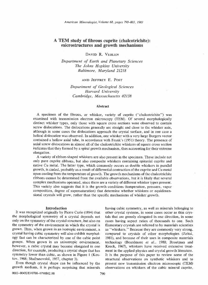
the morphological symmetry of a crystal depends notonly on the symmetry of the crystal structure, but also onthe symmetry of the environment in which the crystal isgrown. Thus, when grown in an isotropic environment, acrystal having cubic symmetry will also exhibit morphol-ogy that can be characterized by one of the cubic pointgroups. When grown in an anisotropic environment,however, a cubic crystal may become elongated in onedirection, for example, producing a crystal shape that hassymmetry lower than cubic, as shown in Figure I (Kos-tov, 1968; Shafranovskii,1957, chapter 3).
Even though crystal shape can be influenced by thegrowth medium, it is perhaps surprising that minerals
having cubic symmetry, as well as minerals belonging toother crystal systems, in some cases occur as thin crys-tals that are grossly elongated in one direction, in somecases having aspect ratios of thousands to one. Suchfilamentary crystals are referred to by materials scientistsas "whiskers." Because they are commonly very strong,compared to crystals of other morphologies (Zoltai,1981), and because of their uses in composite materialstechnology (Beardmore et al., 1980; Broutman andKrock, 1967), whiskers have received extensive treat-ment in the applied physics and crystal growth literature.It is the purpose of this paper to review some of thestructural observations on synthetic whiskers and topresent new transmission electron microscopic (TEM)observations on whiskers of the cubic mineral cuprite,
0003-004x/E3/0708-0790$02. 00 790
VEBLEN AND POST: FIBROUS CUPRITE 79t
a b
Fig. l. a. A dodecahedral crystal ofa cubic mineral grown inan isotropic medium. b. A crystal of the same material that hasgrown in an anisotropic medium (after Kostov, 1968).
which are referred to by the varietal name "chalcotri-chite." These results are discussed in light of possiblegrowth mechanisms for mineral whiskers. In a relatedstudy (Post et al., l98l; and in preparation), we presentmore detailed morphological data on chalcotrichite,based on scanning electron microscopy (SEM).
Whisker growth mechanisms
About forty years ago, it was recognized that crystalgrowth from a vapor phase commonly occurs at levels ofsupersaturation far below those theoretically required forthe nucleation of growth steps on a crystal surface. Toexplain these anomalous results, Frank (1949) proposed amechanism of growth in which a screw dislocation emerg-ing from a crystal face provides a permanent growth step.Such a spiral growth mechanism eliminates the need fornucleation of new layers on a crystal surface and there-fore reduces considerably the degree of supersaturationrequired for crystal growth. Observations of spiral ledgeson growth faces of a wide range of compounds suggestthat Frank's spiral growth mechanism is, in fact, animportant one (see, for example, Verma and Krishna,1966).
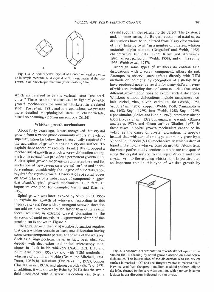
Spiral growth was later invoked by Sears (1953, 1955)to explain the growth of whiskers. According to thistheory, a crystal face with an emergent screw dislocationcan add on new material much faster than other crystalfaces, resulting in extreme crystal elongation in thedirection of rapid growth. A diagrammatic sketch of thismechanism is shown in Figure 2.
The spiral growth theory of whisker formation requiresthat each whisker contain at least one dislocation havingsome screw component parallel to the axis of the whisker.Such axial imperfections have, in fact, been observeddirectly with decoration and optical microscopy tech-niques in alkali halide whiskers (NaCl, KCl, LiF, andKBr: Amelinckx, l958a,b) and with TEM methods inwhiskers of aluminum nitride (Drum and Mitchell, 1964;Drum, 1965a,b), tellurium (Furuta et al., 1972), copper(Hasiguti et al.,1970), and silicon carbide (Shaffer, 1967).In addition, it was shown by Eshelby (1953) that the strainfield associated with a screw dislocation can twist a
crystal about an axis parallel to the defect. The existenceand, in some cases, the Burgers vectors, of axial screwdislocations have been inferred from X-ray observationsof this "Eshelby twist" in a number of diferent whiskermaterials: alpha alumina (Dragsdorf and Webb, 1958),chalcotrichite (Shlichta, 1957; Kirov and Atanassov,1970), silver, palladium (Webb, 1958), and tin (Treuting,1956; Webb et al.,1957).
Although some types of whiskers do contain axialdislocations with a screw component, others do not.Attempts to observe such defects directly with TEMmethods or indirectly by recognition of Eshelby twisthave produced negative results for many different typesof whiskers, including those of some materials that underdifferent growth conditions do exhibit such dislocations.Whiskers without dislocations include manganese, co-balt, nickel, zinc, silver, cadmium, tin (Webb, 1958;Webb et al., 1957), copper (Webb, 1958; Yamamoto etal., 1968;' Regis, 1969), iron (Webb, 1958; Regis, 1969),alpha alumina (Gelles and Binnie, 1969), aluminum nitride(Berezhkova et al., 1972), manganese arsenide (Bdrnerand Berg, 1979), and silicon carbide (Shaffer, 1967). Inthese cases, a spiral growth mechanism cannot be in-voked as the cause of crystal elongation. It appearsinstead that whiskers of this type commonly grow by aVapor-Liquid-Solid (VLS) mechanism, in which a drop ofliquid at the tip of a whisker controls growth. Atoms fromthe vapor perferentially condense into or are transportedalong the crystal surface to the liquid droplet and thencrystallize into the growing whisker tip. Impurities playan important role in this type of whisker growth by
Fig. 2. A schematic representation of a whisker of square crosssection that is forming by spiral growth around an axial screwdislocation. The intersection of the dislocation with the crystalsurface is marked "D" and the Burgers vector is marked "b."New material from the growth medium is added preferentially tothe ledge formed by the screw dislocation, which moves in spiralfashion in the direction indicated bv the arrow.
792 VEBLEN AND POST: FIBROUS CUPRITE
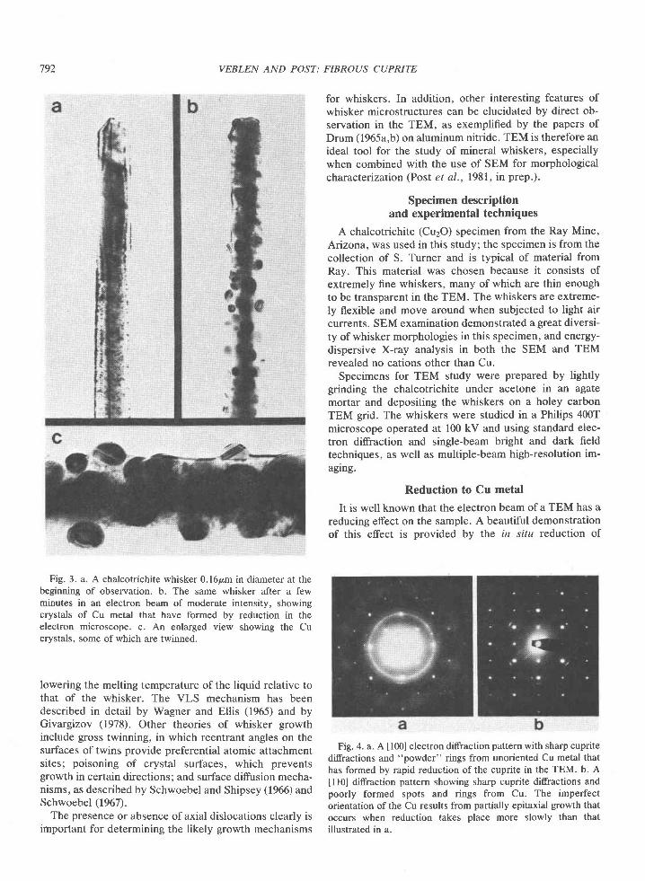
Fig. 3. a. A chalcotrichite whisker 0.l6p,m in diameter at thebeginning of observation. b. The same whisker after a fewminutes in an electron beam of moderate intensity, showingcrystals of Cu metal that have formed by reduction in theelectron microscope. c. An enlarged view showing the Cucrystals, some of which are twinned.
lowering the melting temperature of the liquid relative tothat of the whisker. The VLS mechanism has beendescribed in detail by Wagner and Ellis (1965) and byGivargizov (1978). Other theories of whisker growthinclude gross twinning, in which reentrant angles on thesurfaces of twins provide preferential atomic attachmentsites; poisoning of crystal surfaces, which preventsgrowth in certain directions; and surface diffusion mecha-nisms, as described by Schwoebel and Shipsey (1966) andSchwoebel 1967\.
The presence or absence of axial dislocations clearly isimportant for determining the likely growth mechanisms
for whiskers. In addition, other interesting features ofwhisker microstructures can be elucidated by direct ob-servation in the TEM, as exemplified by the papers ofDrum (1965a,b) on aluminum nitride. TEM is therefore anideal tool for the study of mineral whiskers, especiallywhen combined with the use of SEM for morphologicalcharacteization (Post et al.,1981, in prep.).
Specimen descriptionand experimental techniques
A chalcotrichite (CuzO) specimen from the Ray Mine,Arizona, was used in this study; the specimen is from thecollection of S. Turner and is typical of material fromRay. This material was chosen because it consists ofextremely fine whiskers, many of which are thin enoughto be transparent in the TEM. The whiskers are extreme-ly flexible and move around when subjected to light aircurrents. SEM examination demonstrated a great diversi-ty of whisker morphologies in this specimen, and energy-dispersive X-ray analysis in both the SEM and TEMrevealed no cations other than Cu.
Specimens for TEM study were prepared by lightlygrinding the chalcotrichite under acetone in an agatemortar and depositing the whiskers on a holey carbonTEM grid. The whiskers were studied in a Philips 4007microscope operated at 100 kV and using standard elec-tron diffraction and single-beam bright and dark fieldtechniques, as well as multiple-beam high-resolution im-aglng.
Reduction to Cu metal
It is well known that the electron beam of a TEM has areducing effect on the sample. A beautiful demonstrationof this effect is provided by the in situ reduction of
Fig. 4. a. A [100] electron diffraction pattern with sharp cupritediffractions and "powder" rings from unoriented Cu metal thathas formed by rapid reduction of the cuprite in the TEM. b. A
[10] diffraction pattern showing sharp cuprite diffractions andpoorly formed spots and rings from Cu. The imperfectorientation of the Cu results from partially epitaxial growth thatoccurs when reduction takes place more slowly than thatillustrated in a.
VEBLEN AND POST:
WOr-*, which involves the nucleation and growth ofcrystallographic shear planes (Iijima, 1975; Allpress eral., l97l). Reduction ofcuprite also occurs in the electronmicroscope. However, whereas reasonable reductionrates of WO3-* require the removal of the TEM condens-er aperture, the reduction of cuprite to Cu metal occurseven with a relatively low-intensity electron beam.
Figure 3 shows part of a whisker of chalcotrichite at thebeginning of observation and after exposure to the elec-tron beam for a few minutes. Exposure has resulted in theformation of more-or-less spherical crystals on the sur-face of the whisker; some of the crystals exhibit contrastthat probably arises from twinning. The particles can beidentified from difraction lratterns as copper metal. Fig-ure 4a shows a [100] diffraction pattern of partially-reduced chalcotrichite, in which the diffraction from Cuforms rings and that from cuprite forms sharp spots; thispattern indicates that the Cu is nearly randomly oriented.Such random orientation occurs when irradiation is rela-tively rapid. When reduction occurs in a weaker beam,preferred orientation of the Cu crystals occurs, as a resultof epitaxial growth; Figure 4b is a [l l0] diffraction patternof the whisker shown in Figure 3, exhibiting non-randomCu orientation. Where Cu growth is epitaxial, moir6fringes resulting from the overlapping of the copper andcuprite structures can be observed under multiple-beambright f ield conditions (Fig. 5).
Although most chalcotrichite whiskers that were exam-
Fig. 5. Moird fringes formed by the overlapping of the cupriteand Cu metal structures. The fringes occur at the position ofa Cucrystal that has formed near the whisker tip by reduction in theTEM. The direction of the whisker axis is indicated by the:uTow.
FIBROUS CUPRITE
ined were reduced by the electron beam, it appeared thatsome were more resistant than others. The reason for thisdifference is not fully understood, although it may berelated to whisker shape and size and the quality of theelectrical and chemical contact between the whisker andthe specimen support grid. It was observed in a number ofcases that reduction to Cu metal occurred preferentiallyon parts of whiskers that were in contact with thespecimen grid. This enhanced reduction is probably theresult of reaction with the carbon of the grid, producingCO2. It was also observed that_extremely thin whiskers ofsquare cross section (100-300A) are particularly prone toreduction damage.
The problem of reduction of cuprite to copper metal isan important one, because in some types of whiskersnative copper occurs naturally with cuprite, as discussedin a later section. It is therefore necessary to distinguishbetween primary Cu and secondary Cu that is producedby reduction radiation damage in the electron micro-scope.
Dislocations in whiskers of square cross section
Examination in the SEM of the chalcotrichite sampleused in this study indicates that there are several morpho-logically distinct whisker types (Post et al., 1981, inprep.). These can be separated into two broad categories:(l) whiskers with square or nearly square rectangularcross sections, which can occur as individual crystals orin a parallel growth habit; and (2) ribbons, in which thethickness is much less than the width; there are severaltypes ofribbons, as discussed later. These two categoriesof whiskers could be distinguished readily in the TEM byobserving the whisker outlines while tilting the stage tohigh angles and by observing extinction contours charac-teristic of very thin crystals in the ribbons. In both theribbons and whiskers with square cross section, ditrrac-tion experiments indicate that the fiber axis is parallel toan a-axis, which will henceforth be designated [001]. Inthis section, we discuss the microstructures present in thewhiskers of more-or-less square cross section.
Axial s crew dislocations
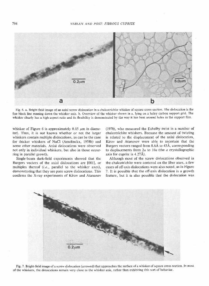
Several hundred whiskers ofsquare cross section wereexamined using standard bright field and, in some cases,dark field techniques. Dislocations were observed in mostof these whiskers (about907o), and experiments in whichthe whiskers were tilted about their axes confirmed thatthe dislocations were, in fact, located on the whisker axisin most cases; such a dislocation is shown in Figure 6a,where it appears as a dark line. A dislocation can also beseen in the whisker in Figure 3a. Figure 6b is an overviewof the whisker in Figure 6a, giving an idea of the highaspect ratio of some of the whiskers and demonstratingtheir flexibility. Only those square whiskers that werevery thin could be examined for dislocations, because thethicker ones are not transparent to the electrons (the
793
794 VEBLEN AND POST: FIBROUS CUPRITE
Fig. 6. a. Bright-field image ofan axial screw dislocation in a chalcotrichite whisker ofsquare cross section. The dislocation is thefine black line running down the whisker axis. b. Overview of the whisker shown in a, lying on a holey carbon support grid. Thewhisker clearly has a high aspect ratio and its flexibility is demonstrated by the way it has bent around holes in the support film.
' : : i
ba
whisker of Figure 6 is approximately 0.15 pm in diame-ter). Thus, it is not known whether or not the largerwhiskers contain multiple dislocations, as can be the casefor thicker whiskers of NaCl (Amelinckx. 1958b) andsome other materials. Axial dislocations were observednot only in individual whiskers, but also in those occur-ring in parallel growth.
Single-beam dark-field experiments showed that theBurgers vectors of the axial dislocations are [001], ormultiples thereof (i.e., parallel to the whisker axes),demonstrating that they are pure screw dislocations. Thisconfirms the X-ray experiments of Kirov and Atanasov
(1970), who measured the Eshelby twist in a number ofchalcotrichite whiskers. Because the amount of twistingis related to the displacement of the axial dislocation,Kirov and Atanosov were able to ascertain that theBurgers vectors ranged from 8.64 to 43A, correspondingto displacements from ?a to lDa (the a crystallographicaxis for cuprite is 4.271r').
Although most of the screw dislocations observed inthe chalcotrichite were centered on the fiber axes, a fewcases of of-axis dislocations were also noted, as in Figure7. It is possible that the off-axis dislocation is a growthfeature, but it is also possible that the dislocation was
' r - , , , ,
Fig. 7. Bright-field image ofa screw dislocation (arrowed) that approaches the surface ofa whiskerofsquare cross section. In mostofthe whiskers, the dislocations remain very close to the whisker axis, rather than exhibiting this sort ofbehavior.
VEBLEN AND POST: FIBROUS CUPRITE 795
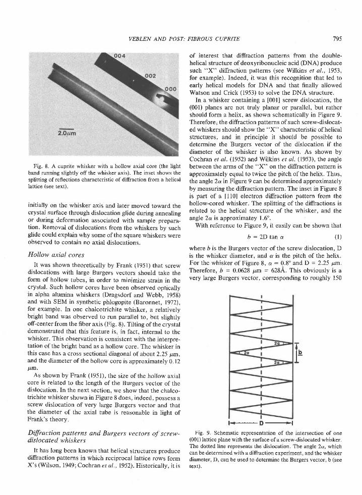
Fig. 8. A cuprite whisker with a hollow axial core (the lightband running slightly off the whisker axis). The inset shows thesplitting of reflections characteristic of ditrraction from a helicallattice (see text).
initially on the whisker axis and later moved toward thecrystal surface through dislocation glide during annealingor during deformation associated with sample prepara-tion. Removal of dislocations from the whiskers by suchglide could explain why some of the square whiskers wereobserved to contain no axial dislocations.
Hollow axial cores
It was shown theoretically by Frank (1951) that screwdislocations with large Burgers vectors should take theform of hollow tubes, in order to minimize strain in thecrystal. Such hollow cores have been observed opticallyin alpha alumina whiskers (Dragsdorf and Webb, 1958)and with SEM in synthetic phlogopite (Baronnet, 1972),for example. In one chalcotrichite whisker, a relativelybright band was observed to run parallel to, but slightlyoff-center from the fiber axis (Fig. 8). Tilting of the crystaldemonstrated that this feature is, in fact, internal to thewhisker. This observation is consistent with the interpre-tation of the bright band as a hollow core. The whisker inthis case has a cross sectional diagonal of about 2.25 p,m,and the diameter of the hollow core is approximately 0.12p.m.
As shown by Frank (1951), the size of the hollow axialcore is related to the length of the Burgers vector of thedislocation. In the next section, we show that the chalco-trichite whisker shown in Figure 8 does, indeed, possess ascrew dislocation of very large Burgers vector and thatthe diameter of the axial tube is reasonable in lieht ofFrank's theory.
Dffiaction patterns and Burgers vectors of screw-dislocated whiskers
It has long been known that helical structures producedifraction patterns in which reciprocal lattice rows formX's (Wilson, 1949; Cochran et a|.,1952). Historicallv. it is
of interest that difraction patterns from the double-helical structure of deoxyribonucleic acid (DNA) producesuch "X" difraction patterns (see Wilkins et al., 1953,for example). Indeed, it was this recognition that led toearly helical models for DNA and that finally allowedWatson and Crick (1953) to solve the DNA structure.
In a whisker containing a [001] screw dislocation, the(fi)l) planes are not truly planar or parallel, but rathershould form a helix, as shown schematically in Figure 9.Therefore; the diffraction patterns of such screw-dislocat-ed whiskers should show the "X" characteristic of helicalstructures, and in principle it should be possible todetermine the Burgers vector of the dislocation if thediameter of the whisker is also known. As shown byCochran et aI. (1952) and Wilkins et al. (1953), the anglebetween the arms of the "X" on the diffraction pattern isapproximately equal to twice the pitch of the helix. Thus,the angle 2a in Figure 9 can be determined approximatelyby measuring the diffraction pattern. The inset in Figure 8is part of a [10] electron diffraction pattern from thehollow-cored whisker. The splitting of the diffractions isrelated to the helical structure of the whisker, and theangJe 2a is approximatey 1.6'.
With reference to Figure 9, it easily can be shown that
b : 2 D t a n a ( l )
where b is the Burgers vector ofthe screw dislocation, Dis the whisker diameter, and a is the pitch of the helix.For the whisker of Figure 8, a : 0.8" and D = 2.25 y.m.Therefore, b = 0.M28 pm = 6284. This obviously is avery large Burgers vector, corresponding to roughly 150
Fig. 9. Schematic representation of the intersection of one(001) lattice plane with the surface ofa screw-dislocated whisker.The dotted line represents the dislocation. The angle 2a, whichcan be determined with a diffraction experiment, and the whiskerdiameter, D, can be used to determine the Burgers vector, b (seetext).
F
t -D.+l
796 VEBLEN AND POST:
times the cuprite cell parameter and much larger than anyof those reported by Kirov and Atanasov (1970). It is notat all surprising that a whisker with a dislocation of suchlarge Burgers vector should contain an axial tube, ratherthan being completely solid!
Frank (1951) gives the approximate equilibrium radiusof an axial tube as
pbzr= *1 Q)
where p is the rigidity modulus of the material, D is theBurgers vector of the dislocation, and y is the surfaceenergy of the material. From this relationship we cancalculate that for the tube diameter and Burgers vectorobserved for the whisker of Figure 8, the ratio /p isapproximately 26 x l0-8 cm. Since this value is of theright order of magnitude (see Frank, 1951), it wouldappear that the Burgers vector and tube diameter in thiswhisker are not inconsistent with the theory.
In whiskers not exhibiting obvious axial tubes, verysmall splitting of diffractions could in a few cases be seenat large values of TEM camera length (i.e., with thediffraction pattern blown up very large). In most cases,however, no splitting associated with the helical structure
FIBROUS CUPRITE
could be observed, consistent with Burgers vectors thatare considerably smaller than the one discussed above.Thus, in agreement with the theory of Frank (1951), itappears that screw dislocations with small Burgers vec-tors do not take the form of hollow axial tubes, whereasthose with very large Burgers vectors do.
Helical dislocations
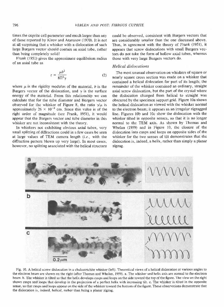
The most unusual observation on whiskers of square ornearly square cross section was made on a whisker thatcontained a helical dislocation for part of its length; theremainder of the whisker contained an ordinary, straightaxial screw dislocation, but the part of the crystal wherethe dislocation changed from helical to straight wasobscured by the specimen support grid. Figure lOa showsthe helical dislocation as viewed with the whisker normalto the electron beam; it appears as an irregular zigzaggedline. Figures lOb and lOc show the dislocation with thewhisker tilted in opposite senses, so that it is no longernormal to the TEM axis. As shown by Thomas andWhelan (1959) and in Figure 10, the closure of thedislocation into cusps and loops on opposite sides of thewhisker for the two senses of tilt demonstrates that thedislocation is, indeed, a helix, rather than simply a planarzigzag.
b
Fig. 10. A helical screw dislocation in a chalcotrichite whisker (left). Theoretical views of a helical dislocation at various angles tothe electron beam are shown on the right (after Thomas and Whelan, 1959). a. The whisker and helix axis are normal to the electronbeam. b. The whisker is tilted so that the helix develops cusps and loops on the side toward the top ofthe figure. The view on the rightshows cusps and loops that develop in the projection ofa perfect helix with increasing tilt. c. The whisker is tilted in the oppositesense, so that cusps and loops appear on the side ofthe whisker toward the bottom ofthe figure. These observations demonstrate thatthe dislocation is, indeed, helical, rather than being a planar zigzag.
VEBLEN AND POST: FIBROUS CUPRITE 797
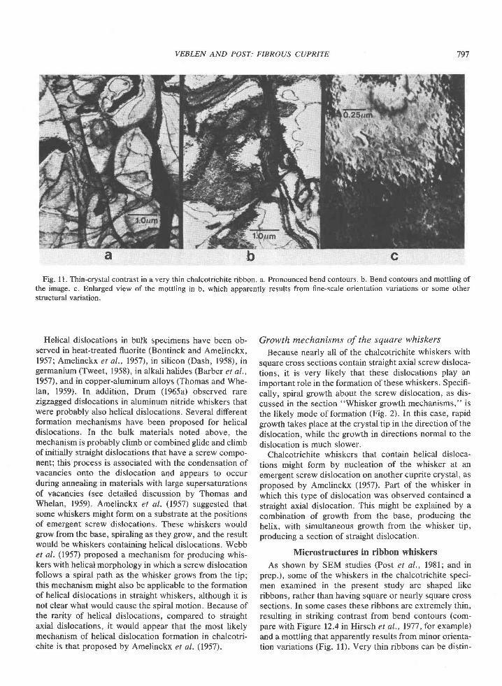
Fig. ll. Thin-crystal contrast in a very thin chalcotrichite ribbon. a. Pronounced bend contours. b. Bend contours and mottling ofthe image. c. Enlarged view of the mottling in b, which apparently results from fine-scale orientation variations or some otherstructural variation.
\r:
cba
Helical dislocations in bulk specimens have been ob-served in heat-treated fluorite (Bontinck and Amelinckx,1957; Amelinckx et al., 1957), in silicon (Dash, 1958), ingermanium (Tweet, 1958), in alkali halides (Barber et al.,1957), and in copper-aluminum alloys (Thomas and Whe-lan, 1959). In addition, Drum (1965a) observed rarezigzagged dislocations in aluminum nitride whiskers thatwere probably also helical dislocations. Several differentformation mechanisms have been proposed for helicaldislocations. In the bulk materials noted above, themechanism is probably climb or combined glide and climbof initially straight dislocations that have a screw compo-nent; this process is associated with the condensation ofvacancies onto the dislocation and appears to occurduring annealing in materials with large supersaturationsof vacancies (see detailed discussion by Thomas andWhelan, 1959). Amelinckx et aI. (1957) suggested thatsome whiskers might form on a substrate at the positionsof emergent screw dislocations. These whiskers wouldgrow from the base, spiraling as they grow, and the resultwould be whiskers containing helical dislocations. Webbet al. (1957) proposed a mechanism for producing whis-kers with helical morphology in which a screw dislocationfollows a spiral path as the whisker grows from the tip;this mechanism might also be applicable to the formationof helical dislocations in straight whiskers, although it isnot clear what would cause the spiral motion. Because ofthe rarity of helical dislocations, compared to straightaxial dislocations, it would appear that the most likelymechanism of helical dislocation formation in chalcotri-chite is that proposed by Amelinckx et al. (1957).
Growth mechanisms of the square whiskers
Because nearly all of the chalcotrichite whiskers withsquare cross sections contain straight axial screw disloca-tions, it is very likely that these dislocations play animportant role in the formation of these whiskers. Specifi-cally, spiral growth about the screw dislocation, as dis-cussed in the section "Whisker growth mechanisms," isthe likely mode of formation (Fig. 2). In this case, rapidgrowth takes place at the crystal tip in the direction of thedislocation, while the growth in directions normal to thedislocation is much slower.
Chalcotrichite whiskers that contain helical disloca-tions might form by nucleation of the whisker at anemergent screw dislocation on another cuprite crystal, asproposed by Amelinckx (1957). Part of the whisker inwhich this type of dislocation was observed contained astraight axial dislocation. This might be explained by acombination of growth from the base, producing thehelix, with simultaneous growth from the whisker tip,producing a section of straight dislocation.
Microstructures in ribbon whiskers
As shown by SEM studies (Post er al., l98l; and inprep.), some of the whiskers in the chalcotrichite speci-men examined in the present study are shaped likeribbons, rather than having square or nearly square crosssections. In some cases these ribbons are extremely thin,resulting in striking contrast from bend contours (com-pare with Figure 12.4 in Hirsch et al., 1977, for example)and a mottling that apparently results from minor orienta-tion variations (Fig. ll). Very thin ribbons can be distin-
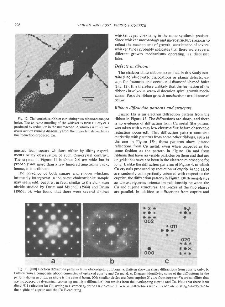
798 VEBLEN AND POST:
Fig. 12. Chalcotrichite ribbon containing two diamond-shapedholes. The extreme mottling of the whisker is from Cu crystalsproduced by reduction in the microscope. A whisker with squarecross section running diagonally from the upper left also exhibitsthis reduction-produced Cu.
guished from square whiskers either by tilting experi-ments or by observation of such thin-crystal contrast.The crystal in Figure 11 is about 2.4 p,m wide but isprobably not more than a few hundred ingstrrims thick;hence, it is a ribbon.
The presence of both square and ribbon whiskersintimately intergrown in the same chalcotrichite samplemay seem odd, but it is, in fact, similar to the aluminumnitride studied by Drum and Mitchell (1964) and Drum(1965a, b), who found that there were several distinct
FIBROUS CUPRITE
whisker types coexisting in the same synthesis product.Since whisker morphology and microstructures appear toreflect the mechanisms of growth, coexistence of severalwhisker types probably indicates that there were severaldifferent growth mechanisms operating, as discussedlater.
Defects in ribbons
The chalcotrichite ribbons examined in this study con-tained no observable dislocations or planar defects, ex-cept for fractures and occasional diamond-shaped holes(Fig. l2). It is therefore unlikely that the formation of theribbons involved a screw dislocation spiral growth mech-anism. Possible ribbon growth mechanisms are discussedbelow.
Ribbon dffiaction patterns and structure
Figure l3a is an electron diffraction pattern from theribbon in Figure 12. The diffractions are sharp, and thereis no evidence of diffraction from Cu metal (the patternwas taken with a very low electron flux before observablereduction occurred). This diffraction pattern contrastsmarkedly with patterns from some other ribbons, such asthe one in Figure l3b; these patterns show intensereflections from Cu metal, even when recorded in thesame fashion as the pattern in Figure 13a and fromribbons that have no visible particles on them and that areon grids that have not been in the electron microscope forlong. Unlike the difraction patterns of Figure 4, in whichCu crystals produced by reduction of cuprite in the TEMare randomly or imperfectly oriented with respect to thecuprite, the diffraction pattern in Figure 13b demonstratesan almost rigorous orientation relationship between theCu and cuprite structures: the a-axes of the two phasesare parallel. In addition to diffractions from cuprite and
Fig. 13. [l00] electron ditrraction patterns from chalcotrichite ribbons. a. Pattern showing sharp ditrractions from cuprite only. b.Pattern from a composite ribbon consisting of epitaxial cuprite and Cu metal. c. Diagram identifying some of the diffractions in thepattern shown in b. Large circle is the central beam, 000; smaller circles are from cuprite; X's are from copper; *'s are satellites thatare introduced by dynamical scattering (multiple diffraction) that results from the overlapping cuprite and Cu. Note that there is nodirect 0l I reflection for Cu, owing to F-centering of the Cu structure. Likewise, difractions with fr + / odd are missing entirely due tothe n-glide of cuprite and the Cu F-centering.
VEBLEN AND POST: FIBROUS CUPRITE 799
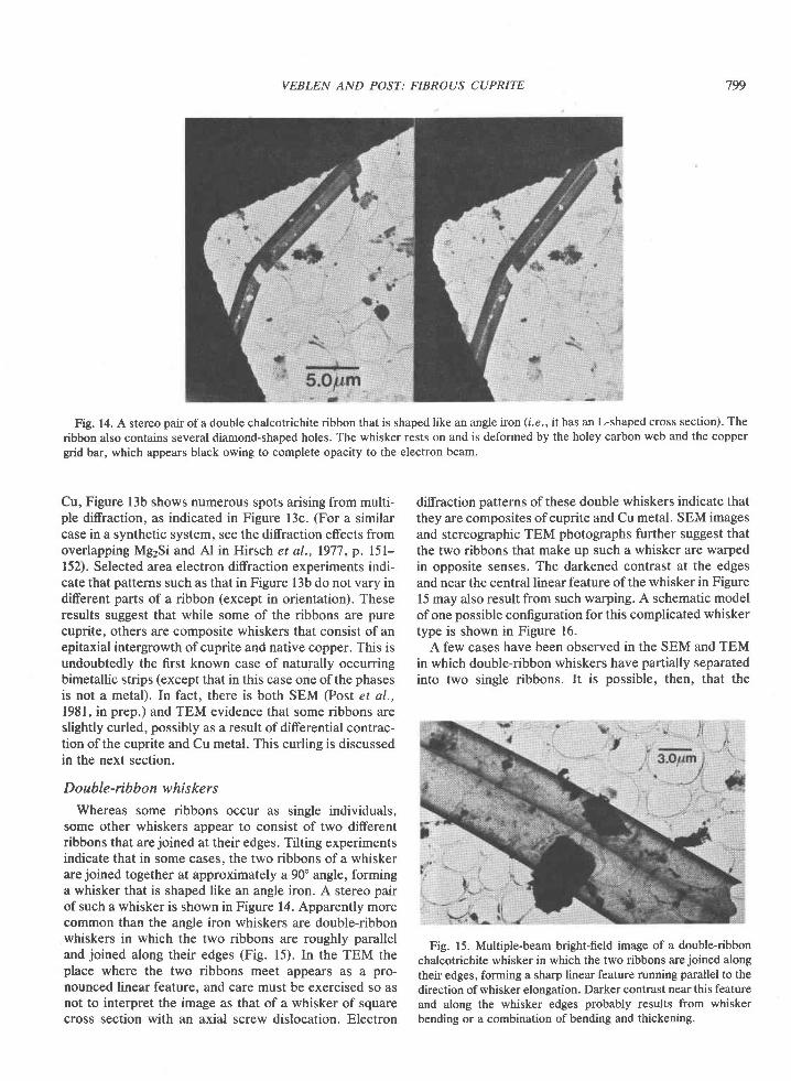
Fig. 14. A stereo pair of a double chalcotrichite ribbon that is shaped like an angle iron (i.e., it has an L-shaped cross section). Theribbon also contains several diamond-shaped holes. The whisker rests on and is deformed by the holey carbon web and the coppergrid bar, which appears black owing to complete opacity to the electron beam.
Cu, Figure l3b shows numerous spots arising from multi-ple diffraction, as indicated in Figure l3c. (For a similarcase in a synthetic system, see the difraction effects fromoverlapping Mg2Si and Al in Hirsch et al., 1977, p. l5l-152). Selected area electron difraction experiments indi-cate that patterns such as that in Figure 13b do not vary indifferent parts of a ribbon (except in orientation). Theseresults suggest that while some of the ribbons are purecuprite, others are composite whiskers that consist of anepitaxial intergrowth of cuprite and native copper. This isundoubtedly the first known case of naturally occurringbimetallic strips (except that in this case one of the phasesis not a metal). In fact, there is both SEM (Post et al.,1981, in prep.) and TEM evidence that some ribbons areslightly curled, possibly as a result of differential contrac-tion of the cuprite and Cu metal. This curling is discussedin the next section.
Double-rib bon whis ke r s
Whereas some ribbons occur as single individuals,some other whiskers appear to consist of two differentribbons that are joined at their edges. Tilting experimentsindicate that in some cases. the two ribbons of a whiskerare joined together at approximately a 90" angle, forminga whisker that is shaped like an angle iron. A stereo pairof such a whisker is shown in Figure 14. Apparently morecommon than the angle iron whiskers are double-ribbonwhiskers in which the two ribbons are roughly paralleland joined along their edges (Fig. l5). In the TEM theplace where the two ribbons meet appears as a pro-nounced linear feature. and care must be exercised so asnot to interpret the image as that of a whisker of squarecross section with an axial screw dislocation. Electron
diffraction patterns of these double whiskers indicate thatthey are composites of cuprite and Cu metal. SEM imagesand stereographic TEM photographs further suggest thatthe two ribbons that make up such a whisker are warpedin opposite senses. The darkened contrast at the edgesand near the central linear feature ofthe whisker in Figure15 may also result from such warping. A schematic modelof one possible configuration for this complicated whiskertype is shown in Figure 16.
A few cases have been observed in the SEM and TEMin which double-ribbon whiskers have partially separatedinto two single ribbons. It is possible, then, that the
Fig. 15. Multiple-beam bright-field image of a double-ribbonchalcotrichite whisker in which the two ribbons are joined alongtheir edges, forming a sharp linear feature running parallel to thedirection ofwhisker elongation. Darker contrast near this featureand along the whisker edges probably results from whiskerbending or a combination of bending and thickening.
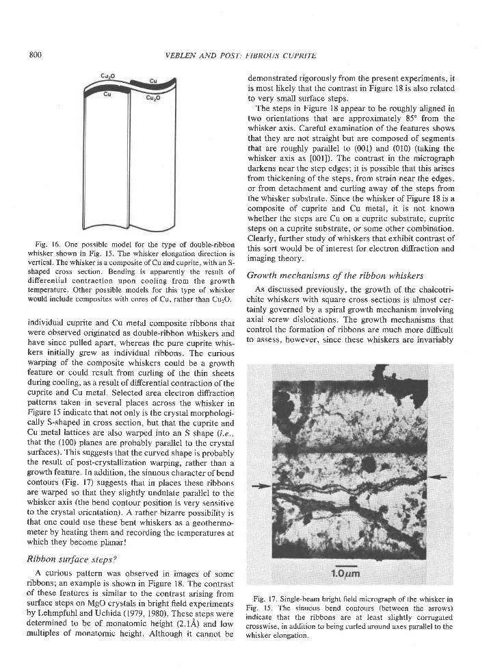
800 VEBLEN AND POST: FIBROUS CUPRITE
Fig. 16. One possible model for the type of double-ribbonwhisker shown in Fig. 15. The whisker elongation direction isvertical. The whisker is a composite of Cu and cuprite, with an S-shaped cross section. Bending is apparently the result ofdifferential contraction upon cooling from the growthtemperature. Other possible models for this type of whiskerwould include composites with cores of Cu, rather than Cu2O.
individual cuprite and Cu metal composite ribbons thatwere observed originated as double-ribbon whiskers andhave since pulled apart, whereas the pure cuprite whis-kers initially grew as individual ribbons. The curiouswarping of the composite whiskers could be a growthfeature or could result from curling of the thin sheetsduring cooling, as a result of differential contraction of thecuprite and Cu metal. Selected area electron diffractionpatterns taken in several places across the whisker inFigure 15 indicate that not only is the crystal morphologi-cally S-shaped in cross section, but that the cuprite andCu metal lattices are also warped into an S shape (1.e.,that the (100) planes are probably parallel to the crystalsurfaces). This suggests that the curved shape is probablythe result of post-crystallization warping, rather than agrowth feature. In addition, the sinuous character ofbendcontours (Fig. 17) suggests that in places these ribbonsare warped so that they slightly undulate parallel to thewhisker axis (the bend contour position is very sensitiveto the crystal orientation). A rather bizarre possibility isthat one could use these bent whiskers as a geothermo-meter by heating them and recording the temperatures atwhich they become planar!
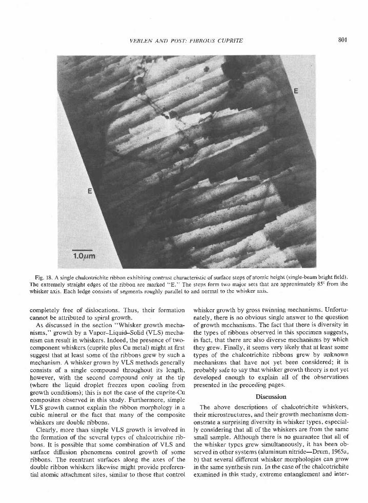
Ribbon surface steps?
A curious pattern was observed in images of someribbons; an example is shown in Figure 18. The contrastof these features is similar to the contrast arising fromsurface steps on MgO crystals in bright field experimentsby Lehmpfuhl and Uchida (1979, 1980). These steps weredetermined to be of monatomic height (2.14) and lowmultiples of monatomic height. Although it cannot be
demonstrated rigorously from the present experiments, itis most likely that the contrast in Figure 18 is also relatedto very small surface steps.
The steps in Figure 18 appear to be roughly aligned intwo orientations that are approximately 85" from thewhisker axis. Careful examination of the features showsthat they are not straight but are composed of segmentsthat are roughly parallel to (001) and (010) (taking thewhisker axis as [001]). The contrast in the micrographdarkens near the step edges; it is possible that this arisesfrom thickening of the steps, from strain near the edges,or from detachment and curling away of the steps fromthe whisker substrate. Since the whisker of Figure 18 is acomposite of cuprite and Cu metal, it is not knownwhether the steps are Cu on a cuprite substrate, cupritesteps on a cuprite substrate, or some other combination.Clearly, further study of whiskers that exhibit contrast ofthis sort would be of interest for electron diffraction andimaging theory.
Growth mechanisms of the ribbon whiskers
As discussed previously, the growth of the chalcotri-chite whiskers with square cross sections is almost cer-tainly governed by a spiral growth mechanism involvingaxial screw dislocations. The growth mechanisms thatcontrol the formation of ribbons are much more difrcultto assess, however. since these whiskers are invariablv
Fig. 17. Single-beam bright field micrograph of the whisker inFig. 15. The sinuous bend contours (between the arrows)indicate that the ribbons are at least slightly corrugatedcrosswise, in addition to being curled around axes parallel to thewhisker elongation.
VEBLEN AND POST: FIBROUS CUPRITE
Fig. 18. A single chalcotrichite ribbon exhibiting contrast characteristic of surface steps of atomic height (single-beam bright field).The extremely straight edges ofthe ribbon are marked "E." The steps form two major sets that are approximately 85" from thewhisker axis. Each ledge consists of segments roughly parallel to and normal to the whisker axis.
801
completely free of dislocations. Thus, their formationcannot be attributed to spiral growth.
As discussed in the section "Whisker growth mecha-nisms," growth by a Vapor-Liquid-Solid (VLS) mecha-nism can result in whiskers. Indeed, the presence oftwo-component whiskers (cuprite plus Cu metal) might at firstsuggest that at least some of the ribbons grew by such amechanism. A whisker grown by VLS methods generallyconsists of a single compound throughout its length,however, with the second compound only at the tip(where the liquid droplet freezes upon cooling fromgrowth conditions); this is not the case of the cuprite-Cucomposites observed in this study. Furthermore, simpleVLS growth cannot explain the ribbon morphology in acubic mineral or the fact that many of the compositewhiskers are double ribbons.
Clearly, more than simple VLS growth is involved inthe formation of the several types of chalcotrichite rib-bons. It is possible that some combination of VLS andsurface diffusion phenomena control growth of someribbons. The reentrant surfaces along the axes of thedouble ribbon whiskers likewise might provide preferen-tial atomic attachment sites, similar to those that control
whisker growth by gross twinning mechanisms. Unfortu-nately, there is no obvious single answer to the questionof growth mechanisms. The fact that there is diversity inthe types of ribbons observed in this specimen suggests,in fact, that there are also diverse mechanisms by whichthey grew. Finally, it seems very likely that at least sometypes of the chalcotrichite ribbons grew by unknownmechanisms that have not yet been considered; it isprobably safe to say that whisker growth theory is not yetdeveloped enough to explain all of the observationspresented in the preceding pages.
Discussion
The above descriptions of chalcotrichite whiskers,their microstructures, and their growth mechanisms dem-onstrate a surprising diversity in whisker types, especial-ly considering that all of the whiskers are from the samesmall sample. Although there is no guarantee that all ofthe whisker types grew simultaneously, it has been ob-served in other systems (aluminum nitride-Drum,1965a,b) that several diferent whisker morphologies can growin the same synthesis run. In the case of the chalcotrichiteexamined in this study, extreme entanglement and inter-
802 VEBLEN AND POST: FIBROUS CUPRITE
twining of the various whisker types also strongly sug-gests that they grew simultaneously. The presence ofseveral different whisker types that have apparentlygrown by different mechanisms therefore suggests that itis the growth conditions (7, P, vapor composition, anddegree of supersaturation) that determine whether whis-ker or equidimensional crystal growth will occur, ratherthan the specific mechanisms of whisker growth. Thus,when conditions appropriate for whisker growth areattained, whiskers form by a variety of mechanisms.
This paper has described a foray into the world ofmorphological crystallography. This may be unusual inthis day of sophisticated scientific instruments, and it isfitting, perhaps, that we close with a brief discussion ofrelevant ideas from the past. First, it is clear from thegreat successes in predicting crystal morphology fromcrystal structure that structure alone in many cases mustdetermine morphology (for example, Donnay andHarker, 1937; Hartman and Perdok, 1956; Dowty, 1976,1977; Hartman, 1977). In some cases, however, as in thepresent case of whisker growth in cuprite, crystal struc-ture is clearly not the only morphological determinant.
As mentioned in the introduction, Pierre Curie (1894)showed that the symmetry properties of a crystal aredependent not only on the symmetry of the crystalstructure, but also on the symmetry of the mediumsurrounding the crystal. It was later shown that morpho-logical symmetry also can be dependent on the symme-tries of molecules dissolved in the growth medium (forexample, Friedel and Weil, 1930). Thus, crystal morphol-ogy is the result not only of crystal structure and symme-try, but also the symmetry of the growth medium (Sha-franovskii, 1957). There are therefore two general ways inwhich we can account for whisker growth in cubicmaterials: (l) anisotropy of the growth medium and (2)some sort ofanisotropy that has been introduced into thecrystal structure.
The case of VLS growth can be classified in the firstcategory, since the direction of whisker elongation resultsfrom the position of the liquid droplet from which thewhisker crystallizes. On the other hand, whisker forma-tion by spiral growth about a screw dislocation can occurin an isotropic growth medium and falls in the secondcategory. In this case, the crystal structure itself pos-sesses cubic symmetry, neglecting strain arising from thedislocation. (Although chalcotrichite morphology has ledsome to conclude that it has tetragonal symmetry, mod-ern X-ray experiments suggest that it is truly cubic-seePost e/ al., l98l; and in prep.) However, the isotropy ofthe crystal as a whole is violated by the screw dislocation.It is indeed remarkable that the reduction in symmetryresulting from a single dislocation can lead to such drasticalteration of a physical property such as crystal shape.
AcknowledgmentsWe thank Dr. Shirley Turner for supplying the chalcotrichite
specimen used in this study. We also thank I. D. R. Mackinnon
for his critical review. This research was supported by NSFgrant EAR8ll5790. Microscopy was performed at the AizonaState University Facility for nneu, which was established withsuppoft from NSF grant CHE79I6D8.
ReferencesAllpress, J. G., Tilley, R. J. D., and Sienko, M. J. (1971)
Examination of substoichiometric WO3-* crystals by electronmicroscopy. Journal of Solid State Chemistry,3, 440451.
Amelinckx, S. (1958a) Decoration of dislocations in alkali halidewhiskers. Journal of Applied Physics, 29, 1610-16ll.
Amelinckx, S. (1958b) Dislocations in alkali halide whiskers. InR. H. Doremus et al., Eds., Growth and Perfection of Crys-tals, p. 139-153. John Wiley and Sons, Inc., New York.
Amelinckx, S., Bontinck, W., Dekeyser, W., and Seitz, F.(1957) On the formation and properties of helical dislocations.Philosophical Magazine, Eth Series, 2, 355-37 8.
Barber, D. J., Harvey, K. B., and Mitchell, J. W. (1957) A newmethod for decorating dislocations in crystals of alkali halides.Philosophical Magazine, 8th Series, 2, 7 M-7 08.
BArner, K. and Berg, H. (1979) The growth figures of MnAs.Journal of Crystal Growth, 46,763J70.
Baronnet, A. (1972) Growth mechanisms and polytypism insynthetic hydroxyl-bearing phlogopite. American Mineral-ogist,57, 1272-1293.
Beardmore, P., Harwood, J. J., and Kinsman, K. R. (19E0)Fiber-reinforced composites: Engineered structural materials.Science, 208, 833-840.
Berezhkova, G. V., Tsvetkova, I. N., Zakharov, N. D., Roz-hanskii, V. N., and Koryukin, V.l. (1972) Growth mechanismof filamentary AIN crystals. Soviet Physics-Crystallography,16 .84E-851.
Bontinck, W. and Amelinckx, S. (1957) Helicoidal dislocationlines in fluorite crystals. Philosophical Magazine, Sth Series, 2,94-96.
Broutman, L. J. and Krock, R. H. (1967) Modern CompositeMaterials. Addison-Wesley Publishing Co., Reading, Massa-chusetts.
Cochran, W., Crick, F. H. C., and Vand, V. (1952) The structureof synthetic polypeptides. L The transform of atoms on ahelix. Acta Crystallographica, 5, 581-586.
Curie, Pierre (1894) On the symmetry in physical phenomena,symmetry of an electrical field and of a magnetic field. (inFrench) Journal de Physique, 1,II1,393415.
Dash, W. C. (1958) The growth of silicon crystals free fromdislocations. In R. H. Doremus. B. W. Roberts. and DavidTumbull, Eds., Growth and Perfection of Crystals, p. 361-385.John Wiley and Sons, Inc., New York.
Donnay, J. D. H. anil Harker, D. (1937) A new law of crystalmorphology extending the law of Bravais. American Mineral-ogist,22, 446467.
Dowty, Enc (976) Crystal structure and crystal growth: I. Theinfluence of internal structure on morphology. American Min-eralogist, 61,448459.
Dowty, Eric (1977) Crystal structure and crystal growth: I. Theinfluence of internal structure on morphology: a reply. Ameri-can Mineralogist, 62, 1036-1037.
Dragsdorf, R. D. and Webb, W. W. (195E) Detection of screwdislocations in a-Al2O3 whiskers. Journal of Applied Physics,29, El7-8r9.
Drum, C. M. (1965a) Axial imperfections in filamentary crystals
VEBLEN AND POST: FIBROUS CUPRITE 803
of aluminum nitride. I. Journal of Applied Physics, 36, 816-823.
Drum, C. M. (1965b) Twist and axial imperfections in filamenta-ry crystals of aluminum nitride. II. Journal of Applied Physics,36,824-829.
Drum, C. M. and Mitchell, J. W. (1964) Electron microscopicexamination of role of axial dislocations in growth of AINwhiskers. Applied Physics Letters, 4, 16/-165.
Eshelby, J. D. (1953) Screw dislocations in thin rods. Journal ofApplied Physics, 24, 176-179.
Frank, F. C. (1949) The influence of dislocations on crystalgrowth. Discussions of the Faraday Society, 5, 48-54.
Frank, F. C. (1951) Capillary equilibria of dislocated crystals.Acta Crystallographica, 4, 497 -501.
Friedel, G. and Weil, R. (1930) Influence of the symmetry of themedium on the symmetry of crystalline forms. Comptes Ren-dus Hebdomadaires des S6ances de L'Acad6mie des Sciences,r%,243-24s.
Furuta, N., Itinose, H., Maruyama, N., and Ohasi, Y. (1972)Morphology and dislocation structure of tellurium whiskersgrown from the vapor. Japanese Journal of Applied Physics,l l , l l l 3 - l l l 8 .
Gelles, I. L. and Binnie, W. P. (1969) High index growth of a-Al2O3 filamentary crystals. Materials Science and Engineering,5 ,71-92.
Givargizov, E. I. (1978) Growth of whiskers by the vaporJiquid-solid mechanism. In E. Kaldis, Ed., Current Topics in Materi-als Science. 1.82-145.
Hartman, P. (1977) Crystal structure and crystal growth: L Theinfluence of internal structure on morphology: a discussion.American Mineralogist, 62, 1034-1035.
Hartman, P. and Perdok, W. G. (1956) An interpretation of thelaw of Donnay and Harker. American Mineralogist, 41, 449-459.
Hasiguti, R. R., Yagi, E., Nishiike, U., and Sakai, T. (1970) Anaxial dislocation in copper whiskers. Journal of CrystalGrowth, 7, ll7-119.
Hirsch, Peter, Howie, A., Nicholson, R. B., Pashley, D. W., andWhelan, M. J. (1977) Electron Microscopy of Thin Crystals.Robert E. Krieger Publishing Company, Malabar, Florida.
Iijima, Sumio (1975) High-resolution electron microscopy ofcrystallographic shear structures in tungsten oxides. Journal ofSolid State Chemistry, 14,52-65.
Kirov, G. N. and Atanasov, V. A. (1970) Cuprite whiskers andthin plates from the occurrence Bartzeto, Malko Turnovo. (inBulgarian) Annuaire de I'Universit€ de Sofia Facultd de G6o-logie at G€ographie, Livre l, Gdologie, 62, l9l-198.
Kostov, Ivan (1968) Mineralogy. Oliver and Boyd. Edinburgh.Lehmpfuhl, G. and Uchida, Y. (1979) Darkfield and brightfield
techniques for electron microscopic observation of atomicsteps on MgO single-crystal surfaces. Ultramicroscopy, 4,275-282.
Lehmpfuhl, G. and Uchida, Y. (1980) Dark- and bright-fieldtechniques for electron-microscopic observation of atomicsteps on MgO single crystal surfaces. In Electron Microscopyand Analysis 1979 (eurc79), Institute of Physics ConferenceSeries.52. 393-396.
Post, J. R., Veblen, D. R., and Buseck, P. R. (1981) Electronmicroscopic study of fibrous cuprite (chalcotrichite). (abstr.)
Geological Society of America Abstracts with Programs, 13,531 .
Regis, M. (1969) Axial screw-dislocation absence in metallicwhiskers made from solid halides. Acta Metallurgica, 17,t28t-1289.
Schwoebel, R. L. (1967) A diffusion model for filamentarycrystal growth. Journal of Applied Physics, 38, 1759-1765.
Schwoebel, R. L. and Shipsey, E. J. (1966) Step motion oncrystal surfaces. Journal of Applied Physics, 37,3682-3686.
Sears, G. W. (1953) Mercury whiskers. Acta Metallurgica, l,457-459.
Sears, G. W. (1955) A growth mechanism for mercury whiskers.Acta Metallurgica, 3, 361-366.
Shaffer, P. T. B. (1967) Whiskers-their growth and properties.In L. J. Broutman and R. H. Krock, Eds., Modern CompositeMaterials, p. 197-216. Addison-Wesley Publishing Co., Read-ing, Massachusetts.
Shafranovskii, I. I. (1957) Crystals of Minerals, Vol. I. (inRussian) Leningrad University.
Shlichta, P. J. (1957) Torsional strain of whiskers. (abstr.)American Physical Society Bulletin, 2, 263.
Thomas, Garreth and Whelan, M. J. (1959) Helical dislocationsin quenched ahtminium-4Vo copper alloys. Philosophical Mag-azine, 8th Series, 4, 5ll-527.
Treuting, R. G. (1956) Torsional strain and the screw dislocationin whisker crystals. (abstr.) American Physical Society Bulle-t i n , 1 , 3 3 3 .
Tweet, A. G. (1958) Evidence for vacancy clusters in disloca-tion-free Ge. Journal of Applied Physics, 29, 1520-1522.
Verma, A. R. and Krishna, P. (1966) Polymorphism and Polytyp-ism in Crystals. John Wiley and Sons, Inc., New York.
Wagner, R. S. and Ellis, W. C. (1965) The vaporJiquid-solidmechanism of crystal growth and its application to silicon.Transactions of the Metallurgical Society of eruB, 233,1053-1964.
Watson, J. D. and Crick, F. H. C. (1953) Molecular structure ofnucleic acids. Nature, l7 l, 737:738.
Webb, W. W. (1958) Dislocation structure of whiskers. In R. H.Doremus et al., Eds., Growth and Perfection of Crystals, p.230-238. John Wiley and Sons, Inc., New York.
Webb, W. W., Dragsdorf, R. D., and Forgeng, W. D. (1957)Dislocations in whiskers. Physical Review, 108, 498-499.
Wilkins, M. H. F., Stokes, A. R., and Wilson, H. R. (1953)Molecular structure of deoxypentose nucleic acids. Nature,t7t,'138-140.
Wilson, A. J. C. (1949) Diffraction by a screw dislocation.Research (succeeded by Research Applied in Industry), 2,541-542.
Yamamoto, M., Gotoh, Y., and Yoshida, K. (1968) Transmis-sion electron-microscopic study on the growth of copperwhiskers by halide reduction. Journal of Crystal Growth, 3-4,705J10.
Zoltai, Tibor (1981) Amphibole asbestos mineralogy. In D. R.Veblen, Ed., Amphiboles and Other Hydrous Pyriboles, Min-eralogical Society of America Reviews in Mineralogy, 9,4,237-278.
Manuscript received, June 25, 1982;acceptedfor publication, November 29, 1982.