a systematic review of the effectiveness and safety of...
TRANSCRIPT
SYSTEMATIC REVIEW OF THE EFFECTIVENESS
AND TOLERABILITY OF HYALURONIC ACID
FOR ACUTE AND CHRONIC WOUNDS
ATIKAH SHAHARUDIN
FACULTY OF MEDICINE
UNIVERSITY OF MALAYA
KUALA LUMPUR
2015

SYSTEMATIC REVIEW OF THE EFFECTIVENESS
AND TOLERABILITY OF HYALURONIC ACID FOR
ACUTE AND CHRONIC WOUNDS
ATIKAH SHAHARUDIN
DISSERTATION SUBMITTED IN FULFILMENT
OF THE REQUIREMENTS FOR THE DEGREE OF
MASTER IN MEDICAL SCIENCES
FACULTY OF MEDICINE
UNIVERSITY OF MALAYA
KUALA LUMPUR
2015

ii
UNIVERSITY OF MALAYA
ORIGINAL LITERARY WORK DECLARATION
Name of Candidate: ATIKAH SHAHARUDIN (I.C/Passport No: 840507-05-5046)
Registration/Matric No: MGN 130002
Name of Degree: MASTER OF MEDICAL SCIENCES
Title of Project Paper/Research Report/Dissertation/Thesis (“this Work”):
SYSTEMATIC REVIEW OF THE EFFECTIVENESS AND TOLERABILITY
OF HYALURONIC ACID FOR ACUTE AND CHRONIC WOUNDS
Field of Study: EVIDENCE-BASED PHARMACOTHERAPY
I do solemnly and sincerely declare that:
(1) I am the sole author/writer of this Work;
(2) This Work is original;
(3) Any use of any work in which copyright exists was done by way of fair
dealing and for permitted purposes and any excerpt or extract from, or
reference to or reproduction of any copyright work has been disclosed
expressly and sufficiently and the title of the Work and its authorship have
been acknowledged in this Work;
(4) I do not have any actual knowledge nor do I ought reasonably to know that
the making of this work constitutes an infringement of any copyright work;
(5) I hereby assign all and every rights in the copyright to this Work to the
University of Malaya (“UM”), who henceforth shall be the owner of the
copyright in this Work and that any reproduction or use in any form or by any
means whatsoever is prohibited without the written consent of UM having
been first had and obtained;
(6) I am fully aware that if in the course of making this Work I have infringed
any copyright whether intentionally or otherwise, I may be subjected to legal
action or any other actions as may be determined by UM.
Candidate’s Signature Date:
Subscribed and solemnly declared before,
Witness’s Signature Date:
Name:
Designation:
iii
ABSTRACT
Hyaluronic Acid (HA) and its derivatives are commonly used for acute and chronic
wounds, but evidence of their effectiveness remains unclear. The aim of the study was
to evaluate the effectiveness of HA (or its derivatives) for promoting healing in acute
and chronic wounds through a systematic review of the available evidence. The
Cochrane Central Register of Controlled Trials and relevant databases were searched.
Drugs companies and experts in wounds were also contacted. Randomised controlled
trials of HA (or its derivatives) compared with control were eligible for inclusion. Ten
randomised controlled trials involving 992 participants with acute and chronic wounds
were included in the review. The research evidence was weak with poor reporting in
many trials. The evidence does not provide strong support for the beneficial effects of
HA (or its derivatives) towards improvement of chronic wounds even though there is
some evidences that they were effective for reducing pain intensity for mixed arterial
and venous ulcers [MD= -6.78 (95% CI: -11.10 to -2.46)]. Evidence to guide decisions
regarding the use of HA (or its derivatives) to promote wound healing is still limited.
More good quality randomised controlled trials are warranted.

iv
ABSTRAK
Asid Hialuronik (HA) dan terbitan-terbitannya biasa digunakan untuk penyembuhan
luka akut dan luka kronik, namun keberkesanan mengenainya masih tidak meyakinkan.
Kajian ini bertujuan untuk menilai keberkesanan HA (atau terbitan-terbitannya) di
dalam merangsang penyembuhan luka akut dan luka kronik melalui kaedah tinjauan
sistematik bagi bukti-bukti yang sedia ada. Pencarian telah dilakukan melalui Cochrane
Central Register of Controlled Trials dan pangkalan data yang berkaitan. Syarikat-
syarikat dan pakar-pakar di dalam bidang luka juga telah dihubungi. Kajian-kajian
rawak terkawal berkaitan HA (atau terbitan-terbitannya) berbanding dengan unsur
kawalan adalah termasuk didalam kriteria. Sepuluh kajian rawak terkawal yang
melibatkan 992 peserta luka akut dan luka kronik termasuk di dalam senarai. Bukti-
bukti sedia ada adalah lemah kerana terdapat kekurangan laporan di dalam kebanyakan
kajian-kajian. Bukti-bukti tersebut tidak menyokong kuat kebaikan efek HA (atau
terbitan-terbitannya) terhadap penyembuhan luka kronik walaupun terdapat satu bukti
yang menunjukkan efek statistikal signifikan di dalam pengurangan kadar kesakitan
bagi ‘mixed arterial and venous ulcers’ [MD= -6.78 (95% CI: -11.10 to -2.46). Bukti
sebagai rujukan berkaitan penggunaan HA (atau terbitan-terbitannya) untuk merangsang
penyembuhan luka masih terhad. Lebih banyak kajian yang mempunyai kualiti yang
tinggi diperlukan.

v
ACKNOWLEDGEMENTS
Alhamdulillah, first and foremost praise is to Allah, the Almighty, the greatest of all, on
whom ultimately I depend for sustenance and guidance.
To my supervisor, Associate Professor Datin Dr. Zoriah Aziz, I would like to express my
deepest gratitude for her encouragement, supervision, advices and patience from the very
beginning to the end of the study. This dissertation would have not been possible without
her generous support. She is my ever-present inspiration. I will keep in mind all her
professional advice to me, because she can understand me as an individual. I really
appreciate it. I am beyond thankful to have her as my supervisor.
I am indebted to Hanisah Azhari who is now studying in New Zealand for giving me a huge
help during the searching for literatures. Also to Chong Nyuk Jet, Hafizeen Omar and Tang
Wei Ling, and those who have contributed constructively to the review work. To my
amazing colleagues at the postgraduate pharmacy department; Syarifah Intan Khadijah,
Dhania, Raymond, Baheerah, Aziah, Masyitah thank you for making me laugh so much and
so often. To all staff in the pharmacy department; Najib, Maria, Salbiah, Nadia, Rustini,
Zuriana thank you for your deep commitment towards quality administration services and
sharing so much time with me and my sons.
I am blessed and truly grateful to my incredibly supportive husband, my sons, and
especially my mom and dad who teach by example and have touched and inspired so many
yet would never admit it. They give my life meaning.
Last but not least, to the major financial support from the Ministry of Health and IPPP,
UM. Without their sponsorship, I might not be able to make my dream come true.

vi
TABLE OF CONTENTS
Abstract ........................................................................................................................... iii
Abstrak ............................................................................................................................. iv
Acknowledgements ........................................................................................................... v
Table of Contents ............................................................................................................. vi
List of Figures ................................................................................................................... x
List of Tables .................................................................................................................... xi
List of Symbols and Abbreviations ................................................................................. xii
List of Appendices ........................................................................................................ xiii
CHAPTER 1: INTRODUCTION .................................................................................. 1
1.1 Aim….. .................................................................................................................... 3
1.2 Specific Objectives .................................................................................................. 3
1.3 Justification of this study ......................................................................................... 3
CHAPTER 2: LITERATURE REVIEW ...................................................................... 5
2.1 Types of wounds ...................................................................................................... 5
2.1.1 Acute wounds ............................................................................................. 5
Surgical wounds ........................................................................................ 5
Burn wounds…. ......................................................................................... 6
2.1.2 Chronic wounds .......................................................................................... 6
Diabetic foot ulcers ................................................................................... 7
Pressure ulcers …………………………………………………………...7
Venous ulcers… ......................................................................................... 7
2.1.3 Prevalence and burden of wounds .............................................................. 8
2.2 Wound-healing Process ........................................................................................... 9
vii
2.3 Wound Managements ............................................................................................ 10
2.3.1 Wound dressings ...................................................................................... 11
2.3.2 Topical preparations ................................................................................. 14
2.4 Hyaluronic acid ...................................................................................................... 15
2.4.1 How hyaluronic acid may acts in wound healing ..................................... 17
2.4.2 Adverse effects of hyaluronic acid-containing dressings and topical
preparations .............................................................................................. 18
2.5 Assessment of Wound ........................................................................................... 18
2.6 Issues for wound healing assessment..................................................................... 19
CHAPTER 3: METHODS ........................................................................................... 21
3.1 Inclusion criteria for considering studies in this review ........................................ 21
3.1.1 Types of studies ........................................................................................ 21
3.1.2 Types of participants ................................................................................ 21
3.1.3 Types of interventions .............................................................................. 21
3.1.4 Types of outcome measures ..................................................................... 22
3.2 Search strategies .................................................................................................... 22
3.2.1 Electronic database ................................................................................... 23
3.2.2 Online publishing site search .................................................................... 23
3.2.3 Specified electronic journals or websites ................................................. 23
3.2.4 Hand searches ........................................................................................... 24
3.2.5 Additional sources of articles ................................................................... 25
3.3 Selection criteria and Data extraction .................................................................... 25
3.4 Data collection and analysis .................................................................................. 26
3.5 Quality assessment of included studies ................................................................. 26

viii
CHAPTER 4: RESULTS .............................................................................................. 28
4.1 Results of the search .............................................................................................. 28
4.2 Description of the studies ...................................................................................... 29
4.3 Risk of bias assessment.......................................................................................... 37
4.3.1 Random sequence generation ................................................................... 38
4.3.1 Allocation concealment ............................................................................ 39
4.3.2 Blinding .................................................................................................... 39
4.3.3 Incomplete outcome data (intention-to-treat analysis) ............................. 40
4.3.4 Selective outcome reporting ..................................................................... 40
4.3.5 Other sources of bias ................................................................................ 40
4.4 Effects of the interventions .................................................................................... 42
4.4.1 Effects of interventions on chronic wounds ............................................. 43
4.4.1.1 Wound area reduction ............................................................... 43
4.4.1.2 Number of wounds healed ......................................................... 46
4.4.1.3 Healing Time ............................................................................. 50
4.4.1.4 Pain Intensity ............................................................................. 51
4.4.1.5 Number of patients with adverse events .................................... 53
4.4.1.6 Quality of wound healing .......................................................... 57
4.4.1.7 Patients’ assessments of tolerability .......................................... 59
4.4.1.8 Oral analgesic consumption ...................................................... 60
4.4.2 Effects of interventions on acute wounds ................................................. 61
4.4.2.1 Wound area reduction ............................................................... 61
4.4.2.2 Number of wounds healed ......................................................... 61
4.4.2.3 Healing time .............................................................................. 61
4.4.2.4 Pain Intensity ............................................................................. 62
4.4.2.5 Number of patients with adverse events .................................... 62

ix
4.4.2.6 Quality of wound healing .......................................................... 63
4.4.2.7 Patients’ assessments on tolerability ......................................... 63
4.4.2.8 Oral analgesic consumption ...................................................... 64
CHAPTER 5: DISCUSSIONS ..................................................................................... 65
5.1 Interpretation of the evidence ................................................................................ 65
5.2 Quality of the evidence .......................................................................................... 68
5.3 Strengths and Limitations ...................................................................................... 69
CHAPTER 6: CONCLUSIONS ................................................................................... 70
6.1 Implication for practice .......................................................................................... 70
6.2 Implication for research ......................................................................................... 70
REFERENCES… .......................................................................................................... 71
APPENDICES…. .......................................................................................................... 82
SUPPLEMENTARY ................................................................................................... 135
List of Publications and Papers Presented..................................................................... 135

x
LIST OF FIGURES
Figure 2.1: Process of wound healing ............................................................................ 10
Figure 4.1: Flow chart of the study selection process .................................................... 29
Figure 4.2: Risk of bias graph ........................................................................................ 37
Figure 4.3: Risk of bias assessment summary ................................................................ 38
Figure 4.4: Chronic wounds (Mixed arterial and venous ulcers); Outcome: Wound
area reduction .................................................................................................................. 44
Figure 4.5: Chronic wounds (Venous leg ulcers); Outcome: Wound area reduction .... 45
Figure 4.6: Chronic wounds (Diabetic foot ulcers); Outcome: Wound area
reduction .......................................................................................................................... 45
Figure 4.7: Chronic wounds (Mixed arterial and venous ulcers); Outcome: Number of
wounds healed ................................................................................................................. 48
Figure 4.8: Chronic wounds (Diabetic foot ulcers); Outcome: Number of wounds
healed .............................................................................................................................. 49
Figure 4.9: Chronic wounds (Diabetic foot ulcers); Outcome: Healing time ................ 50
Figure 4.10: Chronic wounds (Mixed arterial and venous ulcers); Outcome: Pain
intensity ........................................................................................................................... 52
Figure 4.11: Chronic wounds (Mixed arterial and venous ulcers); Outcome:
Number of patients with adverse events ......................................................................... 55
Figure 4.12: Chronic wounds (venous leg ulcers); Outcome: Number of patients
with adverse events ......................................................................................................... 56
Figure 4.13: Chronic wounds (Diabetic foot ulcers); Outcome: Number of patients
with adverse events ......................................................................................................... 56
Figure 4.14: Acute wounds (Burn wounds); Outcome: Healing time ........................... 62

xi
LIST OF TABLES
Table 2.1: Ideal Characteristics of an “ideal dressing” .................................................. 14
Table 2.2: Hyaluronic acid products in dressings and topical preparations ................... 16
Table 4.1: Descriptions of included trials: Study designs, study settings, number and
age of participants ........................................................................................................... 32
Table 4.2: Descriptions of included trials: Types of wounds, interventions & controls
and study durations ......................................................................................................... 34
Table 4.3: Summary for types of outcomes assessed ..................................................... 36
Table 4.4: Number of trials with outcome data .............................................................. 42

xii
LIST OF SYMBOLS AND ABBREVIATIONS
CI
ep
:
:
Confidence Interval
endpoint
HA : Hyaluronic Acid
MD : Mean difference
RR : Relative Risk
VAS : Visual Analogue Scale (VAS)
vs : versus

xiii
LIST OF APPENDICES
Appendix A: Search strategies used for electronic databases……...……………… 82
Appendix B: List of excluded studies and reasons for exclusion…………………. 92
Appendix C: Data extraction form………………………………………………... 93
Appendix D: Quality Assessment Form…………………………………………... 96
Appendix E: Availability of the outcomes data reported…………………………. 97
Appendix F: Details of the risk of bias assessment for included trials…………… 98
Appendix G: Detailed information of included trials……………………………... 104

1
CHAPTER 1: INTRODUCTION
Wounds, especially chronic wounds, which failed to heal in an orderly set of healing
stages and in a predictable time are usually associated with high costs, poor quality of
life and long treatment duration (Beitz & Goldberg, 2005; Ruttermann, Maier-
Hasselmann, Nink-Grebe, & Burckhardt, 2013). A study in 2009 showed that $25
billion is spent annually for 6.5 million Americans patients for wounds care (Sen et al.,
2009). More recently Driscoll (2013b) reported that the number of cases for acute and
chronic wounds in the United States increases every year. It has been shown that, the
costs of treatment increased from $3.5 billion in year 2008 to an estimated $6.0 billion
by the end of 2013 and the total market for wound care products is expected to rise from
$16.8 billion in 2014 to $21 billion in 2015 and will rise further to $4.6 billion by 2016
(Cotthoff & Elder, 2011).
Wound management could be successful when accurate assessment, investigation,
diagnosis and proper product have been selected to achieve faster healing process. An
ultimate factor known to boost wound healing is to keep a moist environment for the
wounds. One recent product increasingly used to keep the wound moist and fasten the
healing process is Hyaluronic acid (HA) containing products. HA is a polysaccharide
which is the main component of the extracellular matrix found in various connective
tissues of different body parts such as skin, heart, eye and synovial fluid. HA is reported
to increase during the periods of rapid tissue regeneration, repair or proliferation
(Manuskiatti & Maibach, 1996). The capacity of HA to retain water has a positive effect
in wound healing as it helps to facilitate the transport of solutes and nutrients (Bansal,
Kedige, & Anand, 2010). Specifically, it plays a critical role in maintaining the structure
and integrity of the skin as well as in the wound healing process (Schultz et al., 2003).

2
Due to the reported beneficial effects of HA and its derivatives in managing wounds,
HA has been formulated in various dosage forms such as cream and dressing containing
HA. The number of researches examining the benefits of HA and its derivatives is also
increasing (Necas, Bartosikova, Brauner, & Kolar, 2008). HA have been claimed to
enhance both the partitioning of drugs into human skin and its retention and localization
in the epidermis, minimise percutaneous absorption of drugs and assists the transport of
drugs to the epidermis (Xie, Upton, Richards, Rizzi, & Leavesley, 2011).
Even though several trials reported the beneficial effects of HA-containing products for
wound healing (Caravaggi et al., 2003; Dereure, Czubek, & Combemale, 2012a;
Humbert, Mikosinki, Benchikhi, & Allaert, 2012; Koller, 2004; Ortonne, 1996;
Taddeucci et al., 2004; Uccioli et al., 2011), evidence of its effectiveness is still
inconclusive. There are trials which showed that there is no significant difference in the
number of wounds healed with the use of HA (or its derivatives) (Dereure, Mikosinki,
Zegota, & Allaert, 2012b; Meaume et al., 2008). Most of these trials differed in
methodological quality and designs which may have affected their findings.
One systematic review published in 2012 examined HA derivatives and their healing
effects on several types of wound such as burns, epithelial surgical wounds and chronic
wounds (Voigt & Driver, 2012). However, since the publication of this review several
new trials are now available. Thus, our systematic review aimed to update the previous
review concerning the effects and tolerability data of all possible HA (or its derivatives).
Additionally we incorporated a quality assessment of included trials using risk of bias
assessment tool to establish the quality of the evidence.

3
1.1 Aim
The objective of this study is to review the evidence of effectiveness and tolerability of
Hyaluronic Acid (or its derivatives) for healing acute and chronic wounds.
1.2 Specific Objectives
i. To describe the characteristics of randomised controlled trials of
hyaluronic acid (or its derivatives) for patients with acute and chronic
wounds
ii. To conduct risk of bias assessment of the included studies
iii. To undertake a meta-analysis of trials of hyaluronic acid (or its
derivatives) for patients with acute and chronic wounds if the data are
appropriate
1.3 Justification of this study
Hyaluronic acid is increasingly used, due to its reported beneficial effects in managing
wounds. Several formulations of HA are available such as cream and dressing
containing HA. Examples of products containing HA marketed specifically for wound
management are Aftamed®, Aloclair®, Atopalm® and Curiosin® gel. Despite the
increasing availability products of, there is no clear evidence of HA’s effectiveness for
patients with acute and chronic wounds.
To date, there is only one review that evaluates the effectiveness of HA for wounds
(Voigt & Driver, 2012). However, the review has several limitations. First, the review
examined all types of wounds and was not focused for specific types of wounds.
Second, the review did not use important outcomes in assessing effectiveness of
interventions. Third, the assessment of the quality evidence did not include all seven
domains as suggested by the Cochrane (Higgins & Green, 2012) in assessing the

4
internal quality of the included trials. Finally, since the publication of this review, newer
trials have been published. Therefore, the aim of this review is to update the previous
review and to do a rigorous assessment of the evidence concerning the effects and safety
of HA in managing both acute and chronic wounds. The findings from this review
would be useful to guide healthcare professionals in their decision-making regarding the
use of hyaluronic acid as an alternative to other standard therapy for managing wounds.

5
CHAPTER 2: LITERATURE REVIEW
2.1 Types of wounds
Wound has been defined as an injury or damage leading to a break in the continuity of
the skin and causes a disturbance or interruption of normal anatomic structure and
function (Lazarus et al., 1994). Wounds and wound healing may be classified in terms
of types of wounds closure, depth of the wounds and onset of duration (Dealey, 1999;
Doughty & Sparks-Defriese, 2007). Generally, depending on the onset and duration of
healing, wounds can be classified into two groups: chronic wounds and acute wounds.
2.1.1 Acute wounds
An acute wound is an injury to the skin that can be repaired or healed in an orderly and
timely process with predictable and expected rate according to the normal wound
healing process (Doughty & Sparks-Defriese, 2007; Robson & Barbul, 2006). Acute
wounds can happen anywhere on the body and vary from superficial scratches to deep
wounds damaging blood vessels, nerves, muscles or other body parts. Examples of
acute wounds include penetrations or bites, abrasions, lacerations, surgical wounds and
burn wounds (Lazarus et al., 1994).
Surgical wounds
Surgical wounds are intentional acute wounds and may be healed either by first
intention, where the skin edges are closed together by using sutures, clips and tape until
the cut edges merge or by second intention, where the wound is left open to heal,
usually to allow drainage of infected material (Dealey, 1999; Vermeulen et al., 2004).
However, the primary intention is usually at risk of infection. The sutures or clips may
be removed and the secondary intention may take over to heal the wound. The healing
process for surgical wounds is classified by their potential for infection. Surgical wound

6
accounts for the highest prevalence for acute wounds which is about 114, 271 millions
worldwide (Driscoll, 2013b).
Burn wounds
Burns are part of traumatic wounds that require special care and usually patients are
treated in a specialised burn unit (Dealey, 1999). It is an injury caused by excessive heat
either by thermal, chemical, electrical or radiation. In reality, a radiation reaction is not
a wound but the skin reaction is akin to a superficial burn and has the potential for
ulceration. Burns can be classified according to the depth of the injury. They are
superficial burns, partial-thickness burns and full-thickness burns (Dealey, 1999).
Burn depth and its assessment
Burns can be classified according to the depth of the injury in the epidermis, dermis,
subcutaneous fat and underlying structures (Dealey, 1999). First-degree (superficial)
burns are injuries confined to the epidermis. Second-degree (partial) burns are injuries
affecting epidermal layer as well as dermis. This category includes superficial partial
burns and deep thickness burns. Third-degree (full) thickness burns are injuries
involving subcutaneous and other structures. Studies have shown that in burnt
management, it is important to measure burn wound depth. Several techniques are used
to assess burnt depth, from the simplest such as thermography and vital dyes
progressing to video angiography, video microscopy and the most accurate predictor
which is a laser Doppler technique (Monstrey, Hoeksema, Verbelen, Pirayesh, &
Blondeel, 2008).
2.1.2 Chronic wounds
Chronic wound has been defined as wound that failed to produce anatomic and
functional integrity of the injured site through an orderly and timely reparative process

7
or in the expected time frame (Sen et al., 2009). Common chronic wounds include leg
ulcers, pressure ulcers, and diabetic foot ulcers (Lazarus et al., 1994).
The cause of chronic wounds varies depending upon the genesis of wounds, its depth,
involvement of the underlying structures, primary wound care and tissue handling.
Wounds are considered to be chronic if time to heal is delayed as a result of impaired
tissue repair due to poor oxygenation, malnutrition or infection. The aetiology of the
wound is one of the factors that affect healing. Basically, the treatment of these ulcers
includes maintenance of a moist wound environment to accelerate wound healing
(McNees, 2006; Ruttermann et al., 2013).
Diabetic foot ulcers
Diabetic foot ulcers are responsible for most foot and leg amputations in the world.
These ulcers are common complications in uncontrolled diabetes mellitus, resulting in
impaired immune function, ischemia (due to poor blood circulation) and neuropathy
(nerve damage), which eventually lead to breakage of skin and ulceration.
Pressure ulcers
Pressure ulcers result from ischemia due to constant pressure and friction resulting from
parts of body weight over a localized area for prolonged duration. The pressure can lead
to breakage of skin and ulceration (also known as bed sores); especially on the back and
on the ankles and feet. They typically occur in paralyzed or unconscious patients who
are unable to sense or response to the need for periodic repositioning (Dealey, 1999).
Venous ulcers
Venous ulcers result from hypoxia in areas of venous congestion in lower extremities.
These ulcers account for more than half of ulcer cases, especially in the lower limbs
8
(mainly the legs) and are also associated with deep vein thrombosis, varicose veins and
venous hypertension.
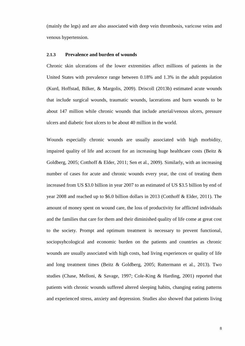
2.1.3 Prevalence and burden of wounds
Chronic skin ulcerations of the lower extremities affect millions of patients in the
United States with prevalence range between 0.18% and 1.3% in the adult population
(Kurd, Hoffstad, Bilker, & Margolis, 2009). Driscoll (2013b) estimated acute wounds
that include surgical wounds, traumatic wounds, lacerations and burn wounds to be
about 147 million while chronic wounds that include arterial/venous ulcers, pressure
ulcers and diabetic foot ulcers to be about 40 million in the world.
Wounds especially chronic wounds are usually associated with high morbidity,
impaired quality of life and account for an increasing huge healthcare costs (Beitz &
Goldberg, 2005; Cotthoff & Elder, 2011; Sen et al., 2009). Similarly, with an increasing
number of cases for acute and chronic wounds every year, the cost of treating them
increased from US $3.0 billion in year 2007 to an estimated of US $3.5 billion by end of
year 2008 and reached up to $6.0 billion dollars in 2013 (Cotthoff & Elder, 2011). The
amount of money spent on wound care, the loss of productivity for afflicted individuals
and the families that care for them and their diminished quality of life come at great cost
to the society. Prompt and optimum treatment is necessary to prevent functional,
sociopsyhcological and economic burden on the patients and countries as chronic
wounds are usually associated with high costs, bad living experiences or quality of life
and long treatment times (Beitz & Goldberg, 2005; Ruttermann et al., 2013). Two
studies (Chase, Melloni, & Savage, 1997; Cole-King & Harding, 2001) reported that
patients with chronic wounds suffered altered sleeping habits, changing eating patterns
and experienced stress, anxiety and depression. Studies also showed that patients living

9
with long-term wounds often have poor psychological wellbeing and a reduced quality
of life (Beitz & Goldberg, 2005).
2.2 Wound-healing Process
Wound healing process consists of a series of overlapping stages; hemostasis and
inflammation, reconstruction or destructive phase, proliferation and maturation or
remodelling (Dealey, 1999). It begins with the phase of hemostasis which includes
vascular constriction, platelet aggregation, degranulation and fibrin formation. Next is
the formation of granulation tissue of inflammatory cells, newly formed blood vessels,
and fibroblast embedded in a loose collageneous extracellular matrix. Then, the
proliferation and remodelling phases take place. The growth factors that participate in
the process are epidermal growth factor (EGF), platelet-derived growth factor (PDGF),
transforming growth factor β (TGF-β), insulin-like growth factor (IGF), and
granulocyte-macrophage colony-stimulating factor (GM-CSF) (Alster & Tanzi, 2003).
Additional group of active compounds important for the healing process are vitamins
and mineral supplements including vitamin A, B, C, D, E, K as well as zinc and copper
(Reynolds, 2001). A high availability of amino acids is necessary to enhance wound
healing due to an increased metabolic activity, thus HA is one of the most important
component (Maggio et al., 2012). Figure 2.1 summarises the process of wound healing.

10
Phase Cellular and Bio-physiologic events Duration of phase
Figure 2.1: Process of wound healing
2.3 Wound Managements
Accurate assessment, investigation, diagnosis and appropriate product choice are the
key to success in the wound management. A major factor known to boost wound
healing is to keep a wound moist environment. Rolstad and Ovington (2007) reported
that providing moisture to the wound and retaining moisture over time are not the same.
The range of available products for wound care includes hydrocolloids, film overlays,
foams, microfiber dressings, alginates and polyacrylates. These are sold in numerous
0-3
days
6 days
24 days
Months
to years
Month-
Years

11
combinations of materials. Some products such as saline-moistened gauze may not
continuously moist the wound, thus the need to occasionally change the dressings while
semi occlusive dressings may be able to keep a wound moist by retaining moisture
vapour on continual basis.
2.3.1 Wound dressings
History of dressings was documented in the Egyptian era since BC 1600 in which
grease-soaked gauze and fabrics were frequently used for dressings (Queen, Orsted,
Sanada, & Sussman, 2004; Scales, 1963). Presently, dressings like cloth, cotton, gauze
have dominated wound dressings and continued to be the main products used. In the
19th
century, several efforts were made to improve wound dressings. These include
Gamgee tissue. Sampson Gamgee of Birmingham discovered that cotton wool would
absorb fluids more rapidly compared to napkin (Scales, 1963). He recommended the
combined use of absorbent cotton wool with compressing gauze in aseptic manner.
Since then, cotton wool, gauze and lint became the established wound dressings.
The beginning of non-adherent dressing started when Lumiere introduced cotton net
impregnated with paraffin wax and balsam which allowed inlet of air to the wound
(Scales, 1963). The concept of moist wound healing began in 1970 when film and
hydrocolloid dressings were introduced. Since then, more absorbent wound dressings
have been developed (Queen et al., 2004).
Traditional wound dressings
Examples of traditional wound dressing which is also known as passive dressing include
dressing pads and tulle dressings. They are either medicated (e.g. containing
clorhexidine or povidone-iodine) or non-medicated (e.g. paraffin gauze dressing).
Winter (1962) introduced the important concept of moist wound healing for interactive

12
dressing category. Currently, the most modern dressing products are formulated as
interactive dressings by adding some agents in contrast to passive dressings which are
dressings alone (Thu, Zulfakar, & Ng, 2012).
Interactive dressings
Another choice of dressings, which is recently used, is interactive dressings. They are
either permeable or semi-permeable like film dressing, xerogel dressing, hydrocolloid
dressing, hydrogel dressing, alginate dressing, bead dressing and foam dressing
(Abdelrahman & Newton, 2011; Scales, 1963; Wardrope & Edhouse, 1999). They are
also known as moist interactive wound dressings with high absorbency to low
absorbency (foams calcium alginates hydrocolloids hydrogels hydrofibre) to
prevent bacterial infection due to accumulation of fluid surrounding the wounds.
Foam dressings: These are opaque dressings with non-adhesive surface for ulcers with
low to medium exudates.
Alginate dressings: Composed of alginic acid, they transform a fibre to gel when in
contact with wound fluid. They are highly absorbent dressings for wounds with medium
to heavy exudates.
Hydrocolloid dressings: These are adhesive dressings containing various polymers that
will form a gel when in contact with wounds, waterproof and indicated for wounds with
low to medium exudates.
Hydrogel dressings: These are amorphous, water-based gels or sheets, moisture
retentive, non-traumatic removal, indicated for wounds with light to medium exudates.
Hydrofibre dressings: These are soft dressings composed of hydrocolloid fibres,
indicated for wounds with heavy exudates.
13
Active dressings
Active dressings have various properties that can change the chemical and cellular
make-up of the wounds. Additionally, bioactive products which have endogenous
activities are also included in this type of dressings (Agrawal, Soni, Mittal, &
Bhatnagar, 2004). Examples of bioactive products include cellular suspensions, growth
factors, skin grafts, and biosynthetic skin substitute dressings such as collagen, chitosan,
peptides and hyaluronic acid (Queen et al., 2004).
Ideal dressings
Generally, the type of dressing selected depends on the size and types of wounds, the
frequency of dressing change, patient comfort and ease of removal and the overall cost
of management. Upton, Johnson, Zelazny, and Dailey (2013) suggested that health
professionals should minimize pain and stress at dressing change by using the most
suitable dressings and techniques which can be easily incorporated into wound care
management to expedite faster healing, promote patient health and eventually, reduce
the costs of care. Additionally, dressings must have a moisture absorptive capacity in
order to manage high drainage levels. The dressing materials should ideally be able to
provide water to the tissue to actively rehydrate the dry wound tissues. One online
survey reported that the ideal properties of wound dressing for burn wounds should be
non-adhesive, absorbent and has anti-microbial properties, easily remove, pain-free
dressing, changes required only once or twice a week and are available in different sizes
(Selig et al., 2012).
The characteristics of an ideal design for dressing are as outlined in Table 2.1
(Abdelrahman & Newton, 2011; Purser, 2009; Sarabahi, 2012; Scales, 1963; Wardrope
& Edhouse, 1999).

14
Table 2.1: Ideal Characteristics of an “ideal dressing”
Ideal Characteristics of an ideal dressings
Promotes a moist environment at wound interface
Allow excess exudates to be removed to the surface of dressings
Provide mechanical protection and thermal insulation
Provide barrier to micro-organisms
Allow for gaseous exchange
Non-adherent and can be removed easily without pain or trauma
Be sterile
Non-allergic, non-sensitising and non-cytotoxic to healthy tissue
Easy to use and cost-effective
2.3.2 Topical preparations
A number of topical agents are available, which aims to change the wound environment
such as topical antibiotics (e.g. neomycin, bacitracin, polymyxin B, gentamycin, fucidic
acid), topical antiseptics (e.g. chlorhexidine, povidone-iodine), topical steroids, and
topical collagen (Vermeulen et al., 2004). These agents were reported to promote the
healing process and prevent bacterial colonization which leads to wound infection
(Costagliola & Agrosì, 2005). Despite the increasing marketing of topical preparations,
conclusive evidence on their efficacy to promote wound healings are unavailable. For
example, to date, products containing iodine such as cadexomer-iodine, PVP-iodine
ointment, PVP-iodine gel or PVP-iodine gauze have no evidence to support their
benefits for wound healing to prevent infection (Rüttermann et al., 2013).

15
2.4 Hyaluronic acid
The molecular formula for HA is (C14H21NO11)n. HA is a polysaccharide composed of
N-acetyl glucosamine and D-glucuronic acid. Karl Meyer and his colleague John
Palmer, scientists at Columbia University, New York, discovered HA in 1934. They
isolated a chemical substance from the vitreous jelly of cow eyes. They proposed the
name hyaluronic acid as it was derived from a Greek word hyalos (glass) and contained
two sugar molecules, one of which was uronic acid (Meyer & Palmer, 1934). HA was
commercialised in 1942 when Endre Balazs used it to replace egg white in bakery
products and patented it. Its discovery was very unique. No other molecule had ever
been discovered that has such unique properties to the human body. Sources of
commercial HA are microbial fermentation, cock combs or chicken cartilage.
Commercial dressings and topical preparations containing HA are shown in Table 2.3.
HA has desirable physicochemical properties which include high viscosity, elasticity,
lubrication and high capacity for holding water (Capila & Sasisekharan, 2004). In
nature, HA is known to be one of the most hygroscopic molecules. Hydrogen bonding
occurs between adjacent carboxyl and an N-acetyl group when it is incorporated into
aqueous solutions, which allow it to maintain conformational stiffness and retain water.
Furthermore, the high concentration of medium and lower molecular weight hyaluronic
acid has the greatest bacteriostatic effect while viscoelastic properties of the material
may slow the penetration of viruses and bacteria (Bansal et al., 2010).

16
Table 2.2: Hyaluronic acid products in dressings and topical preparations
Products Ingredients Applications
Dressings containing hyaluronic acid
Benzyl hyaluronate
membrane
Benzyl hyaluronate esters Wound dressing
HA gauze pad
Sodium hyaluronate 0.05% Cream for wound healing
HYAFF® 11 Esterified HA Hyaluronic acid ester formed as
non-woven, absorbent ,wound
dressing
Hyalofill-F Hyaluronic acid Sheet for wound healing
Hyaloskin ® Hyaluronic acid Wound dressing
Ialuset®
Hyaluronic acid
Gauze pad wound dressing
Jossalind® Hyaluronate sodium Scaffold used in surgery and
wound healing
Silver sulfadiazine-
hyaluronan
collagen membrane
Hyaluronan micropraticles-
silver sulfadiazine (AgSD)
Wound healing
Topical preparations containing hyaluronic acid
Bionect Start® 0.2% w/w bacterial fermented
sodium hyaluronate
Ointment used in surgery and
wound healing
Bionect® Hyaluronic acid (0.98%) Ointment used in surgery and
wound healing
Cicactiv® Hyaluronic acid & zinc Topical cream for solar
keratoses
Coladerm H/HM Collagen/HA temporary
biosynthetic dermal skin
substitute
Apply for wounds and burns
Connettivina® Plus 0.2% hyaluronic acid, 1%
silver-sulfadiazine
Cream use in surgery and
wound healing
HYAL CT1101 3% diclofenac in 2.5% HA Topical gel for actinic keratosis
Hyiodine® Hyaluronan-iodine
complex,KI3
Gel for wound healing
Ialuset®
Hyaluronic acid
Cream for wound healing
Ialugen Plus® Hyaluronic acid Cream for wound healing
Lysial® Lysine-hyaluronate Decubitus Ulcers
(bedsore/pressure sore)
RadiaPlex gel Hyaluronic acid-based Preventing radiation dermatitis
Solaraze 3% diclofenac in 2.5% HA Topical gel for solar keratoses
Vulnamin® Glycine, l-lysine, l-proline, l-
leucine, hyaluronic acid
Gel use in chronic ulcers
Xclair TM
Hyaluronic acid Radiation-induced dermatitis
17
2.4.1 How hyaluronic acid may acts in wound healing
Specific mechanism of action of HA is still unknown. HA is believed to be an
appropriate choice for matrix to support dermal regeneration and augmentation because
it is found naturally in most cells in the body and occurs in high concentrations in
specific body locations especially skin tissues, eyes as well as in bones, cartilages
structures, synovial fluid and connective tissues (Bansal et al., 2010; Price, Berry, &
Navsaria, 2007). In each of these locations, HA serves a different function. Skin
normally will become dry when the capacity of the skin to hold water is reduced due to
the decreasing concentration of hyaluronic acid in the skin (Choulis, 2014).
HA is known to increase cell motility, cell proliferation, cell differentiation, cell
interaction and production of cell physiological substances such as cytokines, PGE2 and
matrix metalloproteinase (Capila & Sasisekharan, 2004). HA stimulates the
development of fibrin, phagocytic activity, neutrophil and macrophage mobility, and the
liberation of chemotactic factors for fibroblasts. Additionally, it induces proliferation of
fibroblasts and stimulates their metabolism during granulation phase of the cicatrisation
process, with a consequent increase in the collagen fibres and deposit of ground
substance (Anderson, 2001). Concentration of HA in cell is reported to increase rapidly
and reaches its peak three days after a wound occurred thus it provides a transitory
matrix for the migration of inflammatory cells and proliferation of fibroblast in the
connective tissue (Tammi & Tammi, 2004).
In summary, HA has been reported to be actively involved in all stages of wound
healing, from the promotion of early inflammation and granulation tissue formation,
through facilitation of cell migration into the wound matrix, to re-epithelialisation, via
its free radical scavenging function and role in keratinocyte proliferation and migration.

18
2.4.2 Adverse effects of hyaluronic acid-containing dressings and topical
preparations
Generally, most studies reported no serious adverse effects directly related to HA
containing dressings and topical preparations (Abbruzzese et al., 2009; Caravaggi et al.,
2003; Dereure et al., 2012a; Falanga et al., 1996; Meaume et al., 2008; Primavera et al.,
2006).
However, the most common side effects reported are pain and discomfort such as
bruising, swelling, redness, itching and tenderness. In one study related to the treatment
of solar keratoses, the number of patients with adverse reactions in HA group was
reported to be larger than the control group (18 vs 3) (McEwan & Smith, 1997). The
local reactions reported for the study were rashes and irritation at the area of gel
application.
HA-derived product responses to immune system are believed to be low due to its
identical chemical structures across different species (Edwards & Fantasia, 2007).
Evidence from available studies seems to indicate that HA is safe and well tolerated.
2.5 Assessment of Wound
Wound assessment is important for diagnosis, treatment and management. The correct
assessment, diagnosis and appropriate treatment will help in managing wound healing.
The common outcomes used in assessing effectiveness of treatment for chronic and
acute wounds are objective measures of healing rate, such as time to complete healing,
rate of change in wound area and volume, proportion of wounds healed within the trial
period/ percentage of wounds healed, reduction of wound size, visual
appearance/quality of the wound surface and patient acceptability (Rüttermann et al.,
2013). Other outcomes used are whether wounds are free of infection and pain.
19
2.6 Issues for wound healing assessment
The definition of wound healing is the most problematic followed by healing
assessment and evaluation. To enhance communication among all parts of society
dealing with this problem, description on definitions and guidelines are the vital steps.
Parameter selection and evaluation frequency should be defined appropriately. For
example, some researchers might describe a healed wound when more than 95% of the
wound has epithelised whilst others described complete wound healing with 100%
epithelialization and 0% residual wound area (Bettinger, Mast, & Gore, 1996;
Costagliola & Agrosì, 2005; Koller, 2004).
Although recently, researches on wound healing have progressed rapidly, standardised
outcome measurements are still lacking, thus making it tough to compare results from
different studies. Lazarus et al. (1994) suggested that the complete wound assessment is
required to include the extent of the wound (parameter involves are perimeter/area,
volume), associated elements of the wound (e.g. duration, blood flow, oxygen,
infection, edema, inflammation), host factors that influence wound status or wound
effects on the host (e.g. wound burden or wound severity), and environmental status that
affects wound management.
The time or duration to measure wound healing is also another issue. Complete healing,
is defined as complete epithelialisation of the wound without drainage (Dereure et al.,
2012a). The time-point on 45 days, is considered as a valid surrogate endpoint for leg
ulcer healing by a board of experts approached for the trial design to assess the
percentage of wound size reduction (Dereure et al., 2012a). However, for diabetic foot
ulcers, 12-week healing rate was the reported time-point in most studies related to
neuropathic ulcers of the foot in diabetes (Ince, Game, & Jeffcoate, 2007).

20
Fife, Carter, Walker, and Thomson (2012) reported that there are many aspects of costs
which are important in wound-healing assessment. Therefore, evaluation of cost
effectiveness should also be parts of the ideal wound management (Fonder et al., 2008).
Abdelrahman and Newton (2011) suggested that minimizing dressing change will help
reduce nursing time demand that lead to the reducing of cost in the overall wound
management.

21
CHAPTER 3: METHODS
3.1 Inclusion criteria for considering studies in this review
3.1.1 Types of studies
Studies reviewed include all randomised controlled trials (RCTs) evaluating the effects
of HA (or its derivatives) used in the form of a dressing or as a topical agent in the
treatment of acute and chronic wounds of any aetiology (i.e. diabetic foot ulcers, partial
thickness burns, traumatic wounds and lacerations, pressure ulcers, arterial/venous leg
ulcers and surgical wounds).
3.1.2 Types of participants
The studies involved people of all ages with acute or chronic wounds of any aetiology
in any care settings. Studies examining the healing of corneal, foetal, acute radiation,
mouth ulcer, bone and joint injuries will be excluded. Studies that do not involve the
outer skin wound will also be excluded.
3.1.3 Types of interventions
The studies assessed the effects of dressing and topical agents containing HA (or its
derivatives) and the likely comparisons were:
a) dressings containing HA (or its derivatives) compared with:
i. any dressings without HA or;
ii. another dressings containing other agents or;
iii. topical preparations of other agents
b) topical preparations of HA compared with:
i. dressings containing other agents or;
ii. topical preparations without HA or:
iii. topical preparations of other agents

22
3.1.4 Types of outcome measures
Two types of outcome measures considered were primary outcomes and secondary
outcomes.
Primary outcomes
a) Healing time or time to complete wound healing
b) Number of wounds healed
c) Wound area reduction or ulcer size reduction or change in wound surface
area
Secondary Outcomes
Trials which reported any of the following secondary outcomes involving the
performance and safety of Hyaluronic Acid:
a) Pain intensity
b) Adverse events
c) Patient acceptability or satisfaction
3.2 Search strategies
Several search strategies were used to identify potentially relevant trials. The search
strategies combined the used of various terms and synonyms for HA and wounds such
as “hyaluronan”, “ulcers” and “RCT’. Details of the search strategies and search terms
used in different databases to retrieve relevant studies are shown in Appendix A. The
multiple strategies used were as follows:
23
3.2.1 Electronic database
For this study, we searched the following electronic databases:
i. CINAHL Plus with Full Text @EBSCOhost (inception to August 2015)
ii. Cochrane Central Register of Controlled Trials (CENTRAL) (inception-
August 2015)
iii. MEDLINE with Fulltext @EBSCOhost (inception to August 2015)
iv. Ovid Full Text (inception to August 2015)
v. PUBMED (inception to August 2015)
vi. EMBASE (inception to August 2014)
The search was limited to humans for MEDLINE, EMBASE and a filter was applied to
identify randomised controlled trials in all databases.
3.2.2 Online publishing site search
The following online sites were also searched:
i. Science Direct (inception to August 2015)
ii. SpringerLink
iii. Wiley Interscience
iv. SAGE Journals
v. Internurse
vi. Karger
vii. DART-Europe E-theses Portal
3.2.3 Specified electronic journals or websites
The following electronic journals or websites were searched:
i. Wound Healing Society (www.woundheal.org)

24
ii. ResearchGate (www.researchgate.net/journal)
iii. Wounds UK (www.wounds-uk.com)
iv. Wounds International (www.woundsinternational.com)
v. Diabetes On the Net.com
vi. European Wound Management Association (www.ewma.org)
vii. Worldwide wounds (www.worldwidewounds.com)
viii. Wounds research (www.woundsresearch.com)
ix. Journal od Wound Care (2000 to August 2015)
x. Journal of European Wound Management Association (2000 to August
2015)
xi. CARE-Science and Practice (2000 to August 2015)
xii. The Australasian Journal of Dermatology
3.2.4 Hand searches
Hand searches on wounds related topics in conferences and proceedings were as
follows:
i. 36th Annual International Urogynecological Association (IUGA) Meeting,
2011
ii. 16th Congress of the Asian Pacific Society of Respirology, 2011
iii. 42nd Annual Meeting of the International Continence Society (ICS), 2012
iv. 28th ESMRMB (European Society for Magnetic Resonance in Medicine
and Biology) Annual Scientific Meeting, 2011
v. American Society of Gene & Cell Therapy Annual Meeting, 2010-2011
vi. XXIX EAACI Congress of the European Academy of Allergy and Clinical
Immunology, 2010
vii. Annual Congress of the British Society for Immunology, 2010
25
viii. 3rd TERMIS (Tissue Engineering & Regenerative Medicine International
Society) World Congress, 2012
ix. First Eastern Asia Dermatology Congress. Fukuoka, Japan, 2010
x. 20th European Conference on General Thoracic Surgery, 2012
xi. 39th Congress of the German Society for Rheumatology, 2011
xii. Annual Meeting of the Society for Investigative Dermatology, 2011
xiii. 10th World Congress on Inflammation, 2011
xiv. Annual Meeting of the German Society for Experimental and Clinical
Pharmacology and Toxicology, 2011-2012
xv. 38th Annual Meeting of the Arbeitsgemeinschaft Dermatologische
Forschung (ADF), 2011
xvi. 47th Annual Meeting of the European Association for the Study of
Diabetes, 2011
3.2.5 Additional sources of articles
The references of published papers on clinical trials and reviews from ClinicalTrials.gov
and DART-Europe E-theses portal were also searched for additional articles.
3.3 Selection criteria and Data extraction
Two review authors (Atikah Shaharudin, AS; Zoriah Aziz, ZA) independently screened
titles and abstracts of studies identified from the searches. We obtained full text articles
if they appeared to satisfy, or to potentially satisfy, the inclusion criteria. The two
review authors then independently checked full papers to identify those trials that were
eligible for inclusion. Any disagreement between the two review authors was resolved
through discussions. One review author (AS) undertook data extraction using a uniform
data extraction form. The second review author (ZA) checked for accuracy. If any data
was missing, attempts were made to obtain it by contacting the authors.

26
3.4 Data collection and analysis
The data were pooled using Review Manager (Revman) 5.3 if heterogeneity, I2 is less
than 80% (Higgins & Green, 2012). We used either a fixed-effect model or random
effect model if pooling seemed appropriate in view of clinical and methodological
similarities between studies. Relative risk (RR) and risk difference (RD) were calculated
for dichotomous data and the results were reported as RR with 95% confidence intervals
(CI). For continuous outcomes, the mean difference (MD), the weighted mean
difference (WMD), or standardised mean difference (SMD) with 95% CI was reported
as appropriate. Statistical significance was set at p < 0.05 for all outcomes. Relative risk
was chosen in preferences to odd ratio (OR) on the basis that OR can be misinterpreted
when event rates are high (>20%) (Deeks, Higgins, & Altman, 2008).
Studies that evaluated similar intervention in a similar population were assessed for the
presence of statistical heterogeneity by using chi-squared, χ2 test. The amount of
heterogeneity was estimated using I2 statistic (which indicates the percentage of
variation between studies that is due to heterogeneity rather than chance). We requested
from authors those relevant outcome results if the data were missing. Alternatively, we
calculated required data from available statistics. We imputed the data for standard
deviation difference (SDdiff) when standard error value was available (Borenstein,
Hedges, Higgins, & Rothstein, 2009; Follmann, Elliott, Suh, & Cutler, 1992).
3.5 Quality assessment of included studies
We assessed the risk of bias in the included RCTs using criteria suggested by the
Cochrane Collaboration (Higgins & Green, 2012). The following methodological
domains were assessed: sequence generation, allocation sequence concealment, blinding
of participants, researchers and outcome assessors, incomplete outcome data, selective
outcome reporting, and other potential threats to validity (Appendix D).

27
We clearly classified risk of bias for each of the domains as either unclear risk of bias,
low risk of bias or high risk of bias. Unclear risk of bias indicates either deficiency of
information or ambiguity over the potential for bias. The two reviewers discussed to
resolve any disagreement at any stages of selecting studies, data extraction, data
analysis and risk of bias assessment.

28
CHAPTER 4: RESULTS
4.1 Results of the search
The search for RCTs from all different sources produced 3466 records out of which
2606 were duplicates (Figure 4.1). We examined their potential relevance by screening
through titles and abstracts of these 860 records. Further 823 records were excluded.
The full texts of the remaining 37 studies were retrieved to assess whether they could be
included in the review. Another 27 studies were excluded for not meeting the inclusion
criteria (Appendix B). The reasons for exclusion include: non-RCT (12 studies),
comparing hyaluronic acid dressing or hyaluronic acid topical with other hyaluronic
products or hyaluronic-added products (7 studies), trials without control group (4
studies), the unit of analysis was wound sites instead of participants (3 studies) and trial
reporting secondary outcome only (1 study). Data from 10 included studies were
extracted by using the data extraction form (Appendix C).
The ten trials included were conducted in three countries (six in France, three in Italy
and one in Slovakia) and were published in English language between 1996 and 2012.

29
Figure 4.1: Flow chart of the study selection process
4.2 Description of the studies
The sample sizes of these trials ranged from 33 to 180 involving a total of 992 patients
where their ages ranged from 18 to 80 years old. One trial did not provide information
on the age of patients (Caravaggi et al., 2003). Out of 992 participants, 503 were
Records identified through
databases searching
(n=3381)
Additional records identified
through other sources
(n=85)
Iden
tifi
cati
on
S
cree
nin
g
Eli
gib
ilit
y
Incl
uded
Records after duplicates
removed (n=860)
(n=860)
Duplicates records
(n=2606)
Records screened
(n=860)
Records excluded
through screening of
titles and abstracts
(n=823)
Full- text articles excluded
with reasons
(n=27)
Full-text articles assessed for
eligibility
(n=37)
Studies included in qualitative
synthesis
(n=10)
Studies included in
quantitative synthesis
(n=10)
30
allocated to hyaluronic acid group and 489 for control group. Five trials enrolled more
than 100 participants and five trials were multi-centred (Table 4.1).
Eight trials assessed chronic wounds: mixed arterial and venous ulcer (n=5), venous leg
ulcer (n=1) and diabetic foot ulcer (n=2). Two trials assessed acute wounds of
superficial and deep partial-thickness burns.
In seven trials, the interventions were HA-containing dressings compared with a variety
of controls. The comparators were non-HA dressings (non-adherent paraffin gauze,
OASIS® dressing, DuoDERME®, hydrocolloid dressings, normal gauze pad) and other
topical agents (dextranomer paste). Three trials compared topical HA with other topical
agent (neutral cream without HA, silver sulfadiazine (SSD) cream). Durations of study
varied between three weeks and 18 months (Table 4.2).
Types of outcomes assessed
All papers reported at least one outcome of healing for their primary outcomes such as
wound area reduction, number of wounds healed and healing time (Table 4.3).
However, healed wounds were defined differently for several trials. Two trials did not
provide any definition (Humbert et al., 2012; Ortonne, 1996). Seven trials defined
complete healing as 100% epithelialisation without residual exudate (Caravaggi et al.,
2003; Costagliola & Agrosì, 2005; Dereure et al., 2012a; Dereure et al., 2012b; Koller,
2004; Romanelli, Dini, Brilli, & Bertone, 2007; Uccioli et al., 2011). One trial
considered the presence of epithelialisation as an indicator of healing and Meaume et al.
(2008) reported that at least 90% reduction of wound area signified healing. Most of the
trials measured wound area reduction either by ‘planimetry and photograph’ or ‘tracing-
paper and digital planimetry (Visitrak®)’.

31
For secondary outcome measures, a few general outcomes were reported in several
trials as shown in the summary on types of outcomes assessed (Table 4.3). Pain was
reported in nine trials (Costagliola & Agrosì, 2005; Dereure et al., 2012a; Dereure et al.,
2012b; Humbert et al., 2012; Koller, 2004; Meaume et al., 2008; Ortonne, 1996;
Romanelli et al., 2007). The majority of trials reported pain by using Huskisson’s Visual
Analogue Scale (VAS) and only two trials measured pain through patients’ complaints
during the treatment. The incident of adverse events was assessed in ten trials by
counting the number of cases or patients with adverse events.
In nine trials acceptability of patients (patients’ assessments) was measured through
counting the number of applications performed and also the use of four-point scale
(“bad”, “fair”, “good”, “excellent”) (Costagliola & Agrosì, 2005; Dereure et al., 2012a;
Dereure et al., 2012b; Humbert et al., 2012; Koller, 2004; Meaume et al., 2008;
Ortonne, 1996; Romanelli et al., 2007).
The measurements of wound appearance varied across trials, therefore this was not
included in the assessment of secondary outcomes. Consumption of oral analgesic was
reported in two trials (Meaume et al., 2008 and Costagliola and Agrosi, 2005) while
local infection was reported in one patient with the application of Hyaluronic cream
(Ialugen Plus®) (Koller 2004).

32
Table 4.1: Descriptions of included trials: Study designs, study settings, number and age of participants
Study Study Design Setting: Country
(number of centres)
Participants
Age in years
(mean (SD)) Intervention (I) &
Control (C)
Chronic wounds
Ortonne (1996) RCT Hospital :
France (NR)
50 patients I: 66.2 (15.8), C:69.7 (17.6)
Caravaggi et al. (2003) Open, RCT Diabetes foot clinic: Italy (6) 79 patients NR
Romanelli et al. (2007) RCT Leg Ulcer Clinic: Italy (1) 54 patients Age: > 18
I: 62 (8), C: 64 (13)
Meaume et al. (2008) Open-label, RCT Hospital: France (15); Italy
(2); Switzerland (1)
125 patients Age: ≥18
I: 73 (11.1), C: 75 (11.0)
Uccioli et al. (2011) Open, RCT Diabetic foot centers: Italy (7) 180 patients I: 61 (10), C: 62 (11)
Note: NR: Not Reported
32
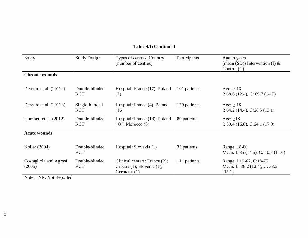
33
Table 4.1: Continued
Study Study Design Types of centres: Country
(number of centres)
Participants
Age in years
(mean (SD)) Intervention (I) &
Control (C)
Chronic wounds
Dereure et al. (2012a) Double-blinded
RCT
Hospital: France (17); Poland
(7)
101 patients
Age: ≥ 18
I: 68.6 (12.4), C: 69.7 (14.7)
Dereure et al. (2012b) Single-blinded
RCT
Hospital: France (4); Poland
(16)
170 patients
Age: ≥ 18
I: 64.2 (14.4), C:68.5 (13.1)
Humbert et al. (2012) Double-blinded
RCT
Hospital: France (18); Poland
( 8 ); Morocco (3)
89 patients
Age: ≥18
I: 59.4 (16.8), C:64.1 (17.9)
Acute wounds
Koller (2004) Double-blinded
RCT
Hospital: Slovakia (1) 33 patients Range: 18-80
Mean: I: 35 (14.5), C: 40.7 (11.6)
Costagliola and Agrosi
(2005)
Double-blinded
RCT
Clinical centers: France (2);
Croatia (1); Slovenia (1);
Germany (1)
111 patients Range: I:19-62, C:18-75
Mean: I: 38.2 (12.4), C: 38.5
(15.1)
Note: NR: Not Reported
33

34
Table 4.2: Descriptions of included trials: Types of wounds, interventions & controls and study durations
Study Wound types
Intervention
(number of participants)
Control
(number of participants)
Study
duration
(week)
Chronic wounds
Ortonne (1996) Venous leg ulcers Gauze pad impregnated with Sodium
hyaluronate cream 0.05% (26)
Dextranomer paste (24) 3
Caravaggi et
al. (2003)
Diabetic foot
ulcers
Hyalograft 3D (HYAFF-11®) (43)
Non-adherent paraffin gauze (36)
11
Romanelli et
al. (2007)
Mixed arterial &
venous ulcers
Dressing consisting of single component
ECM: Hyaluronic acid (Hyaloskin®) (27)
dressing containing all ECM
components (OASIS®) (27)
16
Meaume et al.
(2008)
Mixed arterial &
venous ulcers
Hydrocolloid-Hyaluronic acid 0.2%
dressing (63)
Hydrocolloid dressing (62)
6
Uccioli et al.
(2011)
Diabetic foot
ulcers
HYAFF-Hyalograft 3D(90)
Non-adherent paraffin gauze (90)
72
34

35
Table 4.2: Continued
Study Participants
Intervention
(number of participants)
Control
(number of participants)
Durations
(week)
Dereure et al.
(2012a)
Mixed arterial &
venous ulcers
Hyaluronic acid 0.2% cream (Ialuset®) (50)
Neutral Cream without
Hyaluronic acid (51)
8.6
Dereure et al.
(2012b)
Mixed arterial &
venous ulcers
Hyaluronic acid 0.05% gauze pad (Ialuset®)
(85)
DuoDERME (85)
-Hydrocolloid dressing
8
Humbert et al.
(2012)
Mixed arterial &
venous ulcers
Hyaluronic acid 0.05% gauze pad (Ialuset®)
(45)
Gauze pad without Hyaluronic
acid (44)
8.6
Acute wounds
Koller (2004) Superficial & deep
partial-thickness
burns
Hyaluronic acid-silver sulfadiazine 1% (Ialugen
Plus®) cream (18)
Silver sulfadiazine cream 1%
(15)
4
Costagliola and
Agrosi (2005)
Superficial & deep
partial-thickness
burns
Hyaluronic acid-silver sulfadiazine 1%
(Connettivina® Plus) cream (56)
Silver Sulfadiazine cream 1%
(55)
4
35

36
Table 4.3: Summary for types of outcomes assessed
Note: outcome assessed or reported
- outcome not assessed or not reported
Study Wound
area
reduction
Number
of wounds
healed
Healing
Time
Pain
intensity
Adverse
events
Patients’
assessment
Chronic wounds
Ortonne (1996) - -
Caravaggi et al.
(2003) - -
-
Romanelli et al.
(2007) - -
Meaume et al.
(2008)
-
Uccioli et al.
(2011) -
-
Dereure et al.
(2012a) -
Dereure et al.
(2012b) -
Humbert et al.
(2012) -
Acute wounds
Koller (2004) -
Costagliola and
Agrosi (2005)
-

37
4.3 Risk of bias assessment
Figure 4.2 and 4.3 show the summaries of the qualities of the 10 included trials based on
the Cochrane collaboration tool for assessing risk of bias (Higgins & Altman, 2012).
Appendix F provides the details on the risk of bias assessment for each of the included
RCTs. Five out of ten trials were overall at low to moderate risk of bias for the eight
domains assessed (Dereure et al., 2012a; Dereure et al., 2012b; Humbert et al., 2012;
Meaume et al., 2008; Uccioli et al., 2011). Three trials had more than three domains
judged to have unclear risk of bias (Costagliola & Agrosì, 2005; Koller, 2004; Ortonne,
1996) while the remaining two trials had at least one domain judged as having high risk
of bias (Caravaggi et al., 2003; Romanelli et al., 2007).
Figure 4.2: Risk of bias graph

38
Note: low risk of bias; unclear risk of bias; high risk of bias
Figure 4.3: Risk of bias assessment summary
4.3.1 Random sequence generation
Nine out of ten trials (Caravaggi et al., 2003; Costagliola & Agrosì, 2005; Dereure et al.,
2012a; Dereure et al., 2012b; Humbert et al., 2012; Koller, 2004; Meaume et al., 2008;
Ortonne, 1996; Uccioli et al., 2011) clearly stated the method of generating the
randomisation sequence, thus were classified to have low risk of bias whilst one trial

39
(Romanelli et al., 2007) was judged to have a high risk of bias as the randomisation
sequence was generated through every other patient selected by the clinician. Among
the studies that gave detail of the randomisation, one study stated using sealed envelope
(Meaume et al., 2008), another study stated telephone-randomisation (Caravaggi et al.,
2003) while five other studies reported computer-generated randomisation list
(Costagliola & Agrosì, 2005; Dereure et al., 2012a; Dereure et al., 2012b; Koller, 2004;
Meaume et al., 2008).
4.3.1 Allocation concealment
Only four trials (Caravaggi et al., 2003; Humbert et al., 2012; Meaume et al., 2008;
Uccioli et al., 2011) were judged to be at low risk of bias as they described the method
of concealing allocation for example by the use of sealed envelopes and coding by an
independent department. For another six trials (Costagliola & Agrosì, 2005; Dereure et
al., 2012a; Dereure et al., 2012b; Koller, 2004; Ortonne, 1996; Romanelli et al., 2007),
the risk of bias were considered as unclear because the information provided for this
domain was insufficient to make judgement.
4.3.2 Blinding
The trials had varying levels of blinding. Four trials (Caravaggi et al., 2003; Dereure et
al., 2012a; Dereure et al., 2012b; Humbert et al., 2012) reported blinding of participants,
researcher/healthcare provider and outcome assessor whilst another two trials (Ortonne,
1996; Romanelli et al., 2007) did not mention whether blinding was employed in all
levels. The remaining four trials had insufficient information at least for one level either
on blinding of participants, researcher or assessor (Costagliola & Agrosì, 2005; Koller,
2004; Meaume et al., 2008; Uccioli et al., 2011).

40
4.3.3 Incomplete outcome data (intention-to-treat analysis)
Six trials conducted intention-to-treat (ITT) analysis because the final analysis included
all randomised patients. Therefore, these trials were classified as having low risk of
bias. One trial (Romanelli et al., 2007) did not report whether ITT analysis was carried
out but had a low dropout rate (7.4%), while in another two trials (Koller, 2004;
Ortonne, 1996) it was unknown whether the ITT was carried out and these trials were
thus classified as having unclear risk of bias. Caravaggi et al. (2003) had a high dropout
rate of 22.8% with the number of randomised patients not accounted for in the final
analysis, hence were judged to be at high risk of bias.
4.3.4 Selective outcome reporting
Eight trials were judged as having low risk of bias to selective outcome reporting as the
main outcome measures stated in the method section were included in the result section.
Caravaggi et al. (2003) was judged to be at high risk of selective outcome reporting bias
as they did not report the pain intensity outcome as mentioned in the method section,
while one trial (Costagliola & Agrosì, 2005) had unclear risk of bias for this domain as
the outcome results for wound area reduction and pain intensity were not completely
reported.
4.3.5 Other sources of bias
Two other important sources of risks of bias were assessed. They were baseline
comparability and the financial support received to fund the trials.
Three trials (Dereure et al., 2012a; Dereure et al., 2012b; Meaume et al., 2008) were
judged as having low risk of bias as the baseline characteristics were comparable
between intervention and control group. Additionally, they have no conflict of interest
regarding financial support. One trial (Romanelli et al., 2007) was funded by the
manufacturer of the Oasis product (control group), Healthpoint, Ltd., but we judged the

41
risk of bias to be unclear as we could not determine whether this has affected the
findings of the trial.
Six trials did not provide sufficient information on financial support thus the risk of
potential bias was unclear (Caravaggi et al. 2003; Costagliola and Agrosi 2005;
Humbert et al. 2012; Koller 2004; Ortonne, 1996; Uccioli et al. 2011).
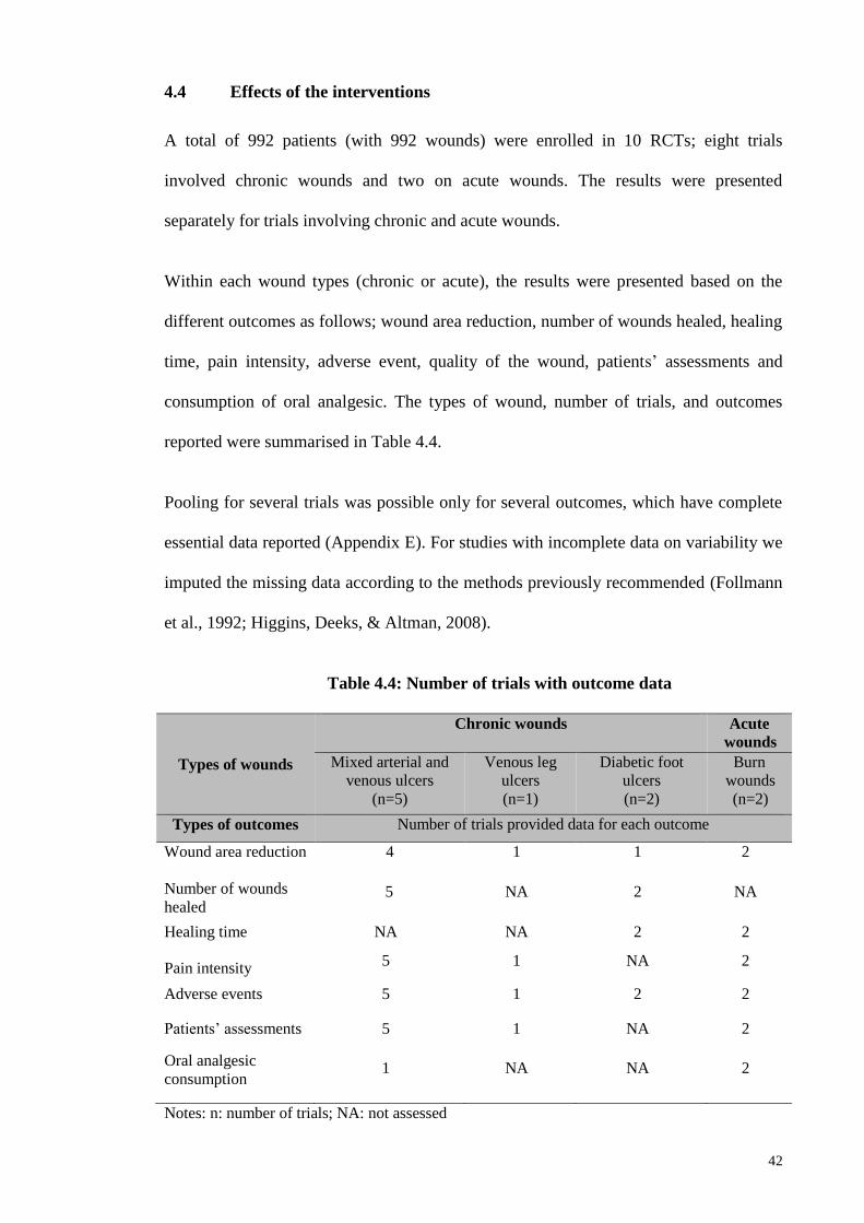
42
4.4 Effects of the interventions
A total of 992 patients (with 992 wounds) were enrolled in 10 RCTs; eight trials
involved chronic wounds and two on acute wounds. The results were presented
separately for trials involving chronic and acute wounds.
Within each wound types (chronic or acute), the results were presented based on the
different outcomes as follows; wound area reduction, number of wounds healed, healing
time, pain intensity, adverse event, quality of the wound, patients’ assessments and
consumption of oral analgesic. The types of wound, number of trials, and outcomes
reported were summarised in Table 4.4.
Pooling for several trials was possible only for several outcomes, which have complete
essential data reported (Appendix E). For studies with incomplete data on variability we
imputed the missing data according to the methods previously recommended (Follmann
et al., 1992; Higgins, Deeks, & Altman, 2008).
Table 4.4: Number of trials with outcome data
Types of wounds
Chronic wounds Acute
wounds
Mixed arterial and
venous ulcers
(n=5)
Venous leg
ulcers
(n=1)
Diabetic foot
ulcers
(n=2)
Burn
wounds
(n=2)
Types of outcomes Number of trials provided data for each outcome
Wound area reduction 4 1 1 2
Number of wounds
healed 5 NA 2 NA
Healing time
NA NA 2 2
Pain intensity 5 1 NA 2
Adverse events 5 1 2 2
Patients’ assessments 5 1 NA 2
Oral analgesic
consumption 1 NA NA 2
Notes: n: number of trials; NA: not assessed

43
4.4.1 Effects of interventions on chronic wounds
4.4.1.1 Wound area reduction
Six trials for chronic wounds reported the outcome wound area reduction. However
only four trials provided quantitative data:
(a) Mixed arterial and venous ulcers
Four out of five trials reported wound area reduction. However, only two trials provided
quantitative data. The results were as follows:
i HA cream (Ialuset®) versus neutral cream (without HA)
Dereure et al. (2012a) reported significantly greater median wound area reduction in
HA group compared to the control group (39% versus 5%).
ii HA gauze pad (Ialuset®) versus Hydrocolloid dressings (DuoDERME®)
Dereure et al. (2012b) reported that there was no significant difference in wound area
reduction between HA group and hydrocolloid dressing group (95% CI: -0.128 to
0.164).
iii HA gauze pad (Ialuset®) versus normal gauze pad (without HA)
Humbert et al. (2012) reported that there was a statistically significant effect in the
wound size reduction favouring HA gauze pad compared to normal gauze pad (Figure
4.4).

44
iv HA-Hydrocolloid dressings versus Hydrocolloid dressings
Meaume et al. (2008) reported that treatment with 0.2% HA-added hydrocolloid
dressings was significantly comparable with the hydrocolloid dressings alone (Figure
4.4).
Figure 4.4: Chronic wounds (Mixed arterial and venous ulcers); Outcome: Wound
area reduction
(b) Venous leg ulcers
Only one trial reported wound area reduction for venous leg ulcers:
i HA gauze pad versus Dextranomer paste
Ortonne (1996) reported that there was no significant difference in the reduction of
wound area between the two groups (Figure 4.5).

45
Figure 4.5: Chronic wounds (Venous leg ulcers); Outcome: Wound area reduction
(c) Diabetic foot ulcers
Two trials compared similar type of dressing, HYAFF-11 with paraffin gauze.
However, only one trial provided the outcome data for wound area reduction.
i Hyalograft 3D (HYAFF-11®) versus non-adherent paraffin gauze
Uccioli et al. (2011) found that the HA group reduced wound area by 29% compared to
14% in the control group. However this difference was not statistically significant
(Figure 4.6).
Figure 4.6: Chronic wounds (Diabetic foot ulcers); Outcome: Wound area
reduction

46
4.4.1.2 Number of wounds healed
Seven trials for chronic wounds reported number of wounds healed. However, only six
trials provided quantitative data:
(a) Mixed arterial and venous ulcers
All five trials reported number of wounds healed and provided quantitative data. Pooled
data from these trials showed that there was no significant difference in the number of
wounds healed between HA (or its derivatives) with control (Figure 4.7). The results
were presented as follows:
i HA cream (Ialuset®) versus neutral cream (without HA)
Dereure et al. (2012a) reported that there was no significant difference between HA
cream and neutral cream in terms of number of wounds healed (Figure 4.7).
ii HA gauze pad (Ialuset®) versus Hydrocolloid dressing (DuoDERME®)
Dereure et al. (2012b) defined ulcer healing as patients having at least 40% decrease of
initial target ulcer surface after eight weeks. On this basis, for patients with mixed
arterial and venous ulcers, the result showed that there was no significant difference
between the two groups (Figure 4.7).
iii HA gauze pad (Ialuset®) versus normal gauze pad (without HA)
Humbert et al. (2012) enrolled 89 patients with mixed arterial and venous leg ulcer.
Eighteen drop-out patients were not included in final analysis. No definition on wound
healing was given. They found an increased number of wounds healed in the HA group
compared to gauze pad group but the effect was not statistically significant (Figure 4.7).

47
iv HA-Hydrocolloid dressing versus Hydrocolloid dressing
Meaume et al. (2008) trial which involved patients with mixed arterial and venous leg
ulcer showed that there was no significant difference in the number of wounds healed
between group with dressing added HA compared to hydrocolloid agent alone (Figure
4.7).
v HA dressing (Hyaloskin®) versus OASIS® dressing
Romanelli et al. (2007) enrolled 54 patients with mixed arterial and venous leg ulcer.
Four patients dropped out from the trial and were not included in the final analysis. In
this trial, complete wound closure was defined by a fully re-epithelialised area. The
result showed that statistically more wounds were healed with the use of OASIS
dressings compared to HA dressings (Figure 4.7)

48
Figure 4.7: Chronic wounds (Mixed arterial and venous ulcers);
Outcome: Number of wounds healed

49
(b) Diabetic foot ulcers
Two trials reported number of wounds healed and provided quantitative data:
i Hyalograft 3D (HYAFF-11®) versus non-adherent paraffin gauze
Two trials (Caravaggi et al. 2003; Uccioli et al. 2011) which involved 239 patients with
diabetic foot ulcer. Both trials defined healed wound as complete re-epithelialisation
without residual exudate, crusting or eschar. The pooled result showed that there was no
significant difference between HA group and non-adherent paraffin gauze in terms of
number of wounds healed (Figure 4.8).
Figure 4.8: Chronic wounds (Diabetic foot ulcers); Outcome: Number of wounds
healed

50
4.4.1.3 Healing Time
Only two trials for chronic wounds with diabetic foot ulcers patients reported the
outcome healing time. However one trial (Caravaggi et al., 2003) did not report the
measure of variability (standard deviation) for both baseline and endpoint values, so the
results of the trial could not be pooled. Another one trial (Uccioli et al., 2011) provided
quantitative data:
(a) Diabetic foot ulcers
The result was presented as follow:
i Hyalograft 3D (HYAFF-11®) versus non-adherent paraffin gauze
Caravaggi et al. (2003) reported the healing time was significantly better for HA group
compared to non-adherent paraffin gauze (median: 57 days versus 77 days). Another
trial, Uccioli et al. (2011) found a tendency towards improvement in healing time in the
HA group but that the difference between the two groups was not statistically
significant (Figure 4.9).
Figure 4.9: Chronic wounds (Diabetic foot ulcers); Outcome: Healing time

51
4.4.1.4 Pain Intensity
Six trials for chronic wounds reported the outcome pain intensity. However, only three
trials with mixed arterial and venous ulcers patients provided quantitative data:
(a) Mixed arterial and venous ulcers
All five trials reported pain intensity. However, only three trials provided quantitative
data. Pain data in these three trials used 100mm Visual Analogue Scale (VAS)..
i HA cream (Ialuset®) versus neutral cream (without HA)
Dereure et al. (2012a) reported pain intensity using the scale 100mm VAS. There was a
significant pain reduction in group treated with Ialuset® cream compared to group
given neutral cream (Figure 4.10).
ii HA gauze pad (Ialuset®) versus Hydrocolloid dressing (DuoDERME)
Dereure et al. (2012b) did not provide quantitative data even though they reported that
there was no statistically significant difference in pain reduction between HA group and
hydrocolloid dressing group (p=0.6658).
iii HA gauze pad (Ialuset®) versus normal gauze pad (without HA)
Humbert et al. (2012) found a tendency on the improvement of pain in patients dressed
with HA gauze pad than patients dressed with normal gauze pad, however the difference
was not statistically significant (Figure 4.10).
iv HA-Hydrocolloid dressing versus Hydrocolloid dressing
Meaume et al. (2008) reported patients dressed with HA-hydrocolloid dressing
experienced significantly less pain than patients dressed with hydrocolloid dressing
alone (Figure 4.10).

52
v HA dressing (Hyaloskin®) versus OASIS® dressing
Romanelli et al. (2007) did not report the baseline value for pain intensity. However,
they reported that there was a significant difference in pain reduction between HA
group (VAS=6.2) and control group (VAS= 3.7).
The pooled data from these trials showed that there was a statistically significant benefit
of HA in improving pain intensity (Figure 4.10)
Figure 4.10: Chronic wounds (Mixed arterial and venous ulcers); Outcome: Pain
intensity

53
(a) Venous leg ulcers
Only one trial reported pain intensity for venous leg ulcers but did not provide the
quantitative data:
i HA gauze pad versus Dextranomer paste
Ortonne (1996) reported severity of pain scale by using Kruskall-Wallis test. The results
showed a significant beneficial effect in reducing severity of pain with HA gauze pad
compared to Dextranomer paste (from 1.00 to 0.24 vs 1.10 to 0.33 respectively).
4.4.1.5 Number of patients with adverse events
Eight trials for chronic wounds reported the outcome adverse events. The majority of
the trials reported the data either in terms of number of adverse events occurred or
number of patients who experienced adverse events. To standardise the results, we used
the number of patients experiencing adverse events as most of the trials provided this
data. Only six trials provided quantitative data:
(a) Mixed arterial and venous leg ulcers
All five trials reported adverse events. However, only four trials provided quantitative
data. The results were presented as follows:
i HA cream (Ialuset®) versus neutral cream (without HA)
Dereure et al. (2012a) has not clearly defined adverse events. However, inflammation
was one of the events reported. The trial found no significant difference in adverse
event between the intervention and control groups (Figure 4.11).
54
ii HA gauze pad (Ialuset®) versus Hydrocolloid dressings (DuoDERME®)
Dereure et al. (2012b) reported number of adverse events instead of number of patients
with adverse events. The trial found that less adverse events occurred in HA group
(n=36) as compared to control group (n=41). Apart from that, 77% of adverse event was
mild to moderate, while 23% was severe.
iii HA gauze pad (Ialuset®) versus normal gauze pad (without HA)
Humbert et al. (2012) reported 75% of the adverse events was mild to moderate, while
25% was severe (total number of patients=27). There was no significant difference in
the number of adverse events reported for both intervention and control groups (Figure
4.11).
iv HA-Hydrocolloid dressing versus Hydrocolloid dressing
The main adverse events reported in Meaume et al. (2008) were itching and edema,
erosion, eczema, rash, and pain. There was no significant difference in the number of
patients experiencing adverse events between the two groups (Figure 4.11).
v HA sheet (Hyaloskin®) versus OASIS® dressing
Romanelli et al. (2007) reported no incidence of ADR during the trial for both groups.
They concluded that both intervention and control were equally safe for the treatment of
chronic wounds.

55
Figure 4.11: Chronic wounds (Mixed arterial and venous ulcers); Outcome:
Number of patients with adverse events
(b) Venous leg ulcers
Only one trial reported adverse events and provided the quantitative data:
i HA gauze pad versus Dextranomer paste
Ortonne (1996) reported local pain, local burning sensation, panniculitis, eczema, and
prickling sensation as the adverse events. The trial found that there was no significant
difference between the two groups (Figure 4.12).

56
Figure 4.12: Chronic wounds (venous leg ulcers); Outcome: Number of patients
with adverse events
(c) Diabetic foot ulcers
Both trials reported adverse events and provided quantitative data:
i. Hyalograft 3D (HYAFF-11®) versus non-adherent paraffin gauze
Caravaggi et al. (2003) reported the main adverse events occurred were wound
infection, inflammation, and worsening of ischemia for both groups. Meanwhile Uccioli
et al. (2011) reported adverse events were mainly due to infections. The pooled result
for both trials showed no significant difference in the number of patients having adverse
events between the two groups (Figure 4.13).
Figure 4.13: Chronic wounds (Diabetic foot ulcers); Outcome: Number of patients
with adverse events

57
4.4.1.6 Quality of wound healing
Five trials for chronic wounds reported on quality of wound healing by description of
the wound appearance. Quality of healing was reported in many ways such as
percentage of fibrous tissue and granulation tissue, skin maceration and presence of
exudate. However no quantitative data were provided for all these trials.
(a) Mixed arterial and venous ulcers
All five trials came from patients with mixed arterial and venous ulcers but no data
could be pooled. The results were presented as follows:
i HA cream (Ialuset®) versus neutral cream (without HA)
Dereure et al. (2012a) reported that there was no statistically significant difference in
the percentage of fibrous tissue and granulation tissue between the HA and control
groups.
ii HA gauze pad (Ialuset®) versus Hydrocolloid dressing (DuoDERME)
Dereure et al. (2012b) reported two different aspects of wound quality. There was no
statistically significant difference between groups in term of ulcer characteristics
(p=0.8544 for fibrinous tissue, p=0.6704 for granulation tissue). However, in the aspect
of peri-ulcerous skin, the trial showed a statistically significant between the two groups
with more favourable result for patients treated with HA gauze pad (p=0.04 for oedema;
p=0.009 for purpura and p=0.003 for maceration).
iii HA gauze pad (Ialuset®) versus normal gauze pad (without HA)
Humbert et al. (2012) reported the percentage of granulation tissue in the patients
treated with HA decreased by 8.5% while it increased in the patients treated with gauze

58
pad (9.0%). This showed that healing was faster in HA groups as granulation tended to
disappear at the end of the healing process.
iv HA-Hydrocolloid dressing versus Hydrocolloid dressing
In Meaume et al. (2008), the quality of wound was not clearly defined. They reported
that the difference between the HA and control groups was statistically significant on
day 28 favouring the HA group (p=0.04). They also stated that skin maceration was less
intense and oozing was less severe in the HA group as compared to the control group
(p=0.05 and p=0.03 respectively).
(b) Diabetic foot ulcers
Only one trial reported quality of wound. The result was presented as follow:
i Hyalograft 3D (HYAFF-11®) versus non-adherent paraffin gauze
Caravaggi et al. (2003) indicated that quality of wound was worst with the presence of
exudate. Quality of wound was better in patients treated with HA as 86% patients had
no exudate as compared to the patients treated with paraffin gauze (69.4% absent of
exudate).
59
4.4.1.7 Patients’ assessments of tolerability
Six trials for chronic wounds reported on patients’ assessments towards the treatments
received. Results were reported in different ways and no data could be pooled:
(a) Mixed arterial and venous leg ulcers
All five trials reported patients’ assessments. The results were presented as follows:
i HA cream (Ialuset®) versus neutral cream (without HA)
Dereure et al. (2012a) reported that there was no significant difference between groups
in terms of patients who did not miss any daily application of the allocated treatment
(>79% of patients).
ii HA gauze pad (Ialuset®) versus Hydrocolloid dressing (DuoDERME)
Dereure et al. (2012b) reported that there was no significant difference between the two
groups in terms of tolerability of application.
iii HA gauze pad (Ialuset®) versus normal gauze pad (without HA)
Humbert et al. (2012) considered patients were compliant if they did not miss any of
their daily applications. Both groups were reported to be highly satisfied with their
treatment as more than 87% patients did not miss their daily applications and the
difference between groups was not statistically significant.
iv HA-Hydrocolloid dressing versus Hydrocolloid dressing
In Meaume et al. (2008), tolerability was assessed using a four-point scale (“very good”,
“good”, “fair” and “poor”). Tolerability of application for the two groups was
comparable.

60
v HA dressing (Hyaloskin®) versus OASIS® dressing
Romanelli et al. (2007) measured patient comfort at dressing change using VAS scale
from 0=excellent to 10=critical. They reported that patients treated as control group had
significantly greater comfort compared to the HA group.
(b) Venous leg ulcers
Only one trial reported patients’ assessments. The result was presented as follow:
i HA gauze pad versus Dextranomer paste
Ortonne (1996) reported patients’ judgement towards tolerability of treatment by using
a four-point scale (good, average, nil, worsened). The difference between groups was
not statistically significant although a higher proportion (58%) in the HA gauze pad
rated good treatment tolerability as compared to Dextranomer paste (42%) at the end of
21-day treatment.
4.4.1.8 Oral analgesic consumption
Of the eight trials for chronic wounds, only one trial reported oral analgesic
consumption (Meaume et al., 2008). They reported that lower proportion of patients
with mixed arterial and venous ulcers took oral analgesic in the HA group compared to
the control group. However the difference between both groups was not statistically
significant.

61
4.4.2 Effects of interventions on acute wounds
4.4.2.1 Wound area reduction
Two trials for acute wounds reported the outcome wound area reduction. However none
of them provided quantitative data:
(a) Burn wounds
Koller (2004) did not report baseline value for intervention and control group, thus data
could not be pooled. The result was presented as follow:
i HA-silver sulfadiazine cream versus silver sulfadiazine (SSD) cream
Both trials (Koller 2004; Costagliola & Agrosi 2005) defined wound healing as 100%
epithelialisation and 0% residual wound area. Koller (2004) (Ialugen Plus® cream)
reported for HA group mean reduction was 5.83cm2
from baseline while for SSD group
reduction was 30.59 cm2. As for Costagliola and Agrosi (2005) (Connettivina® Plus
cream), no data was provided.
4.4.2.2 Number of wounds healed
None of the trials reported the outcome number of wounds healed.
4.4.2.3 Healing time
Both trials reported the outcome healing time and provided quantitative data:
(a) Burn wounds
i HA-silver sulfadiazine cream versus silver sulfadiazine cream
The pooled results of two trials (Koller 2004; Costagliola and Agrosi 2005) showed HA
group shows significantly shorter healing time compared to the control group (Figure
4.14).
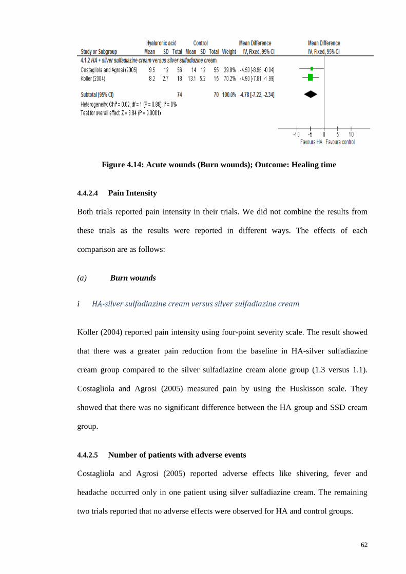
62
Figure 4.14: Acute wounds (Burn wounds); Outcome: Healing time
4.4.2.4 Pain Intensity
Both trials reported pain intensity in their trials. We did not combine the results from
these trials as the results were reported in different ways. The effects of each
comparison are as follows:
(a) Burn wounds
i HA-silver sulfadiazine cream versus silver sulfadiazine cream
Koller (2004) reported pain intensity using four-point severity scale. The result showed
that there was a greater pain reduction from the baseline in HA-silver sulfadiazine
cream group compared to the silver sulfadiazine cream alone group (1.3 versus 1.1).
Costagliola and Agrosi (2005) measured pain by using the Huskisson scale. They
showed that there was no significant difference between the HA group and SSD cream
group.
4.4.2.5 Number of patients with adverse events
Costagliola and Agrosi (2005) reported adverse effects like shivering, fever and
headache occurred only in one patient using silver sulfadiazine cream. The remaining
two trials reported that no adverse effects were observed for HA and control groups.

63
4.4.2.6 Quality of wound healing
None of the two trials on acute wounds reported the outcome quality of wound healing.
4.4.2.7 Patients’ assessments on tolerability
Two trials reported on physician and patients’ assessments of tolerability (Koller 2004;
Costagliola and Agrosi 2005).
(a) Burn wounds
i HA-silver sulfadiazine cream versus silver sulfadiazine cream
Koller (2004) reported tolerability using a four-point scale (none, fair, good, excellent).
The result was not statistically different between the two groups with 80-100% patients
rated treatment as good to excellent. Costagliola and Agrosi (2005) assessed tolerability
by frequency of visits. The result showed that there was no significant difference
between the two groups (76.8% good-excellent in HA-SSD group vs 75.9% good-
excellent in SSD group alone).

64
4.4.2.8 Oral analgesic consumption
Only one trial for acute wounds reported oral analgesic consumption as an outcome.
(a) Burn wounds
i HA-silver sulfadiazine cream versus silver sulfadiazine cream
Costagliola and Agrosi (2005) assessed oral analgesic given other than acetylsalicylic
acid. They found the mean number of oral analgesic taken was greater in the silver
sulfadiazine group compared to HA- silver sulfadiazine group (0.69 vs 0.57) while the
mean number of days patients took analgesic was higher in silver sulfadiazine group
compared to HA- silver sulfadiazine group (4 days vs 2.4 days). However, the
differences between the two groups were not statistically significant for both measures.

65
CHAPTER 5: DISCUSSIONS
Our systematic review included 10 RCTs and we found that HA-containing products
produced some favourable outcomes in acute and chronic wound healing. However, the
available evidence is not consistent enough to make a firm conclusion that HA-
containing dressings and topical preparations are effective in enhancing wound healing
for acute and chronic wounds. However, we found evidence to indicate that, HA (or its
derivatives) are beneficial in reducing pain. This finding is in agreement with one
systematic review published in 2012 (Voigt & Driver, 2012).
5.1 Interpretation of the evidence
It has been suggested that wound healing should be measured using the outcome healed
wound because this measure is more established, objective and quantifiable (Lazarus et
al., 1994; Morison, 1992; Rolstad & Ovington, 2007; Wardrope & Edhouse, 1999).
Even though most of the included studies included outcomes such as wound area
reduction, number of wounds healed and healing time, the results of several trials could
not be pooled because of different definition of healing used or incomplete data given in
the paper. Despite attempts to contact the authors to get relevant information, most
authors did not response to our request. Non response for request of data is a common
problem in most meta-analysis studies (Flather, Farkouh, Pogue, & Yusuf, 1997;
Higgins et al., 2008; Stevens & Wu, 2007).
The findings on the outcome wound area reduction are contradictory. For example, one
trial (Humbert et al., 2012) reported greater reduction in wound area for HA group
compared to the control group. On the other hand, a bigger trial (Meaume et al., 2008)
found that patients given hydrocolloid dressing showed a higher reduction in wound
area compared to HA group. Generally, our findings show little evidence of the
effectiveness of HA-containing dressing in reducing wound area.

66
It is also worth noting that for similar studies, when the outcome “number of wounds
healed” was considered, the findings show little evidence that HA (or its derivatives)
has notable beneficial effects for the three types of chronic wounds. For acute wounds,
none of the trials reported the outcome “number of wounds healed”.
We found that most of the trials involved did not report the healing time as an outcome
although many claims made for hyaluronic acid preparations is to shorten healing time
for chronic wounds. The claims are made because in certain pathological conditions, it
has been shown that there is degradation of local hyaluronic acid, thus leading to
insufficient regeneration of the connective tissues, poor angiogenesis, and deficient
differentiation of histiocyte and fibroblast populations (Edwards & Fantasia, 2007).
Adding exogenous hyaluronic acid will help in speeding up the tissue processing.
The duration of study was short which on average was only 8 weeks and there was even
a study that lasted three weeks. Only two trials assessing the effectiveness for acute
wounds (Costagliola & Agrosì, 2005; Koller, 2004) and one trial for chronic wounds
(Caravaggi et al. 2003) reported significantly better healing time with HA products
compared to the comparators. However, quantitative data given was incomplete.
Therefore we did not find sufficient evidence to support the use of HA dressings and
topical preparations to shorten wound healing time.
Interestingly, pooled data on pain intensity for three similar trials involving patients
with mixed arterial and venous leg ulcers (Dereure et al., 2012a; Humbert et al., 2012;
Meaume et al., 2008) showed that groups receiving HA experienced less intense pain as
compared to the control groups. Additionally, another five trials for acute and chronic
wounds with different pain measurements also reported beneficial effect favouring HA
group (Costagliola & Agrosì, 2005; Dereure et al., 2012b; Koller, 2004; Ortonne, 1996;
Taddeucci et al., 2004). These results support several findings that HA has greater

67
improvement in pain level in any type of ulcer (Goa & Benfield, 1994; Kirova et al.,
2011; Nolan, Badminton, Maguire, & Seymour, 2009; Nolan, Baillie, Badminton,
Rudralingham, & Seymour, 2006; Onesti et al., 2013). The evidence seems to indicate
that HA-containing dressing is effective in reducing pain.
For all seven trials which reported the number of patients with adverse events, there was
no significant difference between HA groups and control groups. However, five trials
assessing chronic wounds (Caravaggi et al. 2003; Dereure at al. 2012a; Humbert et al.
2012; Meaume et al. 2008; Uccioli et al. 2011) found less adverse effects in HA groups
compared to control groups. Given that the occurrence of adverse events was less
frequent in HA groups compared with the control groups it seems that HA is well
tolerated without serious unwanted effects.
Quality of wounds healed was interpreted by the appearance of the wounds. Majority of
the chronic wounds’ patients have better wounds appearance when used HA products
rather than patients who used comparators, thus the authors found a beneficial effect
favouring the HA group (Caravaggi et al. 2003; Dereure et al. 2012b; Humbert et al.
2012; Meaume et al. 2008). However, no differences on the appearance of the wounds
between the HA groups and control groups for acute wounds.
By referring to the two trials reported on consumption of oral analgesic was found to be
less in patients of HA groups compared to control groups in two trials (Costagliola and
Agrosi 2005; Meaume et al. 2008), HA-containing dressings and topical preparations of
HA seem to be better or no worse than the comparators. Thus, overall cost could
eventually be the factor to use HA-containing products to enhance wound healing.
However, all trials did not assess the cost-effectiveness outcome, thus negating the
ability to evaluate the effectiveness of HA on economic scale.

68
5.2 Quality of the evidence
To ensure reliability of the evidence, we have decided to include only randomised
controlled trials (RCTs) for this review. Thus, we were able to include only 10 trials.
Several trials were of moderate quality. Poor reporting of method design and blinding
was common among these trials. For example, six trials (Costagliola & Agrosì, 2005;
Dereure et al., 2012a; Dereure et al., 2012b; Koller, 2004; Ortonne, 1996; Romanelli et
al., 2007) did not adequately report allocation concealment which might lead to
selection bias. Additionally, only four trials completely described how blinding of
participants, researchers or healthcare providers and outcome assessors was carried out
in the trials. Insufficient allocation concealment and poor blinding conducted in any trial
could lead to higher estimation effects of treatment (Egger, Smith, Schneider, &
Minder, 1997). Blinding in a trial should be performed on as many parties as possible
including participants, clinicians, data collectors, outcomes assessors, and data analysts
to minimize differentials interventions and outcome biased assessments (Karanicolas,
Farrokhyar, & Bhandari, 2010).
Of 10 included trials, five trials (Costagliola & Agrosì, 2005; Humbert et al., 2012;
Ortonne, 1996; Romanelli et al., 2007; Uccioli et al., 2011) were financially funded by
product manufacturers. Therefore, the positive outcomes on the company products
might have potential of bias. One review in 2003 found that research sponsored by the
drug manufacturers was four times more likely to produce outcomes favouring the
manufacturer’s product than research sponsored by other sources (Lexchin, Bero,
Djulbegovic, & Clark, 2003).
We were careful as not to pool the results of several trials as pooling results from trials
with clinical and methodological heterogeneity is controversial (Ioannidis, Patsopoulos,
& Evangelou, 2007; Stevens & Wu, 2007). The lack of standard guideline for wound
69
care make, it difficult to assess wound healing. This issue affected wound healing
assessment, thus trials produce different parameters or measurements (Purser, 2009;
Robson & Barbul, 2006). However, the diversity of the studies such as wound types,
and treatments given is useful rather than a problem because the findings could be
generalisable to a broader group of patients (Borenstein et al., 2009).
5.3 Strengths and Limitations
The main strength of this review is the degree of rigour in the conduct of the review.
The methods were in accordance with those proposed by the Cochrane Collaboration for
conducting systematic review of interventions (Higgins & Green, 2012). Additionally
we assessed the quality of the included trials.
This study has several limitations. First, there is difficulty in combining the outcome
effects from various studies due to different parameters used and missing data. Second,
a common problem to most meta-analysis studies is the heterogeneity of the included
RCTs, particularly regarding the definitions of healing, duration of treatment, outcome
measures used and the trials quality might limit the value of evidence for this systematic
review (Flather et al., 1997). As suggested by Lazarus et al. (1994), standard guidelines
in evaluating the healing of wound is crucial in order to organise a uniform outcome in
setting the end points of any study. Third, there are four excluded trials worth to be
included however the combination compounds with HA without a control group (such
as zinc-HA versus normal saline solution) might have confounding effect in wound
healing. With the expanding health care expenditure, future studies should consider the
cost-efficiency of HA in managing wounds in order to help reduce the cost of treating
acute and chronic wounds (Driscoll, 2013a; Landro, 2012).

70
CHAPTER 6: CONCLUSIONS
6.1 Implication for practice
At present, the evidence does not support the benefits of HA or its derivatives to
improve chronic wound healing even though there is some evidence on their
effectiveness especially on reducing pain intensity. The availability of high quality
evidence is still limited.
6.2 Implication for research
The use of relevant measurements is necessary for assessing efficacy to aid the
interpretation of the findings. Different wounds-type might give different therapeutic
effects. Therefore, trials should clearly describe the location of wounds.
Trials also require adequate number of patients to ensure sufficient statistical power to
detect true treatment effects. An economic evaluation should be conducted to determine
if the costs of HA-containing dressings and topical agents justify its potential benefits.
Additionally randomised controlled trials need to use the revised CONSORT statement
to improve the quality of reporting randomised controlled trials (Mills, Wu, Gagnier, &
Devereaux, 2005; Schulz, Altman, & Moher, 2010).
If the trials incorporate all recommendations as outlined, it is anticipated for future
systematic review to be able to provide more conclusive evidence on the effectiveness
of HA-containing dressings and topical agents in enhancing wound healing.

71
REFERENCES
Abbruzzese, L., Rizzo, L., G., F., Tedeschi, A., Scatena, A., Goretti, C., . . . Piaggesi, A.
(2009). Effectiveness and safety of a novel gel dressing in the management of
neuropathic leg ulcers in diabetic patients: a prospective double-blind randomized
trial. The International Journal Of Lower Extremity Wounds, 8(3), 134-140.
doi:10.1177/1534734609344140
Abdelrahman, T., & Newton, H. (2011). Wound dressings: principles and practice. Surgery,
29(10), 491-495.
Agrawal, P., Soni, S., Mittal, G., & Bhatnagar, A. (2004). Role of polymeric biomaterials
as wound healing agents. The International Journal of Lower Extremity Wounds,
13(3), 180-190. doi:10.1177/1534734614544523
Anderson, I. (2001). The properties of hyaluronan and its role in wound healing. Prof
Nurse, 17(4), 232-235.
Bansal, J., Kedige, S. D., & Anand, S. (2010). Hyaluronic acid: a promising mediator for
periodontal regeneration. Indian Journal of Dental Research 21(4), 575-578.
doi:10.4103/0970-9290.74232
Beitz, J. M., & Goldberg, E. (2005). The lived experience of having a chronic wound: a
phenomenologic study. Medsurg nursing, 14(1), 51-62.
Bettinger, D. A., Mast, B., & Gore, D. (1996). Hyaluronic acid impedes reepithelialization
of skin graft donor sites. The Journal of Burn Care Rehabilitation, 17(4), 302-304.
Borenstein, M., Hedges, L. V., Higgins, J. P. T., & Rothstein, H. R. (2009). Introduction to
Meta-Analysis. UK: John Wiley & Sons, Ltd.
Campanati, A., De Blasio, S., Giuliano, A., Ganzetti, G., Giuliodori, K., Pecora, T., . . .
Offidani, A. (2013). Topical ozonated oil versus hyaluronic gel for the treatment of
partial- to full-thickness second-degree burns: A prospective, comparative, single-
blind, non-randomised, controlled clinical trial. Burns, 39(6), 1178-1183.
http://dx.doi.org/10.1016/j.burns.2013.03.002
Capila, I., & Sasisekharan, R. (2004 ). Methods for Analysis of Hyaluronan and Its
Fragments. In H. G. Garg & C. A. Hales (Eds.), Chemistry and Biology of
Hyaluronan (pp. 21-40). UK: Elsevier Ltd.

72
Caravaggi, C., Giglio, R., Pritelli, C., Sommaria, M., Dalla Noce, S., Faglia, E., . . .
Morabito, A. (2003). HYAFF 11-based autologous dermal and epidermal grafts in
the treatment of noninfected diabetic plantar and dorsal foot ulcers: a prospective,
multicenter, controlled, randomized clinical trial. Diabetes Care, 26(10), 2853-
2859.
Caravaggi, C., Grigoletto, F., & Scuderi, N. (2011). Wound bed preparation with a dermal
substitute (hyalomatrix® pa) facilitates re-epithelialization and healing: Results of a
multicenter, prospective, observational study on complex chronic ulcers (the fast
study). Wounds, 23(8), 228-235. Retrieved from
https://www.medscape.com/viewarticle/749515_print. Accessed 21.12.2013.
Cassino, R., & Ricci, E. (2010). Effectiveness of topical application of amino acids to
chronic wounds: a prospective observational study. Journal of Wound Care, 19(1),
29-34.
Cervelli, V., Lucarini, L., Spallone, D., Brinci, L., & de Angelis, B. (2010). Use of platelet
rich plasma and hyaluronic acid on exposed tendons of the foot and ankle. Journal
of Wound Care, 19(5), 186.
Chase, S. K., Melloni, M., & Savage, A. (1997). A forever healing: The lived experience of
venous ulcer disease. Journal of Vascular Nursing, 15(2), 73-78.
Choulis, N. H. (2014). Dermatological drugs, topical agents, and cosmetics. In J. K.
Aronson (Ed.), Side effects of drugs annual (pp. 273-294). US: Elsevier
Cole-King, A., & Harding, K. G. (2001). Psychological factors and delayed healing in
chronic wounds. Psychosomatic Medicine, 63, 216-220. doi:0033-3174/01/6302-
0216
Colletta, V., Dioguardi, D., Di Lonardo, A., Maggio, G., & Torasso, F. (2003). A trial to
assess the efficacy and tolerability of Hyalofill-F in non-healing venous leg ulcers.
Journal of Wound Care, 12(9), 357-360.
Costagliola, M., & Agrosì, M. (2005). Second-degree burns: a comparative, multicenter,
randomized trial of hyaluronic acid plus silver sulfadiazine vs. silver sulfadiazine
alone. Current Medical Research Opinion, 21(8), 1235-1240. doi:
10.1185/030079905X56510

73
Cotthoff, T., & Elder, M. (2011). Market for advanced wound care technologies. Business
Communications Company Research (BCC). Retrieved from
http://www.bccresearch.com/market-research/pharmaceuticals/advanced-wound-
care-technologies-market-phm011g.html. Accessed 13.12.2013.
Cuevas, F. R., Méndez, A. A. V., & Andrade, I. C. (2007). Zinc hyaluronate effects on
ulcers in diabetic patients. Gerokomos, 18(2), 91-101.
Dealey, C. (1999). The Care Of Wounds: A Guide For Nurses (2nd ed.). London, UK:
Blackwell Science.
Dechert, T. A., Ducale, A. E., Ward, S. I., & Yager, D. R. (2006). Hyaluronan in human
acute and chronic dermal wounds. Wound Repair And Regeneration, 14(3), 252-
258. doi:10.1111/j.1743-6109.2006.00119.x
Deeks, J. J., Higgins, J. P. T., & Altman, D. G. (2008). Analysing data and undertaking
meta-analyses. In J. P. T. Higgins & S. Green (Eds.), Cochrane Handbook For
Systematic Reviews Of Interventions (pp. 243-296). UK: John Wiley & Sons.
Dereure, O., Czubek, M., & Combemale, P. (2012a). Efficacy and safety of hyaluronic acid
in treatment of leg ulcers: a double-blind RCT. Journal Wound Care, 21(3), 131-
139.
Dereure, O., Mikosinki, J., Zegota, Z., & Allaert, F. A. (2012b). RCT to evaluate a
hyaluronic acid containing gauze pad in leg ulcers of venous or mixed aetiology.
Journal Wound Care, 21(11), 539-545.
Doughty, D. B., & Sparks-Defriese, B. (2007). Wound-healing Physiology. In R. A. Bryant
& D. P. Nix (Eds.), Acute & Chronic Wounds: Current Management Concepts (pp.
56-81). US: Elsevier.
Driscoll, P. (2013a). Worldwide Wound Management, forecast to 2021: established and
emerging products, technologies and markets in the americas, europe, asia/pacific
and rest of world. Wound Management 2013. CA: MedMarket Diligence, LLC.
Driscoll, P. (2013b). Wound prevalence and wound management, 2012-2020, Advanced
Medical Technologies. Retrieved from
http://blog.mediligence.com/2013/01/29/wound-prevalence-and-wound-
management-2012-2020/. Accessed 19.12.2013.
74
Edwards, P. C., & Fantasia, J. E. (2007). Review of long-term adverse effects associated
with the use of chemically-modified animal and nonanimal source hyaluronic acid
dermal fillers. Clinical Interventions in Aging, 2(4), 509-519.
Egger, M., Smith, G. D., Schneider, M., & Minder, C. (1997). Bias in meta-analysis
detected by a simple, graphical test. BMJ 315(629), 1-16.
doi:10.1136/bmj.315.7109.629
Falanga, V., Carson, P., Greenberg, A., Hasan, A., Nichols, E., & McPherson, J. (1996).
Topically applied recombinant tissue plasminogen activator for the treatment of
venous ulcers: Preliminary report. Dermatologic Surgery, 22(7), 643-644.
Fariba, I., Taghi, G., Mazdak, E. S., Maryam, T., Hossein, S. A., Shahla, E., & Zabiholah,
S. (2005). The efficacy of 3% diclofenac in 2.5% hyaluronan gel base for treatment
of recurrent aphthous stomatitis (RAS): A double blind study. Egyptian
Dermatology Online Journal 1(2 (4)), 1-6. Retrieved from:
http://www.edoj.org.eg/vol001/00102/04/paper.pdf. Accessed 24.12.2013.
Fife, C. E., Carter, M. J., Walker, D., & Thomson, B. (2012). Wound care outcomes and
associated cost among patients treated in us outpatient wound centers: data from the
us wound registry. Wounds, 24(1), 10-17. Retrieved from
http://www.woundsresearch.com/article/wound-care-outcomes-and-associated-cost-
among-patients-treated-us-outpatient-wound-centers-d. Accessed 31.10.2014.
Flather, M. D., Farkouh, M. E., Pogue, J. M., & Yusuf, S. (1997). Strengths and limitations
of meta-analysis: Larger studies may be more reliable. Control Clin Trials, 18(6),
568-579. doi: http://dx.doi.org/10.1016/S0197-2456(97)00024-X
Follmann, D., Elliott, P., Suh, I., & Cutler, J. (1992). Variance imputation for overviews of
clinical trials with continuous response. Journal of Cinical Epidemiology, 45(7),
769-773.
Fonder, M. A., Lazarus, G. S., Cowan, D. A., Aronson-Cook, B., Kohli, A. R., & Mamelak,
A. J. (2008). Treating the chronic wound: A practical approach to the care of
nonhealing wounds and wound care dressings. Journal of the American Academy of
Dermatology, 58(2), 185-206. doi:10.1016/j.jaad.2007.08.048
Gebauer, K., Brown, P., & Varigos, G. (2003). Topical diclofenac in hyaluronan gel for the
treatment of solar keratoses. Australasian Journal of Dermatology, 44(1), 40-43.

75
Goa, K. L., & Benfield, P. (1994). Hyaluronic acid. A review of its pharmacology and use
as a surgical aid in ophthalmology, and its therapeutic potential in joint disease and
wound healing. Drugs, 47(3), 536-566. doi:0012-6667/94/0003-0536
Goodman, G. J., Carlisle, I., Scamp, T., Liew, S., Halstead, M., Muzikants, P., . . . Callan,
P. (2011). A prospective, long-term observational study of the efficacy and safety of
an hyaluronic acid (HA) filler in the correction of mild to severe mid-face volume
deficits: 18 month interim analysis. Paper presented at the The Australasian Journal
Of Dermatology, Australia.
Higgins, J. P. T., Deeks, J. J., & Altman, D. G. (2008). Special topics in statistics. In J.P.T.
Higgins & G. Sally (Eds.), Cochrane Handbook For Systematic Reviews Of
Interventions (pp. 1-39). UK: John Wiley & Son.
Higgins, J. P. T., & Green, S. (2012). Cochrane handbook for systematic reviews of
interventions. The Cochrane Collaboration. UK: John Wiley & Sons.
Humbert, P., Mikosinki, J., Benchikhi, H., & Allaert, F. A. (2012). Efficacy and safety of a
gauze pad containing hyaluronic acid in treatment of leg ulcers of venous or mixed
origin: a double-blind, randomised, controlled trial. International Wound Journal,
10(2), 159-166. doi:10.1111/j.1742-481X.2012.00957.x
Ince, P., Game, F. L., & Jeffcoate, W. J. (2007). Rate of healing of neuropathic ulcers of the
foot in diabetes and its relationship to ulcer duration and ulcer area. Diabetes Care,
30, 660-663. doi:10.2337/dc06-2043
Ioannidis, J. P. A., Patsopoulos, N. A., & Evangelou, E. (2007). Uncertainty in
heterogeneity estimates in meta-analyses. BMJ, 335, 914-916.
Karanicolas, P. J., Farrokhyar, F., & Bhandari, M. (2010). Blinding: Who, what, when,
why, how? Can J Surg, 53(5), 345-348.
Kim, J. Y., Lee, J. Y., Bae, K. H., & Lee, J. H. (2008). Application of 0.2% hyaluronic acid
gel in oral surgical wound: a randomized double-blind study. Journal of Korean
Association of Maxillofacial Plastic and Reconstructive Surgeons, 34(2).
Kirova, Y. M., Fromantin, I., Rycke, Y., Fourquet, A., Morvan, E., Padiglione, S., . . .
Bollet, M. A. (2011). Can we decrease the skin reaction in breast cancer patients
using hyaluronic acid during radiation therapy? Results of phase iii randomised
trial. Radiotherapy Oncology, 100(2), 205-209. doi:10.1016/j.radonc.2011.05.014

76
Koller, J. (2004). Topical treatment of partial thickness burns by silver sulfadiazine plus
hyaluronic acid compared to silver sulfadiazine alone: a double-blind, clinical study.
Drugs Under Experimental Clinical Research, 30(5-6), 183-190.
Kurd, S. K., Hoffstad, O. J., Bilker, W. B., & Margolis, D. J. (2009). Evaluation of the use
of prognostic information for the care of individuals with venous leg ulcers or
diabetic neuropathic foot ulcers. Wound Repair Regen, 17(3), 318-325.
doi:10.1111/j.1524-475X.2009.00487.x
Landro, L. (2012). A burgeoning market for wound care. The World Street Journal: Health
Blog. Retrieved from http://blogs.wsj.com/health/2012/04/16/a-burgeoning-market-
for-wound-care/. Accessed 22.04.2014.
Lazarus, G. S., Cooper, D., Knighton, D. R., Margolis, D. J., Percoraro, R. E., Rodeheaver,
G., & Robson, M. C. (1994). Definitions and guidelines for assessment of wounds
and evaluation of healing. Wound Repair Regeneration, 2, 165-170.
Lexchin, J., Bero, L. A., Djulbegovic, B., & Clark, O. (2003). Pharmaceutical industry
sponsorship and research outcome and quality: systematic review. BMJ, 326, 1-10.
Lobmann, R., Pittasch, D., Mühlen, I., & Lehnert, H. (2003). Autologous human
keratinocytes cultured on membranes composed of benzyl ester of hyaluronic acid
for grafting in nonhealing diabetic foot lesions: A pilot study. Journal of Diabetes
and its Complications, 17(4), 199-204. Retrieved from
http://www.sciencedirect.com/science/article/pii/S1056872702002180. Accessed
27.11.2013.
Maggio, G., Armenio, A., Ruccia, F., Giglietto, D., Pascone, M., & Ribatti, D. (2012). A
new protocol for the treatment of the chronic venous ulcers of the lower limb. Clin
Exp Med, 12(1), 55-60. doi: 10.1007/s10238-011-0136-7
Manuskiatti, W., & Maibach, H. I. (1996). Hyaluronic acid and skin: wound healing and
aging. International Journal Of Dermatology, 35(8), 539-544.
McEwan, L. E., & Smith, J. G. (1997). Topical diclofenac/hyaluronic acid gel in the
treatment of solar keratoses. The Australasian Journal Of Dermatology, 38(4), 187-
189.
McNees, P. (2006). Skin and Wound Assessment and Care in Oncology. Seminars in
Oncology Nursing, 22(3), 130-143. doi:10.1016/j.soncn.2006.04.003

77
Meaume, S., Ourabah, Z., Romanelli, M., Manopulo, R., Vathaire, F., Salomon, D., &
Saurat, J. H. (2008). Efficacy and tolerance of a hydrocolloid dressing containing
hyaluronic acid for the treatment of leg ulcers of venous or mixed origin. Current
Medical Research Opinion, 24(10), 2729-2739. doi:10.1185/03007990802367041
Mekkes, J. R., & Nahuys, M. (2001). Induction of granulation tissue formation in chronic
wounds by hyaluronic acid. Wounds, 13(4), 159-164. Retrieved from
www.medscape.com/viewarticle/407584_print. Accessed 24.12.2013
Mills, E. J., Wu, P., Gagnier, J., & Devereaux, P. J. (2005). The quality of randomized trial
reporting in leading medical journals since the revised CONSORT statement.
Contemporary Clinical Trials, 26(4), 480-487. doi:10.1016/j.cct.2005.02.008
Monstrey, S., Hoeksema, H., Verbelen, J., Pirayesh, A., & Blondeel, P. (2008). Assessment
of burn depth and burn wound healing potential. Burns, 34(6), 761-769.
doi:10.1016/j.burns.2008.01.009
Morison, M. J. (1992). A Colour Guide To The Nursing Management Of Wounds. UK:
Wolfe Publishing Limited.
Necas, J., Bartosikova, L., Brauner, P., & Kolar, J. (2008). Hyaluronic acid (hyaluronan): a
review. Veterinarni Medicina, 53(8), 397-411.
Nolan, A., Baillie, C., Badminton, J., Rudralingham, M., & Seymour, R. A. (2006). The
efficacy of topical hyaluronic acid in the management of recurrent aphthous
ulceration. Journal of Oral Pathology Medicine, 35(8), 461-465.
doi:10.1111/j.1600-0714.2006.00433.x
Nolan, A., Badminton, J., Maguire, J., & Seymour, R. A. (2009). The efficacy of topical
hyaluronic acid in the management of oral lichen planus. Journal of Oral Pathology
Medicine 38(3), 299-303. doi:10.1111/j.1600-0714.2008.00739.x
Onesti, M. G., Fioramonti, P., Carella, S., Fino, P., Sorvillo, V., & Scuderi, N. (2013). A
new association between hyaluronic acid and collagenase in wound repair: an open
study. European Review For Medical and Pharmacological Sciences, 17(2), 210-
216.
Ortonne, J. P. (1996). A controlled study of the activity of hyaluronic acid in the treatment
of venous leg ulcers. J Dermatol Treat, 7(2), 75-81. Retrieved from
http://onlinelibrary.wiley.com/o/cochrane/clcentral/articles/460/CN-
00172460/frame.html

78
Pirard, D., Vereecken, P., lot, C. M., & Heenen, M. (2005). Three percent diclofenac in
2.5% hyaluronan gel in the treatment of actinic keratoses: a meta-analysis of the
recent studies. Arch Dermatol Res, 297, 185-189. doi:10.1007/s00403-005-0601-9
Price, R. D., Berry, M. G., & Navsaria, H. A. (2007). Hyaluronic acid: the scientific and
clinical evidence. Journal of Plastic, Reconstructive & Aesthetic Surgery, 60(10),
1110-1119. doi:10.1016/j.bjps.2007.03.005
Primavera, G., Carrera, M., Berardesca, E., Pinnaró, P., Messina, M., & Arcangeli, G.
(2006). A double-blind, vehicle-controlled clinical study to evaluate the efficacy of
MAS065D (XClair), a hyaluronic acid-based formulation, in the management of
radiation-induced dermatitis. Cutaneous And Ocular Toxicology, 25(3), 165-171.
doi:10.1080/15569520600860009.
Purser, K. (2009). Wound dressing guidelines. Bath: Royal United Hospital Bath NHS
Trust.
Queen, D., Orsted, H., Sanada, H., & Sussman, G. (2004). A dressing history. International
Wound Journal, 1(1), 59-77.
Reynolds, T. M. (2001). The future of nutrition and wound healing. Journal of Tissue
Viability, 11(1), 5-13. doi: http://dx.doi.org/10.1016/S0965-206X(01)80012-7
Rivers, J. (2002). Topical treatment of actinic keratoses with 3á0% diclofenac in 2.5%
hyaluronan gel. Drugs & Aging, 14(4), 320-321.
Robson, M. C., & Barbul, A. (2006). Guidelines for the best care of chronic wounds.
Wound Repair Regeneration, 14, 647-648.
Rolstad, B. S., & Ovington, L. G. (2007). Principles of wound management. In R. A.
Bryant & D. P. Nix (Eds.), Acute & Chronic Wounds: Current Management
Concepts (pp. 391-426). US: Elsevier.
Romanelli, M., Dini, V., Brilli, C., & Bertone, M. (2007). OASIS wound matrix versus
hyaluronic acid in the treatment of difficult-to-heal wounds of mixed arterial/venous
aethiology. International Wound Journal, 4(1), 3-7. Retrieved from
http://onlinelibrary.wiley.com/o/cochrane/clcentral/articles/596/CN-
00605596/frame.html

79
Rossi, S., Marzia, M., Giuseppina, S., Franca, F., Cristina, B. M., Adele, P., . . . Pietro, G.
(2007). Wound Dressings Based on Chitosans and Hyaluronic Acid for the Release
of Chlorhexidine Diacetate in Skin Ulcer Therapy. Pharmaceutical Development &
Technology, 12(4), 415-422. doi: 10.1080/10837450701366903
Ruttermann, M., Maier-Hasselmann, A., Nink-Grebe, B., & Burckhardt, M. (2013). Local
treatment of chronic wounds: In patients with peripheral vascular disease, chronic
venous insufficiency, and diabetes. Dtsch Arztebl Int, 110(3), 25-31. doi:
10.3238/arztebl.2013.0025
Sarabahi, S. (2012). Recent advances in topical wound care. Indian J Plast Surg, 45 (2),
379-387. doi:10.3238/arztebl.2013.0025
Saxen, M. A., Ambrosius, W. T., Rehemtula al, K. F. n., Russell, A. L., & Eckert, G. J.
(1997). Sustained relief of oral aphthous ulcer pain from topical diclofenac in
hyaluronan: a randomized, double-blind clinical trial. Oral surgery, oral medicine,
oral pathology, oral radiology, and endodontics, 84(4), 356-361. Retrieved from
http://onlinelibrary.wiley.com/o/cochrane/clcentral/articles/603/CN-
00144603/frame.html. Accessed 25.11.2013.
Scales, J. T. (1963). Wound healing and dressings. British Journal of Industrial Medicine,
20, 82-94.
Schultz, G. S., Sibbald, R. G., Falanga, V., Ayello, E. A., Dowsett, C., Harding, K., . . .
Vanscheidt, W. (2003). Wound bed preparation: a systematic approach to wound
management. Wound Repair & Regeneration, 11(2), S1-28.
Schulz, K. F., Altman, D. G., & Moher, D. (2010). CONSORT 2010 Statement: updated
guidelines for reporting parallel group randomised trials. BMC Medicine, 8(18), 1-9.
Retrieved from http://www.biomedcentral.com/1741-7015/8/18. Accessed
02.04.2015.
Selig, H. F., Lumenta, D. B., Giretzlehner, M., Jeschke, M. G., Upton, D., & Kamolz, L. P.
(2012). The properties of an "ideal" burn wound dressing--what do we need in daily
clinical practice? Results of a worldwide online survey among burn care specialists.
Burns, 38(7), 960-966. doi: 10.1016/j.burns.2012.04.007.
Sen, C. K., Gordillo, G. M., Roy, S., Kirsner, R., Lambert, L., Hunt, T. K., . . . Longaker,
M. T. (2009). Human skin wounds: A major and snowballing threat to public health
and the economy. Wound Repair Regen, 17(6), 763-771. doi:10.1111/j.1524-
475X.2009.00543.x.

80
Stevens, R. D., & Wu, C. L. (2007). Strengths and limitations of meta-analysis. Journal of
Cardiothoracic Vascular Anesthesia, 21(1), 1-2. doi:10.1053/j.jvca.2006.09.006
Taddeucci, P., Pianigiani, E., Colletta, V., Torasso, F., Andreassi, L., & Andreassi, A.
(2004). An evaluation of Hyalofill-F plus compression bandaging in the treatment
of chronic venous ulcers. Journal Wound Care, 13(5), 202-204.
Tammi, R. H., & Tammi, M. I. (2004). Hyaluronan in the epidermis and other epithelial
tissues. In H. G. Garg & C. A. Hales (Eds.), Chemistry and Biology of Hyaluronan
(pp. 395-413). UK: Elsevier Ltd.
Tankova, T., Dakovska, G., & Koev, D. (2001). Zinc hyaluronate in the treatment of
diabetic foot ulcer: A controlled randomized open-label study. Diabetologia
Croatica, 30(3), 93-96.
Thu, H.-E., Zulfakar, M. H., & Ng, S.-F. (2012). Alginate based bilayer hydrocolloid films
as potential slow-release modern wound dressing. International Journal of
Pharmaceutics, 434(1–2), 375-383. http://dx.doi.org/10.1016/j.ijpharm.2012.05.044
Uccioli, L., Giurato, L., Ruotolo, V., Ciavarella, A., Grimaldi, M. S., Piaggesi, A., . . .
Ghirlanda, G. (2011). Two-step autologous grafting using hyaff scaffolds in treating
difficult diabetic foot ulcers: results of a multicenter, randomized controlled clinical
trial with long-term follow-up. International Journal Lower Extremity Wounds, 10,
80-85. doi:10.1177/1534734611409371
Upton, D. C., Johnson, M., Zelazny, S. K., & Dailey, S. H. (2013). Prospective evaluation
of office-based injection laryngoplasty with hyaluronic acid gel. Annals of Otology,
Rhinology & Laryngology, 122(9), 541-546.
Vermeulen, H., Ubbink, D., Goossens, A., de Vos, R., Legemate, D., & Westerbos, S.
(2004). Dressings and topical agents for surgical wounds healing by secondary
intention. Cochrane Database of Systematic Reviews(4), 1-43. doi:
10.1002/14651858.CD003554.pub2.
Voigt, J., & Driver, V. R. (2012). Hyaluronic acid derivatives and their healing effect on
burns, epithelial surgical wounds, and chronic wounds: A systematic review and
meta-analysis of randomized controlled trials. Wound Repair Regeneration, 20(3),
317-331. doi:10.1111/j.1524-475X.2012.00777.x
Voinchet, V., Vasseur, P., & Kern, J. (2006). Efficacy and safety of hyaluronic acid in the
management of acute wounds. American Journal Of Clinical Dermatology, 7(6),
353-357. doi:1175-0561/06/0006-0353
81
Wardrope, J., & Edhouse, J. A. (1999). The Management Of Wounds And Burns (2nd ed.).
US: Oxford University Press.
Winter, G. D. (1962). Formation of the scab and the rate of epithelization of superficial
wounds in the skin of the young domestic pig. Nature 193, 293-294.
Xie, Y., Upton, Z., Richards, S., Rizzi, S. C., & Leavesley, D. I. (2011). Hyaluronic acid:
Evaluation as a potential delivery vehicle for vitronectin: Growth factor complexes
in wound healing applications. Journal of Controlled Release, 153(3), 225-232.
doi:10.1016/j.jconrel.2011.03.021

82
APPENDICES
Appendix A
Search strategies used for electronic databases
A) CINAHL Plus with Full Text @EBSCOhost
S1 (MH “Wound”)
S2 (MH “Fungating Wounds”)
S3 (MH “Wounds, Penetrating”)
S4 (MH “Wounds, Stab”)
S5 (MH “Wounds, Nonpenetrating”)
S6 (MH “Wounds, Chronic”)
S7 (MH “Wounds, Gunshot”)
S8 (MH “Wounds and Injuries”)
S9 (MH “Surgical Wound”)
S10 (MH “Surgical Wound Infection”)
S11 (MH "Surgical Wound Dehiscence")
S12 (MH "Wound Infection")
S13 acute wound*
S14 surgical wound*
S15 traumatic wound*
S16 incised wound*
S17 contused wound*
S18 lacerated wound*
S19 puncture wound*
S20 avulsion fracture*
S21 burn wound*
S22 chronic wound*
S23 infected wound*
S24 radiation poisoning wound*
S25 ulcer*
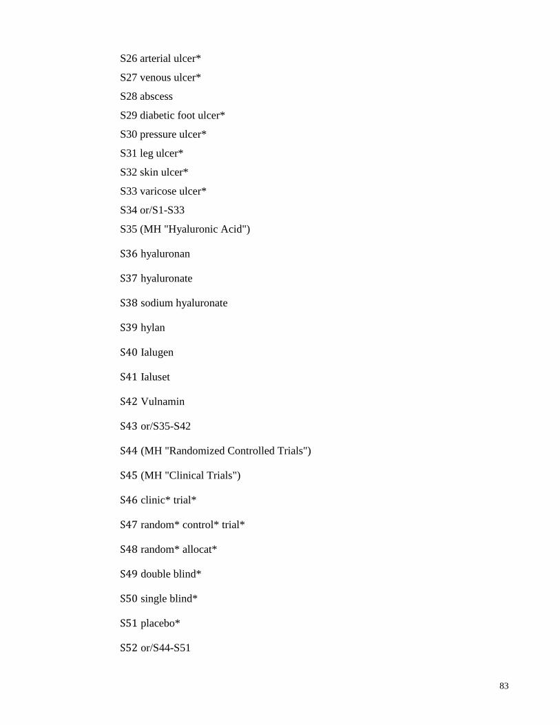
83
S26 arterial ulcer*
S27 venous ulcer*
S28 abscess
S29 diabetic foot ulcer*
S30 pressure ulcer*
S31 leg ulcer*
S32 skin ulcer*
S33 varicose ulcer*
S34 or/S1-S33
S35 (MH "Hyaluronic Acid")
S36 hyaluronan
S37 hyaluronate
S38 sodium hyaluronate
S39 hylan
S40 Ialugen
S41 Ialuset
S42 Vulnamin
S43 or/S35-S42
S44 (MH "Randomized Controlled Trials")
S45 (MH "Clinical Trials")
S46 clinic* trial*
S47 random* control* trial*
S48 random* allocat*
S49 double blind*
S50 single blind*
S51 placebo*
S52 or/S44-S51

84
S53 effective*
S54 efficacy
S55 wound* heal*
S56 therapeutic
S57 safe*
S58 ((effective* OR safe*) AND adj. hyaluronic acid)
S59 or/S53-S58
S60 S34 and S43 and S52 and S60
B) Cochrane Central Register of Controlled Trials (CENTRAL)
#1 MeSH descriptor [Wound and Injuries] explode all trees
#2 acute wound
#3 surgical wound
#4 traumatic wound
#5 incised wound
#6 contused wound
#7 lacerated wound
#8 puncture wound
#9 avulsion fracture
#10 burn wound
#11 chronic wound
#12 infected wound
#13 radiation poisoning wound
#14 ulcer*
#15 arterial ulcer*

85
#16 venous ulcer*
#17 diabetic foot ulcer*
#18 pressure ulcer*
#19 leg ulcer*
#20 skin ulcer*
#21 varicose ulcer*
#22 abscess
#23 (#1 or #2 or #3 or #4 or #5 or #6 or #7 or #8 or #9 or #10 or #11 or #12 or#13 or
#14 or #15 or #16 or#17 or #18 or #19 or #20 or#21 or #22)
#24 MeSH descriptor: [Hyaluronic Acid] explode all tress
#25 hyaluronan
#26 hylan
#27 Ialugen
#28 Vulnamin
#29 (#24 or #25 or #26 or #27 or #28)
#30 MeSH descriptor: [Randomized Controlled Trial] explode all trees
#31 random allocation
#32 clinical trial
#33 double blind
#34 single blind
#35 (#30 or #31 or #32 or #33 or #34)
#36 (#23 and #29 and #35)
C) MEDLINE with Fulltext @EBSCOhost
1. exp Wounds/

86
2. exp Acute Wounds/
3. surgical wound$/
4. traumatic wound$/
5. incised wound$/
6. contused wound$/
7. lacerated wound$/
8. puncture wound$/
9. avulsion fracture/
10. burn wound$/
11. or/1-10
12. exp Chronic Wounds/
13. infected wound$/
14. radiation poisoning wound$/
15. ulcer$/
16. skin abscess$/
17. ((arterial or venous or diabetic foot or pressure or leg or skin or varicose) adj.
ulcer$)
18. or/12-17
19. exp Hyaluronic Acid/
20. hyaluronan/
21. hyaluronate/
22. hylan/
23. “sodium hyaluronate”/
24. Ialuset
25. Vulnamin
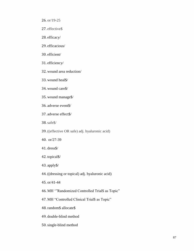
87
26. or/19-25
27. effective$
28. efficacy/
29. efficacious/
30. efficient/
31. efficiency/
32. wound area reduction/
33. wound heal$/
34. wound care$/
35. wound manage$/
36. adverse event$/
37. adverse effect$/
38. safe$/
39. ((effective OR safe) adj. hyaluronic acid)
40. or/27-39
41. dress$/
42. topical$/
43. apply$/
44. ((dressing or topical) adj. hyaluronic acid)
45. or/41-44
46. MH ‘”Randomized Controlled Trial$ as Topic”
47. MH “Controlled Clinical Trial$ as Topic”
48. random$ allocate$
49. double-blind method
50. single-blind method
88
51. comparative study/
52. (clinic$ adj trial$)
53. placebo$/
54. or/46-53
55. case report/
56. letter/
57. review/
58. review of reported case$/
59. or/55-58
60. 54 not 59
61. 11 and 18 and 26 and 40 and 45 and 60
D) PubMed
1. wound$/
2. acute wounds$/
3. surgical wound$/
4. traumatic wound$/
5. incised wound$/
6. contused wound$/
7. lacerated wound$/
8. puncture wound$/
9. avulsion fracture/
10. burn wound$/
11. chronic wound$/

89
12. infected wound$/
13. ulcer$/
14. arterial ulcer$/
15. venous ulcer$/
16. diabetic foot ulcer$/
17. pressure ulcer$/
18. leg ulcer$/
19. skin ulcer$/
20. skin abscess/
21. or/1-20
22. hyaluronic acid/
23. hyaluronan/
24. hyaluronate/
25. hylan/
26. sodium hyaluronate/
27. Ialuset/
28. Ialugen/
29. Vulnamin
30. or/22-29
31. random$ control$ trial$/
32. control$ clinical trial$/
33. random$ allocate$/
34. double-blind method/
35. single-blind method/
36. comparative study/

90
37. placebo$/
38. or/31-37
39. 21 and 30 and 38
E) Journals@Ovid Full Text
1. wound$.af.
2. acute wound$.af.
3. chronic wound$.af.
4. ((surgical or traumatic or incised or contused or lacerated or puncture or avulsion or
burn or infected or radiation poisoning) adj5 wound$).ti.ab.
5. skin abscess$.ti.ab.
6. ulcer$.af.
7. ((arterial or venous or diabetic foot or pressure or leg or skin or varicose) adj5
ulcer$).ti.ab.
8. or/1-7
9. hyaluronic acid.af.
10. hyaluronan.af.
11. hyaluronate.af.
12. hylan.af.
13. sodium hyaluronate.af.
14. Ialuset.af.
15. Ialugen.af.
16. Vulnamin.af.
17. ((effective$ OR safe$) adj hyaluronic acid).af.
18. ((dressing$ or topical$) adj hyaluronic acid).af.

91
19. or/9-18
20. random$ control$ trial$.ti.ab.
21. control$ clinical trial$.ti.ab
22. random$ allocat$.ti.ab.
23. ((double blind or single blind) adj method).ti.ab.
24. comparative study.ti.ab.
25. placebo$.ti.ab.
26. or/20-25
27. 8 and 19 and 26 (274) removes duplicates (205)
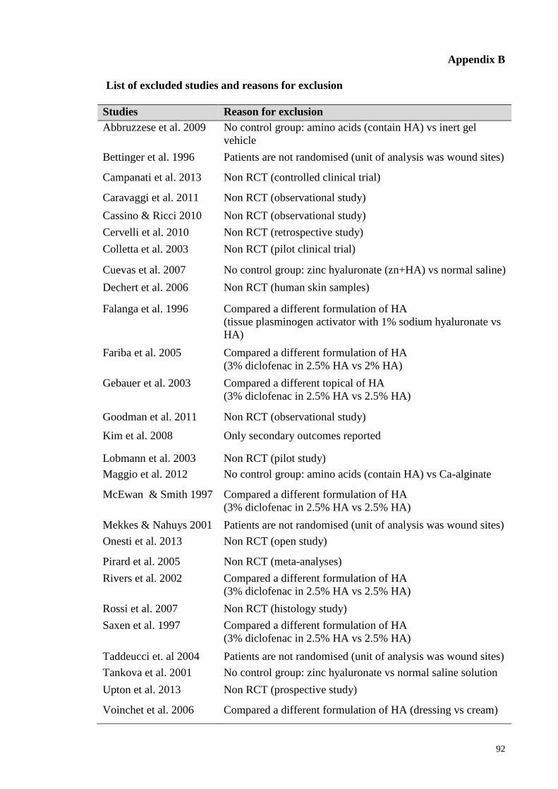
92
Appendix B
List of excluded studies and reasons for exclusion
Studies Reason for exclusion
Abbruzzese et al. 2009 No control group: amino acids (contain HA) vs inert gel
vehicle
Bettinger et al. 1996 Patients are not randomised (unit of analysis was wound sites)
Campanati et al. 2013 Non RCT (controlled clinical trial)
Caravaggi et al. 2011 Non RCT (observational study)
Cassino & Ricci 2010 Non RCT (observational study)
Cervelli et al. 2010 Non RCT (retrospective study)
Colletta et al. 2003 Non RCT (pilot clinical trial)
Cuevas et al. 2007 No control group: zinc hyaluronate (zn+HA) vs normal saline)
Dechert et al. 2006 Non RCT (human skin samples)
Falanga et al. 1996 Compared a different formulation of HA
(tissue plasminogen activator with 1% sodium hyaluronate vs
HA)
Fariba et al. 2005 Compared a different formulation of HA
(3% diclofenac in 2.5% HA vs 2% HA)
Gebauer et al. 2003 Compared a different topical of HA
(3% diclofenac in 2.5% HA vs 2.5% HA)
Goodman et al. 2011 Non RCT (observational study)
Kim et al. 2008 Only secondary outcomes reported
Lobmann et al. 2003 Non RCT (pilot study)
Maggio et al. 2012 No control group: amino acids (contain HA) vs Ca-alginate
McEwan & Smith 1997 Compared a different formulation of HA
(3% diclofenac in 2.5% HA vs 2.5% HA)
Mekkes & Nahuys 2001 Patients are not randomised (unit of analysis was wound sites)
Onesti et al. 2013 Non RCT (open study)
Pirard et al. 2005 Non RCT (meta-analyses)
Rivers et al. 2002 Compared a different formulation of HA
(3% diclofenac in 2.5% HA vs 2.5% HA)
Rossi et al. 2007 Non RCT (histology study)
Saxen et al. 1997
Compared a different formulation of HA
(3% diclofenac in 2.5% HA vs 2.5% HA)
Taddeucci et. al 2004 Patients are not randomised (unit of analysis was wound sites)
Tankova et al. 2001 No control group: zinc hyaluronate vs normal saline solution
Upton et al. 2013 Non RCT (prospective study)
Voinchet et al. 2006 Compared a different formulation of HA (dressing vs cream)

93
Appendix C
Data extraction form
DATA EXTRACTION FORM
(The data extraction form used for each individual study included in this review)
ARTICLE DETAILS
ID
Author(s), Year
Title
Journal, Sources
Country of Origin
Corresponding author &
contact details
Study design
Country
Setting
Treatments:
-Intervention (I)
-Control (C)
Total study duration
Single centre/multicentre trial:
Method used to generate random allocation sequence:
Method used to implement the random allocation
sequence:
Allocation sequence concealment:
Was the participant blinded?
Was the researcher blinded?
Was the assessor blinded?
Inter-assessor reliability measured?
PARTICIPANTS
Total number
Type of participants
Age
Sex
Record of drop-out (with reasons)
All relevant outcomes reported
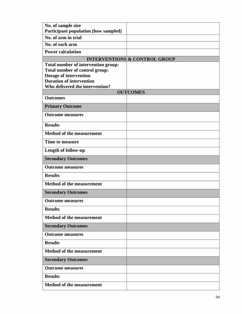
94
No. of sample size
Participant population (how sampled)
No. of arm in trial
No. of each arm
Power calculation
INTERVENTIONS & CONTROL GROUP
Total number of intervention group:
Total number of control group:
Dosage of intervention
Duration of intervention
Who delivered the intervention?
OUTCOMES
Outcomes
Primary Outcome
Outcome measures
Results
Method of the measurement
Time to measure
Length of follow-up
Secondary Outcomes
Outcome measures
Results
Method of the measurement
Secondary Outcomes
Outcome measures
Results
Method of the measurement
Secondary Outcomes
Outcome measures
Results
Method of the measurement
Secondary Outcomes
Outcome measures
Results
Method of the measurement

95
ANALYSIS
Which analysis performed? :
Method of analysis:
Intention-to-treat analysis? :
RESULTS
Sample size:
No. of withdrawals and loss to follow-
up:
Reasons for withdrawals:
Summary of results’ analyses:
MISCELLANEOUS
Funding source:
Key conclusions of the study authors:
AUTHOR’S CONCLUSION
Other comment and interests:
QUALITY ASSESSMENT FORM OF INCLUDED STUDIES
Risk of Bias (Item) Judgement Description
1. Allocation sequence?
2. Allocation concealment?
3. Blinding: participants, researchers, assessors
4. Incomplete outcome data addressed (ITT)
5. Free selective outcome reporting
6. Free from other bias?
i) Baseline comparability
ii) Financial support

96
Appendix D
Quality Assessment Form
QUALITY ASSESSMENT FORM OF INCLUDED STUDIES
METHODOLOGICAL QUALITY
1. Allocation sequence?
Q: was the allocation sequence randomly generated? :
2. Allocation concealment
Q: Was allocation adequately concealed? :
3. Blinding
Q: Was the participants/ researchers/ outcome assessor blinded to the
intervention? :
4. Incomplete outcome data
Q: Were all randomised participants analysed in the group to which they were
collected i.e.by using ITT analysis? :
5. Free selective outcome reporting
Q: Are reports of the trials free of suggestion of selective outcome reporting? :
6. Other sources of potential bias
Q: i) Were the groups similar at baseline for most important prognostic
indicators? :
ii) Was the trial NOT sponsored by a manufacturer who had the potential
interest in the results?

97
Appendix E
Availability of the outcomes data reported
Study Wound
area
reduction
Number
of
wounds
healed
Healing
Time
Pain
intensity
Quality
of the
wound
Adverse
events
Physician
& Patient
assessment
Oral
analgesic
consumption
Chronic Wounds
A) Mixed arterial and venous ulcers
Dereure et
al. (2012a)
(median) - -
Dereure et
al. (2012b)
(ep in
CI)
- (in PV)
(only
cases)
-
Humbert
et al.
(2012)
- -
Meaume
et al.
(2008)
-
Romanelli
et al.
(2007)
- - (baseline
is NR)
- -
B) Venous leg ulcers
Ortonne
(1996) - -
(diff.
scale)
- -
C) Diabetic foot ulcers
Caravaggi
et al.
(2003)
- (SD is
NR)
- - -
Uccioli et
al. (2011)
(SD is
NR)
(SD is
NR)
- - - -
Acute wounds
A) Burn wounds
Koller
(2004)
(baseline
is NR)
- (diff.
scale)
(local
edema)
(none)
Costagliol
a and
Agrosi
(2005)
(ep is
NR)
- (baseline
is NR)
- (in PV)
Notes: NR: Not Reported; SD: standard deviation; ep: endpoint; CI: confidence interval; PV: p-value;
: outcome reported; - : outcome not reported

98
Appendix F
Details of the risk of bias assessment for included trials
Study Random
sequence
generation
Allocation
sequence
concealment
Blinding
Incomplet
e outcome
data (ITT)
Free
selective
outcome
reporting
Free from other bias
participants researcher assessors baseline
comparability
Financial
supports
Chronic wounds- Mixed arterial and venous ulcers
Dereure et
al. (2012a)
Low.
Computer-
generated
randomisati
on list.
Unclear.
Not described
method of
allocation
concealment
Low. Intervention
& Control were
supply in the same
form, external
packaging, shape,
odour & texture.
participants were
assigned to a
treatment group
based on the
sequential order of
the randomisation
Low.
Treatment
allocation &
evaluation
were assessed
by a blinded
physician
Low.
Independent
readers
Low.
The
dropouts
rates are
quite high
but reasons
were
available
Dropouts=
25.7%
Low.
Include all
expected
outcomes
Low.
Patients’
characteristics
are comparable
Low.
No conflict
of interest.
Dereure et
al. (2012b)
Low.
Computer-
generated
randomisati
on list.
Unclear.
Insufficient
detail.
Low. Intervention &
Control were supply
in the same form,
external packaging,
shape, odour &
texture, participants
were assigned to a
treatment group
based on the
sequential order of
the randomisation
Low.
Blinded
observer.
Low.
Treatment
allocation &
evaluation
were
assessed by a
blinded
physician
Low.
All
participants
included
Dropouts=
15.9%
(<20%)
Low.
Include all
expected
outcomes.
Low.
Patients’
characteristics
are comparable
Low.
No conflict
of interest.
98

99
Study Random
sequence
generation
Allocation
sequence
concealment
Blinding
Incomplet
e outcome
data (ITT)
Free
selective
outcome
reporting
Free from other bias
participants researcher assessors baseline
comparability
Financial
supports
Chronic wounds- Mixed arterial and venous ulcers
Humbert
et al.
(2012)
Low.
Parallel-
group
randomised.
Randomisati
on list
balanced
per blocks
of 4 based
on
sequential
order at
each site
Low.
Randomisatio
n list was
prepared by
Data
Management
&Statistics
Unit of IBSA
Low.
Participants are
blinded
Low.
2 independent
readers
(blinded)
measured the
wound size
Low.
2
independent
readers
(blinded)
measured the
wound size
Low.
Missing
outcome
data
balance
across
groups. Dropouts=
19%
(<20%)
Low.
All
outcomes
reported in a
specific
ways
Low.
Baseline are
comparable
Unclear.
Project was
carried out
by the
Sponsor
Laboratoir
es
Genevrier
Meaume
et al.
(2008)
Low.
Computer-
generated
randomisati
on list
Low.
Sealed
envelopes
containing
treatment
code.
Unclear.
No information
provided
Low.
Sealed
envelopes
containing
the treatment
code for each
patient were
given to the
investigator
Low.
Independent
personnel
Low.
All
participants
included
Dropouts=
18.4%
Low.
All outcome
reported in a
specific
ways
Low.
Demographic
characteristics
are comparable
Low.
No conflict
of interest.
99

100
Study Random
sequence
generation
Allocation
sequence
concealment
Blinding
Incomplet
e outcome
data (ITT)
Free
selective
outcome
reporting
Free from other bias
participants researcher assessors baseline
comparability
Financial
supports
Chronic wounds- Mixed arterial and venous ulcers
Romanelli
et al.
(2007)
No.
The
sequence of
randomisati
on was
generated
through
every other
patient
selection by
the clinician
“According
to Cochrane
page 198:
high risk
because of
allocation
by
judgement
of the
clinician”
Unclear.
No
information
provided
Unclear.
No information
provided
Unclear.
No
information
provided
Unclear.
No
information
provided
Unclear.
No ITT
Was
carried out
Low.
Include all
expected
outcomes
Low.
Patients ulcer
characteristics
are comparable
Unclear. Supported
by
Healthpoint
Biopharma
ceutical.
10
0
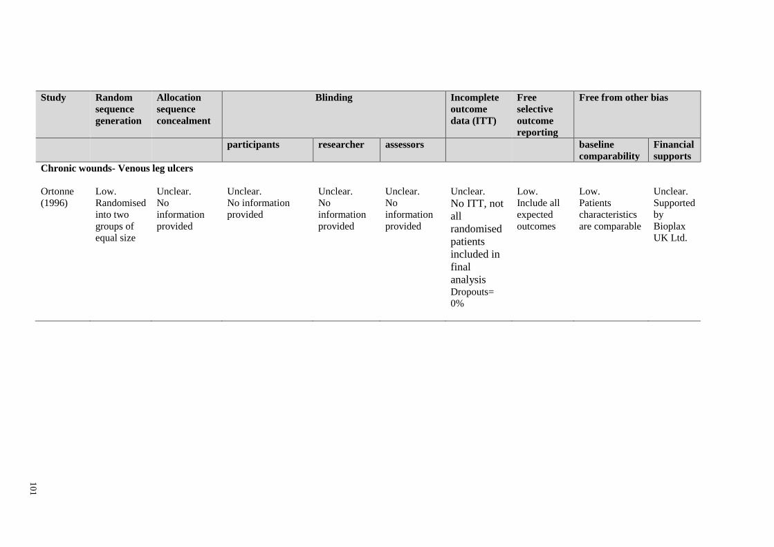
101
Study Random
sequence
generation
Allocation
sequence
concealment
Blinding
Incomplete
outcome
data (ITT)
Free
selective
outcome
reporting
Free from other bias
participants researcher assessors baseline
comparability
Financial
supports
Chronic wounds- Venous leg ulcers
Ortonne
(1996)
Low.
Randomised
into two
groups of
equal size
Unclear.
No
information
provided
Unclear.
No information
provided
Unclear.
No
information
provided
Unclear.
No
information
provided
Unclear.
No ITT, not
all
randomised
patients
included in
final
analysis
Dropouts=
0%
Low.
Include all
expected
outcomes
Low.
Patients
characteristics
are comparable
Unclear.
Supported
by
Bioplax
UK Ltd.
10
1

102
Study Random
sequence
generation
Allocation
sequence
concealment
Blinding
Incomplete
outcome
data (ITT)
Free
selective
outcome
reporting
Free from other bias
participants researcher assessors baseline
comparability
Financial
supports
Chronic wounds- Diabetic foot ulcers
Caravaggi
et al.
(2003)
Low.
Telephone-
randomisati
on
Low.
Central
allocation
(telephone)
Low.
Intervention &
control were
identical
Low.
Blinded
physicians
Low.
Blinded
podiatrist
High.
Total
participants
is 82 were
randomised
but did not
stated
patients in
each of the
arm. Left
only 79
participants.
Dropouts=
22.8%
High.
No
outcomes on
pain
intensity
were
reported
As
mentioned
in method
section
Low.
Patients’
characteristics
are comparable
Unclear.
Insufficient
data about
financial
support
Uccioli et
al. (2011)
Low.
Computer-
generated
randomisati
on list
Low.
Sealed
envelopes
Low.
Patients were
blinded
Unclear.
No
information
provided
Unclear.
No
information
provided
Low.
All
participants
included
dropouts=
11.1%
Low.
All
outcomes
reported in a
specific way
Low.
Baseline are
comparable
Unclear.
Project was
carried out by
the Sponsor,
Anika
Therapeutics
10
2

103
Study Random sequence
generation
Allocation
sequence
concealment
Blinding
Incomplete
outcome data
(ITT)
Free selective
outcome
reporting
Free from other bias
participants researcher assessors baseline
compara
bility
Financial
supports
Acute wounds- Burn wounds
Koller
(2004)
Low.
Computer-
generated
randomisation list.
Quote: “the
recruited patients
were randomly
allocated”.
Unclear.
Insufficient
details
Low.
Same appearance,
colour, consistency,
unmarked tubes of
cream
Unclear .
No
information
provided
Unclear.
Insufficient
details
Unclear.
For wound area
reduction,
baseline value
and endpoint
value were not
equally
reported
Low.
Pre-specified
outcomes
have been
reported.
Low.
Baseline
are
compara
ble
Unclear.
No
information
reported on
financial
support
Costagliola
and Agrosi
(2005)
Low.
Quote: “patients
were randomly
allocated according
to a computer-
generated
randomisation list”
Unclear.
Not reported
Low.
Intervention &
control were
provided in
unmarked, white
tubes containing
25g
Unclear.
Not reported
Unclear.
Insufficient
details.
Low.
All participants
included
Dropouts=
0.9%
Unclear.
Endpoint for
wound area
reduction &
pain intensity
were not
completely
reported
Low.
Patients
character
istics are
compara
ble
Unclear.
Insufficient
details
10
3
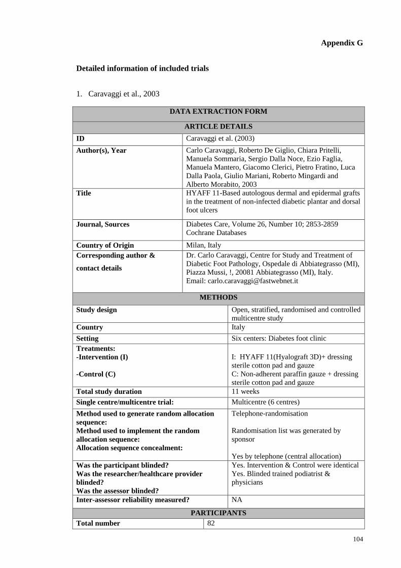
104
Appendix G
Detailed information of included trials
1. Caravaggi et al., 2003
DATA EXTRACTION FORM
ARTICLE DETAILS
ID Caravaggi et al. (2003)
Author(s), Year
Carlo Caravaggi, Roberto De Giglio, Chiara Pritelli,
Manuela Sommaria, Sergio Dalla Noce, Ezio Faglia,
Manuela Mantero, Giacomo Clerici, Pietro Fratino, Luca
Dalla Paola, Giulio Mariani, Roberto Mingardi and
Alberto Morabito, 2003
Title
HYAFF 11-Based autologous dermal and epidermal grafts
in the treatment of non-infected diabetic plantar and dorsal
foot ulcers
Journal, Sources Diabetes Care, Volume 26, Number 10; 2853-2859
Cochrane Databases
Country of Origin Milan, Italy
Corresponding author &
contact details
Dr. Carlo Caravaggi, Centre for Study and Treatment of
Diabetic Foot Pathology, Ospedale di Abbiategrasso (MI),
Piazza Mussi, !, 20081 Abbiategrasso (MI), Italy.
Email: [email protected]
METHODS
Study design Open, stratified, randomised and controlled
multicentre study
Country Italy
Setting Six centers: Diabetes foot clinic
Treatments:
-Intervention (I)
-Control (C)
I: HYAFF 11(Hyalograft 3D)+ dressing
sterile cotton pad and gauze
C: Non-adherent paraffin gauze + dressing
sterile cotton pad and gauze
Total study duration 11 weeks
Single centre/multicentre trial: Multicentre (6 centres)
Method used to generate random allocation
sequence:
Method used to implement the random
allocation sequence:
Allocation sequence concealment:
Telephone-randomisation
Randomisation list was generated by
sponsor
Yes by telephone (central allocation)
Was the participant blinded?
Was the researcher/healthcare provider
blinded?
Was the assessor blinded?
Yes. Intervention & Control were identical
Yes. Blinded trained podiatrist &
physicians
Inter-assessor reliability measured? NA
PARTICIPANTS
Total number
82

105
Type of participants
Diabetic foot ulcers
Age
NA
Sex
Male & Female
Record of drop-out (with reasons)
3, area of ulcer < 1cm2 (n=2), severe acute
ischemia of the foot (n=1)
No. of sample size
Participant population (how sampled)
79 (ITT)
NA
No. of arm in trial
Two
No. of each arm
I: 43 C:36
Power calculation
Based on α=0.005, β=95%, an estimated mean
healing time of 30 days required 78 subjects to
detect any statistically significant.
Inclusion criteria Ulcer > 2cm2, duration of ulcer <1 month
INTERVENTION & CONTROL GROUP
Total number of intervention group:
Total number of control group:
Dosage of intervention & control
Duration of intervention & control
Who delivered the intervention &
control
43
36
NA
NA
NA
OUTCOMES
All relevant outcomes reported No. pain intensity data was not reported
Outcomes
Primary: Percentage of healed ulcers & time to
closure (Complete re-epithelisation)
Secondary: Presence of fibrous slough & necrotic
tissue, Appearance of granulation tissue,
Maceration, Presence & amount of exudate,
Presence of odor & infection, Pain Intensity
Primary Outcome Percentage of participants healed & time for
complete healing (Healing time)
Outcome measures Percentage & days
Results I: 65.3% (28/43), 57 days(median) 47.8 days
(mean)
C: 49.6% (18/36), 77 days (median) 57.8 days
(mean)
Method of the analysis Kaplan-Meier
Secondary Outcomes Number of wounds healed
Outcome measures Percentage & Number of ulcer
Results I: Plantar=55% (12/22), 57 days, Dorsal=66.7%
(14/21), 63days
Total: 60% (28/43)
C: Plantar=50% (10/20), 58.5 days, Dorsal=
31.25% (5/16), complete closure was not apparent
at 77 days
Total: 41.7% (18/36)
Method of the analysis Kaplan-Meier
Secondary Outcomes Complete wound healing (for PP to assess
robustness)
Outcome measures Percentage of participants & days

106
Results I: 63.7%, 59 days
C: 50%, >77days
Method of the analysis Kaplan-Meier
Secondary Outcomes Presence of exudate
Outcome measures Percentage
Results Absent I: 86%, C; 69.4%
Method of the measurement NA
Secondary Outcomes Adverse events
Outcome measures Percentage & number of cases
Results serious AE(I=7, C=10)-wound infection,
inflammation, worsening of ischemia
36.4% (8 cases) severe,
36.4% (8cases) moderate,
36.4% (8cases) low
Method of the measurement NA
ANALYSIS
Method of measurement :
Method of analysis:
Intention-to-treat analysis? :
Computerised morphometric measurement
Continuous variables: by Student’s t-test,
Qualitative variables: by Fisher’s exact test,
Median time to closure: by Kaplan-Meier (log-
rank test). Using SAS statistical software (SAS,
Cary, NC)
Yes. All patients who were randomised included. RESULTS
Sample size:
No. of withdrawals & loss to follow-up:
Reasons for withdrawals:
79
I:8, C:10
Serious adverse events (I=3, C=5)
Investigator decision (C=1)
Protocol violations (I=5, C=4)
MISCELLANEOUS
Funding source:
Key conclusions of the study authors:
Supported by a research grant from Fidia
Advanced Biopolymers (Abano Terme, Italy)
NA
AUTHOR’S CONCLUSION
Other comment and interests: NA
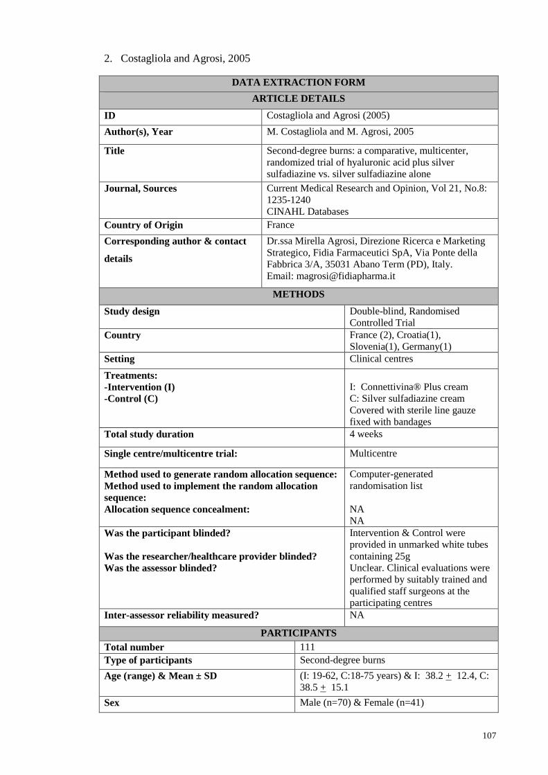
107
2. Costagliola and Agrosi, 2005
DATA EXTRACTION FORM
ARTICLE DETAILS
ID Costagliola and Agrosi (2005)
Author(s), Year
M. Costagliola and M. Agrosi, 2005
Title
Second-degree burns: a comparative, multicenter,
randomized trial of hyaluronic acid plus silver
sulfadiazine vs. silver sulfadiazine alone
Journal, Sources Current Medical Research and Opinion, Vol 21, No.8:
1235-1240
CINAHL Databases
http;//www.cmrojournal.com Country of Origin France
Corresponding author & contact
details
Dr.ssa Mirella Agrosi, Direzione Ricerca e Marketing
Strategico, Fidia Farmaceutici SpA, Via Ponte della
Fabbrica 3/A, 35031 Abano Term (PD), Italy.
Email: [email protected]
METHODS
Study design Double-blind, Randomised
Controlled Trial
Country France (2), Croatia(1),
Slovenia(1), Germany(1)
Setting Clinical centres
Treatments:
-Intervention (I)
-Control (C)
I: Connettivina® Plus cream
C: Silver sulfadiazine cream
Covered with sterile line gauze
fixed with bandages
Total study duration 4 weeks
Single centre/multicentre trial: Multicentre
Method used to generate random allocation sequence:
Method used to implement the random allocation
sequence:
Allocation sequence concealment:
Computer-generated
randomisation list
NA
NA
Was the participant blinded?
Was the researcher/healthcare provider blinded?
Was the assessor blinded?
Intervention & Control were
provided in unmarked white tubes
containing 25g
Unclear. Clinical evaluations were
performed by suitably trained and
qualified staff surgeons at the
participating centres
Inter-assessor reliability measured? NA
PARTICIPANTS
Total number
111
Type of participants
Second-degree burns
Age (range) & Mean ± SD (I: 19-62, C:18-75 years) & I: 38.2 + 12.4, C:
38.5 + 15.1
Sex
Male (n=70) & Female (n=41)

108
Record of drop-out (with reasons)
1 male patient in SSD group (lost at follow-up)
No. of sample size
Participant population (how sampled)
110
Randomly allocated
No. of arm in trial
Two
No. of each arm
n=56, 54
Power calculation
NA
Inclusion criteria Burns not exceed 5% of body surface area,
burns have occurred within 48hours from the
start treatment
INTERVENTION & CONTROL GROUP
Total number of intervention group:
Total number of control group:
Dosage of intervention & control
Duration of intervention & control
Who delivered the intervention & control
56
54
I: 0.2% hyaluronic acid & 1% silver
sulfadiazine (HA-SSD)
C: 1% silver sulfadiazine (HA-SSD)
Applied once daily until 4 weeks
NA
OUTCOMES
All relevant outcomes reported Yes
Outcomes
Primary: Evolution of healing (complete/incomplete)
Secondary: Consumption of analgesics, Pain, Itching,
Impairment to movement, Global comfort, Adverse
events.
Primary Outcome Time to heal/Evolution of healing
(complete/incomplete)
Outcome measures Total time in days required for complete healing of the
wound (time to healing)
Results I: 9.5 days
C: 14 days
A difference of 4.5 days, p=0.0073
Method of the measurement 95% CI, p=0.0073
Secondary Outcomes Area of wound
Outcome measures Wound area size
Results I: baseline 97.3 ± 100.7 cm2 to 39.13% (≤ 50cm
2),
26.09% (51-100cm2), 21.74% (101-150 cm
2), 13.04%
(>150cm2)
C: baseline 91.4 ± 55.9 cm2 to 26.67% (≤ 50cm2),
26.67% (51-100cm2), 30% (101-150 cm2), 16.67%
(>150cm2)
Secondary Outcomes Consumption of analgesics
Outcome measures No. of tablets & days of use
Results I: 0.57 ± 0.89 tablets & 4.0 ± 6.1 days (p=0.5830)
C: 0.69 ± 1.08 tablets & 2.4 ± 3.9 days (p=0.1764)
Method of the analysis Fisher’s exact test
Secondary Outcomes Pain
Outcome measures Means of a visual analog scale
Results No statistically significant difference between groups
(95% CI, -0.43, 1.48)
Method of the measurement Huskisson scale; 0-10cm

109
Time to measure One week after treatment
Secondary Outcomes Itching
Outcome measures Percentage by class
Results I: 87.5% of class Absent-Mild, 12.5% of class
Moderate-Severe
C: 81.5% of class Absent-Mild, 18.5% of class
Moderate-Severe
No statistically significant difference between groups
(p=0.2701, p=0.3551)
Method of the analysis Qualitative approach, Fisher’s exact test
Secondary Outcomes Impairment to movement
Outcome measures Percentage by class
Results I: 73.2% of class No-Mild, 26.8% of class Moderate-
Severe
C: 63% of class Absent-Mild, 37% of class Moderate-
Severe
Method of the analysis Qualitative approach
Secondary Outcomes Global comfort/ Tolerability
Outcome measures Percentage by class
Results I: 76.8% of class Excellent-Good, 23.2% of class Bad-
Very bad
C: 75.9% of class Absent-Mild, 24.1% in class
Moderate-Severe
No statistically significant difference between groups
(p=0.2054, p=0.3122)
Method of the analysis Qualitative approach, Fisher’s exact test
Secondary Outcomes Adverse events
Outcome measures Number of patient
Results I: None C: 1 patient (shivering, fever, headache)
ANALYSIS
Method of measurements :
Method of analysis:
Intention-to-treat analysis? :
Digitalized macrophotography
1. Mean of survival analysis with the Wilcoxon test
2. Qualitative approach with the Fisher’s exact test
3. Descriptive way, visit by visit
Yes. RESULTS
Sample size:
No. of withdrawals and loss to
follow-up:
Reasons for withdrawals:
110
1
Shivering, fever. headache
Fixed combination HA-SSD caused a significantly
more rapid re-epithelialisation of burns
MISCELLANEOUS
Funding source:
Study support was provided by Fidia Farmaceutici
SpA, Abano Terme (PD), Italy
AUTHOR’S CONCLUSION
Other comment and interests: Fixed combination promoted significantly faster
healing of the burn than SSD alone
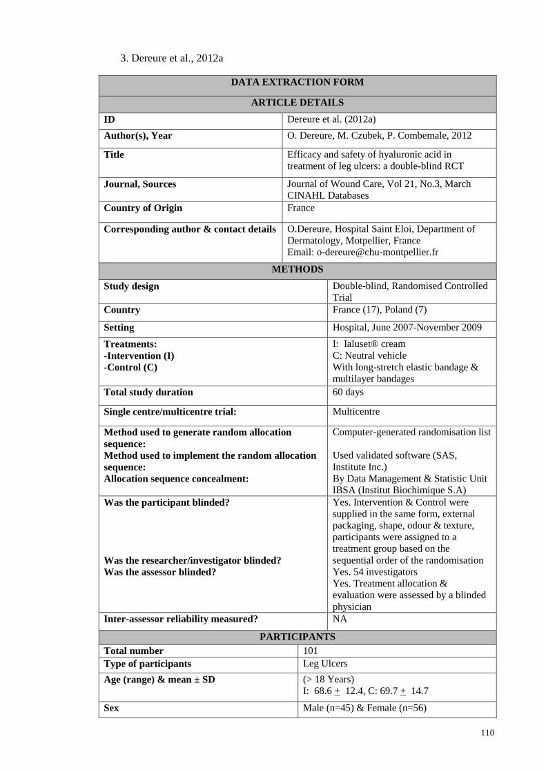
110
3. Dereure et al., 2012a
DATA EXTRACTION FORM
ARTICLE DETAILS
ID Dereure et al. (2012a)
Author(s), Year
O. Dereure, M. Czubek, P. Combemale, 2012
Title
Efficacy and safety of hyaluronic acid in
treatment of leg ulcers: a double-blind RCT
Journal, Sources Journal of Wound Care, Vol 21, No.3, March
CINAHL Databases
Country of Origin France
Corresponding author & contact details O.Dereure, Hospital Saint Eloi, Department of
Dermatology, Motpellier, France
Email: [email protected]
METHODS
Study design Double-blind, Randomised Controlled
Trial
Country France (17), Poland (7)
Setting Hospital, June 2007-November 2009
Treatments:
-Intervention (I)
-Control (C)
I: Ialuset® cream
C: Neutral vehicle
With long-stretch elastic bandage &
multilayer bandages
Total study duration 60 days
Single centre/multicentre trial: Multicentre
Method used to generate random allocation
sequence:
Method used to implement the random allocation
sequence:
Allocation sequence concealment:
Computer-generated randomisation list
Used validated software (SAS,
Institute Inc.)
By Data Management & Statistic Unit
IBSA (Institut Biochimique S.A)
Was the participant blinded?
Was the researcher/investigator blinded?
Was the assessor blinded?
Yes. Intervention & Control were
supplied in the same form, external
packaging, shape, odour & texture,
participants were assigned to a
treatment group based on the
sequential order of the randomisation
Yes. 54 investigators
Yes. Treatment allocation &
evaluation were assessed by a blinded
physician
Inter-assessor reliability measured? NA
PARTICIPANTS
Total number
101
Type of participants
Leg Ulcers
Age (range) & mean ± SD
(> 18 Years)
I: 68.6 + 12.4, C: 69.7 + 14.7
Sex
Male (n=45) & Female (n=56)

111
Record of drop-out (with reasons)
I= 12, C=14
No. of sample size
Participant population (how sampled)
75
Randomly allocated
No. of arm in trial
Two
No. of each arm
I; 50, C:51
Power calculation
140 patients for 90% power (89% for 101
patients)
Inclusion criteria >2 months ulcer < 4 years, ulcer surface area
5-40cm2 with no necrotic tissue
INTERVENTION & CONTROL GROUP
Total number of intervention group:
Total number of control group:
Dosage of intervention & control
Duration of intervention & control
Who delivered the intervention & control
50
51
0.2% hyaluronic acid
Once daily until 60 days
By a nurse and investigator (during evaluation
visit)
OUTCOMES
All relevant outcomes reported Yes
Outcomes
Primary: Percentage of wound size reduction at day
45
Secondary: pain intensity, burden of pain,
cumulative no. healed ulcers, percentage of fibrous
tissue, percentage of granulation tissue,
acceptability, adverse events
Primary Outcome Percentage of wound size reduction at day 45
Outcome measures Median percentage reduction in wound area
Results I: baseline 11.1cm2 mean: 16.08
Endpoint: (day 45=39±6%)mean ± SEM SD
9.81 [4.17]
C: baseline 11.7cm2 mean: 17.04
Endpoint (day 45=5±9%)mean ± SEM 16.55
[10.64] P=0.002
Method of the measurement Traced the wound margins on sterile tracing paper,
measured the wound size based on tracings, using
digital planimetry (Visitrak; Smith & Nephew)
Time to measure Day 15±2, 30±3, 45±3 and 60
Length of follow-up 45 days
Secondary Outcomes Percentage of wound size reduction at day 15, 30
and 60
Outcome measures Mean percentage reduction in wound area
Results I: day 15=27%, day30=33%,
C: day 15=27%, day30=33%,
Method of the measurement using digital planimetry (Visitrak; Smith &
Nephew)
Secondary Outcomes Pain intensity
Results I: 23.0 to 13.2 mm (by 9.8 ± 3.5 mm)
C: 26.6 to 25.8 mm (by 0.8 ± 3.2 mm)
Method of the measurement 0 (no pain) -100 (severe pain) mm VAS
Secondary Outcomes Cumulative no. healed ulcers
Outcome measures Number of ulcer

112
Results I: day15=0, day 30=1, day 45=1, day60=3
C: day15=0, day 30=3, day45=3, day60=4
No statistically significant
Secondary Outcomes Percentage of fibrous tissue (Healing wound
process)
Outcome measures Percentage
Results I: day1=40.9% to day 60=28.6%
C: day1=44.0% to day 60=37.6%
Secondary Outcomes Percentage of granulation tissue (Healing wound
process)
Outcome measures Percentage
Results I: day1=59.1% to day 60=63.5%
C: day1=56.0% to day 60=58.5%
Secondary Outcomes Acceptability
Outcome measures Counting the number of application performed
Results >79% did not miss any daily application
Method of the measurement Excellent=0, Good=0-3, Fair=3-7, Poor= ≥7
Secondary Outcomes Adverse events
Outcome measures Percentage of cases
Results I=20 AE, C=19 AE
Mild to moderate=88%, Severe=11% (application
site burn, inflammation or pain and aggravated
condition)
ANALYSIS
Which analysis performed? :
Method of analysis:
Intention-to-treat analysis? (ITT)
1. Analysis of variance (ANNOVA)
2. Qualitative variables use Chi-square test or
Fisher’s exact test
Yes, 101 patients analysed in the ITT
RESULTS
Sample size:
No. of withdrawals & loss to follow-
up:
Reasons for withdrawals:
Summary of results’ analyses:
20% difference in the % of wound size reduction
between groups at day 45 would be significant,
theoretical sample size was 140 patients, taking
into account 10% of dropouts
I=3, C=5
I: inefficacy(n=2), healing (n=1)
C: inefficacy (n=2), healing (n=3)
These results support hypothesis that hyaluronic
acid significantly contributes to the restoration of
optimal local physiologic conditions which
necessary to promote ulcer healing
MISCELLANEOUS
Funding source:
Key conclusions of the study authors:
Study was sponsored by Laboratoires Genevrier
Hyaluronic acid might have a positive impact on
quality of life through a significant reduction in
pain intensity
AUTHOR’S CONCLUSION
Other comment and interests: Application of hyaluronic acid on leg ulcers is
significantly more effective than a neutral vehicle
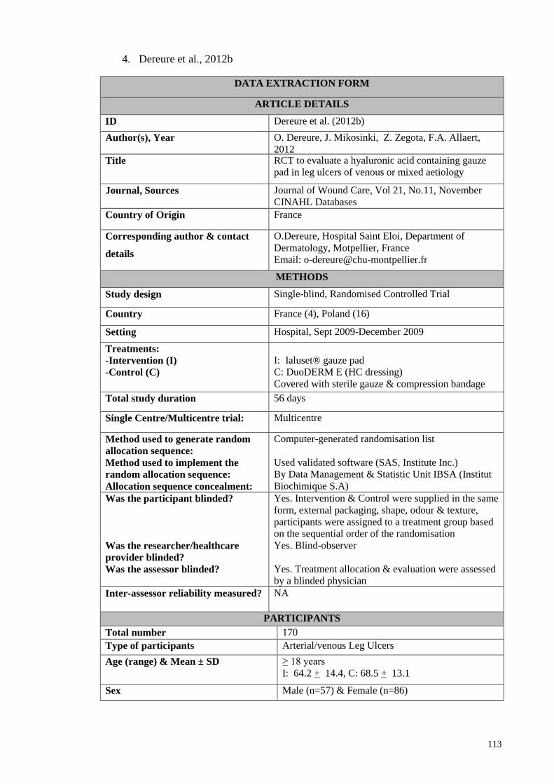
113
4. Dereure et al., 2012b
DATA EXTRACTION FORM
ARTICLE DETAILS
ID Dereure et al. (2012b)
Author(s), Year
O. Dereure, J. Mikosinki, Z. Zegota, F.A. Allaert,
2012 Title
RCT to evaluate a hyaluronic acid containing gauze
pad in leg ulcers of venous or mixed aetiology
Journal, Sources Journal of Wound Care, Vol 21, No.11, November
CINAHL Databases
Country of Origin France
Corresponding author & contact
details
O.Dereure, Hospital Saint Eloi, Department of
Dermatology, Motpellier, France
Email: [email protected]
METHODS
Study design Single-blind, Randomised Controlled Trial
Country France (4), Poland (16)
Setting Hospital, Sept 2009-December 2009
Treatments:
-Intervention (I)
-Control (C)
I: Ialuset® gauze pad
C: DuoDERM E (HC dressing)
Covered with sterile gauze & compression bandage
Total study duration 56 days
Single Centre/Multicentre trial: Multicentre
Method used to generate random
allocation sequence:
Method used to implement the
random allocation sequence:
Allocation sequence concealment:
Computer-generated randomisation list
Used validated software (SAS, Institute Inc.)
By Data Management & Statistic Unit IBSA (Institut
Biochimique S.A)
Was the participant blinded?
Was the researcher/healthcare
provider blinded?
Was the assessor blinded?
Yes. Intervention & Control were supplied in the same
form, external packaging, shape, odour & texture,
participants were assigned to a treatment group based
on the sequential order of the randomisation
Yes. Blind-observer
Yes. Treatment allocation & evaluation were assessed
by a blinded physician
Inter-assessor reliability measured? NA
PARTICIPANTS
Total number
170
Type of participants
Arterial/venous Leg Ulcers
Age (range) & Mean ± SD
≥ 18 years
I: 64.2 + 14.4, C: 68.5 + 13.1
Sex
Male (n=57) & Female (n=86)

114
Record of drop-out (with reasons)
I= 13, C=14
did not meet inclusion criteria (I=6, C=6)
major protocol deviation (I=7, C=8)
No. of sample size
Participant population (how sampled)
143
Randomly allocated according to a central
randomisation list balanced per blocks of four
No. of arm in trial
Two
No. of each arm
n=72, 71
Power calculation
136 patients for 90% power (170 patients if 20%
dropout) Inclusion criteria Ulcer >2months < 4years, surface target ulcer 5-
40cm2
INTERVENTION & CONTROL GROUP
Total number of intervention group:
Total number of control group:
Dosage of intervention & control
Duration of intervention & control
Who delivered the intervention &
control
85
85
0.05% HA-impregnated cotton gauze pad
I: once daily, C: every2-3days until 56 days
By a nurse and investigator (during evaluation visit)
OUTCOMES
All relevant outcomes reported Yes
Outcomes
Primary: Percentages of patients achieving a
reduction of at least 40%of the initial wound surface
after 56 days of treatment
Secondary: percentage of wound size reduction at
day 14, 28& 56, pain intensity, burden of pain,
aspect of peri-ulcerous skin, percentage of complete
healed ulcer, pattern of the wound, patient
acceptability and adverse events
Primary Outcome Percentages of patients achievement a reduction of
at least 40%of the initial wound surface after 56
days of treatment
Outcome measures 95% CI of the difference between the two groups
Results I: baseline 14.4 (9.0) to 74% (53 patients)
C: baseline 15.8 (9.5) to 72% (51 patients)
CI [-0.128, 0.164]
Method of the measurement Traced the wound margins on sterile tracing paper,
measured the wound size based on tracings, using
digital planimetry (Visitrak; Smith & Nephew)
Time to measure Day 14, 28 and 56
Length of follow-up 56 days
Secondary Outcomes Percentage of wound size reduction at day 14, 28 &
56
Outcome measures 95% CI
Results P=0.3931
Secondary Outcomes Pain intensity
Outcome measures 95% CI
Results I: 29.3 ± 25.0 to 11.6 mm
C: 29.7 ± 24.9 to 21.6 mm

115
P=0.6658
Method of the measurement 0 (no pain) -100 (severe pain) mm VAS
Secondary Outcomes Burden of pain
Outcome measures Means of a visual analog scale
Results I: 121.9 ± 20.7 mm
C: 207.4 ± 32.9 mm
P=0.028
Primary Outcomes Percentage of complete healed ulcer (full
epithelialisation)
Outcome measures 95% CI
Results P=0.6007
Secondary Outcomes Aspect of peri-ulcerous skin
Outcome measures Qualitative measure
Results P=0.8544 for fibrinous tissue
P=0.6704 for granulation tissue
Secondary Outcomes Acceptability
Outcome measures Counting the number of application performed
Results >97% had >80% of applications
Method of the measurement Good=>80%, Fair=50%-80%, Poor= <50%
Secondary Outcomes Adverse events
Outcome measures Qualitative measure
Results I=36 AE, C=41 AE
Mild to moderate=77%, Severe=23% (I=4, C=14)
ANALYSIS
Which analysis performed? :
Method of analysis:
Intention-to-treat analysis? (ITT):
1. Analysis of variance (ANNOVA)
2. Qualitative variables use Chi-square test or
Fisher’s exact test
Yes, 170 patients analysed in the ITT
RESULTS
Sample size:
No. of withdrawals and loss to follow-
up:
Reasons for withdrawals:
170.20% difference in the % of wound size
reduction between groups at day 45 would be
significant, theoretical sample size was 170 patients,
taking into account 20% of dropouts
I=13, C=13
Ulcer healing (n=12)
Treatment related AE (n=7)
NA
MISCELLANEOUS
Funding source:
Key conclusions of the study authors:
Study was sponsored by Laboratoires Genevrier
NA
AUTHOR’S CONCLUSION
Other comment and interests: Local application of HA using an impregnated
gauze pad is more favourable as compared with HC
dressing. The study confirmed the good safety
profile of the HA gauze pad.

116
5. Humbert et al., 2012
DATA EXTRACTION FORM
ARTICLE DETAILS
ID Humbert et al. (2012)
Author(s), Year
Philippe Humbert, Jacek Mikosinski, Hakima
benchikhi and Francois-Andre Allaert, 2012
Title
Efficacy and safety of a gauze pad containing
hyaluronic acid in treatment of leg ulcers of venous or
mixed origin: a double-blind, randomised, controlled
trial
Journal, Sources International Wound Journal, CINAHL database
Country of Origin France
Corresponding author & contact
details
P Humbert, Service de Dermatologie, CHU de
Besancon, 2 place Saint-Jacques, 25030 Besancon,
France
Email: [email protected]
METHODS
Study design Double-blind, randomised controlled trial
Country France, Morocco, Poland
Setting 31 investigators from 29 centres
France (18 centres), Morocco (3 centres),
Poland (8 centres) from Nov 2007-
November 2009
Treatments:
-Intervention (I)
-Control (C)
Ialuset® gauze pad
Neutral vehicle gauze pad
Covered with sterile gauze & an appropriate
bandage
Total study duration 60 days
Single centre/multicentre trial: Multicentre
Method used to generate random allocation
sequence:
Method used to implement the random
allocation sequence:
Allocation sequence concealment:
Parallel-group randomised
Validated software from SAS Institute Inc,
Cary, NC
Randomisation list was prepared by Data
Management &Statistics Unit of IBSA
Institut Biochimique SA, Switzerland
Was the participant blinded?
Was the researcher/healthcare provider
blinded?
Was the assessor blinded?
Yes, randomization list balanced per blocks
of 4 based on sequential order at each site
Yes
Yes, 2 independent readers (blinded)
measured the wound size
Inter-rater reliability measured? NA
PARTICIPANTS
Total number
89

117
Type of participants
Arterial/venous Leg ulcer
Age (range) & Mean ± SD ≥18 years
I=59.4 (2.5), C=64.1 (2.7)
Sex
Female=44, Male=45
Record of drop-out (with reasons)
I=7, C=10
Did not meet inclusion criteria (1=5, C=5)
Protocol deviation (I=1, C=3)
Visit outside the specific range for primary evaluation
(I=1, C=1)
Poor compliance (C=1)
No. of sample size
Participant population (how
sampled)
89
In-patients & Out-patients
No. of arm in trial
Two
No. of each arm
I: 45, C:44
Power calculation
Hypothesis: 20% difference between the percentages of
wound size reduction with HA compared with neutral at
D45
Theoretical sample size: 140 patients (with 10%
dropouts)
Inclusion criteria One/several leg ulcers (arterial/venous), present for
>2mths, < 4 years, surface ulcer 5-40cm2 with no
necrotic tissue.
INTERVENTION & CONTROL GROUP
Total number of intervention
group:
Total number of control group:
Dosage of intervention & control
Duration of intervention &
control
Who delivered the intervention &
control
45
44
0.05% Hyaluronic acid
Maximum 60 days
Nurse
OUTCOMES
All relevant outcomes reported Yes
Outcomes
Primary: Percentage of wound size reduction after 45
days, Secondary: Percentage of wound size reduction at
D15, D30, D60, Percentage of patients with healed ulcer
at day 45 & day 60, Pain intensity, Burden of pain,
Tolerance, Systemic anaelgesic & antibiotics used,
Aspect of wound, Aspect of peri-ulcerous skin, Adverse
events
Primary Outcome Percentage of wound size reduction after 45 days
Outcome measures Percentage of wound size reduction
Results I=from 13.8 ± 1.3cm2 decreased by 73±4.6%,
SD (±8.72) to (±8.32)
C= from 12.9 ± 1.3 cm2 decreased by 46±9.6%
SD (±8.62) to (±7.8)
P=0.011 (statistically significant different)

118
Method of the measurement Shape of the wound was drawn by the investigator using
a sterile tracing paper at each evaluation visit for
measure of wound size by a Digital Planimetrics
System, Visitrak®
Time to measure Evaluation visit: day 0, day 45±3
Length of follow-up 60 days
Secondary Outcomes Percentage of wound size reduction at D15, D30, D60
Outcome measures Percentage
Results day15 (I=40%, C=29%), day30 (I=64%, C=36%),
day60 (I=77%, C=52%)
Method of the measurement Shape of the wound was drawn by the investigator using
a sterile tracing paper at each evaluation visit for
measure of wound size by a Digital Planimetrics
System, Visitrak®
Time to measure Evaluation visit: day 0, day 15±2, day 30±3, day 45±3
and day 60±3
Secondary Outcomes Percentage of patients with healed ulcer at day 45 & day
60
Outcome measures Percentage
Results D45 (I=31.1%, C=9.3%) P=0.011 =(14/38, 6/34)
D60 (I=37.8%, C=16.3%) P=0.024 (17/45, 7/44)
Secondary Outcomes Pain intensity
Outcome measures Mean ± SEM
Results Day 60
I= 33.2± 3.7mm to 11.5 ± 2.8mm (SEM)
C=33.4± 4.0mm to 13.7 ±2.9mm (SEM)
Day 30
I= to 12.4 ± 2.6mm
C=to 22.8 ± 3.8mm
Method of the measurement 0-100mm Visual Analogue Scale (VAS)
Secondary Outcomes Aspect of wound
Outcome measures Percentage of necrotic, fibronous or granulation tissue
Results I= 8.5% ±7.6, C=9.0%±8.0
Method of the measurement Semi-quantitative four-point scale (nil=0, slight=1,
moderate=2, important=3)
Secondary Outcomes Aspect of peri-ulcerous skin
Outcome measures Oedema, purpura, erythema, maceration, oozing and
horny edges
Results NA
Method of the measurement Semi-quantitative four-point scale (nil=0, slight=1,
moderate=2, important=3)
Secondary Outcomes Tolerance/Compliance
Outcome measures Counting the number of applications performed
Results 87% =Excellent

119
Method of the measurement Excellent=no day of missed application, Good<3 days,
Fair=3-7 days missed application, Poor>7 days of
missed application
Secondary Outcomes Adverse events
Outcome measures Nature, Severity, Time of onset, Duration, Degree of
relationship to the study treatment & description of any
action/pharmacological treatment undertaken
Results 48 AE in 27 patients
I=22.2%, C=38.6%
I=6.7%, C=18.2% treatment-related AE
75%=mild to moderate, 25%=severe
ANALYSIS
Which analysis performed? :
Method of analysis:
Intention-to-treat analysis? (ITT):
To look at individual outcomes
Yes, on all randomised patients
RESULTS
Sample size:
No. of withdrawals and loss to
follow-up:
Reasons for withdrawals:
Summary of results’ analyses:
89 (ITT), 72(PP)
28 patients did not complete the study (18 HA; 10
control)
17 patients from ITT were reported with major protocol
violations
NA
The results showed significantly greater reduction of
wound size after 45days with HA compared to neutral
vehicle. The proportion of healed ulcer was significantly
higher in the HA group than in the neutral vehicle
group. Pain management also favoured in the HA arm.
MISCELLANEOUS
Funding source:
Key conclusions of the study
authors:
Sponsor, Laboratoires Genevrier.
Study clearly shows that the local application of HA
using an impregnated gauze pad on venous leg ulcers is
significantly more effective than neutral vehicle gauze
pad and effective wound closure with a good safety
profile.
AUTHOR’S CONCLUSION
Other comment and interests: NA

120
6. Koller, 2004
DATA EXTRACTION FORM
ARTICLE DETAILS
ID Koller (2004)
Author(s), Year
Koller J, 2004
Title
Topical treatment of partial thickness burns by silver
sulfadiazine plus hyaluronic acid compared to silver
sulfadiazine alone: a double-blind, clinical study
Journal, Sources Drugs Experimental and Clinical Research, Vol:30,
Issue:5/6; 183-190
Country of Origin Republic Slovakia
Corresponding author & contact
details
Jan Koller, Head of Teaching Department for Burns and
Reconstructive Surgery, Central Tissue Bank,
University Hospital Bratislava Ruzinov, Ruzinoska 6-
821 02 Bratislava, Slovak Republic
Email: [email protected], [email protected]
METHODS
Study design Prospective, Double-blind controlled
Experimental
Country Republic Slovakia
Setting General Hospital, Bratislava
Treatments:
-Intervention (I)
-Control (C)
Ialugen Plus® cream (IBSA Institut
Biochimique SA)
Silver Sulfadiazine cream
Covered with sterile line gauze & bandages
Total study duration 4 weeks
Single centre/multicentre trial: Single centre
Method used to generate random allocation
sequence:
Method used to implement the random
allocation sequence:
Allocation sequence concealment:
Computer-generated randomisation list
NA
NA
Was the participant blinded?
Was the researcher/healthcare provider
blinded?
Was the assessor blinded?
Yes, Same appearance, colour, consistency,
unmarked tubes of cream
Unclear. Performed by suitably trained &
qualified staff surgeons
Inter-rater reliability measured? NA
PARTICIPANTS
Total number
33
Type of participants
Second-degree burns
Age (range) & mean±SD (18-80 years) & I: 35 ± 14.5, C: 40.7 ± 11.6
Sex
Male:24, Female:9
Record of drop-out (with reasons)
None
No. of sample size
Participant population (how
sampled)
33
Selected according to the inclusion and exclusion
criteria

121
No. of arm in trial
Two
No. of each arm
I: 18, C:15
Power calculation
NA
Inclusion criteria Superficial & partial thickness burn exceeding 5% of the
body surface area, 900cm2 in an average adult patient.
INTERVENTION & CONTROL GROUP
Total number of intervention
group:
Total number of control group:
Dosage of intervention & control
Duration of intervention &
control
Who delivered the intervention &
control
18
15
I: 0.2% HA, 1% silver sulfadiazine C: 1% silver
sulfadiazine
Once daily until 4weeks
NA
OUTCOMES
All relevant outcomes reported Yes
Outcomes
Primary: Wound area
Secondary: Time to heal, Local edema, Local infection,
Global response to the treatment, Local tolerability,
Adverse event, Concomitant medications
Primary Outcome Wound area
Outcome measures Percentage of pre-treatment value
Results I: day7= 5.83±14.17, day14=0, day 21=0
C; day7=30.59±28.17, day 14=6.25±12.58, day 21=0
P-value day7=0.002, day14=0.043
Time to measure Day 7, 14, 21
Secondary Outcomes The severity and the extent of the burn
Outcome measures Percentage of the burned skin area
Results I: 9.88 ± 6.89%, C: 11.3 ± 3.53%
P=0.61
Secondary Outcomes Time to heal
Outcome measures Total time in days required for complete healing ,
Record the percentage of the residual wound area
Results I: 8.17± 2.7 days
C: 13.07 ± 5.20 days, P=0.0015
Method of the analysis Student’s t-test for unpaired data
Time to measure Day 1, 7, 14, 21,28
Secondary Outcomes Local edema
Outcome measures Mean local edema, Four-point severity scale (0=absent,
3=severe pain)
Results I: 0.11 ± 0.32
C: 0.50 ± 0.51
P=0.024
Method of the measurement Kruskal-Wallis test
Time to measure Day 7
Secondary Outcomes Pain
Outcome measures Score
Results I: baseline=1.5, day7=0.2

122
C: baseline=1.8, day7=0.7
No stat. significant
Method of the measurement Kruskal-Wallis test
Secondary Outcomes Local infection
Outcome measures Number of patient
Results I: present at day 7 in one patient
C: absent
Method of the measurement Present or Absent
Secondary Outcomes Global response to the treatment
Outcome measures Percentage of participants
Results 80-90%: good/excellent
Method of the measurement Four-point scale (none, fair, good, excellent)
Secondary Outcomes Local tolerability to treatment
Outcome measures Scale
Results Good/Excellent
Method of the measurement Four-point scale (bad, fair, good, excellent)
Secondary Outcomes Adverse event
Outcome measures NA
Results Absence
ANALYSIS
Which analysis performed? :
Method of analysis:
Intention-to-treat analysis? (ITT):
Continous variables were analysed by analysis of
variance (ANOVA) with repeated measures.
Multiple comparisons were performed using Student’s t-
test for unpaired data.
Discrete variables were analyzed using Wilcoxon
Signed Rank Test, Kruskal-Willis test, Fisher’s exact
test, Chi-squared test and linear trend test
Analyzed by independent organisation (IBIS
Informatica & Idee S.r.I, Milan, Italy)
Yes.
RESULTS
Sample size:
No. of withdrawals and loss to
follow-up:
Reasons for withdrawals:
Summary of results’ analyses:
33
None
None
The findings of this study confirmed that the association
of the two compounds in a new topical treatment
significantly reduced the healing time and significantly
accelerated the reduction of local edema occurring
shortly after injury.
MISCELLANEOUS
Funding source:
Key conclusions of the study
authors:
NA
The combination of SS-HA succeeded the wound
healing process. The most favourable effect in SS-HA
is the statistically significant of time to complete healing
AUTHOR’S CONCLUSION
Other comment and interests: NA

123
7. Meaume et al., 2008
DATA EXTRACTION FORM
ARTICLE DETAILS
ID Meaume et al. (2008)
Author(s), Year
Sylvie Meaume, Zohra Ourabah, Marco
Romanelli, Roberto Manopulo, Florent De
Vathaire, Denis Salomon and Jean-Hilaire Saurat,
2008
Title
Efficacy and tolerance of a hydrocolloid dressing
containing hyaluronic acid for the treatment of
leg ulcers of venous or mixed origin
Journal, Sources Current Medical Research and Opinions, Vol: 24,
No:10; 2729-2739
Country of Origin France
Corresponding author & contact
details
S. Meaume, APHP Groupe Hospitalier Charles
Foix, Service de Gerontologie ‘L’ORBE’ 7,
Avenue de la Republique, F-94205 Ivy-sur-Seine
Cedex, France.
Email: [email protected]
METHODS
Study design Open-label, prospective, randomised controlled
trial
Country France, Italy, Switzerland
Setting France: 15 centres, Italy: 2 centres, Switzerland: 1
Centre, November 2001- March 2003
Treatments:
-Intervention (I)
-Control (C)
Hydrocolloid dressing containing Hyaluronic acid
(HC + HA)
Hydrocolloid dressing
Covered with elastic stocking
Total study duration 42 days
Single centre/multicentre trial: Multicentre
Method used to generate random
allocation sequence:
Method used to implement the
random allocation sequence:
Allocation sequence
concealment:
Computer-generated randomisation list
Yes. Sealed envelopes
Yes
Was the participant blinded?
Was the researcher/healthcare
provider blinded?
Was the assessor blinded?
Unclear
Yes. Sealed envelopes containing the treatment
code for each patient were given to the
investigator
Yes, independent personnel
Inter-rater reliability measured? Yes.
PARTICIPANTS
Total number
125

124
Type of participants
Arterial/venous Leg ulcers
Age (range) & Mean ± SD ≥18 years
I: 73± 1.4 (mean ± SEM) 73± 11.11, C: 75 ± 1.4
(mean ± SEM) 75± 11.02 Sex
Male: 54, Female: 71
Record of drop-out (with
reasons)
I: 11, C:11
No. of sample size
Participant population (how
sampled)
125
NA
No. of arm in trial
Two
No. of each arm
I: 63, C:62
Power calculation
120 patients (80% power with 20% drop-outs)
Inclusion criteria Ulcers >2months < 1year and size 5-40cm2 with
no necrotic tissue
INTERVENTION & CONTROL GROUP
Total number of intervention group:
Total number of control group:
Dosage of intervention & control
Duration of intervention & control
Who delivered the intervention & control
63
62
NA
At least once a week until 6 weeks
Investigator
OUTCOMES
All relevant outcomes reported Yes.
Outcomes
Primary: Reduction of wound area
Secondary: Wound bed condition and
Surrounding skin conditions, Complete ulcer
healing, Presence & severity of symptoms pain
and itching, Oral anaelgesic consumption,
Overall efficacy and tolerance, Adverse event
Primary Outcome Reduction of wound area
Outcome measures Cumulative percentage with at least 90% of
ulcer reduction
Results I= SEM 11.7 ± 1.2 to 7.9 ± 1.4 cm2
SD(±9.53) to (±10.1)
C=SEM 12.2 ± 1.7 to 8.3 ± 1.4 cm2 SD
(±13.39) to (±9.9)
Method of the measurement Photographs with graduated scale, Digital
planimeter, PLACOM KP-80 (Koizumi Sokki
MFG. Co.Ltd. Niigata, Japan), t-test
Time to measure Day 1, 7, 14, 28 and 42
Secondary Outcomes Number of patients success
Outcome measures Reduction at least 90% of ulcer area
Results I: 15/63, C: 7/62
Secondary Outcomes Wound bed condition
Outcome measures Percentage of necrotic, fibrinous and
granulation tissues

125
Results Necrotic: I=0.19 ± 0.2 to 0.2 ± 0.2 , C=0.27 ± 3.0 to
0±0
SD: (±1.59) to (±1.59), (±23.6)
Fibrinous: I=29 ± 3.3 to 9.3± 2.3 , C=27 ± 3.0 to
13.5 ± 2.5
SD: (±26.19) to (±18.26),
(±23.62) to (±19.69)
Granulation: I=70.8 ± 3.3 to 89.0 ± 2.7, C= 73 ± 3.1
to 86.5 ± 2.5
SD: (±26.19) to (±21.43), (±24.41) to
(±19.69)
Time to measure Day 1, 7, 14, 28 and 42
Secondary Outcomes Surrounding skin conditions
Outcome measures Intensity of oedema, purpura, erythema, oozing,
maceration, horny edges and smell
Results I: skin maceration p=0.05, oozing p=0.05
Method of the analysis Mantel- Haenszel test
Secondary Outcomes Complete ulcer healing
Outcome measures Day of complete healing
Results I & C= 4 complete healing each group before
42 days
Method of the analysis Wilcoxon non parametric test
Secondary Outcomes Presence & severity of symptoms pain and
itching
Outcome measures Mean ± SD
Results Itching: I=26.8±3.9 to 6.5±2.5 mm, C=14.3±2.9
to 8.4±2.5 mm
SD (±30.95) to (± 19.8), (±23.01) to
(±19.69)
Pain: I=39.4±3.9 to 12.1±3.0 mm, C=27.4 ± 3.9
to 10.0±2.7 mm
SD (±30.96) to (±23.81), (±30.71) to
(±21.26)
Method of the measurement 100mm Huskisson’s Visual Analogue Scale
(VAS)
Secondary Outcomes Oral anaelgesic consumption
Outcome measures Percentage
Results I=52% (33) to 30% (19)
C=42% (26) to 29% (18)
Method of the analysis Wilcoxon’s non-parametric test and Fisher’s
exact test
Secondary Outcomes Overall efficacy and tolerance
Outcome measures Number of patients
Results Efficacy: I day 7 =0/7/44/12 to day 42=
1/13/21/28
Efficacy: C day 7 =1/18/35/8 to day 42=

126
1/12/33/16
Tolerance: I day7= 0/4/44/15 to day
42=2/10/22/29
Tolerance: C day7= 1/8/43/10 to day
42=1/7/38/16
Method of the measurement Four-point scale (nil/poor//good/very good)
Secondary Outcomes Adverse event
Outcome measures Percentage
Results I= 4 patients (6.4%) at least one AE
Itching and oedema (1), Erosion of the peri-
ulcer skin (1)
Rash (1), Pain (1)
C=5 patients (8.1%) at least one AE
Heavy exudates & erosion (1), pruritus &
eczema (1), eczema & purpura (1), site of
application (3)
Method of the analysis Fisher exact test
ANALYSIS Which analysis performed? :
Method of analysis:
Intention-to-treat analysis? (ITT) :
Statistical analysis was performed by an
Independent institute (ECOSTAT, France)
using Wilcoxon’s non-parametric test, ANOVA
for repeated measures. Mantel-Haenszel test for
ordinal variables
Yes, all included patients.
RESULTS
Sample size:
No. of withdrawals and loss to
follow-up:
Reasons for withdrawals:
Summary of results’ analyses:
125
22 patients
8= complete healing, 7= due to AE, 7=other
reasons
Reduction area and changes in wound bed
condition were not statistically significant
between two groups, A reduction of > 90% of
initial ulcer was seen in 15 patients in the
HC+HA group & 7 in HC group. Marked
reduction of fibrinous tissue in the HC+HA
group at day 28 MISCELLANEOUS
Funding source:
Key conclusions of the study
authors:
Supported by IBSA Institut Biochimique SA
(Switzerland) & Laboratoires Genevrier
(France)
The HA+HC was equally well tolerated and
with trend to be more effective than HC group
in the treatment of leg ulcers of venous or
mixed origin.
AUTHOR’S CONCLUSION Other comment and interests: HC+ HA dressing could become an alternative
choice for the treatment of leg ulcers.

127
8. Ortonne, 1996
DATA EXTRACTION FORM
ARTICLE DETAILS
ID Ortonne (1996)
Author(s), Year
JP Ortonne, 1996
Title
A controlled study of the activity of hyaluronic
acid in the treatment of venous leg ulcers.
Journal, Sources Journal of Dermatological Treatment, Volume: 7;
75-81 Country of Origin France
Corresponding author &
contact details
Professor JP Ortonne, Dermatology Department,
Pasteur Hospital, 30 Voie Romaine, BP 69-06002
Nice, France
METHODS
Study design Randomized Controlled Trial
Country France
Setting Hospital, Multicentre
Treatments:
-Intervention (I)
-Control (C)
HA gauze pad
Dextranomer paste
Total study duration 21 days
Single centre/multicentre trial: Multicentre, Hospital
Method used to generate random
allocation sequence:
Method used to implement the random
allocation sequence:
Allocation sequence concealment:
Randomised into two groups of equal size.
NA
NA
Was the participant blinded?
Was the researcher/healthcare provider
blinded?
Was the assessor blinded?
NA
NA
NA
Inter-rater reliability measured? NA
PARTICIPANTS
Total number
51
Type of participants
Venous leg ulcers
Age (range) & mean ± SD I: 66.2 ± 3.1 (mean±SEM)SD 16.11
C: 69.7 ± 3.6 (mean±SEM) SD 17.64 Sex
Male=17, Female=33
Record of drop-out (with reasons)
I=1 (onset of pain and burning sensation)
No. of sample size
Participant population (how sampled)
50
NA
No. of arm in trial
Two
No. of each arm
I=26, C=24
Power calculation
NA
Inclusion criteria Ulcer is not debrided, ulcer between 3-12
cm, >3months

128
INTERVENTIONS & CONTROL GROUP
Total number of intervention group:
Total number of control group:
Dosage of intervention & control
Duration of intervention & control
Who delivered the intervention &
control
26
24
I: 4g of cream containing 0.05% sodium
hyaluronate
C: 6.4g Dextranomer/sachet, once daily until
21days
Investigator/researcher
OUTCOMES All relevant outcomes reported Yes
Outcomes
Primary: Evolution of the ulcer appearance &
dimensions (Reduction of ulcer surface area)
Secondary: Sclerous edges, Re-epitheliased
edges
Primary Outcome Evolution of the ulcer appearance & dimensions
(Wound area reduction)
Outcome measures Mean ± SEM
Results I: 20.8 ± 4.4 to 10.8 ± 4.0cm2 (day 21) (48%)
SD (±22.86) to (±21.56)
C: 23.1 ± 4.4 to 18.9 ± 3.2cm2 (day 21) (18%)
SD (±20.40) to (±15.68)
Stat. Siginificant: I: day 7 (P<0.001) (23%) C:
day 14 (P<0.05)
Method of the analysis Wilcoxon test
Time to measure Day0, 7, 14 and 21
Secondary Outcomes Sclerous edges, Re-epitheliased edges
Outcome measures Area Size
Results Sclerous edges (day21)
I: 18.5 to 9.4 cm C: 25.9 to 13.1cm
Re-epitheliazed edges (day21)
I: 21 to 18.4cm C: 16.3 to 22.2cm
Secondary Outcomes Area of granulation tissue (budding zone)
Outcome measures Area size
Results I: 5.8 to 7.1 cm2
C: 11.1 to 14cm2
Secondary Outcomes Oedema
Outcome measures Severity of oedema
Results I: baseline 15 patients to 8 patients (day7), 1
patient (day21)
C: 15 patients to 12 patients (day7), 6 patients
(day21)
Method of the analysis Kruskall-Wallis test
Secondary Outcomes Oozing
Outcome measures Severity of oozing
Results I: day 14 (P<0.001) C: day 21 (P<0.001)
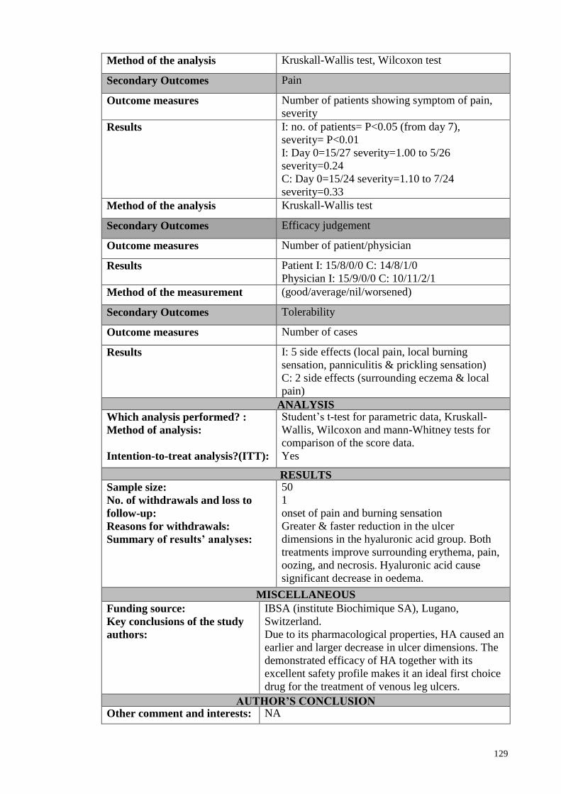
129
Method of the analysis Kruskall-Wallis test, Wilcoxon test
Secondary Outcomes Pain
Outcome measures Number of patients showing symptom of pain,
severity
Results I: no. of patients= P<0.05 (from day 7),
severity= P<0.01
I: Day 0=15/27 severity=1.00 to 5/26
severity=0.24
C: Day 0=15/24 severity=1.10 to 7/24
severity=0.33
Method of the analysis Kruskall-Wallis test
Secondary Outcomes Efficacy judgement
Outcome measures Number of patient/physician
Results Patient I: 15/8/0/0 C: 14/8/1/0
Physician I: 15/9/0/0 C: 10/11/2/1
Method of the measurement (good/average/nil/worsened)
Secondary Outcomes Tolerability
Outcome measures Number of cases
Results I: 5 side effects (local pain, local burning
sensation, panniculitis & prickling sensation)
C: 2 side effects (surrounding eczema & local
pain)
ANALYSIS Which analysis performed? :
Method of analysis:
Intention-to-treat analysis?(ITT):
Student’s t-test for parametric data, Kruskall-
Wallis, Wilcoxon and mann-Whitney tests for
comparison of the score data.
Yes
RESULTS Sample size:
No. of withdrawals and loss to
follow-up:
Reasons for withdrawals:
Summary of results’ analyses:
50
1
onset of pain and burning sensation
Greater & faster reduction in the ulcer
dimensions in the hyaluronic acid group. Both
treatments improve surrounding erythema, pain,
oozing, and necrosis. Hyaluronic acid cause
significant decrease in oedema.
MISCELLANEOUS
Funding source:
Key conclusions of the study
authors:
IBSA (institute Biochimique SA), Lugano,
Switzerland.
Due to its pharmacological properties, HA caused an
earlier and larger decrease in ulcer dimensions. The
demonstrated efficacy of HA together with its
excellent safety profile makes it an ideal first choice
drug for the treatment of venous leg ulcers.
AUTHOR’S CONCLUSION Other comment and interests: NA

130
9. Romanelli et al., 2007.
DATA EXTRACTION FORM
ARTICLE DETAILS
ID Romanelli et al. (2007)
Author(s), Year
Marco Romanelli, Valentina Dini, Mariastefania Bertone,
Sabrina Barbanera, Cinzia Brilli, 2007
Title
OASIS® wound matrix versus Hyaloskin® in the
treatment of difficult-to-heal wounds of mixed
arterial/venous aetiology
Journal, Sources International Wound Journal, Vol: 4, No:1; 3-7
Cochrane database.
Country of Origin Italy
Corresponding author & contact
details
Prof M. Romanelli, Department of Dermatology,
University of Pisa, Via Roma, 67, 56126 Pisa, Italy
Email: [email protected]
METHODS
Study design RCT
Country Italy
Setting Leg ulcer clinic
Treatments:
-Intervention (I)
-Control (C)
Hyaloskin® dressing-Hyaluronic
acid based dressing consisting of
single component ECM
OASIS® dressing-ECM
components
Covered with secondary non
adherent dressing
Total study duration 16 weeks
Single centre/multicentre trial: Single centre
Method used to generate random allocation sequence:
Method used to implement the random allocation
sequence:
Allocation sequence concealment:
The sequence of randomisation
was generated through every other
patient selection by the clinician
Was the participant blinded?
Was the researcher/healthcare provider blinded?
Was the assessor blinded?
NA
NA
NA
Inter-rater reliability measured? NA
PARTICIPANTS
Total number
54
Type of participants
Arterial/Venus ulcer
Age (range) & mean±SD (> 18 years) Mean: I: 62±8, C; 64±13
Sex
Male (26) & Female (28)
Record of drop-out (with reasons)
4
No. of sample size
Participant population (how sampled)
54
NA
No. of arm in trial
2
No. of each arm
27
Power calculation
NA
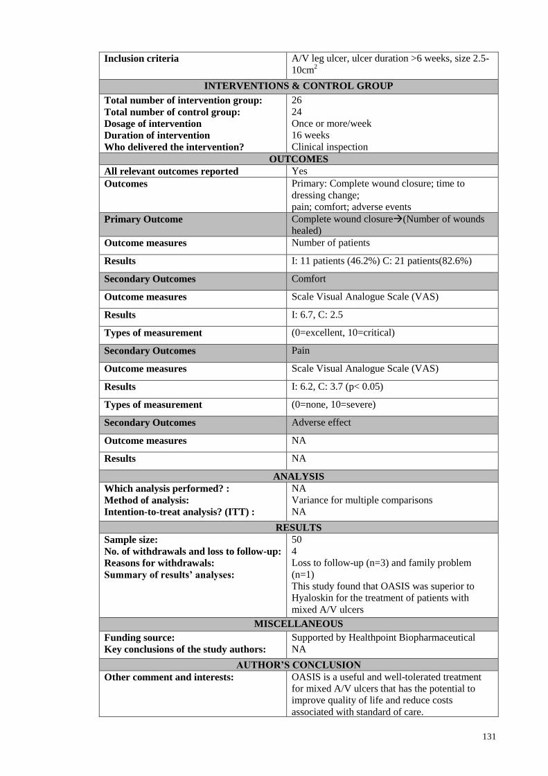
131
Inclusion criteria A/V leg ulcer, ulcer duration >6 weeks, size 2.5-
10cm2
INTERVENTIONS & CONTROL GROUP
Total number of intervention group:
Total number of control group:
Dosage of intervention
Duration of intervention
Who delivered the intervention?
26
24
Once or more/week
16 weeks
Clinical inspection
OUTCOMES
All relevant outcomes reported Yes
Outcomes
Primary: Complete wound closure; time to
dressing change;
pain; comfort; adverse events
Primary Outcome Complete wound closure(Number of wounds
healed)
Outcome measures Number of patients
Results I: 11 patients (46.2%) C: 21 patients(82.6%)
Secondary Outcomes Comfort
Outcome measures Scale Visual Analogue Scale (VAS)
Results I: 6.7, C: 2.5
Types of measurement (0=excellent, 10=critical)
Secondary Outcomes Pain
Outcome measures Scale Visual Analogue Scale (VAS)
Results I: 6.2, C: 3.7 (p< 0.05)
Types of measurement (0=none, 10=severe)
Secondary Outcomes Adverse effect
Outcome measures NA
Results NA
ANALYSIS
Which analysis performed? :
Method of analysis:
Intention-to-treat analysis? (ITT) :
NA
Variance for multiple comparisons
NA
RESULTS
Sample size:
No. of withdrawals and loss to follow-up:
Reasons for withdrawals:
Summary of results’ analyses:
50
4
Loss to follow-up (n=3) and family problem
(n=1)
This study found that OASIS was superior to
Hyaloskin for the treatment of patients with
mixed A/V ulcers
MISCELLANEOUS
Funding source:
Key conclusions of the study authors:
Supported by Healthpoint Biopharmaceutical
NA
AUTHOR’S CONCLUSION
Other comment and interests: OASIS is a useful and well-tolerated treatment
for mixed A/V ulcers that has the potential to
improve quality of life and reduce costs
associated with standard of care.

132
10. Uccioli et al., 2011
DATA EXTRACTION FORM
(The data extraction form used for each individual study included in this review)
ARTICLE DETAILS
ID Uccioli et al. (2011)
Author(s), Year
Luigi Uccioli, Laura Giurato, Valeria Ruotolo, Adolfo
Ciavarella, Michele S. Grimaldi, Alberto Piaggesi, Ilaria
Teobaldi, Lucia Ricci, Luciano Scionti, Cristiana
Vermigli, Roberto Seguro, Lorena mancini and Giovanni
Ghirlanda, 2011 Title
Two-steps autologous grafting using HYAFF scaffolds in
treating difficult diabetic foot ulcers: results of a
multicenter, randomised controlled clinical trial with long-
term follow-up Journal, Sources The International Journal of Lower Extremity Wounds,
Vol:10, Issue: 80; 80-85
Country of Origin Italy
Corresponding author & contact
details
Luigi Uccioli, Policlinico of Tor Vergata, Department of
Internal Medicine, Viale Oxford 81, 00133 Rome Italy
Email: [email protected]
METHODS
Study design Open, Randomised Controlled
Clinical Trial
Country Italy
Setting Diabetic Foot Centers (7)
Treatments:
-Intervention (I)
-Control (C)
HYAFF-Hyalograft -3D
Non-adherent paraffin gauze
Covered with non-adherent
paraffin gauze + bandage of
sterile cotton pads and gauze
Total study duration 18 months
Single centre/multicentre trial: Multicenter
Method used to generate random allocation sequence:
Method used to implement the random allocation
sequence:
Allocation sequence concealment:
Computer-generated
randomization list
In a block of 4 & stratified by
center
Sealed envelope in numerical
order
Was the participant blinded?
Was the researcher/healthcare provider blinded?
Was the assessor blinded?
Yes
NA
NA
Inter-rater reliability measured? NA
PARTICIPANTS
Total number
180
Type of participants
Diabetic foot ulcer
Age Mean ± SD I: 61 ± 10, C: 62 ± 11
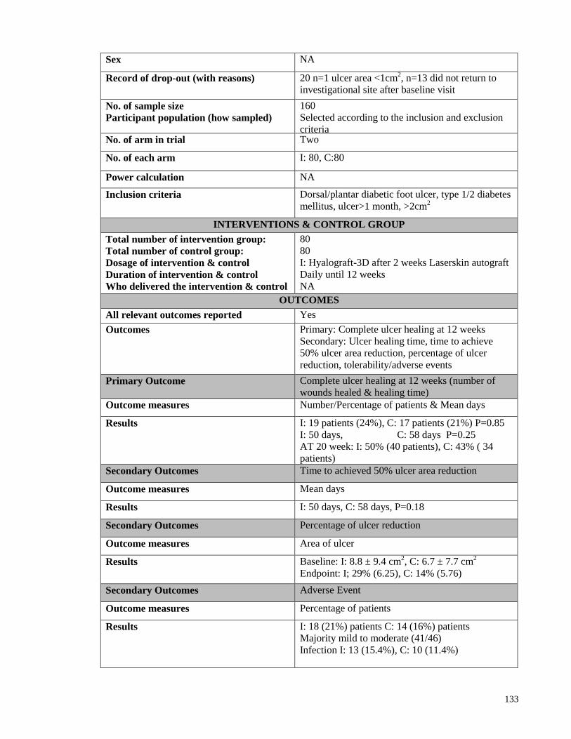
133
Sex
NA
Record of drop-out (with reasons)
20 n=1 ulcer area <1cm2, n=13 did not return to
investigational site after baseline visit
No. of sample size
Participant population (how sampled)
160
Selected according to the inclusion and exclusion
criteria No. of arm in trial
Two
No. of each arm
I: 80, C:80
Power calculation
NA
Inclusion criteria Dorsal/plantar diabetic foot ulcer, type 1/2 diabetes
mellitus, ulcer>1 month, >2cm2
INTERVENTIONS & CONTROL GROUP
Total number of intervention group:
Total number of control group:
Dosage of intervention & control
Duration of intervention & control
Who delivered the intervention & control
80
80
I: Hyalograft-3D after 2 weeks Laserskin autograft
Daily until 12 weeks
NA
OUTCOMES
All relevant outcomes reported Yes
Outcomes
Primary: Complete ulcer healing at 12 weeks
Secondary: Ulcer healing time, time to achieve
50% ulcer area reduction, percentage of ulcer
reduction, tolerability/adverse events
Primary Outcome Complete ulcer healing at 12 weeks (number of
wounds healed & healing time)
Outcome measures Number/Percentage of patients & Mean days
Results I: 19 patients (24%), C: 17 patients (21%) P=0.85
I: 50 days, C: 58 days P=0.25
AT 20 week: I: 50% (40 patients), C: 43% ( 34
patients)
Secondary Outcomes Time to achieved 50% ulcer area reduction
Outcome measures Mean days
Results I: 50 days, C: 58 days, P=0.18
Secondary Outcomes Percentage of ulcer reduction
Outcome measures Area of ulcer
Results Baseline: I: 8.8 ± 9.4 cm2, C: 6.7 ± 7.7 cm
2
Endpoint: I; 29% (6.25), C: 14% (5.76)
Secondary Outcomes Adverse Event
Outcome measures Percentage of patients
Results
I: 18 (21%) patients C: 14 (16%) patients
Majority mild to moderate (41/46)
Infection I: 13 (15.4%), C: 10 (11.4%)

134
ANALYSIS
Which analysis performed? :
Method of analysis:
Intention-to-treat analysis? (ITT) :
Frequency distribution for discrete data
Mean & SD to describe continuous data
Comparisons between group using the chi square-
discrete data
Or Mann-whitney & Student’s T-test –continuous
data
Analysis of covariance model SAS Software.
Yes.
RESULTS
Sample size:
No. of withdrawals and loss to follow-up:
Reasons for withdrawals:
Summary of results’ analyses:
160
20
NA
NA
MISCELLANEOUS
Funding source:
Key conclusions of the study authors:
NA
The results demonstrate the safety and
effectiveness of autologous skin substitutes in the
hard-to-heal diabetic dorsal foot ulcer population
AUTHOR’S CONCLUSION
Other comment and interests: There is a need for larger studies to clearly
demonstrate treatment benefits and how suitable
patients may be Identified

135
SUPPLEMENTARY
List of Publications and Papers Presented
1. A Shaharudin, Z Aziz, NJ Chong. Effectiveness and tolerability of hyaluronic
acid for chronic wounds healing: A systematic Review. Malaysian Journal of
Pharmacy (2014), 1(11), pp 112. (Non-ISI/Non-SCOPUS Cited Publication).
Proceeding (FAPA conference October 2014)
2. A Shaharudin and Z Aziz (2015). Effectiveness of Hyaluronic Acid and its
derivatives for chronic wounds: A Systematic Review. Journal of Wound Care.
(ISI/SCOPUS Cited Publication). Submitted (4 August 2015).