a study on phytochemical composition, gc-ms …
TRANSCRIPT
Swamy et al., IJPSR, 2019; Vol. 10(3): 747-755. E-ISSN: 0975-8232; P-ISSN: 2320-5148
International Journal of Pharmaceutical Sciences and Research 747
IJPSR (2019), Volume 10, Issue 3 (Research Article)
Received on 25 May 2018; received in revised form, 09 September 2018; accepted, 20 September 2018; published 01 February 2019
A STUDY ON PHYTOCHEMICAL COMPOSITION, GC-MS ANALYSIS AND ANTI-
MICROBIAL POTENTIAL OF METHANOLIC LEAF EXTRACT OF ALSTONIA SCHOLARIS
(L.) R. BR.
Nattala Tirupathi Swamy, Gorrepati Rosaiah *, Kakumanu Babu and Kovvada Vijay Kumar
Department of Botany and Microbiology, Acharya Nagarjuna University Nagarjunanagar, Guntur -
522510, Andhra Pradesh, India.
ABSTRACT: The present study was carried out to assess the various
phytochemical composition, GC-MS analysis, and antimicrobial potential of
methanolic leaf extract of Alstonia scholaris (L.) R. Br. by using different
solvents. Phytochemical analysis revealed the presence of alkaloids,
coumarins, flavonoids, glycosides, phenols, quinines, saponins, tannins,
steroids and terpenoids. Susceptibility testing by Agar well diffusion assay
showed significant antimicrobial activity with ethyl acetate and methanol
extracts of leaves against bacterial strains such as Bacillus subtilis, Bacillus
cereus, Pseudomonas aeruiginosa, Staphylococcus aureus, Escherichia coli
and fungal strain Candida albicans. The methanolic leaf extract exhibited
better antimicrobial activity than ethyl acetate extract. The UV-Vis, FTIR,
and GC-MS have been employed to characterize the bioactive compounds
present in methanolic leaf extract of Alstonia scholaris. The crude extract
was scanned in the wavelength ranging from 200-900 nm by using Perkin
Elmer Spectrophotometer, and the characteristic peaks were detected. The
FTIR spectrum confirmed the presence of alcohol, alkanes, amides, alkyl
aldehydes, halogen, and aromatic compounds. The results of the GC-MS
analysis provide different peaks determining the presence of 9 phytochemical
compounds with different therapeutic activities. The major phyto
constituents were oxime-methoxy-phenyl, 2-methoxy 4-vinyl phenol, 2(4H)-
Benzofuranone 5, 6, 7, 7a-tetrahydro-4, 4, 7a trimethyl, (-)-Loliolide,
Neophytadiene, Hexahydrofarnesyl acetone, Phytol, 4, 8, 12, 16-
Tetramethylheptadecan-4-olide, and Squalene.
INTRODUCTION: The use of plants as
therapeutic agents, in addition to being used as
food, is age-long and there is a great awareness in
the use and significance of these medicinal floras 1.
Plants are a rich source of secondary metabolites
with interesting biological activities.
QUICK RESPONSE CODE
DOI: 10.13040/IJPSR.0975-8232.10(3).747-55
The article can be accessed online on www.ijpsr.com
DOI link: http://dx.doi.org/10.13040/IJPSR.0975-8232.10(3).747-55
In general, these secondary metabolites are an
important source with a variety of structural
arrangements and properties 2.
Natural products from microbial sources have been
the primary source of antibiotics. But with the
increasing recognition of herbal medicine as an
alternative form of health care, the screening of
medicinal plants for active compounds has become
very significant 3. The active substances of many
drugs found in plants are secondary metabolites 4
such as alkaloids, phenols, tannins, saponins
flavonoids, terpenoids, glycosides and lactones 5.
Keywords:
Alstonia scholaris, Bio-active
compounds, Spectrophotometer, UV-
Vis, FTIR, GC-MS
Correspondence to Author:
Dr. G. Rosaiah
Associate Professor,
Department of Botany and
Microbiology, Acharya Nagarjuna
University Nagarjunanagar, Guntur -
522510, Andhra Pradesh, India.
E-mail: [email protected]

Swamy et al., IJPSR, 2019; Vol. 10(3): 747-755. E-ISSN: 0975-8232; P-ISSN: 2320-5148
International Journal of Pharmaceutical Sciences and Research 748
The phytochemical analysis of the plants is
commercially very important and holds great
interest for pharma industry which pursues the
discovery of novel drugs for curing various
diseases. This increased importance makes the
study of phytochemicals, and bioactive compounds
are inevitable 6. The knowledge of the chemical
constituents of plants would further be valuable in
discovering the actual value of folk medicines 7.
The plant kingdom holds a great promise for
medicinal substances in many plant species and
still unexplored.
Alstonia scholaris Linn. R. Br. (Apocynaceae)
which is popularly known as the “Saptparni” or
“The Devil tree” is an evergreen tree of the Indian
subcontinent of South Asia and Africa. Different
parts of the plant are used in medicines as
antimicrobial, antihelmenthic, astringent, anti-
periodic, diarrhea, dysentery, antimalarial and
bowel disorders 8. Alstonia scholaris is one such
plant showing multifarious pharmacological
properties viz: diarrhea 9, wounds and earache
10,
Leucorrhoea 11
, dog bite 12
, fever 13
, malignancy,
jaundice, hepatitis, malaria, skin diseases 14
,
astringent, thermogenic, cardiotonic 15
and
antimicrobial activity 16
.
To validate the pharmacological properties of
Alstonia scholaris, the present study was attempted
to evaluate the phytochemical composition and the
antimicrobial properties using various solvent
extracts of leaf material. An attempt was also made
to determine the bioactive compounds present in
the Alstonia scholaris leaves with the aid of UV-
Vis, FTIR and GC-MS techniques, which may
provide an insight into the use of traditional
medicine.
MATERIALS AND METHODS: Healthy
Alstonia scholaris plant with plant authentication
voucher specimen number ANUBH01190 is
selected in the Botanical Garden of Acharya
Nagarjuna University, Nagarjunanagar, Andhra
Pradesh (India). Fresh leaves were collected and
thoroughly washed with distilled water to remove
dust particles and subjected to shade drying at room
temperature for about two weeks. Dried leaf
material was powdered with the help of a
mechanical grinder and sieved. Powdered leaf
material (150g) was Soxhlet extracted with hexane,
chloroform, ethyl acetate, and methanol for about
12-18 h. The crude methanol extracts were
evaporated by a vacuum rotary evaporator (Buchi
Labortech Ag, model l, R-215) under reduced
pressure. The different solvent extracts were
filtered and concentrated under reduced pressure in
a rotary evaporator. The dried extracts were kept in
the refrigerator at 4 °C until use.
Preliminary Phytochemical Screening: Shade
dried plant material was extracted with hexane,
chloroform, ethyl acetate, and 80% methanol, and
preliminary phytochemical screening was done by
using the standard tests 17, 18
.
Alkaloid Test (Dragendroff’s Test): 2 ml plant
extract was acidified with few drops of dilute
hydrochloric acid. To this acidic medium, 1 ml of
Dragendroff’s reagent (Potassium bismuth iodide)
was added. An orange or reddish brown precipitate
produced indicates the presence of alkaloids.
Flavonoid Test (Shinoda Test): The presence of
flavonoids was confirmed by treating the alcoholic
plant extract with few fragments of magnesium
ribbon and hydrochloric acid. The reaction mixture
develops pink, scarlet or crimson red color,
indicating the presence of flavonoids.
Saponin Test (Foam Test): 1 ml of each extract
shaken with 10 ml of distilled water and it was
agitated in a graduated cylinder for 10 min. The
formation of persistent honey-comb like froth
indicated the presence of saponins.
Quinone Test: A small amount of extract was
treated with concentrated HCl and observed for the
formation of a yellow color precipitate.
Tannin Test (Lead Acetate Test): To 2 ml of each
extract add a few drops of 10% Lead acetate were
added. The appearance of white precipitate
indicates the presence of tannins.
Terpenoids and Steroids: 50% H2SO4 is added
along the sides of the test tube containing a mixture
of methanolic HCl and acetic anhydride. If there is
any change in color, from green to blue-green
(sometimes via red or blue) indicates the presence
of terpenoids and steroids.
Phenol Test: When 0.5 ml of FeCl3 (w/v) solution
was added to 2 ml of rest solution, the formation of
an intense color indicated the presence of phenols.

Swamy et al., IJPSR, 2019; Vol. 10(3): 747-755. E-ISSN: 0975-8232; P-ISSN: 2320-5148
International Journal of Pharmaceutical Sciences and Research 749
Coumarin: To the methanolic extract, a few drops
of alcoholic sodium hydroxide was added.
Formation of yellow color indicated the presence of
coumarins.
Test for Glycosides: To the methanolic extract
mixed with a little anthrone on a watch glass. Few
drops of conc. H2SO4 was added and warmed
gently over a water bath. The presence of
glycosides was identified by dark green color
formation.
Resins: Plant extracts were treated with acetone.
To this, a small amount of water was added and
shaken. The appearance of turbidity indicates the
presence of resins.
Antimicrobial Activity: Nutrient agar (NA) was
used for culturing the test bacteria. NA medium
(100 ml) was sterilized at 15 lbs pressure (121 °C)
for 15 min, cooled and inoculated with 0.1 ml of
bacterial test suspension. After thorough mixing,
the inoculated medium was poured into Petri plates
under aseptic conditions. After solidification, wells
of about 5 mm diameter were punched with a
sterilized cork borer. Solvent extract (50 μl, 100 μl,
and 150 μl) was added to each well, and the
addition of solvent alone served as control. The
inoculated plates were incubated at 30 °C, and the
diameter of the inhibition zone was measured after
24 h.
Ultra Violet-Visible Spectrophotometry Analysis
(UV-Vis): UV-Vis Spectrophotometer uses light in
the visible ranges or its adjacent ranges. The color
of the chemicals involved directly affects the
absorption in the visible ranges. Molecules undergo
electronic transitions in these ranges of the
electromagnetic spectrum. Methanolic leaf extract
was examined under UV visible spectrophotometry
in the wavelength ranging from 200 to 900 nm
using Perkin Elmer Spectrophotometer, and the
characteristic peaks were detected.
Fourier Transform Infrared Spectroscopic
Analysis (FTIR): FTIR studies were done using
Schimatzu IR Affinity - 1S spectrophotometer. The
KBr used was of IR grade (SD Fines). About 500
mg of KBr was placed into a mortar and grind it
until there is no evidence of crystallinity. The KBr
powder was transferred into the drying box at a
temperature of 400 °C.
10 mg of solid sample was placed into the mortar
and again grind it until a fine powder is formed.
One milligram of fine solid powder of sample (as
per requirement of the die) and 200 mg of fine dry
powder of KBr were weighed and the quantities
were transferred into a mortar and mixed well with
the help of a spatula. Bottom and the top portion of
KBr were assembled at press assembly, and one of
the 13 mm die with the polished surface up inside
the press. The KBr sample mixture was transferred
to a KBr press assembly. The sample was slowly
compressed in KBr press assembly at a pressure of
2000 kg/cm2 for about 60 sec. The prepared disc
was then subjected for scanning between 500-4000-
1 cm.
Gas Chromatography-Mass Spectrometry
Analysis (GC-MS):
Sample Preparation: A 20 mg of sample was
dissolved in 1ml of methanol, vortex for 5min,
sonicate for 10 min, centrifugation (10000 rpm, 5
min), the supernatant was transferred into a fresh
vial and was subjected (without any dilution) for
GC-MS analysis.
GC-MS Analysis: GC-MS analysis was carried out
on a Perkin Elmer Turbo Mass Spectrophotometer
which includes a Perkin Elmer Autosampler
XLGC. The column used was Perkin Elmer Elite -
5 capillary columns measuring 30 m × 0.25 mm
with a film thickness of 0.25mm composed of 95%
dimethylpolysiloxane. The carrier gas used was
Helium at a flow rate of 0.5 ml/min. 1 μl sample
injection volume was utilized. The inlet
temperature was maintained at 250 °C. The oven
temperature was programmed initially at 110 °C for
4 min, then an increase to 240 °C. And then
programmed to increase to 280°C at a rate of 20 °C
ending with a 5 min. Total run time was 90 min.
The MS transfer line was maintained at a
temperature of 200 °C. The source temperature was
maintained at 180 °C. GCMS was analyzed using
electron impact ionization at 70 eV and data was
evaluated using total ion count (TIC) for compound
identification and quantification. The spectrums of
the components were compared with the database
of the spectrum of known components stored in the
GC-MS library. Measurement of peak areas and
data processing were carried out by Turbo-Mass -
OCPTVS-Demo SPL software.

Swamy et al., IJPSR, 2019; Vol. 10(3): 747-755. E-ISSN: 0975-8232; P-ISSN: 2320-5148
International Journal of Pharmaceutical Sciences and Research 750
RESULT AND DISCUSSION: In the present
study phytochemical screening of the different
solvent extracts like hexane, chloroform, ethyl
acetate and methanol extract of leaves in Alstonia
scholaris revealed the presence of various chemical
compounds such as alkaloids, coumarins,
flavonoids, glycosides, phenols, quinines, saponins,
tannins, steroids and terpenoids Table 1.
Phytochemicals may be effective in combating or
preventing disease due to their antioxidant effect 19
.
The curative properties of medicinal plants are
perhaps due to the presence of various secondary
metabolites 20
. The results obtained in the present
study suggest that the identified phytochemical
compounds may be the bioactive constituents
responsible for the antimicrobial activity.
TABLE 1: PRELIMINARY PHYTOCHEMICAL SCREENING OF LEAF EXTRACTS OF ALSTONIA SCHOLARIS
S. no. Phytochemicals Test name Hexane Chloroform Ethyl acetate Methanol
1 Alkaloids Dragendorff’s test
Mayer’s test
+ + + +
2 Coumarins Sodium hydroxide test - - - +
3 Flavonoids Ferric chloride test + - + +
4 Glycosides Anthrone test - - - +
5 Phenolic compounds Phenol test _ _ _ +
6 Quinones H2SO4 test + + + +
7 Resins Acetone H2O test - _ _ _
8 Saponins Foam test + + + +
9 Tannins Braemer’s test _ + _ +
10 Steroids Salkowski test + + _ _
11 Terpenoids Salkowski test + _ _ +
(+) = positive (present); (-) = negative (absent)
Basing on the analysis of phytochemical
composition in all solvent extractions, ethylacetate,
and methanolic leaf extracts were selected for
antimicrobial activity. The antimicrobial activity of
ethyl acetate and methanolic extracts of the leaf of
Alstonia scholaris were done using Agar well
diffusion method. The zone of inhibition values
was determined for ethyl acetate and methanol leaf
extracts. The tested two extracts showed varying
degree of antimicrobial activities at different
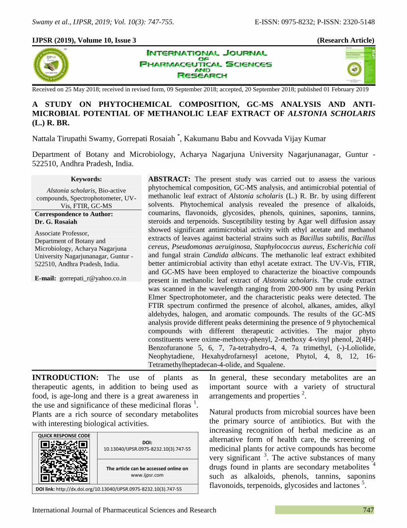
concentrations. The ethyl acetate extract showed a
zone of inhibition at maximum concentration (150
µl) against Pseudomonas aeruginosa (14 mm),
Bacillus cereus and Candida albicans (13 mm)
Table 2. Besides this, methanol extract showed
good activity at same concentration against
Bacillus cereus (21 mm) followed by
Staphylococcus aureus (18 mm), Bacillus subtilis
and Pseudomonas aeruginosa (17 mm) Plate 1A
and 1B; Table 2. The present study is by Deepa 21
and Lakshmi 22
where the activity of methanol
extract of leaves was found to be more pronounced
than the ethyl acetate extract against selected
microbes.
TABLE 2: ANTIMICROBIAL STUDY SHOWING ZONE OF INHIBITION OF ALSTONIA SCHOLARIS LEAF EXTRACTS
Organism Ethyl acetate Methanol
Zone of inhibition (mm)
50 µl 100 µl 150 µl 50 µl 100 µl 150 µl
Bacillus subtilis 10 11 12 12 15 17
Bacillus cereus 12 12 13 18 20 21
Staphylococcus aureaus 10 11 12 15 17 18
Escherichia coli 08 10 11 15 15 16
Pseudomonas aeruginosa 09 12 14 11 14 17
Candida albicans 10 11 13 13 14 16 *zone of inhibition in millimeters
The qualitative UV-Vis spectrum profile of
Alstonia scholaris methanolic extract was selected
from 200-900 nm due to the sharpness of peaks and
proper baseline. The profile showed the peaks at
665, 410, 336 and 277 nm with absorption 0.674,
8.478, 4.263 and 4.612 respectively Fig. 1 and
Table 3. The result confirms the occurrence of
peaks at 277-665 nm reveals that the absorption
bands are due to the presence of phenolic and
alkaloid compounds in Alstonia scholaris 23, 24
.
Swamy et al., IJPSR, 2019; Vol. 10(3): 747-755. E-ISSN: 0975-8232; P-ISSN: 2320-5148
International Journal of Pharmaceutical Sciences and Research 751
PLATE 1: ANTIMICROBIAL ACTIVITY OF METHANOLIC LEAF EXTRACT OF A. SCHOLARIS ON BACILLUS
CEREUS (A) AND STAPHYLOCOCCUS AUREUS (B)
TABLE 3: UV - VIS PEAK VALUES OF ALSTONIA
SCHOLARIS METHANOLIC LEAF EXTRACT
Wavelength (nm) Absorption (O.D)
665.00 0.674
410.00 8.478
336.00 4.263
277.00 4.612
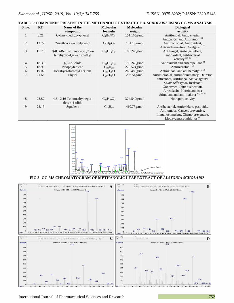
The FTIR spectrum was used to identify the
functional group of the active components based on
the peak value in the region of infrared radiation 25,
26. The results of FTIR peak values and functional
groups were represented in Table 4. The FTIR
spectrum profile as illustrated in Fig. 2. The FTIR
spectrum peaks identified between 509 cm-1
- 3387
cm-1
confirmed the presence of various functional
groups such as alcohol, alkanes, alkynes, amides,
esters, Nitrogen, alkyl halide, halogen and aromatic
compounds 27
.
TABLE 4: FT-IR PEAK VALUES AND FUNCTIONAL
GROUPS OF METHANOLIC EXTRACT OF
ALSTONIA SCHOLARIS
Peak
no.
Group
frequency (cm-1
)
Molecular
motion
Functional
groups
1 3387 O-H stretch Alcohol
2 2922 C-H stretch Alkane
3 2850 C-H stretch Alkane
4 2313 C=C stretch Alkyne
5 1729 C=O stretch Carbonyl
6 1623 N-H stretch Amide
7 1431 N-O stretch Nitrogen
8 1305 C=O stretch Acid
9 1059 C-O stretch Ester
10 887 C-H stretch Aromatic
11 774 C-CI stretch Alkyl halide
12 661 C-I stretch Halogen
compound
13 509 C-I stretch Alkyl halide
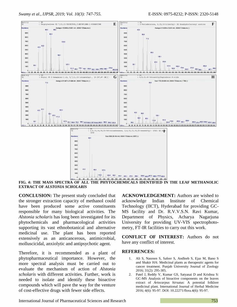
The results about GC-MS analysis lead to the
identification of a number of compounds from GC
fractions of the methanolic extracts of Alstonia
scholaris. They were identified through mass
spectrometry attached with GC. GC-MS is a key
technological tool for secondary metabolites
profiling in plant species 28, 29
. The identification of
the phytochemical compounds was confirmed
based on the peak area, retention time and
molecular formula were presented in Table 5. The
mass spectra of all the phytochemicals identified in
the leaf methanolic extract of Alstonia scholaris
were presented in Fig. 3 and Fig. 4 (a-i). The GC-
MS analysis of Alstonia scholaris methanolic leaf
extract revealed the presence of major phyto-
chemicals. The available phytochemicals and their
biological activity were represented in Table 5.
FIG. 1: UV - VIS SPECTRAL ANALYSIS OF ALSTONIA
SCHOLARIS METHANOLIC LEAF EXTRACT
FIG. 2: IR SPECTRA ANALYSIS OF METHANOLIC
LEAF EXTRACT OF ALSTONIA SCHOLARIS
A B
Swamy et al., IJPSR, 2019; Vol. 10(3): 747-755. E-ISSN: 0975-8232; P-ISSN: 2320-5148
International Journal of Pharmaceutical Sciences and Research 752
TABLE 5: COMPOUNDS PRESENT IN THE METHANOLIC EXTRACT OF A. SCHOLARIS USING GC-MS ANALYSIS
S. no. RT Name of the
compound
Molecular
formula
Molecular
weight
Biological
activity
1 6.21 Oxime-methoxy-phenyl C8H9NO2 151.165g/mol Antifungal, Antibacterial,
Anticancer and Antitumor 30
2 12.72 2-methoxy 4-vinylphenol C9H10O2 151.18g/mol Antimicrobial, Antioxidant,
Anti inflammatory, Analgesic 31
3 15.70 2(4H)-Benzofuranone5,6,7,7a-
tetrahydro-4,4,7a trimethyl
C11H16O2 180.243g/mol Antifungal, Antialgal effect,
antioxidant, antibacterial
activity 32, 33
4 18.38 (-)-Loliolide C11H16O3 196.246g/mol Antioxidant and anti repellant 34
5 18.96 Neophytadiene C20H38 278.524g/mol Antimicrobial 35
6 19.02 Hexahydrofarnesyl acetone C18H36O 268.485g/mol Antioxidant and antihemolytic 36
7 21.66 Phytol C20H40O 296.54g/mol Antimicrobial, Antiinflammatory, Diuretic,
anticancer, Antifungal Active against
Salmonella typhi, Resistant
Gonorrhea, Joint dislocation,
A headache, Hernia and is a
Stimulant and anti-malaria 37, 38, 39
8 23.82 4,8,12,16 Tetramethylhepta-
decan-4-olide
C21H40O2 324.549g/mol No report activity
9 28.19 Squalene C30H50 410.73g/mol Antibacterial, Antioxidant, pesticide,
Antitumour, Cancer, preventive,
Immunostimulent, Chemo preventive,
Lipoxygenase-inhibitor 40
FIG 3: GC-MS CHROMATOGRAM OF METHANOLIC LEAF EXTRACT OF ALSTONIA SCHOLARIS
A B
C D
Swamy et al., IJPSR, 2019; Vol. 10(3): 747-755. E-ISSN: 0975-8232; P-ISSN: 2320-5148
International Journal of Pharmaceutical Sciences and Research 753
FIG. 4: THE MASS SPECTRA OF ALL THE PHYTOCHEMICALS IDENTIFIED IN THE LEAF METHANOLIC
EXTRACT OF ALSTONIA SCHOLARIS
CONCLUSION: The present study concluded that
the stronger extraction capacity of methanol could
have been produced some active constituents
responsible for many biological activities. The
Alstonia scholaris has long been investigated for its
phytochemicals and pharmacological activities
supporting its vast ethnobotanical and alternative
medicinal use. The plant has been reported
extensively as an anticancerous, antimicrobial,
molluscicidal, anxiolytic and antipsychotic agent.
Therefore, it is recommended as a plant of
phytopharmaceutical importance. However, the
more spectral analysis must be carried out to
evaluate the mechanism of action of Alstonia
scholaris with different activities. Further, work is
needed to isolate and identify these bioactive
compounds which will pave the way for the venture
of cost-effective drugs with fewer side effects.
ACKNOWLEDGEMENT: Authors are wished to
acknowledge Indian Institute of Chemical
Technology (IICT), Hyderabad for providing GC-
MS facility and Dr. R.V.V.S.N. Ravi Kumar,
Department of Physics, Acharya Nagarjuna
University for providing UV-VIS spectrophoto-
metry, FT-IR facilities to carry out this work.
CONFLICT OF INTEREST: Authors do not
have any conflict of interest.
REFERENCES:
1. Ali S, Nasreen S, Safeer S, Andleeb S, Ejaz M, Bano S
and Shakir HA: Medicinal plants as therapeutic agents for
cancer treatment. Punjab University Journal of Zoology
2016; 31(2): 295-305.
2. Patel J, Reddy V, Kumar GS, Satyasai D and Krishna S:
GC-MS Analysis of bioactive components on the leaves
extract of Artocarpus hirsutus: A potential folklore
medicinal plant. International Journal of Herbal Medicine
2016; 4(6): 95-97. DOI: 10.22271/flora.4(6): 95-97.
E F
G H
I

Swamy et al., IJPSR, 2019; Vol. 10(3): 747-755. E-ISSN: 0975-8232; P-ISSN: 2320-5148
International Journal of Pharmaceutical Sciences and Research 754
3. Mustafa G, Arif R, Atta A, Sharif S and Jamil A: Bioactive
compounds from medicinal plants and their importance in
drug discovery in Pakistan. Matrix Science Pharma (MSP)
2017; 1(1): 17-26. DOI: 10.26480/msp.01.2017.17.26
4. Wink M: Modes of action of herbal medicines and
secondary plant metabolites. Medicines 2015; 2: 251-286.
DOI: 10.3390/medicines2030251.
5. Cox PA and Balick MJ: The Ethnobotanical approach to
drug discovery. Scientific American 1994; 270(6): 82-87.
6. Pandey P, Mehta R and Upadhyay R: Physico-chemical
and preliminary phytochemical screening of Psoralea
corylifolia. Achieves of Applied Science Research 2013;
5(2): 261-265.
7. Mojab F, Kamalinejad M, Ghaderi N and Vahidipour H:
Phytochemical screening of some Iranian plants. Iranian
Journal of Pharmaceutical Research 2003; 1: 77-82.
8. Nadkarni AK and Nadkarni KM: Indian Materia Medica.
Bombay: Popular Prakashan 1976; 1: 80-83.
9. Khyade MS, Kasote DM and Vaikos NP: Alstonia
scholaris (L.) R. Br. and Alstonia macrophylla Wall. ex G.
Don: A comparative review of traditional uses,
phytochemistry and pharmacology. Journal of
Ethnopharmacology 2014; 153(1): 1-18. DOI: 10.1016/j.
jep.2014.01.025.
10. Singh H, Arora R, Arora S and Singh B: Ameliorative
potential of Alstonia scholaris (Linn.) R. Br. against
chronic constriction injury-induced neuropathic pain in
rats. BMC Complementary and Alternative Medicine
2017; 17: 63. DOI 10.1186/s12906-017-1577-7.
11. Manzur-ul-Kadir MM, Kadir MF, Hossan MS and
Rahmatullah M: Medicinal plants of the Garo tribe
inhabiting the Madhupur forest region of Bangladesh.
American-Eurasian Journal of Sustainable Agriculture
2009; 3(2): 165-171. DOI: 10.22587/aejsa.2009.3.2.165-
171.
12. Khyade M and Kasote DM: Alstonia scholaris (L.) R. Br.
and Alstonia macrophylla Wall. ex G. Don: A comparative
review on traditional uses, phytochemistry and
pharmacology. Journal of Ethnopharmacology 2014;
153(1): 1-18. DOI: 10.1016/j.jep.2014.01.025.
13. Veerachari and Parashurama TR: Ethno-botanical health
care knowledge in Harapanahalli. International Journal of
Current Research 2016; 8(12): 42797-42801. DOI:
https://doi.org/10.24941/ijcr.2017/8(12): 42797-42801.
14. Mollik MAH, Hossan MS, Paul AK, Taufiq-Ur-Rahman
M, Jahan R and Rahmatullah M: A comparative analysis
of medicinal plants used by folk medicinal healers in three
districts of Bangladesh and inquiry as to the mode of
selection of medicinal plants. Ethnobotany Research and
Applications 2010; 8: 195-218.
15. Khanum S: Pharmacological investigation of the
chloroform extracts of Alstonia Scholaris (L.) R. Br.
Journal of Pharmaceutical and Scientific Innovation 2014;
3(1): 14-19. DOI: 10.7897/2277-4572.03198.
16. Khan MR, Omoloso AD and Kihara M: Antibacterial
activity of Alstonia scholaris and Leea tetramera.
Fitoterapia 2003; 74 (7-8): 736-740. PMID: 14630186
17. Harborne JB: Phytochemical methods. A Guide to Modern
Techniques of plant analysis, 1973; Chapman and Hall,
London.
18. Gibbs RD: Chemotaxonomy of flowering plants, 1974;
Mc. Gill Queen’s University Press, Montreal.
19. Halliwell B and Gutteridge JM: Free radicals, antioxidants
and human diseases: Where are now? Journal of
Laboratory and Clinical Medicine 1992; 119: 598-62.
20. Stray F: The Natural Guide to Medicinal Herb and Plants.
Tiger Books International, London 1998; 12-16.
21. Philip D, Kaleena PK, Valivittan K and Girish Kumar CP:
Phytochemical Screening and Antimicrobial Activity of
Sansevieria roxburghiana Schult. and Schult. F. Middle-
East Journal of Scientific Research 2011; 10(4): 512-518.
DOI: 10.5829/idosi.mejsr.
22. Kanthal LK, Satyavathi, K, Naidu AVR, Lakshmi NP,
Sreekanth NP and Madhuri L: Pharmacological Potential
of Lactuca runcinata DC: W Journal of Pharm and
Pharmaceutical Sciences 2015; 4(3): 442-450. DOI:
10.20959/2015/4 (3): 442-450.
23. Karpagasundari C and Kulothungan S: Analysis of
bioactive compounds in Physalis minima leaves using GC
MS, HPLC, UV-VIS and FTIR techniques. J
Pharmacognosy and Phytochemistry 2014; 3(4): 196-201.
DOI: http://dx.doi.org/10.22271/phyto.
24. Bashyam R, Thekkumala M and Sivanandham V:
Evaluation of phytoconstituents of Bryonopsis laciniosa
fruit by UV-Visible Spectroscopy and FTIR analysis;
Pharmacognosy 2015; 7 (3): 165-170. 10.5530/pj.2015.3.4
25. Hawraz IMA, Ahmed AA, Faiq HSH and Giovanni V:
ICP-AES, ICP-MS determination of elemental analysis of
aerial parts and roots of Iris persica L. collected in
Kurdistan Region/Iraq. Journal of Pharmaceutical and
Scientific Innovation 2016; 5(3): 90-92. DOI: 10.7897/
2277-4572.05319.
26. Khalafalah AK, Yousef AH, Esmail AM, Abdelrazik MH,
Hegazy ME and Mohamed AE: Chemical constituents of
Tephrosia purpurea. Pharmacognosy Research 2010; 2(2):
72-75. DOI: 10.4103/0974-8490.62951.
27. Manfred H, Meier H, Zeeh B, Lindon A and Murray M:
Spectroscopic Methods in Organic Chemistry, 1997; New
York: George Theme 365.
28. Merlin NJ, Parthasarathy V, Manavalan R and Kumaravel
S: Chemical investigation of aerial parts of Gmelina
asiatica Linn by GC-MS. Pharmacognosy Research 2009;
1(3): 152-156. DOI: http://www.phcogres.com/text.asp?
2009/1/3/152/58/58128.
29. Janakiraman N, Johnson M and Sathish SS: GC-MS
analysis of bioactive constituents of Peristrophe
bicalyculata (Retz) Nees. (Acanthaceae). Asian Pacific
Journal of Tropical Biomedicine 2012; 2(1): S46-S49.
30. Dahpour AA, Rahdari P and Sobati Z: Chemical
composition of essential oil, antibacterial activity and brine
shrimp lethality of ethanol extracts from Sedum pallidum.
Journal of Medicinal Plants Research 2012; 6(16): 3105-
3109. https://doi.org/10.5897/JMPR11.1270.
31. Gopalakrishnan S and Vadivel E: GC-MS analysis of some
bioactive constituents of Mussaenda frondosa Linn.
International Journal of Pharma and Biosciences 2011;
2(1): 313-320. DOI://doi.org/10.22376/ijpbs.
32. Akhbari M, Batooli H and Kashi FJ: Composition of
essential oil and biological activity of extracts of Viola
odoranta L. from central Iran. Natural Product Research
2012; 26(9): 802-809. DOI: 10.1080/14786419.2011.
558013
33. Yang WD, Liu YR, Liu JS and Liu Z: Inhibitory effects
and chemical basis of Eucalyptus orelliana wood meals on
the growth of Alexanrium tamarense. Huan Jing ke xue,
2008; 29(8): 2296-2301. DOI: 10.13198/j.issn.1001-6929.
0000.
34. Yang X, Min-Cheol K, Ki-Wan L, Sung-Myung K, Won-
Woo L and You-Jin J: Antioxidant activity and cell
protective effect of loliolide isolated from Sargassum
ringgoldianum subsp. Coreanum. Algae 2011; 26(2): 201-
208. DOI: http://dx.doi.org/10.4490/algae.2011.26.2.201
35. Yi Z, Yin-shan C and Hai-sheng L: Screening for
antibacterial and antifungal activities in some marine algae

Swamy et al., IJPSR, 2019; Vol. 10(3): 747-755. E-ISSN: 0975-8232; P-ISSN: 2320-5148
International Journal of Pharmaceutical Sciences and Research 755
from the Fujian coast of China with three different
solvents. Chinese Journal of Oceanology and Limnology
2001; 19(4): 327-331.
36. Al-Snafi AE: The medical importance of Cydonia oblonga
- A Review. IOSR Journal of Pharmacy 2016; 6(2): 87-99.
37. Hema R, Kumaravel S and Alagusundaram: GC-MS
Determination of bioactive components of Murraya
koenigii. Journal of American Science 2011; 7(1): 80-83.
38. Syeda FA, Habib-Ur- Rehman, Choudhary MI and Atta-
Ur-Rahman: Gas Chromatography-Mass Spectrometry
(GC-MS) analysis of petroleum ether extract (oil) and
bioassays of crude extract of Iris germanica. International
Journal of Genetics and Molecular Biology 2011; 3(7): 95-
100. DOI: 10.5897/IJGMB/ C10C17E2743.
39. Okeie W, Ogunlesi M, Ofor E and Osibote EAS: Analysis
of essential oil constituents in hydro-distillates of
Calotropis procera (oil). R. Br Research Journal of
Phytochemistry 2009; 3: 44-53. DOI: 10.3923/rjphyto.
2009.44.53
40. Zih-Rou H, Yin-Ku L and Jia-You F: Biological and
pharmacological activities of squalene and related
compounds: potential uses in cosmetic dermatology.
Molecules 2009; 14: 540-554. DOI: 10.3390/molecules
14010540.
All © 2013 are reserved by International Journal of Pharmaceutical Sciences and Research. This Journal licensed under a Creative Commons Attribution-NonCommercial-ShareAlike 3.0 Unported License.
This article can be downloaded to Android OS based mobile. Scan QR Code using Code/Bar Scanner from your mobile. (Scanners are available on Google Play store)
How to cite this article:
Swamy NT, Rosaiah G, Babu K and Kumar KV: A study on phytochemical composition, GC-MS analysis and anti-microbial potential of
methanolic leaf extract of Alstonia scholaris (L.) R. Br. Int J Pharm Sci & Res 2019; 10(3): 747-55. doi: 10.13040/IJPSR.0975-8232.
10(3).747-55.