a study on comparison of minimal separation …
TRANSCRIPT

“A STUDY ON COMPARISON OF MINIMAL
SEPARATION HYDROCELECTOMY VS.
CONVENTIONAL HYDROCELECTOMY
(JABOULAY’S PROCEDURE)” - AT GOVT.
KILPAUK MEDICAL COLLEGE
HOSPITAL.”
Dissertation submitted to
THE TAMILNADU DR. M.G.R. MEDICAL UNIVERSITY,
CHENNAI
With partial fulfilment of the regulations
for the award of the degree of
M.S (General Surgery)
Branch-I
Government Kilpauk Medical College
Chennai
May -2018

BONAFIDE CERTIFICATE
This is to certify that the dissertation entitled “A STUDY ON
COMPARISON OF MINIMAL SEPARATION HYDROCELECTOMY
VS. CONVENTIONAL HYDROCELECTOMY (JABOULAY’S
PROCEDURE)” AT GOVT. KILPAUK MEDICAL COLLEGE
HOSPITAL.” is a bonafide work of Dr.SANTHI .A, submitted to The
Tamilnadu Dr.M.G.R Medical University in partial fulfilment of requirements
for the award of the degree of M.S. BRANCH I (GENERAL SURGERY)
examination to be held in MAY, 2018.
Prof.M.ALLI, DGO M.S., Prof. R. KANNAN, M.S.,
Professor of General Surgery HOD, Dept. of General Surgery
Govt. Kilpauk Medical College, Govt. Kilpauk Medical College,
Chennai – 600 010. Chennai – 600 010.
Prof.Dr. P. VASANTHAMANI,
M.D., D.G.O.,MNAMS.,DCPSY.,MBA The DEAN
Govt. Kilpauk Medical College
Chennai - 600 010

DECLARATION BY THE CANDIDATE
I hereby declare that this dissertation titled “A STUDY ON
COMPARISON OF MINIMAL SEPARATION HYDROCELECTOMY
VS. CONVENTIONAL HYDROCELECTOMY (JABOULAY’S
PROCEDURE)” AT GOVT. KILPAUK MEDICAL COLLEGE
HOSPITAL.” Is a bonafide and genuine research work carried out by me in
the Department of General Surgery, Government Kilpauk Medical and
Hospital, Chennai-10, under the guidance of our Chief Prof.Dr.M.ALLI,
DGO., MS., Government Kilpauk Medical College and Hospital.
This dissertation is submitted to THE TAMILNADU DR. M.G.R.
MEDICAL UNIVERSITY, CHENNAI in partial fulfilment of the University
regulations for the award of M.S degree (General Surgery) Branch I,
examination to be held in MAY 2018.
Date:
Place: Chennai Dr. A. SANTHI

CERTIFICATE BY THE GUIDE
This is to certify that the dissertation titled “A STUDY ON
COMPARISON OF MINIMAL SEPARATION HYDROCELECTOMY
VS. CONVENTIONAL HYDROCELECTOMY (JABOULAY’S
PROCEDURE)” within General Surgery Department at Govt. Kilpauk
Medical College Hospital.” is a bonafide research work done by post graduate
in M.S. General Surgery, Government Kilpauk Medical College & Hospital,
Chennai-10 under my direct guidance and supervision in my satisfaction, in
partial fulfilment of the requirements for the degree of M.S. General Surgery.
Date: Prof.M.ALLI, DGO., M.S.,
Place: Chennai Professor of General Surgery,
Govt. Kilpauk Medical College,
Chennai-10

CERTIFICATE – II
This is to certify that this dissertation work titled entitled
dissertation “A STUDY ON COMPARISON OF MINIMAL
SEPARATION HYDROCELECTOMY VS. CONVENTIONAL
HYDROCELECTOMY (JABOULAY’S PROCEDURE)” of the candidate
Dr. A. SANTHI. with Registration Number 221511161 for the award of
M.S degree in the branch of GENERAL SURGERY. I personally
verified the urkund.com website for the purpose of plagiarism check. I
found that the uploaded thesis file contains from introduction to
conclusion pages and result shows 7% of plagiarism in this dissertation.
Guide & Supervisor sign with Seal.

ACKNOWLEDGEMENT
I am most thankful to Prof.Dr. P. VASANTHAMANI, M.D.,
D.G.O.,MNAMS.,DCPSY.,MBA, Dean, Kilpauk Medical College and
Hospital for giving me the opportunity to conduct this study in the
Department of General Surgery, Government Kilpauk Medical College &
Hospital, Chennai-10.
I thank Prof.R.KANNAN, M.S, Professor and Head of the
department of General Surgery for his relentless care and concern that he
has provided me to bring out this dissertation.
My deepest gratitude to my guide and mentor Prof.M.Alli, DGO.,
M.S., Professor of the DepartmentDepartment of General Surgery,
Kilpauk Medical College, who has inspired me immeasurably during my
training as a post graduate student.
I also acknowledge the valuable advice and inputs received from
Dr. Sridevi, M.S, Dr. Ramachandran, M.S and Dr.Suguneshwaran,
M.S, in shaping up this study.
This study would have not been possible without the support of my
fellow post graduates and interns who have been a source of help in need.

The most important part of any medical research is patients. I owe
great deal of gratitude to each and every one of them.
I would like to thank God for the things he has bestowed upon me.
I would like to thank my parents for making me who I am today
and for supporting me in every deed of mine.
I thank each and every person involved in making this manuscript
from inception to publication.


CONTENTS
S.NO TITLE PAGE
NO
1 INTRODUCTION 1
2 JUSTIFICATION OF STUDY 5
3 AIM AND OBJECTIVES 6
4 REVIEW OF LITERATURE 7
5 MATERIALS AND METHODS 52
6 ANALYSIS OF OBSERVATIONAL DATA 69
7 RESULTS 70
8 DISCUSSION 81
9 LIMITATIONS 87
10 CONCLUSION 88
11 BIBLIOGRAPHY
12 APPENDIX I - PROFORMA
APPENDIX II – CONSENT FORM
APPENDIX III – MASTER CHART

1
INTRODUCTION
Hydrocele is a abnormal collection of serous fluid in some part
of the process .us vaginalis, genera .lly the tunica. Hydrocele is the most
common benign swelli ing of the scrotum. The occurrences of
hydrocele are estimated as 1% among the adult male population.
“Primary vaginal hydrocele is well-defined as abnormal accumulation
of serous fluid in tunica vaginalis.” Secondary hydrocele occur
subordinate to disease of the testes and epididymis and its
management mainly comprises of treatment of the underlying cause.
Filarial hydrocele and chylocoele account for 80% of hydrocele in
some humid countries where the parasite, Wuchereria Bancrofti, is
endemic.
Hydrocele is very common appearance in tropical countries
especially where filariasis is dom.inant. In India the highest incidence
is seen along the coastal belt where the filariasis is common.
Aspiration and sclerotherapy with doxycycline are the main
nonsurgical treatment option for the hydrocele. Aspiration and
injection of sclerosa .nt can cause severe pain, and simple aspiration
has to be recurrent and carries risk of infection and haematoma
formation. Hydrocelectomy remains the treatment of choice for the

2
managem .ent of hydroceles. Surgery has been the normal and
traditional treatment of choice for hydrocele andwhich is relatively
simple and usually known.
Surgical treatment of idiopathic hydrocele comprises basic
techniques–Winkelmann’s partial excision, Lord’s plicationand
eversion of the sac. Jaboulay’s eversion of the sac and radical excision
of the sac. Congenital hydrocoeleis treated by herniotomy. The most
common surgical procedures for the hydrocele are Lord’s plicatio .n and
Jaboulay’s procedure. The technique, devised by Lord and it may also
apply to repair a hydrocele, and it is quick and relative bloodless since
the sac is not dissected. These operations are minor surgical
procedures and that can be performed in an out-patient setup with the
success rateof 80% to 98%.
Hydrocelectomy through the eversion procedures for hydrocele
may cause postoperative discomfort and temporary limitations of
normal activities. Also the complicationssuch as persistent swelling,
hematoma, infection, chronic pain and decreased fertility.
Complications arisesin the following procedures include
infection, hematoma formation, persistent swelling or recurrence of
the hydrocele and chronic pain. Although hydrocelectomy and

3
spermatocelectomy are done commonly in general urological
practices, there is a definite insufficiency of knowledge describing the
complication rates for this operatio .ns in the peer reviewed literature.
Thereforewe followed all the hydrocele surgeries done in our hospital
to well capture of the incidence of complications following these
procedures.
Since this information appears to be under this reported in the
previous and current literature. However, now days there are few
prospective studies comparing the result .s of the various surgical
techniques.

4
HISTORY OF THE PROCEDURE
The description of the abdominal cavity parietes to th .e tunica
vagina.les and was attributed to Galen in 177 AD. However, the clear cut
explanation of the inguinal anatomy and its association to groi .n hernias
and hydroceles was not documented until the 19th century.

5
JUSTIFICATION OF THE STUDY
Minimal access hydrocelectomy surgery is a novel procedure and
there is an adequate literature about the benefits of this surgical
technique.
In India, still in many hospitals we are practicing only conventional
hydrocelectomy (Jaboulay’s procedure) and Lord’s plication techniques
for the treatment of hydrocele. These techniques have its own
complications.
Only a very few publications have studied the benefits of minimal
access hydrocelectomy over the conventional procedure and there were
no studies which involved Indian population.

6
AIM AND OBJECTIVES
AIM
The aim of this thesis is to compare the operative outcomes among
the primary vaginal hydrocele patients those underwent minimal access
hydrocelectomy and conventional hydrocelectomy.
Postoperative Edema & hardening
Postoperative hematoma
Wound sepsis
Operative time
Hospital stay
OBJECTIVES
A. PRIMARY
The main objective of this thesis is to compare the post-operative
complications among the primary vaginal hydrocele patients those
underwent minimal access hydrocelectomy and conventional
hydrocelectomy
Postoperative Edema & hardening
Postoperative hematoma
Wound sepsis
B. SECONDARY
To compare the operating time and hospital stay among the
primary vaginal hydrocele patients those underwent minimal access
hydrocelectomy and conventional hydrocelectomy.

7
REVIEW OF LITERATURE
HYDROCELE
A hydrocele is an abnormal collection of serous fluid in a part of
the processus vaginalis and the tunica vaginalis. The Acquiredhydroceles
are primary or it is idiopathic, or it is secondary to epididymalor testicular
diseases.
4.1.1. Aetiology
4.1.2. Properties of hydrocele fluid
4.1.3. Anatomy of testis
4.1.4. Types of Hydrocele
Studies done on complications arising out of various surgeries for
hydrocele.
Comparison of the excisional, plication and internal drainage
techniques
Comparison of aspiration-sclerotherapy with hydrocelectomy
Complications following hydrocele surgeries

8
Comparison of minimal Access versus conventional
hydrocelectomy.
Additive evidence regarding hydrocelectomy techniques
HYDROCELE
A hydrocele is an abnormal collection of serous fluid in a part of
the process us vaginalis and the tunica vaginalis. The Acquired
hydroceles are primary or it is idiopathic, or it is secondary to epididymal
or testicular diseases.
Aetiology
Various aetiologies have proposed for the formation of hydrocele
as follows:
The defective absorption of fluid by the tunica vaginalis, possibly
due to damage to the endothelial wall by low grade infection.
Excessive produc.tion of the fluid as in secondary hydrocele.
Interference with the drainage of fluid by lymphatic vessels of the
cord.
Communication with the peritoneal cavity.
i.

9
Properties of hydrocele fluid
It is an amber coloured with specific gravity of 1.022 to 1.024. It
comprises water, salts, albumin, and fibrinogen. Per se, hydrocele fluid
does not clot, but if it comes in contact with the bloo.d, fibrinogen gets
activated and clots decisively. Very often fluid contains cholesterol and
tyrosine crystals.
Fig 1 : Anatomy of the hydrocele sac

10
Anatomy of Testis
The testis is invested by 3 coats, from outside inwards - the tunica
vaginalis, tunica albuginea and tunica vasculosa
Tunica Vaginalis
This is the lower end of the peritoneal process us vaginalis, whose
formation precedes to the descent of the foetal testis from the abdomen to
the scrotum, after this relocation, the tunica’s proximal part from the
intestinal inguinal ring almost to the testis, contracts and eliminates,
leaving a closed distal sac into which the testis is invaginated. The tunica
is reflected from the testis and on to the internal surface of the scrotum,
thus it is forming the visceral and parietal layers of the tunica.
Visceral layer
This covers all aspects of the testis excluding most of the posterior
part. Postero-medially, it is reflected forwards to the parietal layer and
postero-laterally it passes to the medial aspect of the epididymis, lining
the epididymal sinus and then it is laterally to its posterior border where it
is reflected onwards to become continuous with parietal layer. The
visceral and parietal layers are also continuous at both the poles but at the
upper the visceral layer surmounts on the head of epididymis before
reflexion.

11
Fig 2: Anatomy of Testis
Parietal layer
Most extensive than the visceral and it reaches below the testis and
ascends in front of and medial to the spermatic cord. The inner surface of
the tunica vaginalis has smooth, moist mesothelium, the potential space
between its visceral and parie.tal layers being termed as the cavity of
tunica vaginalis.
The tunica albuginea is the stringy covering layer of the testis. It is
a dense blue-grey membrane and it consists of bundles of the white
fibrous connective tissues, from which it derives its name albuginea and
which interlace in every direction.

12
The tunica albuginea is fully covered by the tunica vaginalis,
except at the point where the attachment of the epididymis to the testis,
and along with its posterior border, where the spermatic vessels enters.
The tunica vasculosa is present as the vascular layer of the testis
and it consists of a plexus of blood vessels are held together by delicate
areolar tissue.
Types of Hydrocele
A. Congenital
Vagin.al hydrocele
Infantile hydrocele
True cong.enital hydrocele
Encysted hydrocele of cord
Bilocular hydrocele
B. Acquired
Primary Vaginal Hydrocele
Secondary Hydrocele
Recurrentepididymoorchitis due to filariasis
Tuberculousepididymoorchitis
Testiculartumours
Pyocele
Hematocele

13
A. Congenital Hydrocele
It occurs due to patent processusvaginalis sac either completely or
partially.
Types
1. Vaginal Hydrocele
It occurs when the hydrocele sac is patent only in the scrotum.
Vaginal hydrocele is most common form of the primary hydrocele. It
usually present in the middle aged or elderly men. This is caused by the
collections of amber coloured fluid present between the parietal and
visceral layers of the tunica vaginalis.
2. Infantile Hydrocele
The tunica and processus vaginalis (hydrocele) are inflated up to
internal ring, but sac has no connection with the general peritoneal cavity.
3. True Congenital Hydrocele
In this condition, the scrotal sac communicates with the perito.neal
cavity. It is seen in infants and it may be secondary to TB peritonitis. The
scrotal swelling appears when the child assumes an erect posture for a
long time and it may not reduce due to inverted ink bottle effect. Hence

14
the congenital hydrocele is not reducible. It regresses in size and if the
child assumes supine position while sleeping.
4. Encysted hydrocele of the cord
In this condition, the sac which is obliterated above (inguinal
canal) and below (scrotum) but patent at the root of the scrotum around
spermatic cord.
It presents a soft, cystic, fluctuant and transilluminant swelling
separate from testis, well above the testis.
Diagnosis is established by the traction test. The swelling has got
free mobility but when traction is applied to the testis gently, the swelling
becomes fixed and it moves down when the testis is pulled down. This
variety of hydrocele is treated by the excision of sac.
Fig 3. Image showing Encysted hydrocele of the cord

15
5. Hydrocele-en-Bissac (Bilocular Hydrocele)
In this type of condition, the scrotal sac communicates with another
sac underneath on the anterior abdominal wall musculature. Diagnosis is
made by the eliciting cross-fluctuation test.
Other conditions where cross-fluctuation is elicited
Plunging ranula
Compound palmar ganglion
Psoas abscess
6. Hydrocele of canal of Nuck
It presents as a swelling in the inguinal region in female.
The signs and symptoms of Hydrocele of the Canal of Nuck
contain the presence of a painless mass of variable size in the groin
region. Large cysts may cause abdominal pain and uneasiness. Rarely, the
cystic mass may be get infected leading to abscess formation with pain
and inflammation.
16
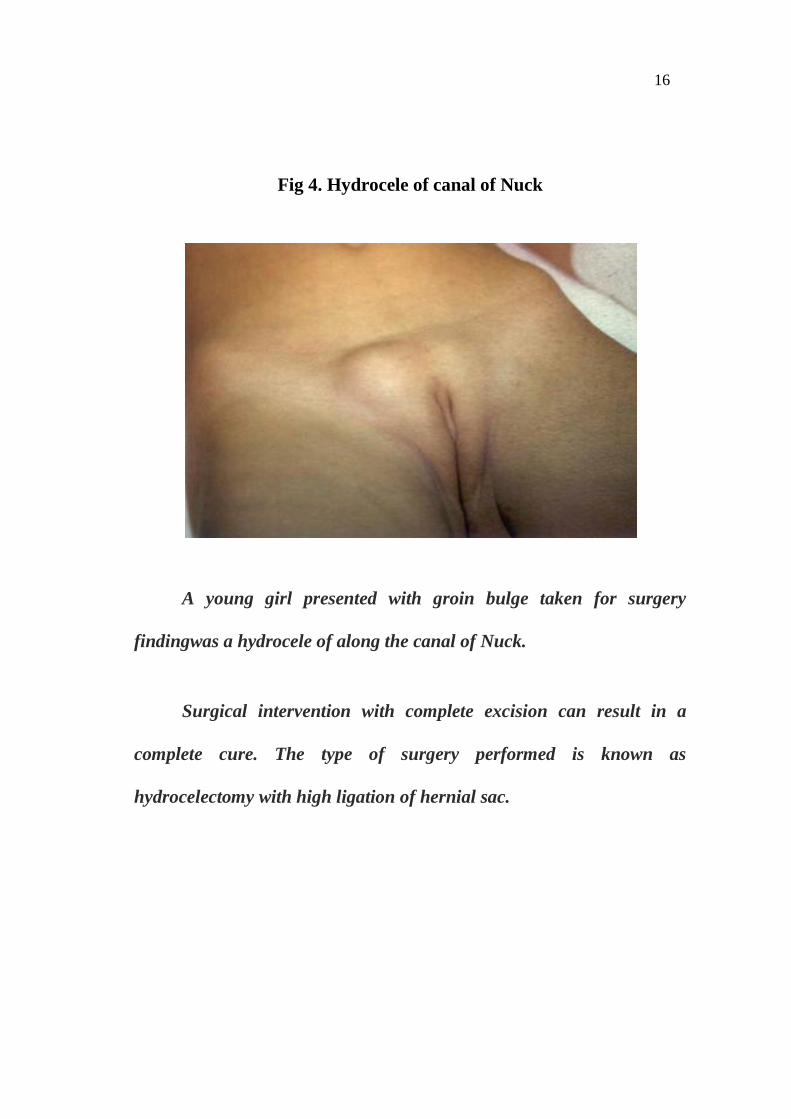
Fig 4. Hydrocele of canal of Nuck
A young girl presented with groin bulge taken for surgery
findingwas a hydrocele of along the canal of Nuck.
Surgical intervention with complete excision can result in a
complete cure. The type of surgery performed is known as
hydrocelectomy with high ligation of hernial sac.

17
Fig 5: Types of Hydrocele

18
B.Acquired Hydrocele
Primary Vaginal Hydrocele
It occurs usually in the middle aged and common in tropical
countries.Testis is not palpable as it is usually attains a large size (unlike
secondary hydroceles, which are very small, except in filarial
hydrocele).The swelling is fluctuant – elicited by the fixing hydrocele
with hand and feeling for the fluid movement by using fingers placed in
two perpendicular directions.
The swelling is also transilluminant and elicited in front of the
swelling, side by side. But long standing hydrocele is not transilluminant
due to the thickened dartos, thickened spermatic fascia, and thickened
hydrocele sac and infected content or chylous fluid.
On examination, we can get above the swelling.Testicular
sensation can be elicited in vaginal hydrocele by transmitting the pressure
sensation through the fluid.

19
Fig 6: Average size of a hydrocele
Image showing the patient presented with average sized
Hydrocele
Skin over the scrotum is stretched and normal rugosity is lost and
subcutaneous vein is very prominent. In case a hydrocele we can see a
constriction around the swelling due to tight tunica albuginea at that level.

20
Fig 7: Get above the hydrocele swelling
The image showings get above the swelling
Get the swelling at the root of scrotum and feel the cord structures.
If there is associated hernia or if the swelling is due to hernia get above
the swelling is not possible.

21
Fig 8: Trans-illumination of the hydrocele swelling
Image showing Transillumination in hydrocele of scrotum
Perform this test in darkness place the pencil torch laterally over
the scrotum blow the light. Place the illuminoscope exactly
perpendicular.
Do not place the illuminoscope posteriorly always place the
illuminoscope anterior and light lateral.
Negative transillumination – pyocele and haemotocele.
Positvetrans illumination implies that the content is clear hydrocele

22
Fig 9: hydrocele in USG scrotum
Image showing hydrocele in USG scrotum
Secondary Hydrocele
1. Recurrent epididymo-orchitis due to filariasis
Fluid that accumulates is due to the obstruction of lymphatics. The
fluid is milky white in colour. Such hydroceles are called as chyloceles
and often do not exhibit intransillumination.

23
Fig 10: Chylocele
Tuberculousepididymo-orchitis
Retrograde infection from the seminal vessels
Craggy epididymis refers to the rough, hard and irregular surface.
This involves the epididymal head and causes fibrosis. So the epididymis
feels craggy. Vas deferens feels like a beads and it is called as beaded
vas. Secondary hydrocele occurs in 32% of the cases. Eventually it forms
a cold abscess which ruptures and results in sinus posteriorly in the
scrotum.It never involves in the testis proper.

24
Testicular tumours
They can present with a swelling of the scrotum, often it is
diagnosed as hydrocele. Any young patient with a rapidly gro.wing
scrotal swelling could be a testicular neoplasm. Fluid within the sac is
hemorrhagic.
Fig 11: Testicular tumours
Testicular ultrasound: the homogeneous tissue of the testicular
teratoma on the left of the image produces multiple ultrasound
reflections.

25
Pyocele
It is an infected hydrocele. Infection in a hydrocele and it is rare
because of the tunica vaginalis sac which is relatively avascular. However
for few cases may get infected resulting in pyocele. These patients
present with fever, chills and rigors.
Fig 12: Pyocele
Pyocele—clinical look. US confirm the diagnosis. TC will be raised
Haematocele
It is a trauma to the hydrocele or spontaneous bleeding into the sac.

26
Fig 13: Haematocele
It is more important to be able to tell if the testis is intact, because
if there is a rupture
Complications of Hydrocele
Hematocele
Pyocele
Calcification of the hydrocele sac
Rupture of the hydrocele sac
Hernia of the hydrocele sac

27
Fig 14: Haematocele
Haematocele in different patients. Orchidectomyis often required
in these patients if testis is not viable.
Studies done on complications arising out of various surgeries for
hydrocele.
Comparision of The excisional, plication and internal drainage
techniques.
A study by Ku et al., “The excisional, plication and internal
drainage techniques - a comparison of the results for idiopathic
hydrocele” comparred the results of numerous techniques done for

28
hydrocele repair. The study was done between January 1992 and June
1998 which was included 131 patients diagnosed as hydrocele. The
patients were randomized in 3 groups using the excisional.Technique in
the first group which included 81 patients, Lord’s plication or eversion
technique in 2nd group which included 24 patients and internal drainage
in 3rd group which included 26 patients. The 1st group underwent the
conventional surgery for idiopat.hic hydrocele and the excision and
eversion of the sac subsequent to it.
The 2nd group underwent Lord’s plication technique and the 3rd
group underwent internal drainage of the accumulated fluid. The basic
principle of internal draina.ge is the fenestration of the hydrocele sac and
by removing parietal tunica vaginalis and layer the opening with the
scrotal skin, so that the hydrocele fluid drains through the `window'
which into the tissues superficial to the parietal layer of the tunica
vaginalis, and then being absorbed by the lymphatics. The possible
complications of the scrotal oedema, hematoma and infection were
monitored for 5 days. The reappearance ratewas also noted for almost 2
years. There was no significa.nt change among the patients in three
groups in the characteristics like age, size of hydrocele, duration of
symptoms and follow up duration. Among the complications, even

29
though the hematoma was seen only among the patients in excision group
the difference was not statistica.lly significant.
The wound infection incidence is difference among the 3 groups
was also not statistically important. The Oedema was more incident
among excision group and followed by plication group and the difference
was significant. The recurrence was significantly high among the patients
who had internal drainage technique and was negligible among other 2
groups. The study concluded that “application is better than the excision,
causing fewer complications, and better than the internal drainage and
giving more favourable results.”
Comparison of aspiration-sclerotherapy with hydrocelectomy
Khaniya et al study titled “Comparison of aspiration-
sclerotherapy with hydrocelecto.my in the management of hydrocele:
A prospective and randomized study” compared the less invasive
aspiration and sclerotherapy technique with surgical hydrocelectomy. The
study was done for a period of a year inclu.ding all unilateral primary
vaginal hydrocele patients. Those patients presented with a spermatocele,
testicular malignancies and scrotal hematocele or other fertility concerns
were excluded.

30
A total of 61 patients aged from 14 – 78 years were included in the
study. Fluctuation and trans-illumination were used for confirmation of
the cases. The 62 patients were randomly allocated into two groups of 31
patients using computer generated random numbers. Group 1 was done
aspiration with sclerotherapy in OP settin.g and group 2 was done with
hydrocelectomy in day care operation theatre. The study states the
procedure done in the both groups.
In group 1
Aspiration of the fluid was done by a 15 gauze intravenous
cannula attached to a 50 ml syringe with the three way stopcock and the
sclerasant was injected through the same cannula in situ. The volume
instilled was 50% of the aspirated fluid up to the maximum of 80 ml. The
sclerasing solution was prepared by diluting the mixture of 4 ml of 3%
sodium tetradecylsulphate and 6 ml of 2% lignocaine hydrochloride with
70 ml of the normal saline. The concentration of sodium
tetradecylsulphate and the lignocaine hydrochloride in the solution was
0.14% each.
In group 2
Jaboulay’s procedure (eversion of tunica without excision of sac)
was performed under local anaesthesia and drain was not placed. After
the intervention, dry dressing with scrotal support was applied for 24 h

31
and oral NSAIDs (the tablet combination of paracetamol 324 mg and
ibuprofen 400 mg) were prescribed 8 hourly for 48 h, then after only on
demand basis.” Follow up of patients was done at 48 hours, 1 week, 1
month, 3 months and 6 months. The patients were noted for the incidence
of fever, infection, pain, hematoma and recurrence of the swelling. There
was no difference in baseline characteristics between the 2 groups. The
incidence of pain and haematocele showed as no significant difference
between those 2 groups. The incidence of fever and infection were very
higher in hydrocelectomy group compared to the sclerotherapy group and
the difference was statistically significant. While considering the
recurrence, the group 1 presented with 34.6% recurrence after initial
sclerotherapy. Out of those 8 patients who underwent the repeat
aspiration and sclerotherapy at 3 months, 6 had a recurrence at 6 months.
The percentage of the patients satisfied with the procedure increased from
66.66% to 94% over the period of time from 48 hours to 6 months in
patients who are all underwent hydrocelectomy whereas in group 1 it
decreased from 66.66% initially to 62.9% at 6 months. The major reason
for dissatisfaction was the recurrence of the swelling.

32
Complications following hydrocele surgeries:
A study by Jin Kyeom Kim et al, on “A 10-Year Retrospective
Study of the Operative Treatment Results of Adult Type Hydrocele”
evaluated the incidence of complications and the outcomes of patients
who underwent hydrocelectomy between January 1996 and December
2005. (12) 289 patients with hydrocele were retrospectively assigned into
three groups according to the degree of dissection or the amount of the
excision of the hydrocele sac. Group 1 had 78 patients who were treated
by surgical dissection and excision of the entire hydrocele sac. Group 2
had 149 patients who were treated by surgical dissection and eversion of
the hydrocele sac. The 62 patients present in group 3 underwent
operations in which there was little or no dissection of the hydrocele.
They analysed the complications, the effects of surgical treatment and the
results according to the various surgical techniques in different groups.
The results showed that the duration of recovery showed no
differences among the three groups. The overall complication rate was
found to be 36.3% among the surgeries. Transient scrotal swelling was
present in 28.0% of the patients, hematoma in 2.7%, wound infection in
1.7%, and injury to the epididymis or testis, chronic pain and persistent
swelling present in 1.3%. The overall incidence of postoperative

33
complications was significantly less among the patients in group 3. The
rate of scrotal swelling was significantly correlated to the volume of the
hydroceles and the amount of the excision of the hydrocele sac.
According to them, The long term results of hydrocelectomy were good.
The most common complications following scrotal surgery for hydroceles
were scrotal swelling, followed by hematoma, wound infection and injury
to the epididymis and testis.
Comparison of minimal Access versus conventional hydrocelectomy:
A study by Saber (2015), titled “Minimally access versus
conventional hydrocelectomy- a randomized trial” aimed at comparing
the new minimally access hydrocelectomy versusJaboulay’s procedure
regarding operative outcome and patient’s satisfaction.
About 123 adult patients of age 17 to 55 years during the interval
of Apr 2008 to Oct 2013 identified as hydrocele were recruited to
thestudy. The study of population was divided into two equal groups.
Group A consisted of 61 patients were subje.cted to conventional surgical
hydrocelectomy (Jaboulay’s procedure) while group B consisted of 61
patients were submitted to the new minimal access hydrocelectomy. The
cases were confirmed by fluctuations and trans-illumination followed by

34
the scrotal ultrasound. The randomization of the patients was done using
the computer generated random numbers which were sealed inside the
opaque envelopes and were opened.up before entering the operation
theatre. Based on their random numbers they were allocated to either
group A (or) B and underwent the corresponding procedure.
Regarding age, duration of symptoms and size of hydroceles, there
was no statistical significant difference between the 2 groups of patients.
The age of the patients were ranged between 18–56 years with a mean
age of 36±11.5 years. The mean operative time in minimal access surgery
group was 16.1 ± 4.24 minutes ranged between 12-17 minutes and the
mean operative time in the conventional hydrocelectomy group was 32.5
± 4.76 minutes ranged between 25-40 minutes.
The difference in the operative time between the two surgical
procedures was statistically significant (P≤0.02). The minimal acce.ss
surgery group had a mean time of hospital stay of 13.47±6.37 hours with
minimum of 10 hours and a maximum of 30 hours and while in
conventional hydrocelectomy it was 21.19 ± 11.65 hours with minimum
of 11 hours and a maximum of 48 hours but the difference work (the

35
number of days between the day of surgery and the first in the
distribution was not significant (P≥0.05).
The time to return to day a patient returned to workwas considered
in both the groups and the mean in minimal access surgery group was 8.5
± 2.1 (7-10) days while in conventional hydrocelectomy group was 12.5
± 3.53 (10-15) days. The mean time off from work in minimal. access
surgery group was 9 ± 2.35 days and in conventional hydrocelectomy
group was 13.5 ± 4 and the difference was significant (P=0.0001).
The postoperative findings taken into account were post-o.perative
hematoma, degree of scrotal oedema, wound infection, patients’
satisfaction and recurrence. The overall complication rate in conventional
hydrocelectomy group was 36% and in minimal access hydrocelectomy
group was 12.88%. Postoperative hematoma was nil in minimal access
hydrocelectomy group while 3 patients (4.7%) had mild hematoma in
conventional hydrocelectomy group.
Mild and moderate scrotal edema usually subsided within a few
days postoperatively while scrotal edema and hardening was considered
when the pain and swelling interfered with their daily activities. The

36
higher incidence of scrotal edema and hardening were found in
conventional hydrocelectomy group while scrotal edema and hardening
occurred only in 3 patients in minimal access hydrocelectomy group and
this difference was statistically significant. (P≤0.05).
Persistent edema & hardening were confined to the ipsilateralhemi
scrotum and required an additional bed rest and anti-inflammatory agents.
Cellulitis was mild to moderate, seen in 4 patients in both groups A and B
(6.45%). Regarding patient satisfaction, only 2 patients (4.83%) were
unsatisfied with the new minimally invasive procedure by the end of
second postoperative week, mainly due to scrotal harden.ing while scrotal
edema and hardening was observed in24.2% those who had conventional
hydrocelectomy. Disease recurrence was equal in two groups which was
also negligent (1.6%).
Additive evidence regarding hydrocelectomy techniques
Ismail Mihmanli et al titled “Testicular Size and Vascular
Resistance Before and After Hydrocelectomy” (2) was done with the
objective of to determine whether there is an association between
hydroceles and testicular size and vascular resistance. The methodology
were done as follows. “Twenty-three consecutive patients with the

37
diagnosis of unilateral idiopathic hydrocele (noncommunicating and
noncongenital) who underwent hydrocelectomy were included in the
study. At physical examination, the physician was unable to palpate the
testis due to the hydrocele. Patients with a history of severe
cardiovascular problems, lymphangitis, previous inguinal radiotherapy,
and hypoalbuminemia were not included in the study. The duration of
scrotal symptoms ranged from 2 to 18 months (mean, 8 months). No
underlying cause for the hydrocele was found in any of the patients.
Informed consent was obtained from all patients before the sonographic
examinations and surgery. The patients were examined in the supine
position, with the scrotum supported over a towel tightly draped over the
thighs. All the examinations were performed in a temperature-controlled
room after the patient had rested for 30 min. All examinations were
performed by the same examiner with a high-resolution
sonographysystem (Sonoline Elegra, Siemens Medical Solutions) using a
4–9- MHz linear array transducer. The examination protocol included the
preoperative evaluation of the hydrocele and pre- and postoperative
evaluations of both testes. Preoperative evaluation consisted of
identifying the testis with the hydrocele and characterizing the nature of
the hydrocele with gray-scale sonography. They also evaluated the
internal septations and loculations within the hydrocele to determine

38
whether it was complicated. The length, width, and anteroposterior
diameter of both testes were measured. At least three separate
measurements were made on different occasions. The mean of these three
separate measurements was used for the calculations. Approximate
volume for ellipsoid structures was calculated by multiplying these three
diameters by 0.523. The parameters of color Doppler sonography were
optimized to display low-flow velocities for evaluating intratesticular
blood flow and low-velocity diastolic arterial flow on both the normal
side and the side with hydrocele. Spectral waveforms were obtained from
at least three different intratesticular arteries. Resistivity index (RI) and
pulsatility index (PI) values were determined from these waveforms. The
sonography scanner is supported with proper software for direct and
automatic calculation of the hemodynamic parameters based on spectral
Doppler waveforms. The spectral waveform was manually traced on the
strip with calipers, and the RI and PI values were calculated automatically
by the software program. Measurements were obtained from three
individual waveforms from separate strips. The mean value of three
measurements was calculated for each testis. Postoperative measurements
included testicular volume and the RI and PI values on both sides. All the
calculations and measurements were performed by the method that was
described earlier. To avoid having early postoperative changes (edema,
39
hyperemia, or inflammation) affect RI and PI values, we performed the
sonographic examinations at least 2 months after the hydrocelectomy. In
all of the patients, the indication for surgery was improvement of the
cosmetic appearance of the testis or the patient’s wish. Hydrocelctomy
with tunical incision in which the fluid is drained and the tunica is everted
was performed. The specimen volume was measured after being collected
in a bowl right after the incision of the tunica. Care was taken to not
manipulate the testes. The program SPSS (version 7.5 for Microsoft
Windows, Statistical Package for the Social Sciences) was used for
statistical analysis. Testicular volumes and RI and PI values for the
normal side and the side with hydrocele were compared before surgery.
Both testicular volumes and intratesticular RI and PI values were
compared after surgery. The Student’s t test for paired samples was used
for statistical analysis. Statistical significance was indicated by a p value
of less than 0.05. The percentage of difference between the normal and
hydrocele testicle measurements (volume and RI and PI values) and the
percentage of change in measurements of a single testicle before and after
hydrocelectomy were calculated and expressed as mean ± SD. A single
analysis of variance (ANOVA) model was created for each measurement.
Volume, RI value, and PI value were included as separate dependent
variables. The independent variables were the hydrocele (presence or

40
absence), surgery (before or after), and patient number (two observations
per patient) for each instance. The interactions between the variables of
the hydrocele and surgery were also studied separately using ANOVA. A
p value of less than 0.05 was regarded as statistically significant.”
The results were stated as “The 23 patients ranged in age from 21
to 72 years (mean age, 42.8 ± 10.8 years). Fourteen patients (60.9%) had
right-sided and nine patients (39.1%) had left-sided hydroceles. None of
the hydroceles appeared complicated on sonography. All the hydroceles
appeared as massive anechoic fluid collections around the testes. The
mean volume of hydroceles was 291 mL (range, 242.7–365.4 mL) at
surgery.
None of the patients was shown to have a testicular tumor,
inflammation, a varicocele, or an inguinal hernia on sonography.
Sonography was performed at a mean follow-up of 4.5 months after
surgery (range, 2–6 months). None of the patients had recurrence of their
hydrocele during this follow-up period. Before surgery, a statistically
significant difference was found between the testicular volumes of both
sides (p < 0.001), and a statistically significant difference in the RI and PI
values was found between the normal side and the side with hydrocele (p
41
< 0.001). After hydrocelectomy, the difference in the testicular volumes
before and after surgery on the side of hydrocele was statistically
significant (p < 0.001). There was not a significant difference in the
testicular volumes before and after surgery on the normal side (p =
0.200). The side with the hydrocele showed a statistically significant
decrease in RI and PI values of intratesticular arteries after
hydrocelectomy (p < 0.001). A statistically significant difference in RI
and PI values was not detected on the normal side (p = 0.549 for RI, p =
0.306 for PI). The results of the single ANOVA test showed that the
volume measurements differed from patient to patient (F = 3.49, p <
0.001). However, the RI (F = 1.51, p = 0.100) and PI (F = 2.60, p =
0.566) values did not. Therefore, although the amount of change in
volume varied among individual patients, the changes in RI and PI values
were more constant. Also, the presence or absence of hydrocele and the
surgical status (before or after) affected all three measurements (p <
0.001). When the presence or absence of a hydrocele and surgical status
were taken into account, the measurements in the normal testicle did not
change after surgery, whereas the measurements in the testicle with the
hydrocele did (F = 67.53 for volume, F = 75.13 for RI value, and F =
25.15 for PI value; p<0.001 for all three measurements).”

42
Ananthakrishnan et al titled “Surgery for vaginal hydroceles:
an update” (13) states “In men, vaginal hydrocele is the most common
morbidity due to Wuchereriabancrofti. The only effective treatment for
hydrocele is surgery, but safe surgery requires adherence to strict
standards for diagnosis, preoperative, intraoperative and postoperative
care of the patient. Other scrotal conditions such as chylocele (collection
of chyle in the tunica vaginalis), hematocele (collection of blood) or a
pyocele (collection of pus) may be mistaken for a hydrocele. These
require appropriate management and need to be excluded when making a
diagnosis of simple uncomplicated hydrocele. The latter three conditions
are characterized by the fact that the contents of the tunica vaginalis sac
are non-transilluminant. This test can be used at the peripheral level for
differentiating uncomplicated hydroceles from other scrotal swellings.
The test is easy to perform, does not require costly equipment other than a
good flashlight and an opaque tube of approximately 6' in length and 1' in
width. The skill of transillumination can easily be taught to physicians at
the appropriate peripheral level. Although there is a report from India
suggesting that diethyl carbamazine (DEC) therapy could reduce the size
of hydroceles, a recent double blind study in Tanzania showed that DEC
has no effect on the size of hydroceles. Hence, surgery remains the
treatment of choice for management of filarial hydrocele. Although there

43
are several publications on surgery of hydrocele and the complications of
surgery, this article presents the consensus obtained in a global meeting
called under the auspices of the WHO. For management of hydroceles,
the levels of health care facilities are classifiable into the following three
levels:
Level I: this is at the community level and is meant for detection of
patients with scrotal swellings either by the community health worker or
the patient presenting himself. Once detected the patient would be
referred to a level II facility.
Level II: this is a centre at which surgery for un-complicated
hydroceles can be performed. In different countries it would be
equivalent to a community health centre or sub-district level hospital with
provision for minor surgery. In addition to oxygen and resuscitative
facilities there should also be facilities for observation of patients for 24-
48 h where required. A trained surgeon or an MBBS physician who is
already performing minor surgical procedures can then be trained to
perform surgery on patients with hydrocele at the level II facility.

44
Level III: this would be equivalent to District Hospitals where
patients with more serious medical problems or complicated hydroceles
can be referred for surgery.
It is essential to examine a patient with a scrotal swelling and
differentiate between a hydrocele and other causes of inguino-scrotal or
scrotal swellings other than hydroceles as per the algorithm. For this
purpose the skill of performing and interpreting a transillumination test is
mandatory. All inguino-scrotal swellings and scrotal swellings that are
not transilluminant, patients in whom the diagnosis is in doubt, children
with hydroceles and those with co-morbid conditions should have
ultrasonography to differentiate these swellings. Indications for hydrocele
surgery at the level II facility would include medical disqualification due
to un-treated hydroceles; interference with work; interference with sexual
function; interference with micturition due to the penis getting buried in
the scrotal sac; negative impact on the patient's family; dragging pain;
liability to trauma in view of nature of patient's work or mode of
transportation such as cycling; possible effect on the testis of long
standing hydroceles.

45
Patients with large hydroceles should be given priority in situations
where resources may be limited. However, if resources are not an issue,
and where the patient may be limited in employment opportunities due to
a hydrocele of any size being considered a disqualification for
Government jobs (as in India) then, surgery should be offered to all.
Preoperative assessment procedures would include evaluation for
systemic illnesses such as history of Diabetes Mellitus, other systemic
illnesses such as angina, drug allergies, sickling tendency and other
problems likely to increase the risk of surgery; haemoglobin, urine and
blood sugar; ensuring adequate scrotal hygiene by preoperative bath and
scrotal washing with soap and water two times daily for 3 days before
surgery, which could reduce the infection rate; surgery should be
rescheduled to at least 4-6 weeks after an acute adenolymphangitis;
It is recommended that all the operations for uncomplicated
hydrocele in patients without serious comorbidity should be performed
under local anaesthesia using either bupivacaine or lignocaine (lidocaine).
The procedure should consist of a spermatic cord block with the drug,
combined with infiltration along the line of incision.

46
The procedure for hydrocele should preferably be done as an
outpatient procedure. However, observation of the patient for 24-48 h
after surgery should be done whenever the situation warrants. The
surgeon who performs the operation should be competent to perform
hydrocelectomies. It is recommended that the operation performed should
be a hydrocelectomy, i.e. a subtotal excision of the parietal layer of the
tunica vaginalis leaving a rim of approximately 1-cm width around the
testis and epididymis. Aspiration with or without injection of sclerosants
was not recommended due to the high recurrence rate and the potential
damage to the testis due to the sclerosant. Likewise the procedure of
eversion of the hydrocele sac (Jaboulay's procedure) is best avoided due
to the following reasons:
In hydroceles, which are larger than tennis balls, the procedure of
eversion of the sac is likely to leave the patient with a significant residual
swelling of the scrotum;
In hydroceles smaller than tennis balls both procedures, (eversion
and excision) are likely to run the same risk of complications;

47
The tunica vaginalis is abnormal in patients with filarial hydrocele
and is best excised. If left behind there are fears in some quarters of
possible complications such as a lymph scrotum or a filarial scrotum in
some patients. It was, however, accepted that there is insufficient
published material to record the instance of such complications, if any;
If improperly performed the procedure of eversion of the sac is
associated with a greater risk of recurrence.
Use of chromic gut sutures was recommended to minimize the
cost.
Postoperative care is done as follows:
1. Analgesics should be administered starting from the morning of
surgery and continued for 48-72 h. The choice of recommended
analgesic was oral acitaminophen or NSAIDs other than aspirin.
2. Antibiotics should be administered starting from the night before
surgery for a total duration of 5 days. This is to forestall the risk of
infection since the patient would return to his home environment to
an ambience, which may be conducive to infection. The
recommended antibiotic in view of cost and the type of bacteria

48
likely to be involved in infection was amoxicillin and
metronidazole.
3. Patients may be allowed to return to their homes a few hours after
surgery except under the following circumstances in which case
they should be observed for 24-48 h. Placement of a drain, which
has to be removed after 24-48 h undue swelling, pain or oozing
from the wound.
4. Hydrocele wounds could be exposed on the third postoperative day
and kept dry resulting in less infection from wet dressings and
sweating,
5. Patients should be asked to return to the centre 7-10 days later for a
follow up visit.
Access issues are addressed as follows:
The issue of patient access for surgery, particularly for hydrocele
needs to be addressed. It is felt that the current level of access to surgery
in most countries is inadequate. The following are the most possible
reasons for the same:
1. Ignorance of patients to the fact that they can be cured of their
condition;

49
2. Fear of surgery and its consequences;
3. Lack of facility or long distance between such a facility and the
patient's home;
4. Cost of surgery, hospitalization, transport, loss of wages during and
in the postoperative stage.
Training for Surgeons
1. Trainers for training of level II surgeons are to be identified by
National Governments/Country co-ordinators. The trainers could
be qualified surgeons with experience in hydrocele surgery
working in endemic areas (they could also be surgeons attached to
teaching or training institutions with experience of hydrocele
surgery). The identified trainers need to be trained on the following
through a workshop (but case demonstration and actual
performance of surgery need not be done during the training),
Surgery protocol for Level II medical officers;
To acquire the ability of Level III surgeons (to be able manage
scrotal swelling cases referred to them from level II); to acquire the
ability to tackle any complications developed in hydrocele cases operated
at level II;

50
2. The trainers will then train the Level II surgeons identified by
national/local health systems. Level II Medical officers need to be
trained on diagnosis, testing for fitness for surgery, all aspects of
the protocol for surgery, postoperative care and follow up.
3. It would be advantageous to encourage actual performance of
surgery during the trainings. This ensures agreement regarding
what is meant by certain terms. When actual surgery cannot be
done, videotapes of surgery may be substituted.
4. Continuing medical education programmes for medical school
teachers, residents and other surgeons, private practitioners on
'Newer developments in the pathogenesis and management of
filariasis, protocol for hydrocele management and available
information on management of other uro-genital manifestations of
filariasis through workshops, round tables, symposia, seminars and
exchange visits.
The author concluded as follows:
In men, vaginal hydrocele is the most common morbidity due to
Wuchereriabancrofti. Diagnosis is straightforward most of the time but
when the diagnosis is in doubt ultrasonography is a useful tool to

51
differentiate these swellings. As the effect of medical treatment with
diethylcarbamazine on the size of hydroceles are doubtful, double blind
randomized clinical trials are required to generate evidence on the effect
of diethylcarbamazine on hydroceles of different grades. The only
effective treatment for hydrocele is surgery as the minimally invasive
therapy like aspiration and sclerotherapy are known to have high
recurrence rates.”

52
MATERIALS AND METHODS
* Study Setting
* Study Duration
* Study Population
* Inclusion Criteria
* Exclusion Criteria
* Study Design
* Sample Size
* Procedure
* Randomization
* Pre-Operative Workup
* Surgical Techniques
* Conventional Hydrocelectomy (Jaboulay’s Procedure)
* Minimal Seperation Hydrocelectomy
* Study Tool
* Data Collection and Methods
* Services rendered

53
Study Setting:
Dept. of General Surgery, Govt. Kilpauk Medical College,
Chennai.
Study Duration:
6 months (April 2017 – August 2017)
Study Population:
Patients attended the surgery OPD with scrotal swelling for
evaluation
Inclusion Criteria
1. Those subjects diagnosed as primary vaginal hydrocele
2. Those who were willing for the surgery
3. Patients aged 18-56 years
4. Male Gender
5. With diagnosis of hydrocele
6. Patient without comorbidity
7. (TB, HT, DM, asthma, seizure)

54
Exclusion Criteria:
1. Those patients presented with spermatocele, testicular
malignancies and scrotal hematocele.
2. Patients having filarial scrotum requiring scrotoplasty were not
included in this study.
3. Secondary hydrocele due to acute infection and malignancy are
excluded from the study.
Study Design
The study is conducted as a single blinded Randomized Control
Trial with two arms – one arm of subjects with hydrocele who underwent
minimal separation hydrocelectomy and the other arm of subjects with
hydrocele who underwent conventional hydrocelectomy (Jaboulay’s
procedure).
Sample Size
Sample size is calculated using the formula:
55
(Z+ Zβ) 2 * 2 * p*(1-p)d2
Where, Z = two tailed deviate for 95% confidence level = 1.96,
Zβ = two tailed deviate for 80% power of the study = 0.84
p = (p1 + p2) / 2
d = difference in incidence of post-operative oedema between
subjects underwent minimal access and conventional hydrocelecetomy.
p1 – incidence of post-operative oedema in subjects underwent
conventional hydrocelectomy
p2 - incidence of post-operative oedema in subjects underwent
minimal access hydrocelectomy
From the study by Aly Saber “Minimal Access versus
Conventional Hydrocelectomy: a Randomized Control Trial”, the
incidences of post-operative oedema are considered as, conventional
hydrocelectomy patients, p1 = 74% and minimal access hydrocelectomy
patients, p2 = 8%.

56
Therefore,
p = (p1 + p2) / 2 = (0.74 + 0.08)/2 = 0.41
The sample size is calculated as
N = (1.96 + 0.84) 2* 2 * 0.41 * (1 - 0.41) / (0.74 – 0.08) 2
N ~ 30
The total sample size estimated is 60 with 30 subjects in each arm.
Procedure:
Randomization
The randomization technique was commenced before the start of
the procedure. There was 60 sealed envelopes were made ready with
sequential number from 1 to 60. Each envelope contained a computer
generated random number inside in it. Based on the last digit of the
random number, the subjects were allocated to respective interventional
group. If the number was between 0 and 4, they were assigned to
conventional hydrocelectomy and if the number was between 5 to 9 and
they were subjected to minimal separation hydrocelectomy. The
envelopes were opened by the investigator after getting the consent from
the patie.nt just prior to the surgery. Based on the random number, the
subjects were allocated and the respective surgeries were done.

57
Pre-Operative Workup:
Each patient was assessed in detail about their history and
complete physical examination was done. Fluctuation and Trans-
illumination was used for confirming the diagnosis of hydrocele. Basic
laboratory investigations like complete blood count and urine routine
examinations were done.
Inj.ceftriaxone 1gm IV at the time of induction of anesthesia or just
after the administration of spinal anesthesia was given followed by
another dose 2 h postoperatively.
Surgical Techniques
After the induction of spinal anaesthesia, antibiotic ceftriaxone
1gm iv was given intravenously followed by one more dose 2 hours post-
operatively.
Conventional Hydrocelectomy (Jaboulay’s Procedure)
The testis was delivered through an incision in the scrotum and the
tunica was opened and everted and most of the hydrocele sac was
resected with electrocautery and leaving a reasonable cuff along the
borders of the testicle.

58
Bleeding was controlled by a running suture closing the free edges
of the hydrocele sac and hemostasis was secured by the aid of
electrocautery. Standard 2 layer closure which was used to close the
scrotum with small tube drain. Patients were followed up on second day
for scrotal edema and hematoma and the drain was removed on third day.
Fig 15: Conventional Hydrocelectomy: Vertical incision of about 6-8
cm in length was made over the scrotum, anteriorly about 1 cm
lateral to the median raphe.

59
Fig 16: Conventional Hydrocelectomy: Bluish hydrocele sac is
identified parietal layer of the tunica vaginalis of testis

60
Fig 17: Conventional Hydrocelectomy: Hydrocele Sac Isolated and
delivered out of scrotum totally

61
Fig 18: Conventional Hydrocelectomy: Fluid is evacuated using
trocar and cannula. Sac is opened.

62
Fig 19: Conventional Hydrocelectomy: Evacuation and eversion of
the sac behind the testis
Testis replace into the scrotal sac lateral sulcus facing laterally a
drain is placed near the root of the scrotum on the lateralaspect because it
becomes the most dependent portion, whenscrotal support is given.
Scrotal support is given to reducethe scrotal oedema.
Wound is closed in layers.
Drain is removed in 48 hours

63
Minimal Separation Hydrocelectomy
A small scrotal incision of about 2cm long was made and incision
of the Dartos muscles in the same line was made using with electro
cautery. The parietal tunica vaginalis (PTV) wasidentif.ied grasped and
minimal blunt dissection was made by the helpof the index finger.
A small hole was made for the aspiration of hydrocele fluid. Then a
disc of tissue was excised of the parietal tunica vaginalisabout double of
the skin incision dimension using electrocautery.
The edge of the visceral surface of the tunica vaginalis was sutured
to the parietal layer of the tunica vaginalis and then to the Dartos muscle
and all was sutured to scrotal skin in an everted manner aim to expose the
visceral tunica toward scrotal skin. If the visceral surface of the tunica
vaginalis is sutured to the Dartos, eversion will be created. Then when
this everted structure is sutured to the scrotal skin, it will be in contact the
sac with lymph-rich subcutaneous tissues.
A drain was kept in place and discharge was allowed for one day.
Patients were followed up on second day for scrotal edema and
hematoma and the drain was removed on same day.

64
Fig 20: Minimal Seperation Hydrocelectomy: length of the scrotal
skin incision about 2cm in size

65
Fig 21: Minimal Seperation Hydrocelectomy: evacuation of
hydrocele fluid through a smallhole made over the tunica vaginalis.
66
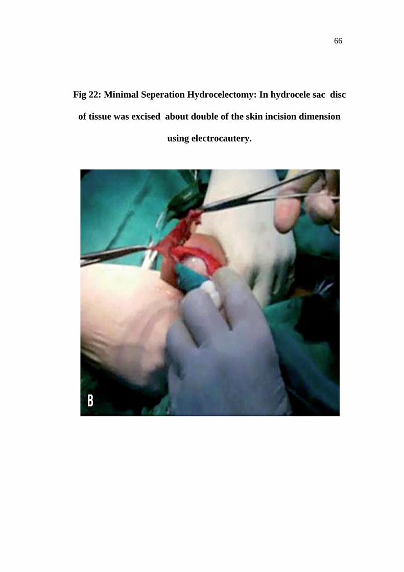
Fig 22: Minimal Seperation Hydrocelectomy: In hydrocele sac disc
of tissue was excised about double of the skin incision dimension
using electrocautery.

67
Fig 23: Minimal Seperation Hydrocelectomy: Completed eversion
technique by suturing the edge of the tunica vaginalis to Dartos and
scrotal skin in an everted manner aiming to expose the visceral
tunica toward scrotal sac

68
Study Tool
A questionnaire was designed which contained the details of
patient’s name, age, sex, symptoms or presenting complaints, duration of
the swelling, site of the swelling, operating time in minutes, post-
operative complications if any and duration of the hospital stay in hours.
Data Collection and Methods
a. Data collection was done in the study area after obtaining prior
permission from the Professor & HOD, Department of Surgery
and The Dean, Kilpauk Medical College and approval of
Institutional Ethical Committee.
b. Each participant was given a brief introduction about the study and
informed consent was obtained from all participants.
c. The information about the study was explained to the patient in the
local language clearly till they understood.
Services rendered
Each participant was assessed and provided treatment for the
clinical condition by either of the two surgical techniques at free of cost.
The patients were followed-up for up to 6 months for complications and
if any noted, treatment and care were provided according to the needs.

69
ANALYSIS OF OBSERVATIONAL DATA
Data Entry
The data collected from the questionnaires were entered in
Microsoft Excel 2013 version and the master chart was framed. The data
entered were double checked for any errors. The data from the master
chart were exported to Statistical Package for Software Solutions (SPSS)
version 21 for analysis. Totally data was collected from 60 patients with
30 from each arm.
Data Analysis
Continuous variables were presented in the form of descriptive
statistics (mean and standard deviation) and categorical variables in the
form of frequency distributions and percentages. Association between
categorical variables are tested using Chi square tests and Fisher exact
tests. Association between continuous variables and a grouping variable
were tested using student ‘t’ test.
Data presentation
The distribution of categorical data in the total study & among
rural and urban population were represented by tables and bar charts. The
continuous variables distribution were depicted by tables, box plot and
error bar chart. The distribution of continuous variables along a grouping
variable with a linear trend is represented by line diagrams.

70
RESULTS
Considering the baseline characteristics, there was no significant
difference between the two groups.
Fig 24 : Distribution of age categories in conventional and minimal
separation hydrocelectomy groups.
The distribution of participants in the both groups of the study
population in different age categories was almost nearly equal with no
much difference.
0
1
2
3
4
5
6
7
8
9
10
21 - 30 years 31 - 40 years 41 - 50 years 51 - 60 years 61 - 70 years 71 - 80 years
2
5
8
7
5
3
6
4
10
8
1 1
Conventional Hydrocelectomy Hydrocelectomy with Minimal Separation

71
Table 1: Distribution of age categories of the subjects in the two
groups of the study population
AGE_CAT
PROCEDURE
Total
Fisher
exact
p
value
Conventional
Hydrocelectomy
Hydrocelectomy
with Minimal
Separation
21 - 30
years 2 (25%) 6 (75%)
8
(100%)
0.332
31 - 40
years 5 (55.55%) 4 (44.44%)
9
(100%)
41 - 50
years 8 (44.44%) 10 (55.55%)
18
(100%)
51 - 60
years 7 (46.66%) 8 (53.33%)
15
(100%)
61 - 70
years 5 (83.33%) 1 (16.66%)
6
(100%)
71 – 80
years 3 (75%) 1 (25%)
4
(100%)
The difference in the distribution of study participants in the both
groups was statistically insignificant.

72
Table 2: Distribution of symptoms of the participants in the two
groups of the study population
SYMPTOMS
PROCEDURE
Total
Fisher exact
p value Conventional Hydrocelecto
my
Hydrocelectomy with Minimal
Separation
Painless scrotal swelling Left
7 (46.66%) 8 (53.33%) 15
(100%)
0.096
Painless scrotal swelling Right
14 (50%) 14 (50%) 28
(100%)
Discomfort with scrotal swelling
Left 4 (50%) 4 (50%) 8 (100%)
Discomfort with bilateral scrotal
swelling 5 (55.55%) 4 (44.44%) 9 (100%)
Total 30 (50%) 30 (50%) 60
(100%)
The presentation of symptoms of the patients is almost equal in
both groups of the study population and the difference in the distribution
is statistically insignificant.

73
Table 3: Distribution of presentation of side of hydrocele of the
participants in the two groups of the study population
SIDE
PROCEDURE
Total
Fisher
exact
p value Conventional
Hydrocelectomy
Hydrocelectomy
with Minimal
Separation
Left 12 (50%) 12 (50%) 24
(100%)
0.143
Right 13 (48.14%) 14 (51.85%) 27
(100%)
Bilateral 5 (55.55%) 4 (44.44%) 9 (100%)
Total 30 (50%) 30 (50%) 60
(100%)
The presentation of side of hydrocele of patients in the both groups
had no much difference with right side more common followed by left
side and a few by both sides. The difference in the distribution is
statistically insignificant.
Table 4: Distribution of duration of hydrocele (in years) of the
participants in the two groups of the study population

74
Variable GROUP N Mean Std.
Deviation
p value
by ‘t’ test
DURATION
OF
HYDROCELE
(Years)
Conventional
Hydrocelectomy 30 7.57 4.08
0.356 Hydrocelectomy
with Minimal
Separation
30 6.63 3.67
Variable Group Minimum Maximum Range
DURATION
OF
HYDROCELE
(Years)
Conventional
Hydrocelectomy 1 17 16
Hydrocelectomy
with Minimal
Separation
1 17 16
The mean duration of hydrocele of patients in the both groups of
the study population had only a mild difference which was not
statistically significant. The range of duration of hydrocele was 16 years
(1 to 17 years) in both the study groups.

75
Fig 25: Percentage of Post-operative complications of the study
subjects in the conventional hydrocelectomy group
93% of the patients presented with oedema and hardening out of
which 33% also presented with wound infection and 3% also presented
with hematoma. Only 7% had no post-operative complications.
1757%
27%1
3%
00%
1033%
Conventional Hydrocelectomy
Edema and Hardening
Edema and Hardening with Wound Infection
Edema and Hardening with Hematoma
Wound Infection
Nil

76
Fig 26: Percentage of Post-operative complications of the study
subjects in the minimal separation hydrocelectomy group
Only 10% of the study participants underwent minimal separation
hydrocelectomy presented with oedema and hardening and only 7%
presented with wound infection. 83% of the patients didn’t experience
any post-operative complications.
Table 5: Distribution of post-operative complications of the
participants in the two groups of the study population
310%
00% 0
0%
27%
2583%
Hydrocelectomy with Minimal Separation
Edema and Hardening
Edema and Hardening with Wound Infection
Edema and Hardening with Hematoma
Wound Infection
Nil

77
POSTOPCOMPLICATIONS
PROCEDURE
Total Fisher
exact p value
Conventional Hydrocelecto
my
Hydrocelectomy with Minimal
Separation
Oedema and Hardening
17 (56.7%) 3 (10%) 20
(100%) <0.001
Oedema and Hardening with
Wound Infection
2 (6.7%) 0 (0%) 2 (100%) 0.246
Oedema and Hardening with
Hematoma 1 (3.3%) 0 (0%) 1 (100%) 0.500
Wound Infection
0 (0%) 2 (6.7%) 2 (100%) 0.246
Edema and hardening was the most common complication and is
more incident in patients who underwent conventional hydrocelectomy.
The difference in the distribution of edema and hardening among the
patients in the two study groups was statistically significant.

78
Table 6: Distribution of overall post-operative complications of the
participants in the two groups of the study population
OVERALL POST-OPERATIVE
COMPLICATIONS
PROCEDURE Fisher exact
p value
Conventional Hydrocelectomy
Hydrocelectomy with Minimal
Separation
YES 20 (66.7%) 5 (16.7%)
<0.001
NO 10 (33.3%) 25 (83.3%)
Taking into account, the overall post-operative complications
suffered by the patients in both groups of the study population, the
conventional hydrocelectomy group had more incidence of post-operative
complications. Around 67% of the patients belonged to conventional
hydrocelectomy group of the study population suffered complications
whereas only 17% of the patients belonged to minimal separation
hydrocelectomy group suffered complications.

79
Table 7: Distribution of operating time of the patients in the two
groups of the study population
GROUP N MEAN
STD.
DEVIA
TION
p VALUE
BY ‘t’ TEST
OPER
ATIN
G
TIME
(Min)
Conventional
Hydrocelectomy 30 30.83 2.94
0.0001 Hydrocelectomy
with Minimal
Separation
30 17.93 1.28
Variable GROUP Minimu
m
Maximu
m Range
OPERATIN
G TIME
(Min)
Conventional
Hydrocelectom
y
25 35 10
Hydrocelectom
y with Minimal
Separation
15 20 5
The difference in the distribution of operative time of the patients
underwent two different surgical procedures were statistically significant
with higher mean operating time in conventional hydrocelectomy than
minimal separation hydrocelectomy.
Table 8: Distribution of time of hospital stay (in hours) of the patients
in the two groups of the study population

80
Variable GROUP N MEAN
STD.
DEVIATI
ON
p VALUE
BY ‘t’
TEST
HOSPITAL
STAY
(Hours)
Conventional
Hydrocelecto
my
30 80.50 13.45
0.0001 Hydrocelecto
my with
Minimal
Separation
30 48.57 21.19
Variable GROUP Minimum Maximum Range
HOSPITAL
STAY
(Hours)
Conventional
Hydrocelectomy 48 98 50
Hydrocelectomy
with Minimal
Separation
25 95 70
The difference in the distribution of time of hospital stay of the
patients underwent two different surgical procedures was statistically
significant with higher mean time of hospital stay in conventional
hydrocelectomy than minimal separation hydrocelectomy.
DISCUSSION

81
The mean age of the participants in the study population was 47.7
± 14.15 years with a minimum of 21 years to a maximum of 80 years.
This age distribution was almost close to the Saber study which was
included participants from 18 to 56 years with a mean of 37 ± 11.4 years.
The mean operating time among those patients who underwent
conventional hydrocelectomy was 30.83 ± 2.9 minutes with the range of
25 to 35 minutes and those who underwent the Minimal seperation
hydrocelectomy was 17.93 ± 1.28 minutes with a range of 15 to 20
minutes. The difference in the mean time between the two surgical
procedures was statistically significant (p <0.01).
Similarly in Saber study, the operating time for conventional
hydrocelectomy was slightly higher with mean of 32.5 ± 4.76 minutes
upto a maximum of 40 minutes and the operating time for minimal access
hydrocelectomy was slightly lower with mean of 15.1 ± 4.24 minutes
with a range of 12 to 18 minutes. The difference in mean operating time
between the two procedures was statistically significant (p < 0.02).
The mean time of hospital stay among the patients who underwent
conventional hydrocelectomy was 80.5 ± 13.45 hours with a range of 48

82
to 98 hours and those who underwent Minimal access hydrocelectomy
was 48.57 ± 21.19 hours with a range of 25 to 95 hours.
The difference in the mean time between the two surgical
procedures was statistically significant (p <0.01). In Saber study, the
mean time of hospital stay for conventional hydrocelectomy was lower
with mean of 21.19 ± 11.65 hours with a range of 12 to 48 hours and the
mean time of hospital stay for minimal access hydrocelectomy was lower
with mean of 13.48 ± 6.38 hours with a range of 10 to 30 hours. But the
difference in the above mean time of hospital stay between two
procedures was not statistically significant (p > 0.05). This could be
attributed to the geographical differences in the protocol management of
the cases in the hospital. The differences may be due to available
resources and sufficient health care providers.
The overall complicat.ion rate (percentage of patients experienced
any complication) among the patients underwent conventional
hydrocelectomy was 66.6% whereas it was very low among patients
underwent minimal separation hydrocelectomy of 16.6% and the
difference in this distribution was statistically significant (p<0.001). The
low complication rate among the minimal separation group was
supported by the Saber study which states an overall complication rate

83
among patients underwent minimal access hydrocelectomy was 12.7%
and also showed a statistically significant difference from the
complication rate among patients underwent conventional
hydrocelectomy (37%).
The most common complication of the patients undergoing
hydrocelectomy is edema and hardening. In the present study, 57% of the
patients who underwent conventional hydrocelectomy suffered from
edema and hardening over the surgical site post-operatively compared to
10% incidence in the patients who underwent minimal separation
hydrocelectomy.
This difference in the distribution was also statistically significant.
This is additive to the evidence produced by Saber study which also
showed a significant differe.nce in the distribution of edema and
hardening among the patients between conventional hydrocelectomy
(25%) and minimal access hydrocelectomy (5%). The next common
complication following hydrocelectomy is hematoma over the surgical
site. Only 3% of the patients who underwent conventional
hydrocelectomy had incidence of hematoma whereas there was no
incidence of hematoma in patients underwent minimal separation

84
hydrocelectomy. In the Saber study alsothere was zero incidence of the
hematoma in patients who underwent minimal access hydrocelcetomy.
Oedema and hematoma are the most common in excision and
eversion technique (conventional hydrocelectomy). This is because of
wide dissection and excessive handling of the hydrocele sac during the
surgery. In the minimal separation hydrocelectomy a disc of the
hydrocele sac is pulled and resected through a small scrotal incision with
minimal dissection. The other complications following hydrocelcetomy
are wound infection which is very negligent among both groups of
patients.

85
Fig 27.Left side 3rd post-operative day
Left side 3rd post-operative day picture shows healthy woun.d with no
scrotal edema in minimal seperation hydrocelectomy…right si.de shows
scrotal edema with discomfort felt by the patient in conventional
hydrocelectomy….

86
Fig 28: 3rd post-operative day picture shows healthy wound
3rd
post-operative day picture shows healthy wound with no scrotal edema
in minimal seperation hydrocelectomy

87
LIMITATIONS
Due to availability of limited resources, the trial was single blinded
and so there would have been a few chances of interviewer bias. If the
study was done double or triple blinded, the results would have been
much better.
Due to availability of limited resources, the patients were followed
up for only up to the post-operative period of hospital stay only. So that
long term complications could not be evaluated.

88
CONCLUSION
1. The overall complication rate among patients underwent minimal
access hydrocelectomy (17%) is very less compared to
conventional hydrocelectomy (67%).
2. The operating time of hydrocelctomy was around 13 minutes
significantly lesser in minimal access hydrocelectomy (17.93 ±
1.28 minutes) compared to conventional hydrocelectomy (30.83 ±
2.9 minutes).
3. The patients underwent minimal access hydrocelectomy (48.57 ±
21.19 hours) had a significantly lesser hospital stay of around 32
hours compared to conventional hydrocelectomy (80.5 ± 13.45
hours).
BIBLIOGRAPHY
1. Christopher Fowler. The Testes and Scrotum.Chapter 79. In: Bailey
and Love’s Short Practice of Surgery, Norman S Williams, Christopher
J K Bulstrode, P Ronan O Connell(eds). 25th Edition, London: Arnold
publishers 2008; 1377-1416.
2. Mihmanli I, Kantarci F, Kulaksizoglu H, Gurses B, Ogut G, Unluer E,
et al. Testicular size and vascular resistance before and after
hydrocelectomy. AJR Am J Roentgenol. 2004;183:1379-85.
3. Agbakwuru EA, Salako AA, Olajide AO, Takure AO, Eziyi
AK.Hydrocelectomy under local anaesthesia in a Nigerian adult
population.Afr Health Sci. 2008;8:160-2.
4. Ku JH. The Excision, Plication and Internal drainage techniques-a
comparison of the results for idiopathic Hydrocoele. British Journal of
Urology International 2001; 87:82-84.
5. Rodriguez WC, Rodriguez DD. The operative treatment for hydrocoele
- A comparison of 4 basic techniques.Journal of Urology 1981;
125:804-805.
6. Lord PH. A Bloodless operation for the radical cure of idiopathic
hydrocoele.British Journal of Surgery 1964; 51:914-916.

7. Miroglu, C., Tokuc, R. and Saporta, L.: Comparison of an extrusion
procedure and eversion procedures in the treatment of hydrocele.
IntUrolNephrol, 26: 673, 1994
8. Kiddoo DA, Wollin TA, Mador DR. A population based assessment of
complications following outpatient hydrocelectomy and
spermatocelectomy. J Urol. 2004;171:746-8.
9. Onol SY, Ilbey YO, Onol FF, Ozbek E, Arslan B, Akbaş A.A novel
pull-through technique for the surgical management of idiopathic
hydrocele. J Urol. 2009;181:1201-5.
10. Khaniya S, Agrawal CS, Koirala R, Regmi R, Adhikary S.
Comparison of aspiration-sclerotherapy with hydrocelectomy in the
management of hydrocele: a prospective randomized study.
International Journal of Surgery. 2009 Dec 31;7(4):392-5.
11. Onol SY, Ilbey YO, Onol FF, Ozbek E, Arslan B, Akbaş A.A novel
pull-through technique for the surgical management of idiopathic
hydrocele. J Urol. 2009; 181 : 1201 - 5.
12. Rioja J, Sanchez-Margallo FM, Uson J, Rioja LA. Adult hydrocele
and spermatocele. BJU Int. 2011;107:1852-64.
13. Saber A, Ellabban GM, Gad MA, Elsayem K. Open preperitoneal
versus anterior approach for recurrent inguinal hernia: a randomized
14. Lasheen A. Hydrocelectomy through the inguinal approach versus
scrotal approach for idiopathic hydrocele in adults. J Arab Soc Med
Res. 2012;7 :68–72.
15. Cimador M, Castagnetti M, De Grazia E. Management of hydrocele
in adolescente patients. Nat Rev Urol. 2010;7:379- 85.
16. Shan CJ, Lucon AM, Arap S. Comparative study of sclerotherapy
with phenol and surgical treatment for hydrocele. J Urol.
2003;169:1056-9.
17. Swartz MA, Morgan TM, Krieger JN. Complications of scrotal
surgery for benign conditions.Urology. 2007;69:616-9.
18. Thomas G, Richards FO Jr, Eigege A, Dakum NK, Azzuwut MP,
Sarki J, et al. A pilot program of mass surgery weeks for treatment of
hydrocele due to lymphatic filariasis in central Nigeria. Am J Trop
Med Hyg. 2009;80:447-51.
19. Camacho FT, Feldman SR, Balkrishnan R, Kong MC, Anderson RT.
Validation and reliability of 2 specialty care satisfaction scales. Am J
Med Qual. 2009;24:12-8.
20. Francis JJ, Levine LA. Aspiration and sclerotherapy: a nonsurgical
treatment option for hydroceles. J Urol. 2013;189:1725-9.

APPENDIX I - PROFORMA
Questionnaire:
Name :
Age :
Sex :
IP No :
Date of admission :
Date of surgery :
Date of discharge :
Address :
Phone number :
Co-morbid illness(DM, TB, HT, BRONCHIAL ASTHMA,
SEIZURE):
Elective/ Emergency :
Diagnosis :
Procedure done :
Duration of surgery :
Post-operative complaints if any:-
At 3rd, 5th post-operative day, do you have any increased swelling
in the operative site?
Do you have fever?
Do you have persistent pain in the operative site?
Do you have any discharge from the operative site?
Do you have any discomfort in doing your daily activities?
Do you find any difference in those symptoms during 1st, 3rd and
6th month?
Are you satisfied with this procedure done.

APPENDIX II–CONSENT FORM
சசசசசசசசசசசசசசசசச
சசசசச சசசசசசசசசசசச சசசசசசச“A study on comparison
of Minimal Separation Hydrocelectomy Vs. conventional Hydrocelectomy
(Jaboulay’s procedure), KMCH
சசசசசசசசசசசசசசசசசசசச :
சசசசசசசசசசசசசசசசசசச :
சசசசசசசசசசசசசசசசசச :
சசசசசசசசசசசசசசசசசசசசசசசசசசசசசசசசசசசசசசசசசசசச
சசசசசசசசசசசசசசசசசசச.சசசசசசசசசசசசசசசசசசசசசசசச
சசசசசசசசசசசசசசசச.சசசசசசசசசசசசசசசசசசசசசசசசசசசச
சசசசசசசசசசசசசசசசசசசசசசசசசசசசசசசசசசசசசசசசசசச
சசசசசசசசசசசசசசசசசசசசசசசசசசசசசசசசசசசசச.
சசசசசசசசசசசசசசசசசசசசச,
சசசசசசசசசசசசசசசசசசசசசசசசசசசசசசசசசசசசசசசசசசசச
சசசசசசசசசசசசசசசசசசசசசசசசசசசசசசசசசசசசசசசசசசசச
சசசசசசசசசசசசசசசசசசசசசசசசசசசசசசசசசசசசசசசசசசசச
சசசசசசசசசசசசசசசச.
சசசசசசசசசசசசசசசசசசசசசசசசசசசசசசசசச,
சசசசசசசசசசசசசசசசசசசசசசசசசசசசசசசசசசசசசசசச.
சசசசசசசசசசசசசசசசசசசசசசசசசசசசசசசசசசசசசச.சசசசச
சசசசசசசசசசசசசசசசசசசசசசசசசசசசசசசசசசசசசசசசசசசச
சசசசசசசசசசசசசசசசசசசசசசசசசசசசச.
சசசசசசசசசசசசசசசசசசசசசச:
சசசச :
சசசச :
சசசசசசசசசசசசசசசசசசசசசசசசசசசசசசசச:
சசசசசசசசசசசசசசசசசசச:

APPENDIX III–MASTER CHART
