a study on clinical presentation and outcome of …jmscr.igmpublication.org/v4-i12/132 jmscr.pdf ·...
TRANSCRIPT
Rajendra Kumar Verma et al JMSCR Volume 4 Issue 12 December 2016 Page 15116
JMSCR Vol||04||Issue||12||Page 15116-15127||December 2016
A Study on Clinical Presentation and Outcome of Concurrent Malaria and
Dengue Infection from a Malaria Endemic Zone of North India
Authors
Rajendra Kumar Verma1, Richa Giri
2, Nirmala Singh
3, Chirag Gupta
4, Arisht Jain
5
1Assistant Professor, Department of Internal Medicine, GSVM Medical College, Kanpur, India- 208002
Email: [email protected], 0917499339329 2Professor and Head, Department of Internal Medicine, GSVM Medical College, Kanpur, India- 208002
Email: [email protected], 0918400331045 3Clinical Practitioner, Pediatrics, Naman Clinic, Kanpur, India- 208002
Email: [email protected], 0917839261937 4Junior Resident, Department of Internal Medicine, GSVM Medical College, Kanpur, India- 208002
Email: [email protected], 0918755488009 5Junior Resident, Department of Internal Medicine, GSVM Medical College, Kanpur, India- 208002
Email: [email protected], 0919616476455
Abstract
This study has been done to differentiate clinical and biological parameter of co-infections from infections
alone and to find out whether patients infected by both malaria and dengue were more severe than either
infection alone.
Material and Methods: All febrile adult patients (> 18 years of age) were investigated for malaria, dengue
and other causes of fever. Patients of concurrent dengue and malaria (Group A) were compared with malaria
mono-infection (Group B) and dengue mono-infection (Group C). Frequencies of alterations in clinical,
biochemical, haematological parameters and outcome were determined in various groups. Data was analysed
using appropriate statistical tests.
Results: Out of these 340 patients, there were 52 (15.29%), 132 (38.82%), and 156 (45.88%) patients in
Groups A, B, and C respectively. Prevalence of co-infection in our study was 25%. Features like prolonged
fever (7.18+/- 3.91), severe anemia (23.07%) and respiratory distress (7.69%) were comparable to malaria
mono-infection whereas features like bleeding manifestations(15.38%), frontal or retro-orbital headache
(80%), arthralgia (15.38%) and / elevated haematocrit were more like dengue mono-infection.
Conclusion: In endemic areas for dengue and malaria, prolonged fever, severe anemia and respiratory
distress (in dengue patients) and bleeding manifestations, frontal or retro-orbital headache, arthralgia and /
elevated haematocrit level (in malaria patients) should raise the suspicion of co-infection and since a high
prevalence of co-infection was found in our geographical area. We recommend that all the patients suspected
for dual infections should be treated concomitantly for dengue and malaria in malaria endemic areas.
Keywords: Malaria, Dengue, Co infection, India.
www.jmscr.igmpublication.org
Impact Factor 5.244
Index Copernicus Value: 83.27
ISSN (e)-2347-176x ISSN (p) 2455-0450
DOI: https://dx.doi.org/10.18535/jmscr/v4i12.132

Rajendra Kumar Verma et al JMSCR Volume 4 Issue 12 December 2016 Page 15117
JMSCR Vol||04||Issue||12||Page 15116-15127||December 2016
Introduction
Dengue fever and malaria are the two most
common arthropod-borne diseases in India and
they represent major public health problem.
Dengue virus (family Flaviridae, genus Flavivirus)
and Plasmodium parasites are widespread in Asian
tropical regions. The former is a viral disease
transmitted by Aedes mosquito and the latter is a
parasitic disease transmitted by Anopheles
mosquito. In a geographical area where both the
vectors co-exist, simultaneous occurrence of
dengue and malaria in an individual cannot be
ruled out. Our region is situated on the bank of
Ganges River and therefore there are lots of
marshy places which provide excellent places for
mosquito breeding, besides this there is unplanned
urbanization with unchecked construction active-
ities and poor sanitation facilities that contribute
to fertile breeding grounds for mosquitoes
Studies on concurrent infections are scarce. Since
the first case reported in 2005 [1]
, only few case-
reports and studies have been published. They
have been reported with Plasmodium
falciparum and/or Plasmodium vivax in India and
Pakistan [2–7]
, Southeast Asia [8][9]
French Guiana [10][11]
and Brazil [12] [13] [14].
The very first report of existence of dengue fevers
in Kanpur was way back in 1968 and 69 [15][16]
.
Thereafter, for the next 40 years, there was no
significant dengue activity reported in and around
Kanpur. But in last 6 years dengue epidemic is
occurring every alternate year. Malaria is endemic
in Kanpur with P. vivax being the predominant
species. This finding is supported by the data of
NVBDC 2015 which reported the presence of
only 351(0.85%) P .falciparum cases out of total
41264 malaria cases in Uttar Pradesh, the state
where our institution is situated [45]
. There is
insufficient data on the severity of co-infections
worldwide and none from our region, therefore,
the aim of this study was to differentiate clinical
and biological parameter of co-infections from
infections alone and determine whether patients
infected by both malaria and dengue were more
severe than either infection alone.
Material & Methods
1.1 Setting and Selection Criteria
This was a prospective observational study
conducted in the K P S Institute of Medicine,
GSVM Medical College, Kanpur from July 2015
to Nov 2015. It is a tertiary care teaching hospital
and provides a full range of medical, surgical and
super specialty facilities.
Inclusion Criteria: All Febrile adult (>18yrs)
patients admitted in the department were screened
for malaria, dengue and other causes of fever and
those positive for malaria and/or Dengue were
included in the study.
Exclusion Criteria: Following patients were
excluded from the study: Pregnant females,
patients with age less than 18 yrs, typhoid,
kalaazar, Bacterial and viral meningitis,
HIV/HbSag positive patients, abnormal liver
function test due to hepatotoxic drugs or any other
cause, abnormal renal function test in acute or
chronic renal failure due to any other cause,
bleeding diathesis and patients with P. vivax
infection with primaquine-induced hemolysis.
2.2. Malaria and Dengue Diagnosis
Briefly, 4 to 6ml of blood was collected from each
patient by nursing personnel, male orderlies or
physicians using strict aseptic precautions and
serum was collected using standard methods. All
the samples were collected after obtaining the
informed consent from the patients.
Malaria was diagnosed, and the species and
number of parasites determined on Giemsa –
stained thick and thin peripheral blood films
examined under oil immersion. A slide was
considered negative when there were no parasites
in the 100 high power fields. Each blood film was
reviewed by two experienced microscopists. For
the diagnosis of dengue, Serum collected was
tested for IgM and IgG anti dengue antibodies by
Dengue IGM capture enzyme linked immune
sorbent assay (MAC ELISA) and IgG MAC
ELISA (Panbio Pty limited, Queensland,
Australia) rapid diagnostic test.
Rajendra Kumar Verma et al JMSCR Volume 4 Issue 12 December 2016 Page 15118
JMSCR Vol||04||Issue||12||Page 15116-15127||December 2016
After diagnosis, patients were grouped into
dengue and malaria co-infection (Group A),
malaria mono-infection (Group B) and dengue
mono-infection (Group C).
2.3. Clinical and laboratory assessment
After the diagnosis, Clinical evaluation was done
by a physician. The patient demographics and
clinical details were recorded in a standard
proforma. These included name, age, sex, address,
symptoms and signs of the patients. Other
standard evaluation included assessment of blood
pressure, axillary temperature, systemic examina-
tion and description of the other general
examination of the patients. Complications and
outcome of every patient was also noted. A
complete blood count, RBS, liver function test,
renal function test, serum electrolytes, USG-
abdomen, were done in all patients.
2.4 Statistical Analysis
Data was compiled using Microsoft Excel.
Percentages, proportion were calculated. χ2 was
used as test of significance.
Results
A total of 340 febrile patients with malaria and /
dengue positive were admitted in the medicine
department of GSVM Medical college, out of
these 340 patients, concurrent infection of dengue
and malaria was found in 52 patients(Group A),
malaria mono-infection in 132 (38.82%) patients
(Group B), and dengue mono-infection in 156
(45.88%) patients(Group C).
Of total 340 patients 256 (75.29%) cases were
males and 84(24.70%) were females.
Similar was the finding in all the three groups too
as shown in the table below.
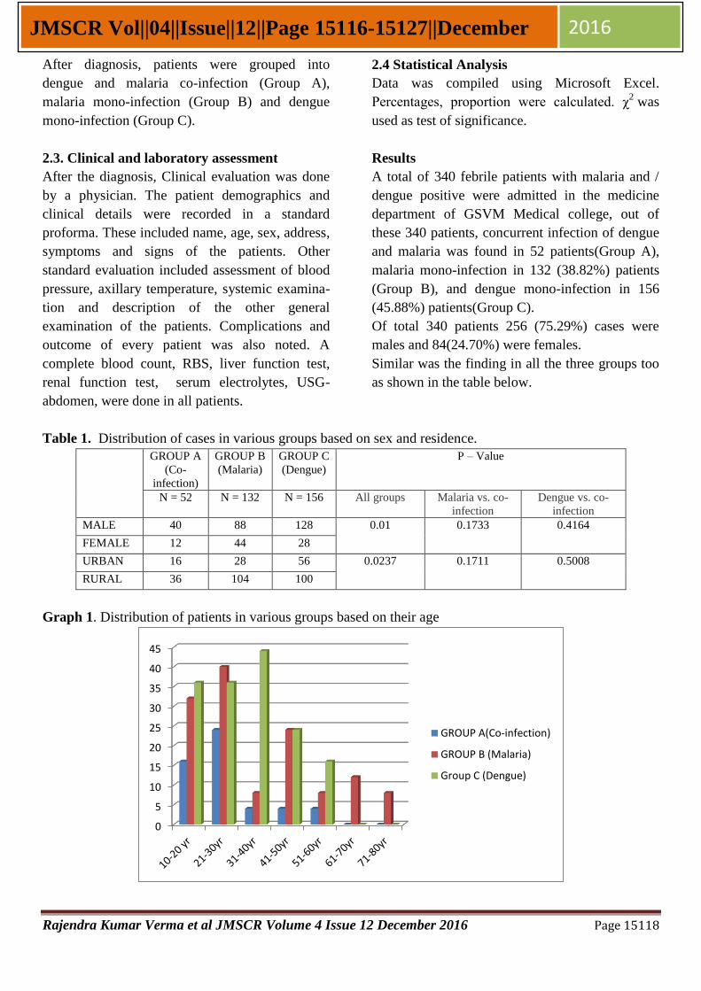
Table 1. Distribution of cases in various groups based on sex and residence.
GROUP A
(Co-
infection)
GROUP B
(Malaria)
GROUP C
(Dengue)
P – Value
N = 52 N = 132 N = 156 All groups Malaria vs. co-
infection
Dengue vs. co-
infection
MALE 40 88 128 0.01 0.1733 0.4164
FEMALE 12 44 28
URBAN 16 28 56 0.0237 0.1711 0.5008
RURAL 36 104 100
Graph 1. Distribution of patients in various groups based on their age
0
5
10
15
20
25
30
35
40
45
GROUP A(Co-infection)
GROUP B (Malaria)
Group C (Dengue)

Rajendra Kumar Verma et al JMSCR Volume 4 Issue 12 December 2016 Page 15119
JMSCR Vol||04||Issue||12||Page 15116-15127||December 2016
100 (29.41%) cases were from urban area while
240 (70.58%) were from rural areas. In all the
three groups [A= 36(69.23%), B= 104(78.78%),
C= 100(64.10%)] majority of the patients hailed
from rural areas.
Out of 340 patients fever was present in all
patients (100%) followed by headache in 67.05%
and body ache in 48.23% of total patients. Fever
was the chief presentation in all the groups A
[52(100%)], B [132(100%)] and C [156(100%)]
with a mean duration of 7.18+/- 3.91, 8.09+/-4.4,
and 5.26+/-3.49 days in group A, B and C
respectively.
Headache was most common in patients with
group A 44(80.00%) followed by group C
120(76.92) whereas it was least in group B
(malaria) 64(48.48). This difference between
group A and B is statistically significant(p=
.00007) while that in between group A and C (p=
.2394) is comparable .Generalized body ache
symptom was presenting complaint in 82.05% of
group C patients followed by 46.15% cases of
group A and 9.09% of group B patients. Pain in
the joints was present in group C (61.53%)
patients followed by group A (15.3%) patients. No
patients of group B had joint pain.
Bleeding manifestations in the form of purpuric
rashes over skin, melaena, epistaxis were present
in 8 (15.38%) cases of co infection, 4(3.03%)
cases of malaria (Grp B)1 and 32(20.51%) cases
of dengue (Grp C). Bleeding manifestations were
significantly more in group A as compared to
group B (p = 0.0048) but was not different
significantly with group C (p= 0.5428), hence,
comparable with dengue.
Table 2. Clinical features of various groups in our study.
GROUP
A(Co-
infection)
%
GROUP
B
(Malaria)
%
GROUP
C
(Dengue)
%
P value
N = 52 N = 132 N = 156 A x B A x C
FEVER 52 132 156 100 - -
HEADACHE 44 80 64 48.48 120 76.92 0.00007 0.23949
BODYACHE 24 46.15 12 9.09 128 82.05 0.00001 0.00001
JOINT PAIN 8 15.38 0 0 96 61.53 - 0.00001
ABDOMINAL SYMPTOM
AND SIGN
20 38.46 32 24.24 64 46.03 0.0000 0.8705
BLEEDING 8 15.38 4 3.03 32 20.51 0.0048 0.5428
CNS Altered Sensorium 0 0 12 9.09 2 1.28 0 0
HYPOTENSION 3 5.35 4 3.03 28 17.59 0.381865 0.032697
REPIRATORY DISTRESS 4 7.69 20 15.15 4 2.56 0.2688 0.095846
Abdominal symptoms and sign in the form of
nausea and vomiting, pain abdomen, hepatosple-
nomegaly was present maximum in Group C
(46.03%) followed by Group A (38.46%) and then
Group B (24.24%). These abdominal findings
were comparable between co infection and dengue
group.
Out of 340 patients, 35 (10.29%) cases were in
hypotension (BP < 90/60 mmHg) on admission.
Of these 28 patients were from group C followed
by 4 cases in Group B and 3 cases in Group A.
Respiratory distress in the form of acute lung
injury or ARDS was present in 28 patients. Of
these maximum cases were in Group B (20)
followed by group A(4) and C(4). This difference
was statistically not significant.
Enchephalitis due to dengue virus is rare but there
are case reports which have documented this

Rajendra Kumar Verma et al JMSCR Volume 4 Issue 12 December 2016 Page 15120
JMSCR Vol||04||Issue||12||Page 15116-15127||December 2016
finding. In our study also Enchepahalitis was seen
in 1.28% cases of Group C whereas Cerebral
malaria was present in 12(9.09%) of Group B
cases. Of these three patients expired.
Laboratory Parameters
Table 3 shows various haematological
abnormalities in different groups. Out of 340 total
cases, severe anemia (i.e. haemoglobin <7mg %)
was seen in 40(11.76%) patients. Severe anemia
was seen in 23.07% cases of Group A, 15.15%
cases of Group B, and only 5.12% cases of Group
C cases. Hematocrit levels were low in group A
and B whereas it was elevated in group C.
Table 3. Hematological parameters of cases in our study.
GROUP A(Co-
infection)
GROUP B (Malaria)
Group C (Dengue)
N = 52 % N = 132 % N = 156 %
Duration of fever 7.18+/- 3.91 8.09+/-4.4 5.26+/-3.49
Heamoglobin
<7mg/dl 12 23.07 20 15.15 8 5.12
7.0 - 10
mg/dl
28 53.84 60 45.45 34 21.79
>10mg/dl 12 23.07 52 39.4 114 73.07
Hematocrit (%)
<20 8 15.38 16 12.12 4 2.56
20 - 40 40 76.92 92 69.69 56 35.89
>40 4 7.69 24 18.18 96 61.53
TLC (No./mm3) 6661+/- 2842 9189+/-5605 5000+/-2719
Platelets
(No./mm3)
20000 -
50000
20 38.46 48 36.36 108 82.05
50001-
100000
28 53.85 44 33.34 40 25.64
100001 -
150000
4 7.69 32 24.24 8 5.12
>150000 - - 8 6.06 - -
Thrombocytopenia was present in all the groups.
Out of 340 patients, 332 patients (i.e.97.64%) had
thrombocytopenia (platelet count <150000/µL). It
was present in all the patients of Group A and C
while in group B it was seen in 93.93% cases.
Severe thrombocytopenia (<50000) was most
commonly seen in dengue monoinfection
group(C)(82.05%), while platelet count between
50000 – 100000 was mostly seen in Group A
(53.85%). Mean leucocyte count in Group A
(6661+- 2842) was lesser than Group B (9189+-
5605) but higher than Group C (5000+-2719).
There were no other important differences in the
haematological profile.
Rajendra Kumar Verma et al JMSCR Volume 4 Issue 12 December 2016 Page 15121
JMSCR Vol||04||Issue||12||Page 15116-15127||December 2016
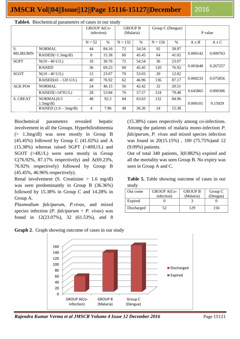
Table4. Biochemical parameters of cases in our study
GROUP A(Co-
infection)
GROUP B
(Malaria)
Group C (Dengue)
P value
N = 52 % N = 132 % N = 156 % A x B A x C
S.
BILIRUBIN
NORMAL 44 84.16 72 54.54 92 58.97
0.000142
0.000763 RAISED(>1.3mg/dl) 8 15.38 60 45.45 64 41.02
SGPT N(10 - 40 U/L) 16 30.76 72 54.54 36 23.07
0.003648
0.267257 RAISED 36 69.23 60 45.45 120 76.92
SGOT N(10 - 40 U/L) 12 23.07 70 53.03 20 12.82
0.000233
0.075856 RAISED(41 - 120 U/L) 40 76.92 62 46.96 136 87.17
ALK PO4 NORMAL 24 46.15 56 42.42 32 20.51
0.645865
0.000306 RAISED(>147IU/L) 28 53.84 76 57.57 124 79.48
S. CREAT NORMAL(0.5 -
1.5mg/dl)
48 92.3 84 63.63 132 84.96
0.000101
0.15929
RAISED (1.6 – 3mg/dl) 4 7.96 48 36.36 24 15.38
Biochemical parameters revealed hepatic
involvement in all the Groups. Hyperbilirubinemia
(> 1.3mg/dl) was seen mostly in Group B
(45.45%) followed by Group C (41.02%) and A
(15.38%) whereas raised SGPT (>40IU/L) and
SGOT (>4IU/L) were seen mostly in Group
C(76.92%, 87.17% respectively) and A(69.23%,
76.92% respectively) followed by Group B
(45.45%, 46.96% respectively).
Renal involvement (S. Creatinine > 1.6 mg/dl)
was seen predominantly in Group B (36.36%)
followed by 15.38% in Group C and 14.28% in
Group A.
Plasmodium falciparum, P.vivax, and mixed
species infection (P. falciparum + P. vivax) was
found in 12(23.07%), 32 (61.53%), and 8
(15.38%) cases respectively among co-infections.
Among the patients of malaria mono-infection P.
falciparum, P. vivax and mixed species infection
was found in 20(15.15%) , 100 (75.75%)and 12
(9.09%) patients.
Out of total 340 patients, 3(0.882%) expired and
all the mortality was seen Group B. No expiry was
seen in Group A and C.
Table 5. Table showing outcome of cases in our
study
Out come GROUP A(Co-
infection)
GROUP B
(Malaria)
Group C
(Dengue)
Expired 0 3 0
Discharged 52 129 156
Graph 2. Graph showing outcome of cases in our study
0
20
40
60
80
100
120
140
160
GROUP A(Co-infection)
GROUP B (Malaria)
Group C (Dengue)
Discharged
Expired
Rajendra Kumar Verma et al JMSCR Volume 4 Issue 12 December 2016 Page 15122
JMSCR Vol||04||Issue||12||Page 15116-15127||December 2016
Discussion
In our study we found a high (25%) prevalence of
the co-infection, among those with dengue. In
Pakistan, however, the prevalence found was as
high as 23.2% [18]
. In Brazil, a prospective study
performed in 2009 on 132 patients with vivax
malaria found 11 co-infected and the prevalence
was 8.3% [19]
. In the French Guiana, the
prevalence of co-infection was 7.1% (17 of 238)
among patients with dengue [11]
. Thus, the
prevalence of co-infection may fluctuate,
depending on local endemicity. In these studies,
the prevalence was estimated on hospitalized
patients; therefore it could not be extrapolated to
the community-based level.
In the present study, P. Vivax was present in
majority of the cases (61.53%). However, P.
falciparum and mixed (P. vivax and P.
falciparum) infection was found in 12 and 8 cases
respectively. Similarly Plasmodium vivax was the
common species found in both French Guiana [10]
and Pakistan [18]
. In the former study P. vivax was
found in 63.9% and P. falciparum in 33.8% cases
where as in the latter P. vivax was found in 96.2%
(25 of 26) cases. This is due to the species
prevalence in a particular locality.
In our study co-infected group followed similar
pattern [like male predominance, 21- 30 years as
commonest age group, and maximum cases from
rural areas] to that of both mono-infection and this
finding is statistically similar.
The clinical features of concurrent infection had a
mixed presentation of both dengue mono-infection
and malaria mono-infection. Therefore clinically it
is difficult to diagnose concurrent dengue and
malaria. Features like headache with retroorbital
pain, abdominal symptoms (pain, nausea and
vomiting) and bleeding manifestation were
statistically similar to dengue mono-infection
whereas hypotension, respiratory symptoms were
more like malaria mono-infecton.
Symptom analysis of all the patients showed that
fever was the commonest symptom (100%) in co-
infected group and both mono-infection groups.
The duration of fever was longer in Co- infected
patients and they were hospitalized more
frequently than dengue patients but lesser than
malaria mono-infection group. That means that a
patient with the diagnosis of dengue presenting
with prolonged evolution should raise the
suspicion of malaria co-infection. Almost Similar
findings were seen in studies by M.K. Mohapatra
et al [7]
Abbasi A et al [5]
. Loïc Epelboin et al [11]
also has similar finding but differs in that the
duration of fever in co-infected group was higher
than both the groups.
Bleeding is reported as an infrequent finding in
malaria, despite common platelet depletion [20] [21]
.
Conversely, bleeding is the most feared
complication of dengue fever, where in addition to
platelet depletion, virus-induced endothelial and
liver injury concur to the risk of coagulopathy [22] [23] [24]
. In our study severe thrombocytopenia
(platelets count <20,000/mm3) was predominantly
seen in dengue mono-infection group whereas it
was similar in both malaria mono-infection and
co infection group, but bleeding manifestation
was uncommon in malaria mono-infection group,
and was significantly present in the form of rash
and maleana in both co infected and dengue
mono-infection group. This finding is in
accordance to several studies [5] [7] [11] [13]
. This
suggests a possible synergistic pathogenic
mechanism, which could be related to both
capillary fragility and coagulation disorders.
Headache in co infected patients followed the
pattern similar to dengue mono infection group. It
was moderate to severe in intensity associated
with retro orbital pain in majority of the patients.
Arthralgia was present in few patients of co
infected group as compared to dengue mono
infection group while not a single patient in
malaria mono-infection group had arthralgia.
Hypotension, a well known clinical feature in
dengue shock syndrome was present chiefly in
17.59% of dengue mono-infection group but it
was found only in 5.35% of co infected patients.
This feature in co infected patients was
comparable to that of malaria mono-infection.

Rajendra Kumar Verma et al JMSCR Volume 4 Issue 12 December 2016 Page 15123
JMSCR Vol||04||Issue||12||Page 15116-15127||December 2016
Dyspnoea was frequent in all groups, particularly
in malaria followed by co-infected patients.
Dyspnoea is an early clinical feature of plasma
leakage and, in dengue, may be due to fluid
accumulation in the pleural cavity [25] [26]
. In
malaria, dyspnoea is due to acute lung injury
leading to acute lung edema [27]
. which is one of
the severity criteria for falciparum malaria [28]
. In
a study conducted in Timor East, one patient co-
infected with falciparum malaria and dengue
presented respiratory distress with radiographic
findings compatible with the presentation of acute
lung injury [29]
. The clinical management of these
cases is difficult, as the inappropriate fluid therapy
for dengue treatment may induce fluid overload
and large fluid effusion to the lungs.
CNS involvement was not found in any patient of
co infected group whereas CNS involvement in
the form of cerebral malaria is one of the
severities not only for falciparum [28]
malaria but
there are reports in which p. vivax too is
presenting with such severe clinical features [32-35]
.
In our study 9.09% patients with malaria mono-
infection presented with cerebral malaria out
which, 3 (0.882%) patients expired. Dengue
encephalopathy is a well-recognized entity with
the incidence ranging from 0.5 to 6.2 % [30] [43]
.
The possible mechanisms are deranged electrol-
ytes, liver dysfunction (hepatic encephalopathy),
cerebral hypo perfusion (shock), cerebral edema
(vascular leak), and intracranial bleeding due to
thrombocytopenia or coagulopathy, which is
secondary to hepatic dysfunction [31]
. There are
subsets of patients in whom the cause for
neurological injury remains unknown even after
excluding the above-mentioned indirect
mechanisms.
Anaemia was more frequent in patients with dual
infection. Haematological profile reveal that
severe anemia was present both in co infected
group and malaria mono-infection group to a
significant extent (23.05% and 15.15%
respectively) whereas only 5.12% patients with
dengue mono-infection were present with severe
anemia (Hb < 7 mg/dl). Similar finding was
reported in a study from Pakistan in which co
infected and malaria group patient had lower
haemoglobin as compared to dengue mono-
infection group [5][11]
. Similarly Haematocrit levels
were lower in co infected and malaria group
where as it was elevated in dengue mono-infection
patients due to plasma leakage syndrome [25]
. An
explanation for this fact can be attributed to
malaria-induced anemia, a common complication
in vivax malaria [36]
. For this reason, the malaria
clinical manifestation may be a confounder for
health care professionals during the interpretation
and application of dengue severity criteria, in
areas where both diseases occur. The proper
clinical management of co-infected patients may
be compromised due to delay in investigations or
misinterpretation, and inappropriate treatment
may result in fatal complications [37] [38]
.
In our study, moderate elevation of
aminotransferase was seen in all the three groups
including co infected group and this elevation in
co infected group was comparable to that of
dengue mono-infection group. Hyperbilirubinemia
(>1.3mg/dl) was predominantly present in malaria
mono-infection group followed by dengue mono-
infection group and least in co infected group.
Hyperbilirubinemia (>1.3mg/dl) was predomin-
antly present in malaria mono-infection group
followed by dengue mono-infection group and
least in co infected group. These findings suggest
that hepatic cell injury rather than cholestatic
injury is the predominant mechanism involved in
the pathology of co infected infection.
Acute renal failure is one of the commonest cause
in severe malaria patients of our region. In a study
from India, acute kidney injury was seen in 16.6%
patients with P. falciparum and 12.3% patients
with P.vivax [35]
. The possible pathogenic factors
are renal damage through renal hypo perfusion or
endothelial injury through release of various
circulating substances (intravascular haemolysis
and sepsis). As far as dengue is concerned
Lizarraga KJ et al [39]
in his study Dengue-
associated kidney disease concluded that Dengue
infection has been associated with a variety of

Rajendra Kumar Verma et al JMSCR Volume 4 Issue 12 December 2016 Page 15124
JMSCR Vol||04||Issue||12||Page 15116-15127||December 2016
renal disorders. Acute renal failure, hematuria,
proteinuria,, and glomerulonephritis have been
reported during or shortly after acute dengue
infection. It complicates severe dengue infection
in 2-5% of the cases and carries a high mortality
rate [40]
. Studies on renal injury in patients co
infected with dengue and malaria to the best of
our knowledge are very rare; we found only one
case report by Youg K P et al in PubMed [40]
. In
our study renal injury (S.creatinine > 1.5mg/dl)
was seen predominantly in malaria group
(36.36%) followed by dengue mono-infection
group (15.38%) and co infected group (7.96%).
Renal injury in 4 patients with co infection was
comparable to that of dengue mono-infection.
These patients also had hypotension and
respiratory distress. The possible pathogenic
factors might be due to synergistic effect of both
infection resulting in renal damage through renal
hypo perfusion or endothelial injury through
release of various circulating substances.
In our study mortality was seen only in malaria
mono-infection group [3(0.882%)]. Death was due
to multiorgan dysfunction. They had deranged
hepatic, haematological and renal parameters.
Conclusion
In our study we found a very high
prevalence of co-infection which indicates
that it is relatively a common event in our
region.
Patients with the diagnosis of dengue
presenting with prolonged fever, severe
anemia and respiratory distress should
raise the suspicion of malaria co-infection.
Whereas patients with the diagnosis of
malaria presenting with bleeding manifest-
ations, frontal or retro-orbital headache,
arthralgia and / elevated haematocrit level
should raise the suspicion of dengue co-
infection.
Thus, there is a need for clinicians serving
in geographical areas that are endemic to
both dengue and dengue to be vigilant of
co-infection, especially when the patient
presents with atypical clinical features,
unexpected laboratory findings or the
response to treatment is unsatisfactory and
should treated concomitantly for both the
infection.
Rapid diagnostic kits for detecting dengue
or malaria are available and are helpful for
early diagnosis and treatment of the
patients but a single combined rapid
diagnostic kit which will detect both
dengue and malaria is the need of the hour
in our region.
Declarations
Consent: Written Informed consent was taken
from all the patients included in the study.
Acknowledgments: The authors thank Residents
of medicine department and Mr Rahul Singh for
their help in the acquisition of data.
Funding: No Funding Sources.
Competing interests: The authors declare no
competing interests.
References
1. Charrel RN, Brouqui P, Foucault C, de
Lamballerie X: Concurrent dengue and
malaria. Emerg Infect Dis 2005, 11:1153–
1154.
2. Arya CS, Mehta KL, Agarwal N, Agarwal
BK, Mathai G, Moondhara A: Episodes of
concurrent dengue and malaria. Dengue
Bulletin2005, 29:208–209.
3. Manish Bhagat, Sujata Kanhere, Varsha
Phadke, and Riya George: Concurrent
Malaria and Dengue Fever: A Need for
Rapid Diagnostic Methods. J Family Med
Prim Care. 2014 Oct-Dec; 3(4): 446–
448.doi: 10.4103/2249 863.148146
PMCID: PMC4311362.
4. Deresinski S: Concurrent Plasmodium
vivax malaria and dengue. Emerg Infect
Dis 1802, 2006:12.
5. Abbasi A, Butt N, Sheikh QH, Bhutto AR,
Munir SM, Ahmed SM: Clinical features,
diagnostic techniques and management of
Rajendra Kumar Verma et al JMSCR Volume 4 Issue 12 December 2016 Page 15125
JMSCR Vol||04||Issue||12||Page 15116-15127||December 2016
dual dengue and malaria infection. J Coll
Physicians Surg Pak 2009, 19:25–29.
6. Ali N, Nadeem A, Anwar M, Tariq WU,
Chotani RA: Dengue fever in malaria
endemic areas. J Coll Physicians Surg
Pak 2006, 16:340–342.
7. M.K. Mohapatra, P. Patra & R. Agrawala :
Manifestation and outcome of concurrent
malaria and dengue infection . J Vector
Borne Dis 49, Dec 2012, pp. 262–265.
8. Ward DI: A case of fatal Plasmodium
falciparum malaria complicated by acute
dengue fever in East Timor. Am J Trop
Med Hyg2006, 75:182–185.
9. Thangaratham PS, Jeevan MK, Rajendran
R, Samuel PP, Tyagi BK: Dual infection
by dengue virus and Plasmodium vivax in
Alappuzha District, Kerala, India. Jpn J
Infect Dis 2006, 59:211–212.
10. Carme B, Matheus S, Donutil G, Raulin O,
Nacher M, Morvan J: Concurrent dengue
and malaria in Cayenne Hospital, French
Guiana.Emerg Infect Dis 2009, 15:668–
671.
11. Loïc Epelboin, Matthieu Hanf, Philippe
Dussart, Sihem Ouar Epelboin, Félix
Djossou, Mathieu Nacher and Bernard
Carme: Is dengue and malaria co-infection
more severe than single infections? A
retrospective matched-pair study in French
Guiana. Malaria Journal201211:142
DOI: 10.1186/1475-2875-11-142.
12. Dos Santos Santana V, Lavezzo LC,
Mondini A, Terzian AC, Bronzoni RV,
Rossit AR, Machado RL, Rahal P,
Nogueira MC, Nogueira ML:Concurrent
Dengue and malaria in the Amazon
region. Rev Soc Bras Med Trop 2010,
43:508–511.
13. Belisa M. L. Magalhães, André M.
Siqueira, Márcia A. A. Alexandre, Marcela
S. Souza,
João B. Gimaque,Michele S.
Bastos, Regina M. P. Figueiredo,Gisely C.
Melo, Marcus V. G. Lacerda, and Maria P.
G. Mourão: P. vivax Malaria and Dengue
Fever Co-infection: A Cross-Sectional
Study in the Brazilian Amazon. PLoS
Negl Trop Dis. 2014 Oct; 8(10): e3239.
Published online 2014 Oct
23. doi: 10.1371/journal.pntd.0003239.
PMCID: PMC4207662
14. Vitor R R Mendonça, Bruno B Andrade, L
igia C L Souza, Belisa M L Magalhães, M
aria P G Mourão, Marcus V G Lacerda and
Manoel Barral-Netto : Unravelling the
patterns of host immune responses in
Plasmodium vivax malaria and dengue co-
infection. Malaria Journal 201514:315
DOI: 10.1186/s12936-015-0835-8.
15. Chaturvedi UC, Mathur A, Kapoor AK,
Mehrotra NK, Mehrotra RML. Virological
study of an epidemic of febrile illness with
haemorrhagic manifestations at Kanpur,
India, during 1968. Bull World Health
Organ 1970; 43 : 289-93. 23.
16. Chaturvedi UC, Kapoor AK, Mathur A,
Chandra D, Khan AM, Mehrotra RML. A
clinical and epidemiological study of an
epidemic of febrile illness with
haemorrhagic manifestations which
occurred at Kanpur, India in 1968. Bull
World Health Organ 1970; 43 : 281-7
17. Gulati S, Maheshwari A (2007): Atypical
manifestations of dengue. Trop Med Int
Health 12: 1087–1095.
18. Abbasi ABN, Sheikh QH, Bhutto AR,
Munir SM, Ahmed SM (2009): Clinical
features, diagnostic techniques and
management of dual dengue and malaria
infection. J Coll Physicians Surg Pak 19:
25–29.
19. Magalhaes BM, Alexandre MA, Siqueira
AM, Melo GC, Gimaque JB: Clinical
profile of concurrent dengue fever and
Plasmodium vivax malaria in the Brazilian
Amazon: case series of 11 hospitalized
patients. Am J Trop Med Hyg 87: 1119–
1124.
20. Kochar DK, Das A, Kochar A, Middha S,
Acharya J: Thrombocytopenia in

Rajendra Kumar Verma et al JMSCR Volume 4 Issue 12 December 2016 Page 15126
JMSCR Vol||04||Issue||12||Page 15116-15127||December 2016
Plasmodium falciparum, Plasmodium
vivax and mixed infection malaria: a study
from Bikaner (Northwestern India).
Platelets 21: 623–627.
21. Lacerda MV, Mourao MP, Coelho HC,
Santos JB (2011) Thrombocytopenia in
malaria: who cares?Mem Inst Oswaldo
Cruz 106 Suppl 1: 52–63.
22. Mourao MP, Lacerda MV, Macedo VO,
Santos JB (2007) Thrombocytopenia in
patients with dengue virus infection in the
Brazilian Amazon. Platelets 18: 605–612.
23. Alonzo MT, Lacuesta TL, Dimaano EM,
Kurosu T, Suarez LA, et al. (2012) Platelet
apoptosis and apoptotic platelet clearance
by macrophages in secondary dengue virus
infections. J Infect Dis 205: 1321–1329.
24. Costa VV, Fagundes CT, Souza DG,
Teixeira MM (2013) Inflammatory and
Innate Immune Responses in Dengue
Infection: Protection versus Disease
Induction. Am J Pathol 182: 1950–1961.
25. WHO (2009) Dengue: Guidelines for
diagnosis, treatment, prevention and
control. Geneva, Switzerland.
26. Sen MK, Ojha UC, Chakrabarti S, Suri JC
(1999) Dengue hemorrhagic fever (DHF)
presenting with ARDS. Indian J Chest Dis
Allied Sci 41: 115–119.
27. Lomar AV, Vidal JE, Lomar FP, Barbas
CV, de Matos GJ, et al. (2005) Acute
respiratory distress syndrome due to vivax
malaria: case report and literature
review. Braz J Infect Dis 9: 425–430.
28. World Health Organization (2006)
Guidelines for the treatment of malaria.
Geneva, Switzerland.
29. Ward DI (2006): A case of fatal
Plasmodium falciparum malaria
complicated by acute dengue fever in East
Timor. Am J Trop Med Hyg 75: 182–185.
30. Misra UK, Kalita J, Syam UK, Dhole TN :
Neurological manifestations of dengue
virus infection. J Neurol Sci. 2006 May
15; 244(1-2):117-22.
31. Cho Naing, Maxine A. Whittaker, Victor
Nyunt Wai, and Joon Wah Mak.
Is Plasmodium vivax Malaria a Severe
Malaria?: A Systematic Review and Meta-
Analysis. PLoS Negl Trop Dis. 2014 Aug;
8(8): e3071. Published online 2014 Aug
14. doi: 10.1371/journal.pntd.0003071
PMCID: PMC4133404.
32. Singh H, Parakh A, Basu S, Rath B.
Plasmodium vivax malaria: is it actually
benign?. J Infect Public Health. 2011
Jun;4(2):91-5. doi: 10.1016/j.jiph.2011.-
03.002. Epub 2011 May 26.
33. Vivek B. Kute, Hargovind L. Trivedi,
Aruna V. Vanikar, Pankaj R. Shah, Manoj
R. Gumber, Himanshu V. Patel,Jitendra G.
Goswami, and Kamal V. Kanodia.
Plasmodium vivax Malaria–associated
Acute Kidney Injury, India, 2010–2011.
Emerg Infect Dis. 2012 May; 18(5): 842–
845. PMCID: PMC3358071.
34. D. K. Kochar, A. Das, S. K. Kochar et al.,
“Severe Plasmodium vivax malaria: a
report on serial cases from Bikaner in
northwestern India,” American Journal of
Tropical Medicine and Hygiene, vol. 80,
no. 2, pp. 194–198, 2009.
35. Verma RK, Giri R, Singh N,Verma S,
Srivastav V : A Study ON Clinical
Presentation and Outcome of Malaria from
an Underreported, P.vivax Predominant
Region of North India. Sch. J. App. Med.
Sci., 2016; 4(1C):233-243.
36. Costa AP, Bressan CS, Pedro RS, Valls-
de-Souza R, Silva S, et al. (2010) Delayed
diagnosis of malaria in a dengue endemic
area in the Brazilian extra-Amazon: recent
experience of a malaria surveillance unit in
state of Rio de Janeiro. Rev Soc Bras Med
Trop 43: 571–574.
37. Ward DI (2006) A case of fatal
Plasmodium falciparum malaria
complicated by acute dengue fever in East
Timor. Am J Trop Med Hyg 75: 182–185.

Rajendra Kumar Verma et al JMSCR Volume 4 Issue 12 December 2016 Page 15127
JMSCR Vol||04||Issue||12||Page 15116-15127||December 2016
38. Simmons CP, Farrar JJ, Nguyen vV, Wills
B (April 2012). "Dengue". N Engl J
Med366 (15): 1423–32. doi:10.1056/NEJ-
Mra1110265.PMID 22494122.
39. Lizarraga KJ, Nayer A: Dengue-associated
kidney disease. J Nephropathol. 2014;3
(2):57-62. doi: 10.12860/jnp.2014.13.
Epub 2013 Dec 28. Review. PMID:
24772398.
40. Yong KP, Tan BH, Low CY: Severe
falciparum malaria with dengue
coinfection complicated by
rhabdomyolysis and acute kidney injury:
an unusual case with myoglobinemia,
myoglobinuria but normal serum creatine
kinase. BMC Infect Dis.2012 Dec
20;12:364.doi: 0.1186/1471-2334-12-364.
PubMed PMID: 23256803; PubMed
Central PMCID: PMC3557149.
41. Anand AC, Puri P (2005): Jaundice in
malaria. J Gastroenterol Hepatol 20: 1322–
1332.
42. Seneviratne SL, Malavige GN, de Silva HJ
(2006): Pathogenesis of liver involvement
during dengue viral infections. Trans R
Soc Trop Med Hyg 100: 608–614.
43. Gulati S, Maheshwari A (2007): Atypical
manifestations of dengue. Trop Med Int
Health 12: 1087–1095.
44. Varatharaj A: Encephalitis in the clinical
spectrum of dengue infection. Neurol
India. 2010 Jul-Aug; 58(4):585-91.
45. Malaria, National Vector Borne Disease
Control Programme (NVBDCP).
Available from: http://www.nvbdcp.gov.i-
n/malaria3.html. [Last accessed on 2015
Dec 27].