a study of the effects of titanium dioxide nanoparticles
TRANSCRIPT

East Tennessee State UniversityDigital Commons @ East
Tennessee State University
Electronic Theses and Dissertations Student Works
12-2011
A Study of the Effects of Titanium DioxideNanoparticles on the Fluorescent Intensity ofFluorescent Compounds in the Presence of KnownQuenchers.Vivian Dzigbodi KokaEast Tennessee State University
Follow this and additional works at: https://dc.etsu.edu/etd
Part of the Chemistry Commons
This Thesis - Open Access is brought to you for free and open access by the Student Works at Digital Commons @ East Tennessee State University. Ithas been accepted for inclusion in Electronic Theses and Dissertations by an authorized administrator of Digital Commons @ East Tennessee StateUniversity. For more information, please contact [email protected].
Recommended CitationKoka, Vivian Dzigbodi, "A Study of the Effects of Titanium Dioxide Nanoparticles on the Fluorescent Intensity of FluorescentCompounds in the Presence of Known Quenchers." (2011). Electronic Theses and Dissertations. Paper 1384. https://dc.etsu.edu/etd/1384

A Study of the Effects of Titanium Dioxide Nanoparticles on the Fluorescent Intensity of
Fluorescent Compounds in the Presence of Known Quenchers
_____________________
A thesis
presented to
the faculty of the Department of Chemistry
East Tennessee State University
In partial fulfillment
of the requirements for the degree
Master of Science in Chemistry
____________________
by
Vivian Dzigbodi Koka
December 2011
_____________________
Dr. Chu-Ngi Ho, Chair
Dr. Peng Sun
Dr. Peter Zhao
Keywords: Titanium Dioxide, Nanoparticles, Fluorescence, Quenching, Methyl Iodide

2
ABSTRACT
A Study of the Effects of Titanium Dioxide Nanoparticles on the Fluorescent Intensity of
Fluorescent Compounds in the Presence of Known Quenchers
by
Vivian Dzigbodi Koka
Titanium Dioxide is a naturally occurring oxide of titanium. It has a wide range of uses in
commercial products for providing whiteness and opacity. It has photocatalytic properties and
can also be used to produce electricity in its nanoparticles form. This research is focused on
investigating the effect of titanium dioxide nanoparticles in analysis of compounds using
luminescence-based techniques. Quenching, which is one of the basic problems of fluorescent
measurements, was studied in the presence of molecular oxygen and methyl iodide. The rutile
phase of titanium dioxide nanoparticles was synthesized by the acid hydrolysis of titanium
isobutoxide at low temperatures with nitric acid. The crystalline powder was dissolved at
different concentrations and used to monitor the fluorescence intensities of carbazole, pyrene,
and fluoranthene in the presence of methyl iodide and oxygen. Quenching by molecular oxygen
was studied by comparing the fluorescence intensities of compounds with and without degassing
the solutions. Titanium Dioxide was found to exhibit interesting effects on the fluorescent
intensities of these compounds in the presence of quenchers.

3
DEDICATION
To God Almighty who made this possible, and to my beloved family.

4
ACKNOWLEDGEMENTS
My sincere thanks and appreciation goes to Dr. Chu-Ngi Ho for his guidance and
encouragement throughout this project and the entire graduate school experience.
I would also like to thank Dr. Peter Zhao and Dr. Peng Sun for serving on my committee.
I am also grateful to Dr. Yu-Lin Jiang for letting me use his fluorometer when ours was
down, and the entire faculty and staff of the chemistry department for their support during my
graduate studies.
I thank God for the continuous support of my parents and my husband, whose friendship
and encouragement has brought me this far.
Finally, I am grateful to Dr. Nyarambi and his family of the College of Education ETSU
for their mentorship. To all friends and course mates who have contributed in various ways to
make this dream a reality, I say God richly bless you all.

5
CONTENTS
Page
ABSTRACT .................................................................................................................................... 2
DEDICATION ................................................................................................................................ 3
ACKNOWLEDGEMENTS ............................................................................................................ 4
CONTENTS .................................................................................................................................... 5
LIST OF TABLES .......................................................................................................................... 8
LIST OF FIGURES ...................................................................................................................... 10
CHAPTER
1. INTRODUCTION ................................................................................................................... 12
Fluorescence .......................................................................................................................... 12
Phosphorescence .................................................................................................................... 13
Chemiluminescence and Bioluminescence ........................................................................... 13
Types of Fluorescence Quenching ............................................................................................ 16
Collisional or Dynamic Quenching ....................................................................................... 16
Static Quenching.................................................................................................................... 17
Comparism of Dynamic and Static Quenching ..................................................................... 17
Concentration Quenching ...................................................................................................... 18
Heavy Atom Substituent Effects ........................................................................................... 18
Dark Chemical Quenchers ..................................................................................................... 19
Types and Applications of Dark Quenchers .......................................................................... 19
Mechanism of Energy Transfer Quenching ........................................................................... 20
Oxygen Quenching ................................................................................................................ 21
Other Quenchers .................................................................................................................... 21
Applications of Fluorescence Quenching ................................................................................. 22
6
Iodide ..................................................................................................................................... 22
Oxygen................................................................................................................................... 23
Acrylamide ............................................................................................................................ 23
Some Recent Applications of Fluorescence Quenching ........................................................ 24
2. EFFECTS OF SELECTED SUBSTANCES ON LUMINESCENCE .................................... 26
Surfactants and Micellar Mediums ........................................................................................ 26
Beta- Cyclodextrins Effects on Luminescence ...................................................................... 27
Titanium Dioxide Nanoparticles ........................................................................................... 28
Applications and Uses of Titania ........................................................................................... 28
Luminescence Characteristics of Titanium Dioxide ............................................................. 29
New Developments and Future Trends ..................................................................................... 29
Quantum Dots Nanoparticles ................................................................................................. 29
Fullerenes as a Cushion for Nanoparticles ............................................................................ 30
Research Proposal.................................................................................................................. 30
Scope of Research ................................................................................................................. 31
3. EXPERIMENTAL PROCEDURE .......................................................................................... 32
Reagents Used ....................................................................................................................... 32
Synthesis of Rutile TiO2 ........................................................................................................ 32
Preparation of Working Solutions ......................................................................................... 33
Fluorescence Instrumentation ................................................................................................ 33
Data Analysis ......................................................................................................................... 34
4. RESULTS AND DISCUSSION .............................................................................................. 35
Dynamic Quenching of Fluorophores ....................................................................................... 35
Studies of Oxygen Quenching................................................................................................... 39

7
Studies of Fluorescence of Carbazole in Mixture of Methyl Iodide and TiO2 Nanoparticle
Solutions .................................................................................................................................... 48
Studies on Fluorescence of Pyrene in Mixtures of Methyl Iodide and TiO2 Nanoparticles ..... 52
Studies of the Fluorescence of Fluoranthene in Mixture of Methyl Iodide and TiO2
Nanoparticles ............................................................................................................................. 55
CONCLUSION ............................................................................................................................. 59
REFERENCES ............................................................................................................................. 61
VITA ............................................................................................................................................. 67

8
LIST OF TABLES
Table Page
1. Ratios of fluorescent intensities of solutions of carbazole with varying concentrations of
methyl iodide added compared to one without any methyl iodide present............................... 35
2. Ratios of fluorescent intensities of solutions of pyrene with varying concentrations of
methyl iodide added compared to one without any methyl iodide present............................... 37
3. Ratios of fluorescent intensities of solutions of fluoranthene with varying concentrations of
methyl iodide added compared to one without any methyl iodide present............................... 38
4. Fluorescence intensities found in normal and degassed solutions of carbazole with varying
concentrations of methyl Iodide. ............................................................................................... 39
5. Fluorescence intensities found in normal and degassed solutions of carbazole with varying
concentrations of titanium dioxide added. ................................................................................ 41
6. Fluorescent intensities of normal and degassed solutions of pyrene with varying
concentrations of added methyl iodide. .................................................................................... 42
7. Fluorescent intensities obtained on normal and degassed solutions of pyrene with varying
concentrations of titanium dioxide nanoparticles added. .......................................................... 44
8. Fluorescence intensity of normal and degassed solutions of fluoranthene with varying
concentrations of methyl iodide added. .................................................................................... 45
9. Fluorescent intensities obtained from normal and degassed solutions of fluoranthene with
varying concentrations of titanium dioxide nanoparticles added. ............................................ 47
10. Fluorescent intensities of carbazole solutions with varying concentrations of titanium
dioxide and with fixed concentrations of methyl iodide added. ............................................. 49
11. Fluorescence intensities of carbazole solutions with varying concentrations of methyl
iodide containing added fixed concentrations of titanium dioxide nanoparticles................... 50
12. Fluorescent intensities of pyrene solutions with varying concentrations of titanium dioxide
nanoparticles containing fixed concentrations of methyl iodide added. ................................. 52
13. Fluorescent intensities of pyrene solutions with varying concentrations of methyl iodide
containing fixed concentrations of titanium dioxide nanoparticles added. ............................. 54
14. Fluorescent intensities of fluoranthene solutions with varying concentrations of methyl
iodide containing fixed concentrations of titanium dioxide nanoparticles added................... 56

9
15: Fluorescent intensities of fluoranthene solutions with varying concentrations of titanium
dioxide nanoparticles containing fixed concentrations of 2.0 x 10⁻³
M and 2.0 x 10⁻⁵
M
methyl iodide added. ............................................................................................................... 57

10
LIST OF FIGURES
Figure Page
1. Stern-Volmer plot for carbazole showing the ratios of fluorescence intensity in the absence
(If0) and presence (If) of methyl iodide. .................................................................................... 36
2. Stern-Volmer plot for pyrene showing the ratios of fluorescence intensity in the absence
(If0) and presence (If) of varying concentrations of methyl iodide. .......................................... 37
3. Stern-Volmer plot of the ratios of the fluorescence intensities for fluoranthene in the
absence (If0) and presence (If) of methyl iodide. ....................................................................... 38
4. Plots showing the effect of methyl iodide of varying concentrations on the fluorescence
intensity of normal (blue) and degassed (red) solutions of carbazole. .................................... 40
5. Plots of the intensities of normal (blue) and degassed (red) solutions of carbazole with
varying concentrations of titanium dioxide nanoparticles added. ............................................ 41
6. Plot of fluorescent intensities of normal (blue) and degassed (red) solutions of pyrene with
varying concentrations of added methyl iodide. ....................................................................... 43
7. Plots of fluorescent intensities obtained on normal (blue) and degassed (red) solutions of
pyrene with varying concentrations of titanium dioxide nanoparticles added. ........................ 44
8. Plot showing the fluorescence intensity of solutions of normal (blue) and degassed (red)
solutions of fluoranthene with varying concentrations of methyl iodide added. ...................... 46
9. Plots of Fluorescent intensities obtained from normal (blue) and degassed (red) solutions
of fluoranthene with varying concentrations of titanium dioxide added. ................................. 47
10. Structure of Carbazole ............................................................................................................ 48
11. Plots of fluorescence intensities obtained from carbazole solutions with varying
concentrations of titanium dioxide and with 2.0 x 10⁻³
M (blue) and 2.0 x 10⁻⁶
M (red) methyl
iodide added. ........................................................................................................................... 49
12. Fluorescent intensities of carbazole solutions with varying concentrations of methyl
iodide containing fixed concentrations of 2.0 x 10⁻⁷
M (red) and 2.0 x 10⁻⁴
M TiO2 (blue)
added. ...................................................................................................................................... 51
13. Structure of Pyrene ................................................................................................................. 52
14. Plots of the fluorescence intensities of solutions of pyrene with varying concentrations of
titanium dioxide with 2.0 x 10⁻⁵
M (red) and 2.0 x10⁻³
M (blue) of methyl iodide added. .... 53

11
15. Fluorescent intensities of pyrene solutions with varying concentrations of methyl iodide
containing fixed concentrations of 2.0 x 10⁻⁷
M (red) and 2.0 x 10⁻⁴
M TiO2 nanoparticles
(blue) added. ........................................................................................................................... 54
16. Structure of Fluoranthene ....................................................................................................... 55
17. Fluorescent intensities of fluoranthene solutions with varying concentrations of methyl
iodide containing fixed concentrations of 2.0 x 10⁻⁷
M (red) and 2.0 x 10⁻⁴
M TiO2
nanoparticles (blue) added. ..................................................................................................... 56
18. Plots of the fluorescence intensity of solutions of fluoranthene with varying
concentrations of titanium dioxide nanoparticles with 2.0 x 10⁻⁵
M (red) and 2.0 x 10⁻³
M
(blue) of methyl iodide added. ................................................................................................ 58
12
CHAPTER 1
INTRODUCTION
Luminescence is the name given to three related optical methods: fluorescence,
phosphorescence, and chemiluminescence. Luminescence techniques are widely used in the
analysis of many compounds in recent years. Their diverse application in other fields has made
luminescence methods almost indispensible. Luminometry has several advantages over other
analytical techniques. Excellent sensitivity and selectivity makes luminescence very popular.
Luminescence is 100,000 more sensitive than absorption spectroscopy. A wide dynamic range
and inexpensive instrumentation are also some of features that make this technique highly
recognized. The technique is classified according to the means by which energy is supplied to
excite the luminescent molecule. This technique is hence an excellent way to study unique
properties of compounds.
Fluorescence
Molecules excited by interactions with photons of electromagnetic radiation give rise to
fluorescence. This normally occurs when the release of electromagnetic radiation is from the
singlet energy state. Every fluorescent molecule has two distinctive spectra; the excitation
spectrum and the emission spectrum. The fluorescent spectrum of a compound results from the
reemission of radiation absorbed by the molecule. Fluorometric methods can detect
concentration of substances as low as one part in 10 billion, a sensitivity 1000 times greater than
most spectrophotometric methods. High sensitivity is however a result of direct measurement of
emitted radiation. Hence a zero background signal is measured, which is not the cases in
spectrophotometric methods, as their measurements are done indirectly as a difference between
incident and transmitted beams. Fluorescence measurements are also very specific. This is
mainly because very few compounds fluoresce compared to absorbing ones. This is however
because not all compounds that absorb radiation essentially emit (1). Fluorescence is a short-
lifetime phenomenon with its emission occurring between the same states with similar
multiplicities. The primary demerit of fluorescence as an analytical technique is its serious
dependence on environmental factors, such as temperature, concentration, and pH. The

13
fluorescent lifetime of most fluorophores is about 10-9
s. Within this range collisional
interactions also occur around 10-4
-10-3
M. Thus collisional deactivation of the excited state is
mostly a competitive process at room temperature. Other competing deactivation processes
mostly known as radiationless transitions also lead to reduction of fluorescence. Vibrational
relaxation, and internal conversion are the results of the collisional interactions that lead to
fluorescence quenching.
Phosphorescence
This is the delayed emission of light after the absorption of light. A change in electronic
spin accompanies phosphorescence, with longer excited state. One unique feature of
phosphorescence is the persistence of the emitted radiation after the excitation beam is removed,
which is not the case with fluorescence. Phosphorescence is hence associated with the
phenomenon of “after glow”. The delayed time scale of re-emission is due to the forbidden
energy state transitions in quantum mechanics. Phosphorescence, just as fluorescence, adds to
one of the wonders of nature. Humans have taken advantage of this wonder and created items
for vital use and entertainment, some of which are glow sticks for parties or matching bands,
highway exist signs, pathway markings, and glow sticks used for military purposes amongst
others (2-4).
Chemiluminescence and Bioluminescence
Production of visible illumination by a chemical reaction is called Chemiluminescence.
Such chemical reactions occurring in living organisms are called bioluminescence. Three
conditions are normally required for Chemiluminescence to occur; the chemical reaction must
release sufficient energy to populate an excited energy state; the reaction pathway must favor the
formation of an excited state complex; and the excited state product must be capable of emitting
a photon itself or transferring its energy to another molecule that can emit Chemiluminescence
occurs when an energetic (exothermic) reaction produces a molecule in an electronically excited
state. That molecule, as it returns to the ground state, releases its energy as a photon of light.
The rate of production of light and concentration of chemiluminescent molecule, often coupled
to concentration also of a catalytic reagent, imposes limits on the amount of time that this
luminescence can be usefully observed from a sample volume. (5) Some samples will generate

14
a relatively bright signal for a short period of time (until the entire chemiluminescent reagent is
used up); others will yield a weaker signal over a longer period. In some occurrences the
excited species is the product of a reaction between an analyte and a suitable reagent, mostly
strong oxidants. These normally result in an emission spectrum characteristic of the oxidation
product of the analyte or the reagent rather than the analyte itself. However, in other instances
the analyte is not directly involved in the reaction. Instead, the analyte inhibits or has a catalytic
effect on the chemiluminescence reaction. Technically the chemical reactions results in an
electronically excited species that emits a photon in order to reach the ground state.
Chemiluminescence and bioluminescence methods are highly sensitive, hence offer important
advantages for chemical analysis. It is very easy to measure low levels of light emission using
these techniques and the calculation of the theoretical detection limits from the efficiency of
chemiluminescence is quite simple.
Currently developed methods for atmospheric ozone and nitric oxide provide a good
example of the advantages of chemiluminescence generating reactions for chemical analysis.
Ozone can be determined either by its reaction with rhodamine-B adsorbed on an activated silica
gel surface or by its gas phase-phase reaction with ethylene. The rhodamine-B method however
has all the advantages of Chemiluminescence that are linear response up to 400ppb, sensitivity
up to less than 1-ppb, simple instrumentation in the form of a gas flow system for pulling sample
over the surface and a photomultiplier tube to measure Chemiluminescence intensity. This
method however needs frequent re-calibration as the sensitivity of the chemiluminescence
surface changes with time as the rhodamine is consumed.
Nitric oxide can be determined determined by involment of ozone in the chemiluminescence
reaction;
NO+O3→NO2* +O2 (1)
NO2* →NO2+hv (2)
As shown in the reactions 1and 2, the total oxides of nitrogen (NO+NO2) is determined by
reducing NO2 to NO with carbon before reacting with ozone. The NO2 concentration is equal is
equal to the difference between the total oxides of nitrogen and the NO concentration.

15
Bioluminescence is an extensively dispersed phenomenon in nature, mostly found in
marine organisms. The most widely studied bioluminescent orgamisms are the firefly, marine
bacteria such as Beneckea harveyi and Photobacterium fischeri, the comb jelly Renilla, and the
jelly fish Aequorea. Studies of other organisms are limited by their availability. Some of these
contain bioluminescent substances that have considerable analytical potentials, for instance the
Pholas dactylus. Their scarcity coupled with their complex nature of extraction and purification
of the active bioluminescent substances has not allowed the serious consideration of such
substances as reagents for routine analysis. Rapid growth in genetic engineering techniques is
leading to the reappraisal of these bioluminescent substances as they could be prepared in large
amounts by such techniques in theory. In the firefly, an enzyme called luciferase triggers a
reaction that energy emitted as light, normally a flashing beacon from the insects lower
abdomen. Fireflies reactions below;
Luciferase+LH2 +MgATP→luciferase: LH2: AMP+Pyrophosphate (3)
Luciferin: LH2AMP+O2 → Oxyluciferin +CO2 +AMP+Light. (4)
In equation 3 and 4, firefly luciferase catalyses the ATP-dependent oxidation of D-
luciferin (LH2). Initially a luciferase:luciferyl adenylate (LH2:AMP) complex is formed and this
reacts with oxygen to produce oxyluciferin(the emitter), carbon dioxide, AMP, and light. Firefly
luciferase is prepared by extraction of firefly tails and purified by crystallization. (6)
Luciferin is also contained in the tails of fireflies but is more conveniently obtained by
chemical synthesis. Both luciferase and Luciferin are available at chemical suppliers.
These light emitting reactions have been explored by chemists in a large number of
laboratories and clinical tests. Some of its applications are in: lights from emergency “light
sticks” used by campers and glowing necklaces seen at concerts and sporting events.
16
Types of Fluorescence Quenching
Fluorescence quenching occurs when the fluorescent intensity of a given substance is
decreased. A variety of processes can result in quenching. Normally it is engineered by the
presence of a competing de-activating process resulting from specific interaction between a
fluorophore and another substance present in the system. The substance that causes this
phenomenon is called a quencher. Different types of quenchers and their mechanisms are
discussed below.
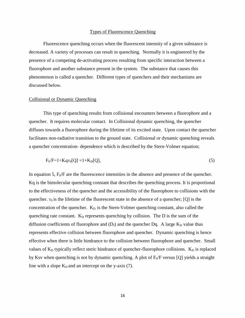
Collisional or Dynamic Quenching
This type of quenching results from collisional encounters between a fluorophore and a
quencher. It requires molecular contact. In Collisional dynamic quenching, the quencher
diffuses towards a fluorophore during the lifetime of its excited state. Upon contact the quencher
facilitates non-radiative transition to the ground state. Collisional or dynamic quenching reveals
a quencher concentration- dependence which is described by the Stern-Volmer equation;
F0/F=1+Kqт0[Q] =1+KD[Q], (5)
In equation 5, F0/F are the fluorescence intensities in the absence and presence of the quencher.
Kq is the bimolecular quenching constant that describes the quenching process. It is proportional
to the effectiveness of the quencher and the accessibility of the fluorophore to collisions with the
quencher. т0 is the lifetime of the fluorescent state in the absence of a quencher; [Q] is the
concentration of the quencher. KD is the Stern-Volmer quenching constant, also called the
quenching rate constant. KD represents quenching by collision. The D is the sum of the
diffusion coefficients of fluorophore and (Df) and the quencher Dq. A large KD value thus
represents effective collision between fluorophore and quencher. Dynamic quenching is hence
effective when there is little hindrance to the collision between fluorophore and quencher. Small
values of KD typically reflect steric hindrance of quencher-fluorophore collisions. KD is replaced
by Ksv when quenching is not by dynamic quenching. A plot of F0/F versus [Q] yields a straight
line with a slope KD and an intercept on the y-axis (7).

17
Static Quenching
This quenching occurs as a result of the formation of a non-fluorescent ground state
complex between the fluorophores and the quencher. Upon absorption of radiation this complex
returns to the ground state almost immediately without the emission of a photon. The association
constant for the quencher-fluorophore complex describes the effectiveness of a static quencher.
The association constant has an expression:
Ks = [F-Q]/ [F] [Q], (6)
where [F-Q] is the concentration of the non-fluorescent complex and [F] is the concentration of
the uncomplexed fluorophore.
Comparism of Dynamic and Static Quenching
Static and Dynamic quenching are basically distinguished by their fluorescent lifetime
measurements. Temperature is also used to distinguish between these two quenching
mechanisms. Dynamic quenching depends upon diffusion, and because higher temperatures
result in larger diffusion coefficients, the bimolecular quenching constant is expected to increase
with increasing temperatures. Diffusion rate increases with temperature and this leads to an
increase in dynamic quenching at higher temperatures. In contrast however, complex formation
tends to be inversely proportional to temperature. Increased temperature mostly results in
decreased stability of complexes, thus lower values of quenching constants. As a result, static
quenching tends to be higher at lower temperatures (8).
One other unique feature in distinguishing dynamic and static quenching is by careful
examination of the absorption spectra of the fluorophores. Collisional quenching only affects the
excited states of the fluorophores, and thus no change in the absorption spectra are predicted. In
contrast, ground state complex formation frequently results in perturbation of the absorption
spectrum of the fluorophores (9).

18
Concentration Quenching
Absorption is necessary for fluorescence to occur. Hence, fluorescence intensity is
proportional to the molar absorptivity. This implies the more highly absorbing a substance the
greater the fluorescence. However, when absorption is too much, no light can pass through to
cause excitation. Thus, at low concentrations, when the absorbance is less than about 0.05, a
linear relationship exists between fluorescence and concentration. At very high concentrations
of a sample, front-face absorption occurs. In front-face absorption, the portion of the sample
nearest the radiation source absorbs so much radiation due to high number of molecules. The
result is that less radiation is available for the rest of the solution. The incident radiation is thus
attenuated as it goes through the sample towards the viewing area of the cuvet. This leads to a
depression in fluorescence intensity of the sample. This phenomenon is known as Inner filter
effect or concentration quenching. Concentration of solutions should therefore be taken into
consideration in the preparation samples for fluorescence analysis, in order to avoid this
phenomenon.
Heavy Atom Substituent Effects
Research has shown that the halide ions are good chemical quenchers (7, 8). Iodine,
chlorine, and bromine, which are also heavy atom elements, reduce the fluorescent intensity of
fluorophores. This is as a result of intersystem crossing to an excited triplet state, promoted by
spin-orbit coupling of the excited singlet fluorophores and the halogen. Intersystem crossing is
very competitive with the presence of this substituent. The pattern of quenching ability of these
ions was found to be iodine˃bromine˃chlorine, an order which is the same as their ionization
energies (10). This could be attributed to the increase in size of the ions as they go down the
group 7, with the radius of sphere of action getting bigger too, with iodine having the biggest
size, thus more prone to collision, leading to quenching. Quenching by these halide ions are
mostly by dynamic or collisional quenching or energy transfer. Methyl Iodide was used as the
quencher for this research.

19
Dark Chemical Quenchers
Dark chemical quenchers are substances that absorb excitation energy from fluorophores
and dissipate the energy as heat, as opposed to a typical fluorescent quencher that re-emits much
of this energy in the form of light. Dark quenchers are largely used in molecular biology in
conjunction with fluorophores. When a fluorophore and a dark quencher are close together, such
as in a molecule or protein, the fluorophores emission is suppressed. This effect is used to study
molecular geometry and motion (11).
Dark quenchers also normally known as dyes with no native fluorescence, (non-
fluorescent dyes) offer a solution to background noise, caused by quencher fluorescence, due to
overlap between the quencher and the fluorescence spectra (12). They exhibit lower background
fluorescence, causing larger signal-to-noise ratio with great dynamic range. In the absence of
secondary fluorescence arising from a dark quencher, multiple fluorophores can be
simultaneously spectrally resolved, making dark quencher probes amendable to multiplex assays.
In addition, dark quenchers enable multiplexing when two or more reporter-quencher probes are
used together(13).
Types and Applications of Dark Quenchers
Fluorescence resonance energy transfer (FRET) is a highly distance-dependent
interaction between a reporter dye in an excited state and a quencher in its ground state. Energy
is transferred from one molecule (the fluorophore) to the other (the quencher) without the
emission of a photon. In order for efficient FRET quenching to take place: a) the fluorophore
and quencher molecules must be close to each other (approx. 10 - 100 Å) and, b) the absorption
spectrum of the quencher must overlap with the emission spectrum of the fluorophore.
Dark quenchers are mostly used in fluorescent dye-quencher probes requiring
suppression of the dye’s fluorescence under one set of circumstances. Common examples of
dark quenchers are Dabcyl and the three Black Hole Quenchers (BHQ). Taken together, the
absorption spectra of these four dark quenchers span the entire visible range, which provides a
researcher with broad flexibility in choice of fluorescent dye, along with the ability to search a
sample for multiple targets in a multiplex quantitative, polymerase chain reaction (PCR) format.

20
Fluorescent quenchers, such as tetramethyl-6-carboxyrhodamine (TAMRA) dye are typically
used in fluorescence resonance energy transfer (FRET)-based applications. FRET probes
contain a donor (fluorescent dye)-acceptor (fluorescent quencher) pair in close proximity. After
absorbance of light by the donor moiety the donor’s fluorescence emission energy is absorbed
(quenched) by the acceptor moiety and subsequently emitted at the acceptor’s emission
wavelength. The result is a final fluorescence emission at a substantially longer wavelength
than would be expected if only the donor moiety were present. FRET probes thus are useful in
cases where a substantial shift in final emission wavelength is desirable. This type of FRET-
based system is typically used to determine intra- and inter-molecular distances at very high
resolution (1-10 nm). For instance, FRET oligo probes have been used to measure the dynamic
changes in intermolecular distances between tRNAs bound at the A and P sites of ribosomes
during mRNA translation (14). FRET oligo primers have also been used to obtain direct
evidence of strand slippage during in vitro synthesis of poly (dG)-poly (dC) duplexes by the
Kleno exo- fragment of DNA polymerase I (15).
Mechanism of Energy Transfer Quenching
In this, mechanism sometimes known as resonance energy transfer quenching applies.
Energy from an excited state is transferred to an acceptor molecule. The transfer normally
occurs without the emission of a photon, but the process is somehow related to absorbance. The
rate transfer depends on the following processes:
1. Spectral overlap between the emission spectrum of the donor and the absorption
spectrum of the acceptor molecule.
2. The quantum yield of the donor.
3. The relative orientation of the transition dipoles of the donor and acceptor.
4. Distance between donor and acceptor.
Resonance energy transfer is essential during photosynthesis, as light molecules use this
mechanism to transfer collected energy to the photosynthetic reaction centre. It is also applied in
the study of proteins resonance energy quenching is used the measure the distance within or
between molecules. These studies are either carried out with single donor or acceptor functions
or by the use of instruments capable of time resolved measurements.

21
Oxygen Quenching
Oxygen is one of the most notorious of all quenchers. It is a ubiquitous quencher of both
fluorescence and phosphorescence. Dissolved oxygen has a very large diffusion coefficient
especially in polar solvents. The mechanism by which oxygen quenches fluorescence has been
the subject of considerable research (17) and it is clear that contact between the oxygen molecule
and the fluorophores is a requirement for quenching. The studies so far published show that
quenching by oxygen is by diffusion –controlled process in which virtually every collision with
the excited fluorophores is effective in quenching (18). Unsubstituted aromatics are severely
affected by oxygen. The enhancement of intersystem crossing by oxygen is the basic route of
oxygen quenching. Oxygen is also an effective triplet state quencher because oxygen gas is
triplet in its ground state. Fluorescence intensity is enhanced with the removal of oxygen. This
is achieved by bubbling an inert gas through the solution for about 5-10 minutes or by a freeze-
thaw cycle. In this study helium gas was bubbled through the sample solution in order to study
the extent of oxygen quenching of the fluorophores (19).
Other Quenchers
Aromatic and aliphatic amines are efficient quenchers of most unsubstituted aromatic
hydrocarbons (20). Anthracene fluorescence is for example effectively quenched by
diethylaniline. The mechanism of quenching is based on the formation of a charge transfer
complex. The excited state fluorophores accepts an electron from the amine. In nonpolar
solvents fluorescence emission from the charge transfer complex (exciplex) is frequently
observed, and this process is often regarded as an excited state reaction rather than quenching. In
polar solvents however the exciplex emission is often quenched so that the fluorophores-amine
interaction appears to be that of simple quenching (21). Other collisional quenchers include
hydrogen peroxide, acrylamide, BrO-4, I
-, nitrous oxide, nitromethane, and some olefins. Many
halogen-containing substances act as collisional quenchers. Examples of such are chloroform,
trichloretanol, bromobenzene, methylmercuric chloride, and a variety of substances containing
more than a chorine atom. Indole, carbazole, and their derivatives(22) are uniquely sensitive to
quenching by chlorinated hydrocarbons and by electron scavengers such as protons, histidine,
cysteine, NO-3, fumarate, Cu
2+, Pb
3+, Cd
3+, and Mn
2+. Indole, tryptophan, and its derivatives are

22
quenched by succinimide, dichloroacetamide, pyridinium hydrochloride, imidazolium
hydrochloride, methionine, Eu3+
, Ag+, and Cs
+. A variety of quenchers are hence essential in the
study of protein fluorescence, especially regarding the surface accessibility of tryptophan
residues and the permeation of proteins by the quenchers. The last but not the least group of
quenchers includes purines, pyrimidines, N-methyl-nicotinamide, N-alkyl pyridinium, and
picolinium salts. An example is the fluorescence of both flavin adenine dinucleotide(FAD) and
reduced nicotinamide adenine dinucloetide (NADH) are quenched by the adenine moiety. Flavin
fluorescence is quenched by both static and dynamic processes, whereas the quenching of
dihydronicotinamide appears to be primarily dynamic. These aromatic substances appear to
quench by the formation of charge-transfer complexes. Depending however upon the structure
involved, the ground state complex can be reasonably stable. Hence both dynamic and static
quenching are frequently observed. Because most substances act as quenchers, fluorophore-
quencher combinations are identified for a desired purpose. Thus the occurrence of quenching
depends on the mechanism, which in turn depends on the structure of the individual molecules
(23).
Applications of Fluorescence Quenching
Quenching of fluorescence has been explored in biochemical and bioanalytical research
(24). In the study of proteins and other macro-bimolecules, some of the quenchers that have
been tried and found useful for bioanalytical studies are iodide, oxygen, and acryl amide. A
great number of quenchers are still being studied for their usefulness.
Iodide
Iodide and its applications in quenching tryptophan fluorescence using model
compounds and the protein lysozyme have been studied by Lehrer, a biochemist (25). He found
iodide to be an efficient dynamic quencher due to its size and charge. In the protein studied,
iodide was found to quench mainly the surface tryptophanyl fluorescence. Iodide is very
sensitive to local charge densities. Hence the nature of the microenvironment can be estimated
because the presence or absence of charged groups in the vicinity of the fluorophore can affect
the local concentration of iodide and hence alter the quenching efficiency. The viscosity of the
microenvironment can be estimated for dynamic quenching because the viscosity of the solvent

23
influences the quenching rate by affecting the diffusion rate of quenchers towards the
fluorophore. Diffusion within the protein shows less dependence on the viscosity of the
environment. Denatured protein is mostly quenched by iodide with 100% efficiency. Hence the
quenching of protein fluorescence by iodide can indicate the degree of exposure and accessibility
of the fluorescent residue. Structural information can be inferred from these data.
Oxygen
Oxygen has been proven to be an efficient quencher of proteins by most researchers (26).
Oxygen, a small, neutral, and polar molecule, has been found to quench dynamically and
effectively with a large quenching constant. Oxygen devoid of charge effects eliminates
uncertainties in its usage where charge effects may play a role in observing fluorescence
quenching. Oxygen in this regard is contrasted with iodide quenching in the interpretation of
quenching data by oxygen of the ethidium bromide DNA complex. This complex contains
positive charged ethidium bromide and negatively charged phosphate groups that can affect the
local concentration of charge-sensitive quenchers due to attraction of opposite charges. Some
researchers have unambiguously concluded that the fluorophore is intercalated and protected by
the helix and that of oxygen must collide with the fluorophore for quenching to occur (27).
Molecular oxygen is an important quencher for many fluorophores because of its high solubility
in aqueous solutions and organic solvents. Fluorescence quenching by molecular oxygen is
usually a diffusion-controlled mechanism (28). Lakowicz and Weber also proposed non-rigid
conformations for proteins. However the rather undergo very rapid fluctuations on a
nanosecond time scale. They hence concluded that oxygen effectively quenches tryptophanyl
residues that were known to be buried deep into the known protein structures. Hence some of
these conformations allow oxygen to diffuse into the proteins.
Acrylamide
The use of acrylamide as a quencher of indole fluorescence was investigated by Eftink
and Ghiron (29). They found that acrylamide is an efficient quencher and quenches by both
dynamic and static quenching. It’s a neutral quencher like oxygen. However it is larger, more
polar, and very soluble aqueous solution. In their evaluation of acrylamide as a quencher, they

24
used an indole-micelle complex to stimulate a simple a simple protein structure. They were able
to obtain information about the microenvironment of the fluorescence probe and the general
structure of the complex. An additional advantage of acrylamide over other neutral and non-
polar quenchers like oxygen and trichloroethanol is that it does not interact with proteins to any
significant extent. In another study of trichloroethanol as a quencher, Eftink et al. (30) found
that tricloroethanol can localize in the polar region of the macromolecules resulting in observable
changes in quenching efficiencies.
Some Recent Applications of Fluorescence Quenching
The myriad applications of fluorescence quenching is still being explored and used in all
areas of analytical chemistry, biomedical, and biochemistry in recent years. Fluorescence
quenching methods have been seen to be useful in obtaining information about conformation and
dynamic changes of proteins in complex macromolecular systems (31).
A study by Limei et al. employed the mechanism of fluorescence quenching for the
detection of antigen. In the study, a highly specific fluoroimmunoassay system for antigen
detection was developed using gold and magnetic nanoparticles (32). The assay used was based
on the fluorescence quenching of fluorescein isothiocyanate resulting from gold nanoparticles
coated with monoclonal antibody. The analytical properties of the gold nanoparticles were
explored by coating the magnetic nanoparticles with anti-α-fetoprotein polyclonal antibodies. A
sandwich-type-immunocomplex was formed, with the α-fetoprotein captured by the magnetic
nanoparticles probes. The supernatant liquid form the immunocomplex containing unbound gold
nanoparticles probes was used to quench the fluorescence. The fluorescence intensity of
fluorescein isothiocyanate observed was at 516 nm, which was proportional to the α-fetoprotein
concentration. The results gave the limit of detection 0.17 nm for the α-fetoprotein. From their
finding Limei et al. concluded on the possibilities of extension of the fluoroimmunoassay system
to detect target molecules with matched antibodies with potential applications in immunoassay
and disease diagnosis.
Fluorescence quenching mechanisms has also been explored by Zhuang et al. (33) to
study protein folding at a single molecule level. In the study titin molecules were used as a

25
model system. Fluorescence resonance energy transfer (FRET) was used to fold titin molecule
with multiple dye molecules attached to a native folded state. Fluorescence from the dye
molecules was quenched in the native state due to the closeness of the dye molecules. The titin
molecule was unfolded after this state which led to a dramatic in the fluorescence intensity. The
folded and unfolded states of the titin molecules were clearly differentiated hence permitted the
measuring of the folding dynamics of the individual titin molecules in real time. Their finding
hence demonstrated the use of fluorescence quenching for signal folding and unfolding of a
small protein with only one immunoglobulin domain.
Another useful application of fluorescence quenching is its use in optical halide sensing
(38). A review article by Chris D. Geddes outlines some of the unique applications of
fluorescence quenching in optical halide sensing. He discusses the versatility of the analytical
technique for the determination of halide ions or organic halides. Some of the advantages of this
technique over other methods such as ion-selective electrode were mentioned in that halide
concentrations can be determined in very small sample volumes with no pre-treatment of
samples required, and measurements can be non-evasive and very quick and sometimes
continuous. The possibility of the determining the halide concentration using either intrinsic or
extrinsic fluorescence probes is another advantage of halide sensing by fluorescence quenching.
The addition of extrinsic probe molecules to livingsystems and some industrial processes is
however not desirable and therefore, for physiological samples, intrinsic fluorescent probes, such
as protein tryptophans or even yellow or green fluorescent proteins, are sometimes preferred. An
alternative to the introduction of extrinsic probes has been to immobilize halide sensitive
fluorophores onto or within a support that, when immersed into the desired sample, readily
allows the diffusion and therefore the sensing of aqueous halide ions, or, in the case of halothane,
gaseous alkyl halide (38).

26
CHAPTER 2
EFFECTS OF SELECTED SUBSTANCES ON LUMINESCENCE
Surfactants and Micellar Mediums
Surfactants have different levels of impact on the fluorescence of compounds, mainly due
to their ionic properties. Surfactants in the form of micelles serve as organized media that have
the tendency of protecting the molecules of a compound from impurities and quenching
characteristics. Micelles have the tendency to arrange solutes into their interior or on their
colloidal surface. When solutes move from aqueous medium to the micellar medium, changes in
several properties such as solubility, reactivity, and spectroscopic characteristics result. These
change in properties lead to increase in fluorescence intensity due to increase in sensitivity. The
relative viscosity of micellar microenvironments also inhibit quenching by molecular oxygen
(40).
Research by Charles Odame Ankra of the chemistry department, under Dr Ho
(June,2009), came out with the finding that surfactants such as CTAB, SDS, and Triton X-100,
singly and in combination with titanium dioxide enhanced the fluorescence of fluorophores, such
as Anthracene and fluoranthene, but they quenched that of carbazole and phenanthrene.
Surfactants are generally known to affect the fluorescence intensity through micelle formation.
Micelles enhance fluorescence by increasing sensitivity, reduction in number of potential
interferences and result in greater experimental convenience (41).
Research by Kim et al. (42), showed that surface modification of CdS and CdMnS
quantum dots by micelles using the reverse-micelle method enhanced the luminescence
properties of the quantum dots. In their findings, before the surface modification a broad
luminescence band from defects was observed for CdS quantum dots. After the modification
however intensity of the band-edge was remarkably enhanced. They also reported an increase in
the intensity of Mn2+
luminescence after its surface was modified by CdMnS quantum dots. A
study of the quantum yield of CdS quantum dots with broadband luminescence also recorded an
increase in quantum yield after treatment with surfactant. The quantum dots were synthesized by

27
classic inverse micelle method using dioctyl sulfosuccinate sodium salt in heptanes. The
quantum yield of CdS quantum dots was doubled to a 20% instead of the 10-13% quantum yield
without the modification (43). Other research by Robert Leif and others concluded that
luminescence of lanthanide ion complexes increased by using the micellar solution medium to
enhance luminescence. The Resonance Energy Transfer Enhanced Luminescence (RETEL)
effect was used as a mechanism of luminescence enhancement (44). Two methods were used to
achieve the enhancement; firstly a complex of a second lanthanide ion was added in a micellar
solution. In the other method a dry preparation by evaporation of a homogeneous solution
containing an added complex of a second lanthanide ion in excess of an unbound antenna ligand
was obtained. Both methods lead to an increase in luminescence by the RETEL method.
Beta- Cyclodextrins Effects on Luminescence
Cyclodextrins are cyclic oligosaccharides obtained from degradation of starch by Bacillus
macerans. They were first isolated in the late nineteenth century. Applications for cyclodextrins
and their derivatives are sought in various areas of chemistry, including the sensing of organic
molecules (45). Their ability to form complexes has lead to its wide array of applications. Beta-
cyclodextrin is added to compounds as complexes to enhance their luminescence. In a study by
Turnball and Walker, beta-cyclodextrin was added to para amino benzoic acid (PABA). The
fluorescence intensity was increased upon complexation of the para amino benzoic acid with
beta-cycodextrin at 298K. This was attributed to the prevention of Collisional deactivation of
the para amino benzoic acid. At a lower temperature of 77K however, the fluorescence intensity
was reduced due to the vibrational deactivation modes available to the complex relative to the
free PABA. The luminescence effects were interpreted as inclusion complexation of the PABA
anion (46).
Another study by Vazquez et al, recorded a substancial enhancement of the fluorescence
emission of aflatoxins by complexation with cyclodextrin. In their study fluorescence properties
of four main aflatoxins; B1, B2, G1, and G2 in solution was investigated alone and in the presence
of various cyclodextrin derivatives. An enhanced fluorescence emission of the aflatoxins with an
unsaturated furan ring (B1 and G1) in the presence of acqueous solutions of α,-β-heptakis-di-O-
methyl-β-cyclodextrin and hydroxypropyl-β-cyclodextrin was observed. The selectivity of the

28
interaction was attributed to the partial involvement of a non-inclusion process because of the
high molar ratio of cyclodextrin to aflatoxin needed for the fluorescence enhancement (47).
Titanium Dioxide Nanoparticles
Titania is an oxide of titanium, TiO2, that occurs naturally. This naturally occurring
oxide can be mined and used as a source of commercial titanium. Titania exists in three basics:
tetragonal rutile (stable at room temperature), tetragonal anatase (metastable for kinetic reasons),
and orthorhombic brookite (not commercially viable). Each of the above forms exhibits
different physical properties such as refraction or chemical or photochemical reactivity that
allows its use in particular applications normally requiring a specific particle size. (48)
Applications and Uses of Titania
Titanium dioxide is widely used white pigment in the world. It’s not toxic and
chemically stable, used mainly to achieve opacity and whiteness of commercial products.
Titania has a myriad of applications.
Titanium dioxide is the most widely used white pigment as a result of its brightness and
high refractive index of about 2.7. Titania is an efficient opacifier in its powder form, mostly
used as pigment to provide whiteness and opacity to common products such as paints, papers,
plastics, inks, food , drugs (in pills and tablets), and also in most toothpastes. In food it is
mostly used to whiten skimmed milk, which increases its palatability (49).
Titania is used in sunscreen due to its high refractive index, its strong ultraviolet light
absorbing capability, and its resistance to discoloration under ultraviolet light. This property
enhances its stability and ability to protect the skin from ultraviolet radiation. Sunscreens
designed for infants and adults with sensitive skin contain titanium dioxide or zinc oxide as they
cause less skin irritation (50).
Titanium dioxide has a wide range of uses as a photocatalyst and catalytic support. It is
also used as a heterogeneous catalyst in solar cells and gas sensors. The rutile and anatase phase
exhibit photocatalytic properties. However the anatase form shows higher photocatalytic
efficiency compared with the rutile phase. (51) Titanium dioxide, when spiked with nitrogen

29
ions or doped with metal oxides such as tungsten trioxide, also exhibit photocatalytic properties
under either visible or ultraviolet radiation (52). The strong oxidative potential of the positive
holes oxidizes water to create hydroxyl radicals. It also oxidizes oxygen or organic materials
directly. Titanium dioxide is hence added to cements, paints, windows, tiles, and other products
for its deodorizing, sterilizing, and anti-fouling properties. It is also used as a hydrolysis
catalyst. (53).
Luminescence Characteristics of Titanium Dioxide
Titanium dioxide in its different phases exhibit luminescence characteristics under
various conditions. Research by Zhang and others reported anatase titanium dioxide
nanocrystals possessed interesting luminescence characteristics and have potential applications
in photocatalysis and photoelectric chemical conversion using visible light (54). Another study
by E. Zaleta-Alejandre et al. (2009) recorded strong luminescence properties of a mixture of
tetragonal anatase and rutile crystals. The unique properties were determined by x-ray
diffraction. The crystals exhibited strong luminescence under ultraviolet and electron beam
excitation. These characteristics were attributed to f-f transitions (55).
New Developments and Future Trends
Quantum Dots Nanoparticles
Quantum dots are semiconductors whose electronic characteristics are closely related to
the size and shape of the individual crystal. Generally, the smaller the size of the crystal, the
larger the band gap, the greater the difference in energy between the highest valence band and
the lowest conduction band. Therefore, more energy is needed to excite the dot, and
concurrently, more energy is released when the crystal returns to its resting state (56).
Researchers have studied quantum dots in transistors, solar cells, LEDs, and diode lasers.
They have also investigated quantum dots as agents for medical imaging and hope to use them as
qubits (57). For instance, in fluorescent dye applications, this equates to higher frequencies of
light emitted after excitation of the dot as the crystal size grows smaller, resulting in a color shift
from red to blue in the light emitted. In addition to such tuning, a main advantage with quantum
dots is that, because of the high level of control possible over the size of the crystals produced, it

30
is possible to have very precise control over the conductive properties of the material. Quantum
dots of different sizes can be assembled into a gradient multi-layer nanofilm (58).
Fullerenes as a Cushion for Nanoparticles
Nanoparticles are known as promising building blocks for future applications.
Depositing them however on surfaces or in a matrix is a major task (59). Stefanie et al. (60)
have suggested a double layer of spherical C60, carbon molecules called fullerenes as an ideal
substrate for these microscopic particles. They discussed that by tuning the size and
composition of the nanoparticles, one can 'tailor' the chemical, optical, or magnetic properties,
and obtain features different from any bulk material. But for an application of this potential in
the fields of catalysis, magnetic storage technology, or optoelectronics, one has to deposit the
nanoparticles on surfaces or in matrixes. However during this process, the interaction with the
surface or matrix can destroy the unique properties of the nanoparticles. Therefore it is
important to develop techniques for a 'gentle' yet secure fixation of nanoparticles. The
nanoparticles were deposited on a layer of spherical C60carbon-molecules and their properties
investigated. It was shown that a double layer of fullerenes on a metal surface is an ideal
substrate for the fixation of nanoparticles. The size and shape of the particles stayed unchanged
for days even at room temperature, which is a high demand for nanoscale processes. On a single
layer of fullerenes, however, the nanoparticles shrank fast and disappeared within a few hours.
Using atomic simulations this was traced back to temporary contacts bridging the fullerene layer
and transporting atoms from the nanoparticles to the supporting metal surface. On the basis of
these results it might be possible to control the contact between nanoparticles by thin films that
can either be penetrated or stay isolating. The report demonstrated how to deposit nanoparticles
on surfaces without destruction of their geometric structure and also characterized a decay
process for nanoparticles by the penetration of nanoscopic barriers in detail. These findings
improve significantly the understanding of nanoparticle stability, which is an important step
towards the application of tailor-made nanosystems (61).
Research Proposal
The unique characteristics of luminescence as an analytical tool, due to its selectivity and
sensitivity, drive the desire to explore its applications and properties in the presence of different

31
substances. This research was developed to explore the luminescence properties of chosen
compounds in the presence of substances that are known to exhibit luminescent characteristics.
The myriad characteristics and applications of titanium dioxide leads to curiosity about what
more it can do. Previous research showed that titanium dioxide to an extent enhanced the
fluorescent intensity of some chosen fluorescent aromatic compounds on its own and also in the
presence of surfactants. This discovery lead to the quest to investigate the possible effects of
titanium dioxide in the presence of a known quencher on selected organic compounds. Beta-
Cyclodextrin, also known to possess luminescence characteristics, was explored to investigate its
properties in the presence of chosen fluorophores.
The following scope of analysis and findings were hence developed to discover the
effects of titanium dioxide nanoparticles on the fluorescent intensities of the selected organic
compounds in the absence and presence of a known quencher.
Scope of Research
1. To explore the quenching of the above aromatic compounds in the presence of methyl
iodide and also titanium dioxide nanoparticles.
2. To study the effects of the presence of oxygen and degassed solutions of the solutions
of these fluorophores in varying concentrations of methyl iodide and varying
concentrations of titanium dioxide nanoparticles.
3. To study the effects of the fixed concentrations of methyl iodide (higher and low) on
the quenching of the solutions of the fluorophores with varying concentrations of
titanium dioxide nanoparticles.
4. To study the effects of the fixed concentrations of titanium dioxide nanoparticles
(higher and low) on the quenching of the solutions of the fluorophore with varying
concentrations of methyl iodide.

32
CHAPTER 3
EXPERIMENTAL PROCEDURE
In this chapter the materials and methods used to synthesize titanium dioxide
nanoparticles, prepare various working solutions, and determine fluorescent intensities of various
compounds are described.
Reagents Used
All reagents used were of highest purity grade obtained from commercial sources hence
no further purifications were needed.
1. Titanium isobutoxide Ti (OC4H9)4, 97% Aldrich (Milwaukee,WI).
2. Methyl Iodide ( 99% Alfa Aesar) A Johnson Matthey Company(Ward Hill, MA)
3. Carbazole, pyrene and fluoranthene from Aldrich Chemical Company (Milwaukee, WI).
4. 95% Ethanol, Laboratory grade from Fischer Scientific (Pittsburgh, PA)
5. Deionized Water, from US Filter Company Pittsburg, PA).
Synthesis of Rutile TiO2
TiO2 nanocrystals were prepared by following the procedures described by Tang et al.
(62). This procedure involves the combination of titanium isobutoxide and nitric acid in water.
Titanium isobutoxide Ti(OC4H9)4 was added drop- wise to 2 M nitric acid (HNO3) with
continuous stirring of the solution until a transparent solution was obtained. The molar ratios of
Ti (OC4H9)4: (HNO3): H2O were 1: x: 50 with 2≤x≤5 ratio held constant. This ratio ensured that
pure rutile phase TiO2 was obtained, as the rutile phase can easily be converted to the anatase
phase when the solution is too acidic. The transparent solution was hydrolyzed by heating to a
temperature of about 50◦C in a water bath. A two-layered solution (an upper organic and a lower
sol) was obtained after hydrolysis. The sol was separated from the organic layer by decantation
and heated to about 50◦C without stirring for about 6-7 hours. A transparent gel was obtained
and dried in the oven overnight at temperatures of about 50◦C. White crystalline TiO2 was
obtained and used for analysis (64).

33
Preparation of Working Solutions
The working solutions were prepared using 95% ethanol as the solvent. All the
compounds used in the preparations were organic in nature with the exception of TiO2. TiO2 has
good dispersibility in water but is insoluble in ethanol. The 5% water in the ethanolic solution
ensured even distribution of the nanoparticles in the solution. Stock concentrations of all
fluorophores were prepared and further diluted for desired concentrations for each analysis.
1. Carbazole: 0.083g of carbazole (C12H9, 167.21 g/mol) was dissolved in an amount of
95% ethanol solution and made up to 50 ml to obtain a 1.0 x 10⁻²
M stock solution and
was kept in the refrigerator until analysis. Further dilutions of the stock solution were
made for various studies.
2. Pyrene: 0.010 g of pyrene (C16H10, 202.25 g/mol) was dissolved in an amount of 95%
ethanol and made up to 50 ml to obtain a 1.0 x 10⁻²
M stock solution and was kept in the
refrigerator until analysis. Further dilutions of the solution were made for various
studies.
3. Fluoranthene: 0.010 g of fluoranthene (C16H10, 202.26 g/mol) was dissolved in an amount
of 95% ethanol and made up to 50 ml to obtain a 1.0 x 10⁻²
M stock solution and was
kept in the refrigerator until analysis. Further dilutions of the solution were made for
various studies.
4. Iodomethane: 1.0 x 10⁻²
M working solution was prepared by pipetting 30 µL of the
16.05 M stock solution and diluted to 50 ml with 95% ethanol in a volumetric flask. This
was kept in the refrigerator until measurements were done.
5. TiO2 Nanoparticles; 0.040 g of TiO2 (80 g/mol) was dissolved in 95% ethanol and made
up to 50 ml to afford a 10-2
M solution. This was kept for analysis, with further dilutions
made as desired.
Fluorescence Instrumentation
A Perkin Elmer Model 650 Spectrometer (Perkin Elmer Corporation, Waltham,
Massachusetts) was used. It has a xenon arc lamp as its radiation source with a photomultiplier
tube as a detector. Signals were recorded on a built-in readout attached.

34
It has an excitation and emission wavelength selectors that were varied to obtain the appropriate
signal level for each sample analyzed. The excitation and emission wavelengths used for all
aromatic compounds studied were: for carbazole, excitation wavelength of 290 nm and emission
wavelength of 350 nm; for pyrene, excitation wavelength of 300 nm and emission wavelength of
371 nm; and for fluoranthene, excitation wavelength of 322 nm and emission wavelength of 395
nm. A fused silica fluorescence cuvette was used for all measurements. The excitation and
emission slits were both 2 nm wide all measurements. Background corrections were done using
a 95% ethanol blank solution and all final readings were corrected with this.
Data Analysis
All measurements were conducted in triplicates and the data reported with average
values. Data were processed using Microsoft Office Excel 2007 software for Windows Vista
(Microsoft Corp., Redmond, WA). The results obtained from the experimental measurements,
were tabulated, graphed, and analyzed using Excel.

35
CHAPTER 4
RESULTS AND DISCUSSION
The results of fluorescence measurements on the selected compounds alone and in the
presence of added methyl iodide and titanium dioxide of various concentrations are presented
and discussed in this chapter. Various concentrations of the selected fluorophores were prepared
as well. To these fluorophores were added the various concentrations of methyl iodide and
titanium dioxide, either alone or together, to establish how these compounds and their
concentrations affect the fluorescent intensities of the select fluorophores. The solutions of these
fluorophores were also degassed to establish whether oxygen quenching had occurred and to
what extent.
Dynamic Quenching of Fluorophores
The first study we would like to do is to confirm that the quenching of the fluorophores
that occurred involving methyl iodide and titanium dioxide nanoparticles is that of dynamic
quenching mostly. Table 1 is the ratios of the results obtained for carbazole solutions with
various concentrations of methyl iodide to them, and Figure 1 is the corresponding plot of the
ratios calculated.
Table 1: Ratios of fluorescent intensities of solutions of carbazole with varying concentrations of
methyl iodide added compared to one without any methyl iodide present
Log Molar Concentration Of Methyl Iodide If0/If
-5.0 1.09
-4.0 1.08
-3.0 1.28
-2.0 2.22
-1.0 4.31

36
Figure 1: Stern-Volmer plot for carbazole showing the ratios of fluorescence intensity in the
absence (If0) and presence (If) of methyl iodide
The Stern-Volmer plot for carbazole in Figure 1 indicates that quenching by methyl
iodide did not occur significantly at very low concentrations. The fluorescent intensities did not
change much in the presence of quencher as the ratios were pretty much constant until a
reasonably high concentration of methyl iodide was reached. Then when the concentration was
above 1.0 x 10⁻³
M dynamic quenching did occur. The plot is not as linear as it should be. This
is most likely due to experimental error, such as volume measurements and other quenching
process such as oxygen quenching that might also occurred to some extent.
The same study was conducted for pyrene solutions. Table 2 tabulated the data obtained
for pyrene in the presence of varied concentrations of methyl iodide. The corresponding Stern-
Volmer plot is shown in Figure 2. Again, at the lowest concentration of methyl iodide quenching
was not linear. Because the quenching even at this low concentration was quite noticeable, it
most likely was due to oxygen quenching as pyrene was well known to be susceptible to oxygen
quenching (76). However, the quenching was linear after methyl iodide was higher than 1.0 x
10-4
M. The linear plot after that concentration clearly indicates that pyrene was dynamically
quenched by methyl iodide.
1
1.5
2
2.5
3
3.5
4
4.5
-5 -4 -3 -2 -1
I fo/I
f
Log [CH3I]

37
Table 2: Ratios of fluorescent intensities of solutions of pyrene with varying concentrations of
methyl iodide added compared to one without any methyl iodide present
Log Molar Concentration Of Methyl Iodide If0/If
-5.0 1.21
-4.0 1.28
-3.0 1.46
-2.0 1.76
-1.0 2.02
Figure 2: Stern-Volmer plot for pyrene showing the ratios of fluorescence intensity in the
absence (If0) and presence (If) of varying concentrations of methyl iodide
The same study was finally carried out for fluoranthene solutions. Table 3 shows the
ratio data obtained for fluoranthene in the absence and presence of varied concentrations of
methyl iodide. The corresponding Stern-Volmer plot is shown in Figure 3.
1
1.2
1.4
1.6
1.8
2
2.2
-5 -4 -3 -2 -1
I fo/I
f
Log [CH3I]

38
Table 3: Ratios of fluorescent intensities of solutions of fluoranthene with varying concentrations
of methyl iodide added compared to one without any methyl iodide present
Log Molar Concentration Of Methyl Iodide If0/If
-5.0 1.16
-4.0 1.21
-3.0 1.26
-2.0 1.31
-1.0 1.46
Figure 3: Stern-Volmer plot of the ratios of the fluorescence intensities for fluoranthene in the
absence (If0) and presence (If) of methyl iodide
The Stern-Volmer plot can be used to figure out the kind of fluorescence quenching that
is taking place in the solutions by methyl iodide. Also the possibility that at high quencher
concentrations a fraction of the fluorophores were adjacent to the quencher at the moment of
excitation and thus the chance that they were immediately deactivated was high. Research
findings by Lakowicz, (7) concluded that positive deviations were observed when the extent of
quenching was large. He discussed further that oxygen molecules adjacent to the fluorophore at
the moment of excitation also contributed to such positive deviations. The large deviation could
1.1
1.15
1.2
1.25
1.3
1.35
1.4
1.45
1.5
-5 -4 -3 -2 -1
I fo/I
f
Log [CH3I]

39
also be an indication of inner-filter effect at higher concentrations of the fluorophore-quencher
mixture if the excitation wavelength used could also be absorbed by the quencher.
The plot for fluoranthene in Figure 3 was interesting in that it was linear at the lower
concentration range and that the slope abruptly changes when the concentration of methyl iodide
was 1.0 x 10⁻²
M. The initial linearity in the curve at lower concentrations of methyl iodide
confirms the occurrence of dynamic quenching. The positive deviation from the normal linear
plot of Stern-Volmer observed at 1.0 x 10⁻²
M methyl iodide seems to point to the presence of
some level of quenching by molecular oxygen, and there may be some degree of inner-filter
effect
Studies of Oxygen Quenching
Oxygen is a well-known ubiquitous dynamic quencher, as it is small, has a very high
diffusion coefficient, and is present everywhere. This set of studies would like to find out the
extent to which it quenches the fluorophores on top of the added methyl iodide and titanium
dioxide. Table 4 shows the results obtained for carbazole with various concentration of methyl
iodide added in the presence and absence of oxygen (by degassing) and Figure 4 is the
corresponding plot of the results.
Table 4: Fluorescence intensities found in normal and degassed solutions of carbazole with
varying concentrations of methyl Iodide
Log Molar Concentration
of Methyl Iodide
Fluorescent Intensity of
Normal Carbazole Solution
Fluorescent Intensity of
Degassed Carbazole Solution
-5.0 76.4 84.8
-4.0 70.6 81.0
-3.0 65.8 77.4
-2.0 37.8 44.5
-1.0 19.8 23.3

40
Figure 4: Plots showing the effect of methyl iodide of varying concentrations on the fluorescence
intensity of normal (blue) and degassed (red) solutions of carbazole
The results from Table 4 and Figure 4 showed an initial gradual decrease in the
fluorescent intensity of carbazole in the presence of methyl iodide. However when the
concentration of the quencher was increased to 1.0 x 10⁻³
M, a sharp decrease in intensity was
observed. This severe quenching of carbazole can be attributed to the charge-transfer
interactions between the electronically excited carbazole and the ground state quencher acceptor.
These charge-transfer interactions lead to the delocalization of electrons, leading to the formation
of a hydrogen bonded complex, causing intense quenching by methyl iodide (76). Carbazole is
also quenched by molecular oxygen because an increase in the signal intensities was observed
after the solution was degassed. Also the two lines are somewhat parallel, and the amount of
oxygen quenching was from the range of about 8% at 1.0 x 10⁻⁵
M to about 13% at 1.0 x 10⁻¹
M.
Oxygen quenching of carbazole could be linked to the complex formation between oxygen
molecules and the molecules of carbazole at the ground state (63).
Table 5 shows the measured fluorescent intensities in normal and degassed solutions of
carbazole containing varying concentrations of titanium dioxide. Figure 5 is the plot of the
corresponding results obtained
18
28
38
48
58
68
78
88
-5 -4 -3 -2 -1
Inte
nsi
ty
Log [CH3I]
Carbazole
Carbazole (degased)

41
Table 5: Fluorescence intensities found in normal and degassed solutions of carbazole with
varying concentrations of titanium dioxide added
Log Molar Concentration of TiO2
Nanoparticles
Fluorescent Intensity of
Normal Carbazole Solution
Fluorescent Intensity
of Degassed Carbazole
Solution
-5.0 80.4 87.9
-4.0 78.7 87.0
-3.0 75.2 86.0
-2.0 71.0 79.8
-1.0 54.7 70.7
Figure 5: Plots of the intensities of normal (blue) and degassed (red) solutions of carbazole with
varying concentrations of titanium dioxide nanoparticles added
A similar trend is observed for the varying concentration of titanium dioxide
nanoparticles added under the same conditions as in the case where varying concentrations of
methyl iodide were added in the presence and absence of oxygen. The gradual quenching of
carbazole by titanium dioxide nanoparticles at two different rates occurring at 1.0 x 10⁻³
M was
also seen. This trend of quenching by titanium dioxide is not as abrupt and severe as in the case
by methyl iodide. Quenching of carbazole by titanium dioxide nanoparticles could be linked to
50
55
60
65
70
75
80
85
90
-5 -4 -3 -2 -1
Inte
nsi
ty
Log Molar Concentration TiO2
Carbazole Carbazole (degased)

42
the possible charge-transfer complexation between carbazole and titanium dioxide. (72).
However quenching observed with titanium dioxide was not as severe as compared to quenching
by methyl iodide. Oxygen quenching was again of about 9% to 22% on top of quenching due to
TiO2.
Table 6 shows the results of the same study on normal and degassed solutions of pyrene
with varying concentrations of methyl iodide added. Figure 6 is the plot of the corresponding
results that show the effects of oxygen quenching on top of quenching by varying concentrations
of methyl iodide on the fluorescence of pyrene.
Table 6: Fluorescent intensities of normal and degassed solutions of pyrene with varying
concentrations of added methyl iodide
Log Molar Concentration of
Methyl Iodide
Fluorescence Intensity of
Normal Pyrene Solution
Fluorescence Intensity of
Degassed Pyrene Solution
-5.0 58.0 67.5
-4.0 55.0 64.8
-3.0 48.0 55.0
-2.0 40.0 49.8
-1.0 34.8 45.6

43
Figure 6: Plot of fluorescent intensities of normal (blue) and degassed (red) solutions of pyrene
with varying concentrations of added methyl iodide
The gradual decrease of the fluorescence intensity of pyrene solutions with varying
concentrations of methyl iodide added was almost linear with increasing concentration of methyl
iodide. The presence of the heavy atom in methyl iodide can lead to quenching of pyrene due to
heavy atom effect. The closeness of heavy atom to the fluorophore influences spin-orbit
coupling that increases the rate of intersystem crossing (S1-T0). Ethanol is a polar solvent and
could also influence the quenching efficiency. Dynamic quenching is hence very likely to have
occurred. Dynamic quenching by diffusion is about 75% of total quenching of the fluorescence
of pyrene (75). Quenching by molecular oxygen was also evident by the increase in fluorescence
intensity of pyrene after degassing the solutions even though it was quenched by methyl iodide.
In fact, from the difference in the fluorescence intensity of pyrene in normal and degassed
solutions, one can estimate that oxygen quenching can account for about 14% of the loss in
intensity at the lowest methyl iodide concentration to as high as about 24% at the highest methyl
iodide concentration. Oxygen quenching can be attributed to the longer lifetime of pyrene
fluorescence in the absence of oxygen, which renders it particularly susceptible to oxygen
quenching (64). The higher apparent percentage of quenching at very high methyl iodide
concentration may be due to other added factors such as charge interaction or fitter effect.
30
35
40
45
50
55
60
65
70
-5 -4 -3 -2 -1
Inte
nsi
ty
Log [CH3I]
Pyrene Pyrene (degased)

44
Table 7 and Figure 7 show the fluorescent intensities obtained on normal and degassed
solution of pyrene with varying concentrations of titanium dioxide nanoparticles added now
instead of methyl iodide.
Table 7: Fluorescent intensities obtained on normal and degassed solutions of pyrene with
varying concentrations of titanium dioxide nanoparticles added
Log Molar Concentration of
TiO2 Nanoparticles
Fluorescence Intensity of
Normal Pyrene
Fluorescence Intensity of
Degassed Pyrene
-5.0 61.7 68.9
-4.0 59.1 65.6
-3.0 50.0 60.5
-2.0 45.0 52.6
-1.0 41.6 48.0
Figure 7: Plots of fluorescent intensities obtained on normal (blue) and degassed (red) solutions
of pyrene with varying concentrations of titanium dioxide nanoparticles added
The results shown in Table 7 and plots in Figure 7 indicate a continuous decrease in the
fluorescence intensity of pyrene with increasing concentration of titanium dioxide in normal and
40
45
50
55
60
65
70
-5 -4 -3 -2 -1
Inte
nsi
ty
Log [TiO2]
Pyrene
Pyrene (degased)

45
degassed solutions. The plots were not entirely linear particularly when oxygen was present
because quenching by factors other than titanium dioxide nanoparticles were also taking place.
The cause of quenching by titanium dioxide may be the result of complex formation by titanium
dioxide and aromatic hydrocarbons upon contact by physical adsorption (65). UV/VIS spectra
indicate that the complex formation is by charge-transfer interactions. Comparing the
fluorescence intensity seen in solutions with added titanium dioxide nanoparticles with that of
pyrene solutions with added methyl iodide, the fluorescence intensity observed with titanium
dioxide nanoparticles was slightly higher. This indicates that quenching of pyrene by methyl
iodide was stronger. The results also showed that quenching by molecular oxygen were about
10%-13% on top of that due to titanium dioxide compared to that by methyl iodide of 14%-24%.
As mentioned before the high concentration of methyl iodide might introduce other factors that
quench more.
The results for a similar study of oxygen quenching on top of other quenchers for
fluoranthene are shown in Table 8 and plotted in Figure 8.
Table 8: Fluorescence intensity of normal and degassed solutions of fluoranthene with varying
concentrations of methyl iodide added
Log Molar Concentration of
Methyl Iodide
Fluorescent Intensity of
Normal Fluoranthene
Fluorescent Intensity of
Degassed Fluoranthene
-5.0 71.1 82.2
-4.0 68.3 79.8
-3.0 65.2 76.7
-2.0 62.8 74.6
-1.0 56.7 72.0

46
Figure 8: Plot showing the fluorescence intensity of solutions of normal (blue) and degassed
(red) solutions of fluoranthene with varying concentrations of methyl iodide added
The intensities of fluoranthene showed a gradual decrease with increasing concentration
of methyl iodide. It is interesting to note that fluoranthene was only slightly quenched by methyl
iodide. Research done by Berlman et al. (74) reported the anomalous quenching characteristics
of fluoranthene in cyclohexane solution. They reported that fluoranthene had a very large Stokes
shift and appeared to be less prone to quenching and excimer formation. From the data in Table
8, it appeared that fluoranthene was relatively resistant to quenching by methyl iodide even at
high concentrations. Quenching by oxygen was also slightly less in fluoranthene, 13.5%-21% as
compared to pyrene. In degassed solution, the plot is quite linear indicating that quenching is
mostly due to added methyl iodide alone.
Table 9 is the fluorescent intensities obtained from normal and degassed solutions of
fluoranthene with varying concentrations of titanium dioxide added. Figure 9 is the plot of the
corresponding results
55
60
65
70
75
80
85
-5 -4 -3 -2 -1
Inte
nsi
ty
Log [CH3I]
Fluoranthene
Fluoranthene (degased)

47
Table 9: Fluorescent intensities obtained from normal and degassed solutions of fluoranthene
with varying concentrations of titanium dioxide nanoparticles added
Log Molar Concentration of
TiO2 Nanoparticles
Fluorescent Intensity of
Normal Fluoranthene
Fluorescent Intensity of
Degassed Fluoranthene
-5.0 81.5 115
-4.0 76.0 99.5
-3.0 72.1 87.8
-2.0 57.2 72.2
-1.0 46.1 53.5
Figure 9: Plots of Fluorescent intensities obtained from normal (blue) and degassed (red)
solutions of fluoranthene with varying concentrations of titanium dioxide added
Table 9 and Figure 9 show a gradual decrease in the fluorescence intensity of
fluoranthene in the presence of titanium dioxide. Again, in degassed solutions, the plot is
mostly linear, while the plot for the data in the normal un-degassed solutions was not. This
difference can only be surmised by noting that other quenching factors besides titanium dioxide
had taken place. Fluoranthene seemed to resist concentration quenching even at high titanium
dioxide concentrations, compared to pyrene and carbazole. Quenching by oxygen was estimated
40
50
60
70
80
90
100
110
120
-5 -4 -3 -2 -1
Inte
nsi
ty
Log [TiO2]
Fluoranthene
Fluoranthene (degased)

48
to be about 29% at the lowest concentration of titanium dioxide and about 14% at the highest
concentration. This is different from the previous results as apparent oxygen quenching was
higher at the highest concentration of quenchers. Also two distinct slopes in the plot are seen in
normal solutions of fluoranthene with added titanium dioxide. It seems that higher concentration
of titanium dioxide nanoparticles may have mitigated the effect of oxygen quenching by having
some interaction with oxygen.
Studies of Fluorescence of Carbazole in Mixture of Methyl Iodide and TiO2 Nanoparticle
Solutions
Carbazole is an aromatic heterocyclic organic compound. It has nitrogen atom in its
dibenzopyrrole system. Carbazole and its derivatives are widely used as an intermediate in
synthesis of pharmaceuticals. It is used in luminescence as a photosensitizing as an additional
charge transport material. The structure of carbazole is given below:
Figure 10: Structure of Carbazole
Solutions of different concentrations of carbazole were prepared and its fluorescence
measured in the presence of varying concentrations of methyl iodide and titanium dioxide.
Table 10 shows the results obtained and Figure 11 the corresponding plot of the
fluorescence intensities when varied concentration of titanium dioxide and in the presence of a
constant concentration of 2.0 x 10⁻³
M and 2.0 x 10⁻⁶
M methyl iodide were mixed with aliquots
of carbazole solution.

49
Table 10: Fluorescent intensities of carbazole solutions with varying concentrations of titanium
dioxide and with fixed concentrations of methyl iodide added
Varying Log Molar
Concentration of TiO2
Nanoparticles
Fluorescent Intensity of
Carbazole + Methyl Iodide
(2.0 x 10⁻³
M)
Fluorescent Intensity of
Carbazole + Methyl Iodide
(2.0 x 10⁻⁶
M)
-5.0 64.1 71.1
-4.0 52.4 65.8
-3.0 46.9 56.8
-2.0 37.2 13.2
-1.0 25.4 10.5
Figure 11: Plots of fluorescence intensities obtained from carbazole solutions with varying
concentrations of titanium dioxide and with 2.0 x 10⁻³
M (blue) and 2.0 x 10⁻⁶
M (red) methyl
iodide added
The results show a moderate gradual quenching of carbazole for both constant
concentration of methyl dioxide added. For the carbazole solutions with 2.0 x 10⁻³
M CH3I
added, varying the concentration of TiO2 nanoparticles showed that quenching was rather linear
as was observed in the case of carbazole solutions without added CH3I. However, for the

50
solutions in which 2.0 x 10⁻⁶
M of methyl dioxide was present, when the titanium dioxide
concentration reached 1.0 x 10⁻³
M, carbazole was abruptly and substantially quenched. This
anomaly cannot be readily explained and may have to await further research.
Table 11 shows the results obtained and Figure 12 the corresponding plot of the
fluorescence intensities when varied concentration of methyl iodide and in the presence of a
constant concentration of 1.0 x 10⁻⁴
M and 1.0 x 10⁻⁷
M titanium dioxide nanoparticles were
mixed with aliquots of carbazole solution.
Table 11: Fluorescence intensities of carbazole solutions with varying concentrations of methyl
iodide containing added fixed concentrations of titanium dioxide nanoparticles
Log Molar Concentration of
Methyl Iodide
Fluorescent Intensity of
Carbazole + 2.0 x 10⁻⁴
M TiO2
Nanoparticles
Fluorescent Intensity of
Carbazole + 2.0 x 10⁻⁷
M TiO2
Nanoparticles
-5.0 71.2 76.4
-4.0 75.6 32.5
-3.0 81.4 28.6
-2.0 48.8 15.7
-1.0 35.1 11.8

51
Figure 12: Fluorescent intensities of carbazole solutions with varying concentrations of methyl
iodide containing fixed concentrations of 2.0 x 10⁻⁷
M (red) and 2.0 x 10⁻⁴
M TiO2 (blue) added
The results from this set of experiments are puzzling, especially in the case where the
relatively high concentration of 2.0 x 10⁻⁴
M of TiO2 nanoparticles were added to the aliquots of
carbazole. An increase in the fluorescence intensities actually occurred instead of been quenched
in the range of 10-5
M to 10-3
M TiO2 nanoparticles and carbazole solutions. The possible
explanation for this initial increase could be that at low concentrations of methyl iodide the
amount of titanium dioxide nanoparticles present interacts with methyl iodide and inhibit its
ability to quench carbazole. The presence of the titanium dioxide is known to lessen the effect of
methyl iodide quenching so far with pyrene and carbazole.
At low concentration of titanium dioxide of 2.0 x 10⁻⁷
M added to the carbazole, varying
concentrations of methyl iodide did not affect the quenching ability of methyl iodide on
carbazole and the fluorescence intensities of carbazole was quenched in proportional to
concentrations. The nonlinearity may be due to additional factors like differing degree of
oxygen quenching on top of quenching by methyl iodide.

52
Studies on Fluorescence of Pyrene in Mixtures of Methyl Iodide and TiO2 Nanoparticles
Pyrene, a fluorescent compound, and its derivatives are commercially used for dyes and
dye precursors. Some of its derivatives are also important molecular proves through
fluorescence spectroscopy. The chemical structure of pyrene is shown below:
Figure 13: Structure of Pyrene
Table 12 shows the fluorescence intensities of pyrene with varying concentrations of
TiO2 nanoparticles with 2.0 x 10⁻³
M and 2.0 x 10⁻⁵
M CH3I added and Figure 14 the
corresponding plots of the results.
Table 12: Fluorescent intensities of pyrene solutions with varying concentrations of titanium
dioxide nanoparticles containing fixed concentrations of methyl iodide added
Varying Log Molar
Concentration of TiO2
Nanoparticles
Fluorescence Intensity of
Pyrene+ 2.0 x 10⁻³
M CH3I
Fluorescence Intensity of
Pyrene + 2.0 x 10⁻⁵
M CH3I
-5.0 49.6 61.5
-4.0 52.0 55.0
-3.0 47.5 50.4
-2.0 41.3 44.8
-1.0 34.9 37.7

53
Figure 14: Plots of the fluorescence intensities of solutions of pyrene with varying concentrations
of titanium dioxide with 2.0 x 10⁻⁵
M (red) and 2.0 x10⁻³
M (blue) of methyl iodide added
At a high methyl iodide concentration of 2.0 x 10⁻³
M added to pyrene solutions, pyrene
showed, a gradual decrease in intensity except for the anomalous pyrene solution at 1.0 x 10⁻⁵
M
TiO2 nanoparticles concentration, the quenching of pyrene was quite linear with varying
concentrations of titanium dioxide nanoparticles. In the case with 2.0 x 10⁻⁵
M CH3I present, the
quenching by TiO2 nanoparticles was practically linear with concentrations, with a very small
different in degree of oxygen quenching taking place as the two plots are almost parallel and
close to one another.
Table 13 shows the fluorescence intensities of pyrene with varying concentrations of
CH3I with 2.0 x 10⁻⁴
M and 2.0 x 10⁻⁷
M TiO2 nanoparticles added and Figure 15 is the
corresponding plots of the results.
The results showed a similar quenching trend as before. It seems that the addition of
either methyl iodide or titanium dioxide nanoparticles to the fluorophore solutions did not affect
how much quenching occurs. Thus the fluorescence intensities of the pyrene solutions decreased
with increasing concentrations of quenchers practically linearly.

54
Table 13: Fluorescent intensities of pyrene solutions with varying concentrations of methyl
iodide containing fixed concentrations of titanium dioxide nanoparticles added
Varying Log Molar
Concentration of Methyl
Iodide
Fluorescent Intensity of
Pyrene + 2.0 x 10⁻⁴
M TiO2
Nanoparticles
Fluorescent Intensity of
Pyrene + 2.0 x 10⁻⁷
M TiO2
Nanoparticles
-5.0 62.1 75.7
-4.0 53.4 70.8
-3.0 47.5 68.4
-2.0 45.7 63.6
-1.0 42.6 59.5
Figure 15: Fluorescent intensities of pyrene solutions with varying concentrations of methyl
iodide containing fixed concentrations of 2.0 x 10⁻⁷
M (red) and 2.0 x 10⁻⁴
M TiO2 nanoparticles
(blue) added
The plots are not as linear as they should be, indicating that some other quenching factors
might be present causing variations. Pyrene solutions containing a higher concentration of TiO2
nanoparticles of 2.0 x 10⁻⁴
M however do have lower measured fluorescence intensities. This

55
greater quenching could be attributed to some inner-filter effect due to the presence of lots of
molecules in the solution. But this should only occur more at combined higher methyl iodide
concentration, not throughout the whole series. Also compared to the previous case where the
concentrations of the TiO2 nanoparticles are varied instead of CH3I, the lines are further apart,
that is, the difference in quenching was greater. Thus, the simultaneous presence of a quencher
containing a heavy atom like iodide and titanium dioxide nanoparticles must have led to some
charge transfer interactions that cause differences in the degree of quenching by either of them
alone by themselves.
Studies of the Fluorescence of Fluoranthene in Mixture of Methyl Iodide and TiO2 Nanoparticles
Fluoranthene, a polycyclic aromatic hydrocarbon, consists of a benzene and naphthalene
unit connected by a five membered ring. It is a nonalternate conjugated hydrocarbon that has
been of lots of interest in theoretical and experimental studies (66). It is not thermodynamically
stable as compared to pyrene. It is highly fluorescent and its structure is shown below:
Figure 16: Structure of Fluoranthene
Table 14 shows the fluorescence intensities of fluoranthene with varying concentrations
of CH3I with 2.0 x 10⁻⁴
M and 2.0 x 10⁻⁷
M TiO2 nanoparticles added and Figure 17 is the
corresponding plots of the results.

56
Table 14: Fluorescent intensities of fluoranthene solutions with varying concentrations of methyl
iodide containing fixed concentrations of titanium dioxide nanoparticles added
Varying Log Molar
Concentration of
Methyl Iodide
Fluorescent Intensity of
Fluoranthene + 2.0 x 10⁻⁴
M
TiO2 Nanoparticles
Fluorescent Intensity of
Fluoranthene + 2.0 x 10⁻⁷
M
TiO2 Nanoparticles
-5.0 74.6 97.3
-4.0 70.7 84.0
-3.0 67.6 77.5
-2.0 66.1 73.8
-1.0 61.2 68.0
Figure 17: Fluorescent intensities of fluoranthene solutions with varying concentrations of
methyl iodide containing fixed concentrations of 2.0 x 10⁻⁷
M (red) and 2.0 x 10⁻⁴
M TiO2
nanoparticles (blue) added
From the results shown in the table and plots in the figure, methyl iodide seemed not to
quench fluoranthene much when the added concentration of TiO2 nanoparticles was high, while
at the lower added TiO2 nanoparticles concentration, quenching by varying concentrations of
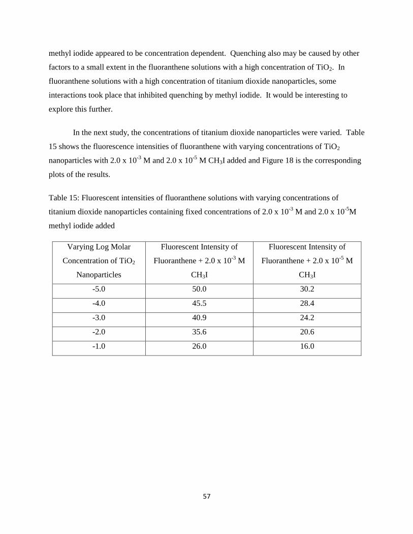
57
methyl iodide appeared to be concentration dependent. Quenching also may be caused by other
factors to a small extent in the fluoranthene solutions with a high concentration of TiO2. In
fluoranthene solutions with a high concentration of titanium dioxide nanoparticles, some
interactions took place that inhibited quenching by methyl iodide. It would be interesting to
explore this further.
In the next study, the concentrations of titanium dioxide nanoparticles were varied. Table
15 shows the fluorescence intensities of fluoranthene with varying concentrations of TiO2
nanoparticles with 2.0 x 10⁻³
M and 2.0 x 10⁻⁵
M CH3I added and Figure 18 is the corresponding
plots of the results.
Table 15: Fluorescent intensities of fluoranthene solutions with varying concentrations of
titanium dioxide nanoparticles containing fixed concentrations of 2.0 x 10⁻³
M and 2.0 x 10⁻⁵
M
methyl iodide added
Varying Log Molar
Concentration of TiO2
Nanoparticles
Fluorescent Intensity of
Fluoranthene + 2.0 x 10⁻³
M
CH3I
Fluorescent Intensity of
Fluoranthene + 2.0 x 10⁻⁵
M
CH3I
-5.0 50.0 30.2
-4.0 45.5 28.4
-3.0 40.9 24.2
-2.0 35.6 20.6
-1.0 26.0 16.0

58
Figure 18: Plots of the fluorescence intensity of solutions of fluoranthene with varying
concentrations of titanium dioxide nanoparticles with 2.0 x 10⁻⁵
M (red) and 2.0 x 10⁻³
M (blue)
of methyl iodide added
The results and plots show that quenching by varying concentrations of the titanium
nanoparticles were quite linear with other quenching factors superimposed upon it. Also the
fluorescent intensities started at a much lower values indicating that methyl iodide present in all
the fluoranthene solutions exerted its quenching effect quite strongly especially at its
concentration of 2.0 x 10⁻³
M. However, interestingly at the low 2.0 x 10⁻⁵
M methyl iodide
quenching by varying concentrations of titanium dioxide was at a steeper rate than in the case
with methyl iodide at 2.0 x 10⁻³
M. Why the high concentration of methyl iodide with
increasing concentration of titanium dioxide did not quench as much is not fully understood.
These observations were peculiar to fluoranthene. This could once again be attributed to the
tendency of fluoranthene to be less susceptible to quenching.

59
CHAPTER 5
CONCLUSION
The fluorescence intensities of fluorescent compounds are susceptible to quenching by
diverse compounds and environmental factors. In this study, the fluorophores; carbazole,
pyrene, and fluoranthene were used to study the effects of oxygen, a well know quencher methyl
iodide and, finally, titanium dioxide nanoparticles. The findings from the experiments fulfilled
most of the expectations of the research. Some results however, were not obvious and need
further investigation.
Methyl Iodide the quencher used for this research was proven to be an efficient quencher
of all three compounds and quenched via dynamic quenching. Methyl iodide contains a heavy
atom giving rise to the well-known heavy atom effects that quench by promoting inter system
crossing. Quenching by molecular oxygen was also evident with all three compounds, as can be
seen with considerable increase in the fluorescent intensities after the removal of oxygen from
the solutions by degassing with helium gas. Titanium dioxide was also found to quench
dynamically and found in most cases that quenching to be linear with increasing concentrations.
Carbazole was quenched by both methyl iodide and titanium dioxide, to a lesser degree
by titanium dioxide. Carbazole was also quenched by molecular oxygen because a considerable
increase in the signal intensities was observed after the solution was degassed. The effects on
quenching by varying concentration of methyl iodide in the presence of 2.0 x 10⁻⁴
M TiO2
nanoparticles gave anomalous results as compared to when 2.0 x 10⁻⁷
M of it was added to the
solutions of carbazole, and the cause is not clear and need further work to confirm them. In the
case when the concentration of titanium dioxide nanoparticles was varied with a given
concentration of methyl iodide added to the solution of carbazole, the results obtained were close
to what was expected.
The fluorescence of pyrene was quenched dynamically by methyl iodide, titanium
dioxide nanoparticles, and oxygen. Quenching of pyrene by titanium dioxide nanoparticles was
again to a lesser degree by methyl iodide as in the case of carbazole. Oxygen quenching of
pyrene is well known and pyrene was quenched to a larger extent by oxygen than was carbazole.

60
Interestingly, the quenching by varying concentrations of titanium dioxide nanoparticles with 2.0
x 10-3
M and 2.0 x 10⁻⁵
M methyl iodide added to the solutions did not show much difference.
However quenching by varying concentrations of methyl iodide with the addition titanium
dioxide nanoparticles to the pyrene solutions were quite different and to a larger extent at higher
concentration of 2.0 x 10⁻⁴
M than 2.0 x 10⁻⁷
M. This might be attributed to the tendency of
titanium dioxide to form a complex with pyrene, based on a charge transfer interaction. High
concentrations of both titanium dioxide nanoparticles thus might exacerbate quenching by
methyl iodide.
Fluoranthene, a highly fluorescent fluorophore, was slightly quenched by both methyl
iodide and titanium dioxide. Quenching by molecular oxygen was also observed. Quenching by
varying concentrations of methyl iodide, in the case of fluoranthene, was quite different and to a
much lower degree by the presence of added titanium dioxide nanoparticles at both higher
concentration of 2.0 x 10⁻⁴
M or the lower concentration of 2.0 x 10⁻⁷
M. Also the quenching did
not change greatly with changes in the methyl iodide concentration. However, when the
concentration of titanium dioxide nanoparticles was varied while the added concentration of
methyl iodide was kept at 2.0 x 10⁻³
M or 2.0 x 10⁻⁵
M, the fluorescence intensities varied much
more greatly and the overall intensities were much lower. It was found, in the case of
fluoranthene, titanium dioxide nanoparticles quench more strongly in the presence of methyl
iodide. This was not studied or reported before, so should be explored further.
From the results obtained for all compounds, it can be concluded that methyl iodide is an
efficient dynamic quencher for all three compounds studied. Oxygen also quenched all three
fluorophores but to a greater extent for pyrene. Also all fluorophores were quenched to a lesser
degree by titanium dioxide as compared to quenching by methyl iodide. Finally, varying
concentrations of titanium dioxide nanoparticles in the presence of methyl iodide, and vice versa,
had different effects on the quenching of these fluorophores. Fluoranthene, however, could be
an interesting focus of further research as some of its fluorescence characteristics in the presence
of both methyl iodide and titanium dioxide were not expected.

61
REFERENCES
1. Guilbault, G. Practical Fluorescence, 2nd
ed.; rev. and expanded. Marcel Dekker, Inc., New
York, (1990), 1-120.
2. McQuarrie, D.A., and Simon, J.D. (1997). Physical Chemistry, Molecular Approach.
Sausalito, CA; University Science Books.
3. International Union of Pure and Applied Chemistry, .n.d.Potochemistry IUPAC Gold Book.
Compendium of Chemical Terminology edition.
4. Haddock, S.H.D., McDougall, C.M and Case, J.F., (2006). The bioluminescence web page.
University of California, Santa Barbara.
5. Seitz Rudolf W. and Neary Michael P. Chemiluminescence and Bioluminescence in
Chemical Analysis, Analytical Letters (1974), 2(46), pp 189-195.
6. Kricka, Larry J. and Thorpe Gary H.G. Chemiluminescent and Bioluminescent Methods in
Analytical Chemistry, Analyst, November, (1983) vol. 108, pp 1278-1280.
7. J. Lakowicz, Principles of Fluorescence Spectroscopy, 2nd ed., Plenum, New York, (1999),
pp.257-301.
8. Peng, X., Chen, H., Draney, D.R., Volcheck, W.M., A Non-fluorescent, Broad Range
Quencher Dye for FRET Assays, Analytical Biochemistry, (2009), Vol. 388, pp. 220–228
9. Geddes Chris D. Optional halide sensing using fluorescence quenching: theory, simulations
and applications-a review, Institute of Physics Publishing, Meas.Sci.Technol. 12(2001) R53-
R88.
10. Giri Rajesh, Fluorescence Quenching of Coumarins by Halide Ions, Spectrochimica Acta
Part A: Molecular & Biomolecular Spectroscopy, March (2004), Vol 60(4), pp.757-763.
11. Osterman, H., The Next Step in Near Infrared Fluorescence: IRDye QC-1 Dark Quencher,
(2009); Review Article
12. Lakowicz, J.R. Quenching of Fluorescence in Principles of Fluorescence Spectroscopy, 3rd
Edition (2006), Springer-Verlag, Berlin, 278-330.
13. Peng, X., Chen, H., Draney, D.R., Volcheck, W.M., A Non-Fluorescent, Broad Range
Quencher Dye for FRET Assays, Analytical Biochemistry, (2009),(vol.388), pp.220-228.
14. Munro, J.B., Altman, R.B., O’Connor, N., Blanchard, S.C. Molecular Cell (2007), 25: 505-
517.

62
15. Kotlyar, A.B., Borovok, N., Molotsky, T., Fadeev, L., Gozin, M. In vitro synthesis of
uniform poly (dG)-poly(dC) by Klenow exo(-) fragment of polymerase I. Nucleic Acids Res.
(2005), 33: 525-535.
16. J-B_ B&man. Handbook of fluorescence spectra of are- matic molecules (Academic Press.
New York, 1971); IV_ Ware. J_ Phys- Chem- 66 1962 4.55; R_ Potashnik. CO_
Goklschmidt and Sf_ Ottolenghi, Chem- Phys. Letters 9 (1971) 424; LX_ Patterson. G.
Porter and XI-R_ Topp. Chem. Phys. Letters 7 (1970) 612; F_ Wiinson. In: Organic
molccuku photophysics, Vol. 2, ed. J- Birks (Wiley, New York. 1975) p_ 95.
17. Turro, J. Nicholas, Alkawa, M. and Yekta A. Dynamics of Molecular Oxygen in Micellar
Solutions. Chemical Physical Letters, (1979), 64(3) p 473-478.
18. Chong Lee-Gau, P and Thompson T.E. Oxygen Quenching of Pyrene-Lipid Fluorescence in
Phosphatidylcholine Vesicles.(A probe for membrane organization) Biophysical Journal,
(1985), vol.47, pp-613-621.
19. Gijzeman, O.L.J., Kaufman, F. and Porter G. Oxygen Quenching of Aromatic Triplet States
in Solution. Journal of Chemical Society, Faraday Transitions 2, (1973), 69, pp 703-720.
20. Tacko, Niwa Inada et al, A comparative study on electron-transfer fluorescence quenching by
aliphatic and aromatic amines, Journal of photochemistry and photobiology A: Chemistry
vol. 137(2-3), December (2000), pp 93-97.
21. Bernhardt, P. V.; Moore, E. G.; Riley, M. J. Inorg. Chem. (2001), 40, 5799-580
22. Goryaeva, E. M.; Shablya, A. V.; Ermolaev, V. L. Opt. Spectrosc. (2001), 99, 508-515
23. East, A. L.; Lim, E. C. J. Chem. Phys. (2000), 113, 8981-8993.
24. Lakowicz, J.R and Weber. Biochemistry, New York; Luwer Academic/Plenum Press (1999).
25. Lakowicz, J.R and Weber, G. Biochemistry, 12, (1973) 4161.
26. S. Hirayama, H. Yasuda, A.D. Scully, M. Okamoto, J. Phys. Chem. 98 (1994) 4609
27. K.K. Pandey, M. Okamoto, S. Hirayama, Chem. Phys. Lett. 224(1994) 417.
28. Arik M., Celebi N., Onganer Y., Fluorescence Quenching of Fluorescein with molecular
Oxygen in Solution, Journal of Photochemistry and Photobiology A: Chemistry 170 (2005)
pp. 105-111.
29. Eftink, M.R and Ghiron, C.A., J.Phys.Chem. 80, (1976) 486.
30. Eftink,M.R, Zajicek,J.L. and Ghiron, C.A., Biochem.Bio-phys.Acta,491, (1977) 473.

63
31. Matyus,L., Szollosi,J., Jenei,A. Steady-State Fluorescence Quenching Applications for
Studying protein structure and Dynamics. Journal of Photochemistry Photobiol B. Epub
(2006) 83(3), pp 223-236.
32. Limei Ao, Feng Gao, Bifeng Pan, Rong He, and Daxiang Cui. Fluoroimmunoassay For
Antigen Based on Fluorescence Quenching Signal of Gold Nanoparticles Analytical
Chemsitry,( 2006), 78(4), pp 1104-1106.
33. Xiaowei Zhuang, Taekjip Ha, Harold D. Kim, Thomas Centner, Siegfried Labeit and Steven
Chu. Fluorescence Quenching: A tool for single-molecule protein-folding study study. PNAS
December, (2000), vol.97 (26), pp 14241-14244.
34. Oesterhelt, D., Oesterhelt, F., Pfeiffer, M., Engel, A., Gaub, H. E. & Mu¨ller, D. J. (2000)
Science 288, 143–146.
35. Schoefer, L., Braune, A., and B., Michael. A Fluorescent Quenching, Test for the Detection
of Flavonoid Transformation. FEMS Microbiology Letters (2001), 204(2) pp 277-280.
36. Halide (Cl-) Quenching of Quinine Sulfate Fluorescence: A Time-Resolved e
FluorescencExperiment for Physical Chemistry Jonathan H. Gutow J. Chem. Educ., (2005),
82 (2), p 302.
37. Peng, X., Chen, H., Draney, D.R., Volcheck, W.M., A Non-fluorescent, Broad Range
Quencher Dye for FRET Assays, Analytical Biochemistry, (2009), Vol. 388, pp. 220–228.
38. Geddes D Chris. Optical Halide Sensing Using Fluorescence Quenching; Theory
Simulations and Applications-A review. Institute of Physics Publishing. Meas. Sci.Technol.
12(2001) R53-R88.
39. Geddes C.D., A halide sensor based on the quenching of fluorescence of an immobilized
indolium salt Photochem. Photobiol. (2000B) A 137145–53
40. U. Diebold, Surf. Sci. Rep., (2003), 48, 53
41. Hinze, W.Organized assemblies in chemical analysis. Overview of the utilization of reversed
micellar media in analytical spectroscopy, Jai Press Inc.: Greenwich, CT, (1994), vol.1, pp.3.
42. Kim, D. Miyamoto, M., Nakayama, M. (2003), Surface modification effects on luminescence
properties of CdS and CdMnS quantum dots prepared by a reverse-micelle method. Physica
status solidi(c), O: 1233-1236, DOI: 10.1002/pssc.200303055.
43. Lauren E., Shea-Rohwer, James E.Martin and Kelly F. David. Increasing the luminescent
quantum yield of CdS nanoparticles having broadband emission. J. Electrochem. Soc.,
(2010) vol. 157(1) pp.J1-J7.

64
44. Leif C.Robert, Vallarino M. Lidia, Becker C. Margie & Yang Sean. Increasing the
luminescence of lanthanide complexes for cytometry by use of RETEL effect cytometry
(2006).
45. Ueno, A. Supramol. Sci. (1996), 3, 31-36.
46. Turnbull, J.H., Walker R.G., The effects of β-cyclodextrin on the luminescence of para amino
acids. IL NUOVO CIMENTO B (1997-1996) vol. 63, (1), pp. 441-445, DOI:
10:1007/BF02721450.
47. Vazquez,M.L., Cepeda, A., Prognon,P., Mahuzier,G.and Blais,J. Cyclodextrins as modifiers
of the luminescence characteristics of aflatoxins. Analytica Chimica Acta, (1991), vol.,
255(2), pp.343-350.
48. Tomczak, M.M., Slocik, J.M., Stone, M.O. and Naik, R.R. Biofunctionalized Nanoparticles
and Their Uses. Mrs. Bulletin (2008), 33 pp 519-520.
49. Lance G. Phillips and David M. Barbano. The Influence of Fat Substitutes Based on Protein
and Titanium Dioxide on the Sensory Properties of Low fat Milk. Journal of Dairy
Science 80 (11)2726.
50. Ju-Young Park, Changhoon Lee, Kwang-Woo Jung and Dongwoon Jung. Structure Related
Photocatalytic Properties of TiO2. Bull. Korean Chemical Society (2009), 30(2) pp 402-404.
51. Abrahams Jeffrey, Davidson Stephen R., Morrison L. Colin., Optimization of the
photocatalytic properties of titanium dioxide. Journal of photochemistry, (2001), 29(3-4),
pp.353-361.
52. Wold Aaron, Photocatalytic properties of titanium dioxide. Chem.Mater, (1993), 5(3),
pp.280-283.
53. Zhang, W.F., Zhang,M.S., Yin,Z., Chen, Q. Photoluminescence in anatase titanium dioxide
nanocrystals. Appl.Phys. (2000), B70, 261-265.
54. E Zaleta-Alejandre et al. Structural and luminescent properties of europium doped Titanium
dioxide thick films synthesized by the ultrasound spray pyrolysis technique. J. Phys. D: Appl.
Phys. 42 0955102.
55. K. Robbie and M.J. Brett, J. Vac. Sci. Technol., (1997), A15, 1460.
56. J. C. Sit, D. Vick, K. Robbie and M. J. Brett, J. Mater. Res., (1999), 14, 1197.
57. Kumar, S. A., Chen, S. M., Lo, P. H., Nanotechnology 19, (2008), 255501(7pp).

65
58. Fraunhofer-Gesellschaft (2010, April 17). Using fullerenes as cushion nanoparticles Science
Daily. Retrieved July 16, 2011.From:
http://www.sciencedaily.com/releases/2010/04/100412112901.htm
59. M. Grätzel, Nature, (2001), 414, 338
60. K. Robbie and M.J. Brett, J. Vac. Sci. Technol., (1997), A15, 1460.
61. Tang, Z., Zhang, J., Cheng, Z., Zhang, Z., Mat. Chem. And Phy. 77, (2002), 314-317
62. Gary, Christian D., Analytical Chemistry, 6th ed., John Wiley & Sons Inc. (2003), pp 1‐803.
63. Reyes –Corondo, D., Rodriguez-Gattorno, G., Espinosa-Pesqueira, M.E., Cab, C., de Cross,
R., Oskam, G. Nanotechnology. (2008), 19 (145605) 10pp
64. Harnandan, S., Hinze, W.Micellar Enhanced spectrofluorimetric methods: application to the
determination of pyrene. Analytical letters, (1982), 15(A3)221-243.
65. Rubinson, K. A., Rubinson, J. F. Contemporary Instrumental Analysis 1st ed Prentice Hall
Inc: New Jersey, (2000) pp 330‐350.
66. O’Haver, T. C. The Development of Luminescence Spectrometry as an Analytical Tool. J.
Chm Edu. (1978), 55(7), 423.
67. Association of Official Analytical Chemists, (1984), in official Methods of Analysis.
AOAC, Arlington, VA. 43.039-43.047.
68. Fetzer, J. C. (2000). The Chemistry and Analysis of the Large Polycyclic Aromatic
Hydrocarbons. New York: Wiley.
69. Fischkoff, S. and Vanderkooi, J.M. Oxygen Diffusion in Biological and Artificial
Membranes Determinated by the Fluorochrome pyrene. Journal of Gen.Physiol (1975), 65 pp
663-676.
70. Shatalov, I.A., Zavarin, I.S., Zaitsev, N.K. and Malkin, J.N. The Effect of the Potential of
Colloid Titanium Dioxide on Fluorescence Quenching. Journal of Photochemistry and
Photobiology. (1996), 94(1) pp 63-66.
71. You Seok Seo, Changhoon Lee, Kee Hag, Lee Prof. Dr., Kyung Byung Yoon Prof.Dr. 1:1
and 1:2 Charge Transfer Complexes Between Aromatic Hydrocarbons and Dry Titanium
Dioxide. Angewandte Chemie International Edition, (2005), 44(6) pp 910-913
72. Camyshun, S.V., Gritsan, N.P., Korolev, V.V., and Bazhin, N.M. Quenching of the
Luminescence of Organic Compounds by Oxygen in Glassy Matrices. Journal of Chemical
Physics. (1990), 142(1) pp

66
73. Berlman, I.B., Wirth, H.O. and Steingraber, O.J., Anomalous Fluorescence Characteristics of
Fluoranthene and Some of its Derivatives. Journal of the American Chemical Society.
(1968), 9(3) pp 566-570.
74. Michel Deumie, Mohamed El Baraka, Edwin Quinones. Fluorescence Quenching of pyrene
derivatives by iodide compounds in erythrocyte membranes: an approach of the proble
location. Journal of photochemistry and Photobiology A: Chemistry (1995) vol. 87(2) pp
105-113.
75. Margaret W., Geiger, Nicholas J.Turro. Pyrene Fluorescence Lifetime as probe for Oxygen
Penetration of Micelles. Photochemistry and Photobiology (2008) vol. 22(6) pp 121-125

67
VITA
VIVIAN DZIGBODI KOKA
Personal Data: Date of Birth: January 12, 1982
Place of Birth: Nkwakubew-Anum, Ghana
Marital Status: Married
Education University of Cape Coast, Ghana, Chemistry, B.S. 2005
East Tennessee State University (ETSU), Johnson City,
Tennessee, USA; Chemistry, M.S. 2011
Professional Experience: Assistant Chemist, American Crystal Sugar Company,
Crookston, MN, USA, 2011
Graduate Teaching Assistant, ETSU, Johnson City, TN,
USA, 2009‐2011
Chemist, Ghana Palm Oil Development Co. Ltd, Ghana
2007-2009
Quality Analyst, Pinora Ghana Ltd, Ghana 2006 – 2007