a study of ki-67, c-erbb2 and cyclin d-1 expression in cin ... · 50% of the lsil, hgsil and scc...
TRANSCRIPT

Summary. The histological criteria for cervicalintraepithelial neoplastic lesions and their follow-upshave been established, but their reproducibility,specificity and sensibility are not certain.Immunohistochemical markers provide moreinformation on each specific case, in order to facilitateits classification and, eventually, its prognosis. Usingimmunohistochemical techniques, this study analyzesthe prognostic value of three markers (Ki-67, c-erbB2and Cyclin D1) in cases of low grade squamousintraepithelial neoplasia (CIN-I), high grade squamousintraepithelial neoplasia (CIN-III), and infiltratingsquamous cell carcinoma (SCC) taken from a group ofcervical samples. In situ hybridization was performed inorder to detect high-risk HPV.High risk HPV was demonstrated in 82%, 89% and
100% of the LGSIL, HGSIL and SCC cases,respectively.C-erbB2 expression was detected in 9%, 33% and
50% of the LSIL, HGSIL and SCC cases, respectively.The Ki-67 LI was 25%, 68% and 65.5% in the LGSIL,HGSIL and SCC cases, respectively. Nuclear Cyclin D1expression was seen in 82%, 11% and 30% of the CIN-I,CIN-III and SCC cases, respectively. We observed thatthe cytoplasmic cyclin D1 expression increased with theseverity of the lesion instead of the nuclear expressiondecreasing with the progression of the pathology.Nuclear and cytoplasmic Cyclin D1 expression seemedto be related to HPV high risk infection. We concluded that Cyclin D1, cerbB2 and The Ki-
67 LI expression changed in relation to the severity ofthe lesion and that they could be helpful in making adifferential diagnosis.Key words: CIN, Squamous cell carcinoma, Cervix, Ki-67 LI, Cyclin D1
Introduction
In recent years, the mortality rate of patients withcervical squamous cell carcinoma (SCC) has decreased,probably due to the use of the Papanicolau test. Thehistological criteria for cervical intraepithelial neoplasticlesions and their follow-ups have been established, buttheir reproducibility, specificity and sensibility are notcertain (Robertson et al., 1989). Only around 20% of thecases with low cervical intraepithelial neoplastic lesions(CIN-I) will progress to moderate or severe cervicalintraepithelial neoplastic lesions (CIN-III). In fact, wehaven’t any criteria to predict the follow-up of CIN-Icases. The oncogenic role of HPV in cervical
carcinogenesis, is actually Indisputable. High-risk HPVproduces two proteins E6 and E7, called oncoproteins,which link p53 and pRb, respectively. Consequently,E2F is liberated of pRb, independently of its(CD1/CDK4) function. E2F induces p16 synthesis,which deactivates the CD1/CDK4 complex. On the otherhand, E2F drives the infected cells to begin cell-cycling,(without the check point induced by p53, deactivated bythe action of E6). In this process, cells are submitted togenomic instability and can accumulate a great quantityof mutations. Furthermore, genomic instability increasesthe possibility that viral DNA can integrate into the cellDNA. In the integration process, the viral DNA breaksinto the E2 region, and the E2 protein is not produced.The absence of E2 deregulates E6 and E7 production.The viral DNA integration is produced in differentchromosomal loci, and it is possible that someoncogenes or tumor suppressor genes can alter theirfunctions. The CD1 gene maps to 11q13, and it showsthe characteristics of a cellular oncogene. CD proteinshave CDK-independent properties, which are importantfor cell growth, metabolism, and cellular differentiation.The over-expression of CD1 is one of the mostcommonly observed alterations in cervical carcinomas;however, their role in cervical carcinogenesis is still not
A study of Ki-67, c-erbB2 and cyclin D-1 expression inCIN-I, CIN-III and squamous cell carcinoma of the cervixR. Carreras2, F. Alameda1, G. Mancebo2, P. García-Moreno1, M.L.M. Mariñoso1, C. Costa1, P. Fusté2, T. Baró1 and S. Serrano11Pathology service and 2Obstetry and Gynecology service, Hospital del Mar, IMAS, Universitat Autónoma de Barcelona, Barcelona, Spain
Histol Histopathol (2007) 22: 587-592
Offprint requests to: F. Alameda, MD, Department of Pathology,Hospital del Mar, Passeig Maritim 25-29, 08003 Barcelona, Spain. e-mail: [email protected]
DOI: 10.14670/HH-22.587
http://www.hh.um.es
Histology andHistopathologyCellular and Molecular Biology

clear (Kim and Zhao, 2005).The immunohistochemical expression of CD1, in
cases of SIL and SCC, has been widely studied. It hasbeen detected in cell nuclei. A review of the literatureshows variable results ranging from an absence ofexpression in normal cervical epithelia to a variabledegree of expression in SCC (Bae et al., 2001; Kedzia etal., 2002; Skomedal et al., 1999; Cheung et al., 2001). Inaddition, normal CD1 expression has been observed innormal and CIN-I epithelia, and an absence ofexpression has been observed in CIN.III cases (Nichols,et al., 1996; Cho et al, 1997; Southern and Herrington,1998). CD1 expression has been associated with the HPV
infection: expression is higher when HPV is consideredto be low risk and is lower when HPV is considered tobe high risk (Southern et al, 1998). A good correlationhas been observed between CD1 and E7 oncoproteinexpression (Crish et al, 2000). In cervical lesions and incell cultures, the expression of CD1 has been detected inthe nuclei, but this expression increases in the cytoplasmand decreases in the nuclei, when the cells enter into Sphase (Baldin et al., 1993).C-erbB2 maps to 17q12 and encodes a protein with
tyrosine-kinase activity, homologous to the epidermalgrowth factor receptor (EGF-R). It is expressed in agreat number of epithelial tumors, and it is known that c-erbB2 expression is increased in approximately 20-25%of ovarian and/or breast carcinomas, 35-45% ofpancreatic carcinomas, and 90% of colorectalcarcinomas. In cases of SIL and SCC, it has beenobserved that there is greater c-erbB2 expression whenthe severity of the lesion increases. In CIN-I cases, c-erb-B2 expression is low (Brumm et al., 1990). In SCC,the greater expression has been correlated to poorprognosis, an increase in aggression, and lymph nodemetastases. These data allow us to suspect that c-erb-B2expression is a late event in cervical carcinogenesis andthat it probably acts in a great number of functions andin the activation of the Cyclin D1 function (Chang et al.,1999; Niibe et al., 2003).The antibody Ki67 reacts with the nuclear Ki67
antigen, a protein encoded by ki67, on 10q25. Ki-67 isonly present in proliferating cells (Gerdes et al., 1984).In normal exocervical epithelia, it is only expressed inthe suprabasal layer (Konishi et al., 1991; Resnick et al.,1996), and in CIN cases, it is expressed throughout thedifferent epithelial layers. The Ki-67 labeling index (Ki-67 LI) increases, according to the degree of squamouscervical neoplasia (Payne et al., 1996; McCluggage etal., 1998; Maeda et al., 2001; Alameda et al., 2004).Our aim in this study was to analyze CD1, c-erbB2
and Ki-67 expression in a group of cervical samples ofdifferent pathological stages (CIN-I.CIN-III and SCC). Materials and methods
Patients
A group of 30 cervical biopsies from the Department
of Pathology at the Hospital del Mar in Barcelona,Spain, were selected. There were 11 cases diagnosed asCIN-I, 9 cases diagnosed as CIN-III and 10 casesdiagnosed as SCC. The diagnosis of each samplespecimen was established on the basis of a routinehistopathological examination (Wright et al., 2002). Allbiopsy specimens were examined by two experimentalpathologists. Cases where there was disagreement on thediagnosis were excluded from the study.HPV detection
HPV detection was realized using in situhybridization techniques. It was performed using anautomated system (Inform HPV, Ventana), based on aprobe for high risk HPV (HPV-HR) and was carried outon paraffin-embedded tissue. After sampledeparaffination and rehydration, we incubated the slidesin salt sodium citrate buffer with formamide at 75°C.Later, we incubated the slides with the high risk HPVprobe, conjugated with FITC. The post-hybridizationwashes were performed using salt sodium citrate buffer20x formamide. For amplification and detection, weused an anti-FITC antibody, a second anti-IGbiotinylated antibody, an avidin-alkaline phosphataseconjugate that reacted to BICP (5 brome- 4 chlore- 3indolyl phosphate), and an NBT (Nitro BlueTetrazolium). The post-hybridization washes were doneusing salt sodium citrate buffer with formamide. Nucleiwere counterstained with ISH red (Ventana). Weconsidered a positive (high-risk HPV) result to be whenwe observed diffuse or granular, blue nuclei. Positiveand negative controls were used. Immunohistochemical study
The immunohistochemical study was performedusing the DakoThechMate Immunostainer (Dako-Cytomation, Glostrup, Denmark). The immuno-histochemical staining was developed for the followingprimary antibodies: CD1 (DCS-6; 1:10; Novocastra,Newcastle, UK), Ki-67 (Mib-1; 1:100; DakoCytomation,Glostrup Denmark), and c-erb-B2 (Polyclonal; 1:350;DakoCytomation; Glostrup, Denmark). The detectionwas carried out using the EnVision system (DakoCytomation, Glostrup, Denmark). Positive and negativecontrols for each antibody were used. The Ki-67 LI index (percentage of cells with a
brown coloration in the nucleus) was obtained bycounting 200 cells in a 25 HPF, which included all of theepithelial layers. The positive expression of C-erb-B2 isusually established using a scale of cross-like +1, +2 or+3, and in our study, its expression was consideredpositive when there was immunoexpression around all ofthe cytoplasmic membrane. We only considered aspositive the cases that showed a score of +3. CD1expression is usually considered positive in the nucleus,and its positivity is expressed with a percentage of cells.In this study, we observed CD1 expression in the cellcytoplasm, and it was evaluated in both locations.
588Ki-67, c-erbB2, & cyclin and squamous cervical lesions
Results
The results of this study are summarized in Tables 1-3.High risk HPV was demonstrated in 82% of the
cases of LGSIL, 89% of the cases of HGSIL and in all ofthe SCC cases.CD1 expression
In the CIN-I group, nuclear CD1 expression (Figure)was seen in 9/11 cases (82%): In 4 cases, all of themhigh-risk HPV positive (36%), the expression was onlydetected in the nuclei with an 11% mean of positivenuclei, and in 5 cases (45%), 4 of them high-risk-HPVpositive, we observed expression in the nuclei and thecytoplasm with a mean of 11% positive cells. In theremaining 2 cases, CD1 expression was onlycytoplasmic with a mean of 20% positive cells (Table 1).In the CIN-III group, only one case (high-risk HPV
positive) showed nuclear and cytoplasmic positivity witha mean of 40% positive cells. The remaining eight casesonly showed the cytoplasmic expression of CD1 with a76% mean of positive cells (Table 1).In the SCC group, three cases showed nuclear and
cytoplasmic expression of CD1 with a 37% mean ofpositive cells, and the remaining seven cases showedonly cytoplasmic positivity with a 57% mean of positivecells (Table 1).We observed that nuclear CD1 expression decreased
with the progression of the pathology (9/11 LSIL, 1/9CIN-III, and 3/10 SCC cases) instead of the cytoplasmicCD1 expression increasing with the severity of the lesion(2/ 11 CIN-I 8/9 CIN-III, and 7/10 SCC cases) (Table 1).Furthermore, the percentage of nuclear and cytoplasmicpositive cells increased with the severity of the lesion.C-erbB2 expression (Fig. 1) was detected in only
one CIN-I case that was HPV-HR positive (9%), in 33%of the CIN-III cases, all of them HPV-HR positive, andin 50% of the SCC cases (Table 1). We observed that theexpression of c-erbB2 increased with the intensity of thelesion and seemed to be related to the HPV-HRinfection, due to the fact that in the HPV-HR negativecases of CIN-Iand CIN-III, the expression of c-erbB2was not detected (Table 1). The mean Ki-67 LI (Fig. 1) was 25% in the CIN-I
cases, 68% in the CIN-III cases and 65.5% in the SCCcases. In the HPV-HR positive cases, the mean Ki-67 LIwas 26% in the CIN-I and 70% in the CIN-III HPV-HRcases. In the HPV-HR negative cases, the mean KI-67 LIwas 21% in the cases of CIN-I and 65% in the CIN-IIIcases (Table 1).In Table 2 we compared the mean of the Ki-67 LI
related to c-erb.B2 expression. In those cases, the Ki-67LI in c-erbB2 positive cases was greater than in the c-erbB2 negative ones.In Table 3, we summarized the relationship between
the Ki-67 LI and CD1 expression. In all of thepathological groups, the Ki-67 LI was higher when CD1expression was detected in the cytoplasm (Table 3).
589Ki-67, c-erbB2, & cyclin and squamous cervical lesions
Table 1. Summary of results.
LESION HPV-HR CD1 N CD1 N+C CD1 C c-erbB2 + KI-67 LI
CIN-I POS 9/11(82%) 4/9 (44%) 4/9 (44%) 1/9 (11%) 1/9(11%) 26%NEG 2/11 (18%) 0/2 (0%) 1/2 (50%) 1/2 (50%) 0/2 (0%) 21%
TOTAL 11 CASES 4/11 (36%) 5/11 (45%) 2/11 (19%) 1/11 (9%) 25%
CIN-III POS 8 (89%) 0/8 (0%) 1/8 (13%) 7/8 (87%) 3/8(27%) 70%NEG 1 (11%) 0/1 (0%) 0/1 (0%) 1/1 (100%) 0/1 (0%) 65%
TOTAL 9 CASES 0/9 (0%) 1/9 (11%) 8/9 (89%) 3/9 (33%) 68%
SCC POS 10 (100%) 0/10 (0%) 3/10 (30%) 7/10 (70%) 5/10 (50%) 65.5%
CD1, c-erbB2, Ki-67 LI and HPV-High risk positive and negative cases
Table 2. A comparative study between c-erbB2 expression and the Ki-67 LI.
KI-67 LI
c-erbB2 EXPRESSIONPOS 62.6%NEG 47.7%
Table 3. Relationship between the Ki-67 LI and CCND1 expression.
KI-67 LICIN-I CIN-III SCC
CD1 expressionN 19.3% - -N+C 22% 68.5% 58.3%C 45% 80% 68.6%
N: nuclear; C: cytoplasmic

Discussion
Our results revealed different expression patterns forthe studied markers related to the lesions, mainly whenwe considered the CIN-I cases as compared to the CIN-III and SCC cases.Ki-67 is expressed only in proliferating cells (Kim
and Zhao, 2005). Our cases showed an increase in theKi-67 LI in relation to increases in the severity of thelesions. Our results showed a Ki-67 LI of 25% in theCIN-I cases, a Ki-67 LI of 70% in the CIN-III cases, anda Ki67 LI of 65.5% in the SCCs. These data are in
accordance with the literature. The Ki-67 LI can beuseful in distinguishing the different grades of dysplasia,though not in predicting their behavior (Alameda et al.,2004). There were no differences in the Ki67 LI in thehigh-risk HPV positive cases and the rest of the studiedcases.Keeping in mind that only the cases with a +3 score
were considered positive, in our cases c.erb-B2expression, increased according to the severity of thelesions as shown in the literature (Brumm et al., 1990). We observed a relationship between the Ki-67 LI
and c-erb-B2 immunoexpression. C-erbB2 is a protein
590Ki-67, c-erbB2, & cyclin and squamous cervical lesions
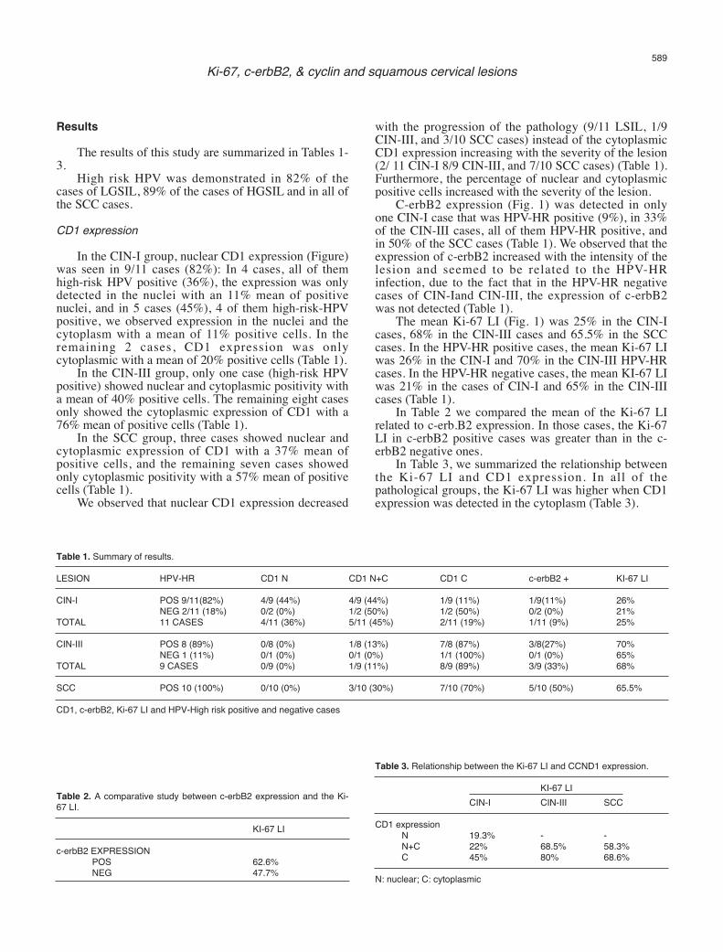
Fig.1. C-erb-B2. Strong immunoreactivity in the profound half of the epithelium in a case of CIN-I (A), and weak immunoreactivity, but throughout theepithelium, in the cases of CIN-III (B) and a squamous cell carcinoma (C).
Ki-67. Immunoreactivity in parabasal and some intermediate cells in a CIN-Icase (D) and throughout the epithelium of a CIN-III case (E) and in a caseof infiltrating squamous cell carcinoma (F).
Cyclin D1. Nuclear and cytoplasmic immunoreactivity in a CIN-I case (G), and mainly cytoplasmic immunoreactivity in the cases of a CIN-III (H) and aninfiltrating squamous cell carcinoma (I). x 10

with tyrosine-kinase activity, homologous to theepidermal growth factor receptor (EGF-R). C-erb-B2acts by stimulating cell proliferation. In CIN-I and CIN-III cases there is a relationship between high-risk HPV-HR positivity and c-erb-b2 immunoexpression. All ofthe cases without c-erb-B2 immunoexpression werenegative for HPV-HR. We do not have an explanationfor this fact, as it appears that c-erb-B2 amplification is alate event in cervical carcinogenesis (Chang et al., 1999;Niibe et al., 2003). The nuclear expression of CD1 decreases with an
increase in the severity of the lesion. In the literature, aloss of nuclear CD1 expression has been reported inSCC (Nichols et al., 1996; Bae et al., 2001). Our casesshowed nuclear CD1 expression in 9/11 cases of CIN-I,in only 1/9 cases of CIN-III and in 3/10 cases of SCC.The percentage of positive CD1 increased with theseverity of the lesion. Nuclear CD1 expression has beeninversely associated with HR-HPV infection (Southernand Herrington, 1998). Our cases showed CD1 nuclearexpression in all of the CIN-I cases that were HPV-HRnegative and only in 77% of the LGSIL HR-HPVpositive cases. There is no data in the literature related tocytoplasmic CD1 expression. A possible explanation forthis fact may be the following: CD1 binds CDK4 andphosphorylates pRB in the G1 cell phase (Diehl et al.,1997a,b). In HPV-HR related lesions the E7 oncoproteinbinds pRb and deactivates it directly by ubiquitinization.Under these conditions CD1 is not necessary for the cellto enter into S phase (Cho et al., 2002). Under normalconditions CD1 is located in the nucleus in the G1phase, and it is phosphorylated, ubiquitinized, andcarried to the cytoplasm in the S phase (Diehl et al.,1997a,b). In our cases, the cytoplasmic expression ofCD1 increased according to the severity of the lesion (7of 11 CIN-I cases and all of the CIN-III and SCC cases),and the CD1 expression changed to be predominantlynuclear in the CIN-I cases and predominantlycytoplasmic in the CIN-III and SCC cases. An increasein the severity of the lesion is in correlation with anincrease in the number of cells in the S phase, when CD1is found in the cytoplasm of the cell (Baldin et al., 1993).Consequently, if we accept that neoplastic tissuecontains a greater number of cells than normal tissue,then the neoplastic cells would be able to showcytoplasmic CD1 expression, and the cytoplasmicimmunoexpression of CD1 would be seen with moreintensity and more extensively in cases of SCC than incases of CIN-I.We concluded that CD1, cerbB2 and the Ki-67 LI
markers increased in relation to the severity of thelesions and that they could be helpful in making adifferential diagnosis. The cytoplasmic expression ofCD1 and the absence of nuclear CD1 expression in SCCcould also be of help in diagnosis. More studies shouldbe conducted to elucidate the importance of cytoplasmicCD1 expression.
Acknowledgements. This study was supported in part by grants from theRedes de Centros de Genética, C03/07, and from Cáncer, C03/10, andfrom the Instituto de Salud Carlos III del Ministerio de Sanidad yConsumo.
References
Alameda F., Fuste P., Boluda S., Ferrer L., Baro T., Marinoso L.,Mancebo G., Carreras R. and Serrano S. (2004). The Ki67 labelingIndex is not a useful predictor for the follow up of cervicalIntraepithelial Neoplasia-1. J. Low Gen. Tract Dis. 8, 313-316.
Bae D.S., Cho S.B., Kim Y.J., Whang J.D., Song S.Y., Park C.S., KimD.S. and Lee J.H. (2001). Aberrant expression of cyclin D1 isassociated with poor prognosis in early stage cervical cancer of theuterus. Gynecol. Oncol. 81, 341-347.
Baldin V., Lukas J., Marcote M.J., Pagano M. and Draetta G. (1993).Cyclin D1 is a nuclear protein required for cell cycle progression inG1. Genes Dev. 7, 812-821.
Brumm C., Riviere A., Wilckens C. and Loning T. (1990).Inmunohistochemical investigation andnorthern blot analysis of c-erbB2 expression in normal, premalignant and malignant tissues ofthe corpus and cervix uteri. Virchows Arch (A). 417, 477-484.
Chang J.L., Tsao Y.P., Liu D.W., Han C.P., Lee W.H. and Chen S.L.(1999). The exprssion of type I growth factor receptors in squamousneoplastic changes of the uterine cervix. Gynecol. Oncol. 73, 62-71.
Cheung T.H., Yu M.M., Lo K.W., Yim S.F., Chung T.K. and Wong Y.F.(2001). Alteration of cyclin D1 and CDK4 gene in carcinoma ofuterine cervix. Cancer Lett. 166, 199-206.
Cho N.H, Kim Y.T. and Kim J.W. (1997). Correlation between G1 cyclinsand HPV in the uterine cervix. Int. J. Gynecol. Pathol. 16, 339-347.
Cho N.H., Kim Y.T. and Kim J.W. (2002). Alteration of cell cycle incervical tumor associated with human papillomavirus: cyclin-dependent kinase inhibitors. Yonsei Med. J. 43, 722-728.
Crish J.F., Bone F., Balasubramanian S., Zaim T.M., Wagner T., Yun J.,Rorke E.A. and Eckert R.L. (2000). Suprabasal expression of thehuman papillomavirus type 16 oncoproteins in mouse epidermisalters expression of cell cycle regulatory proteins. Carcinogenesis21, 1031-1037.
Diehl J.A., Zindy F. and Sherr C.J. (1997a). Inhibition of cyclin D1phosphorylation on threonine286 prevents its rapid degradation viathe ubiquitin-proteasome pathway. Gen. Dev. 11, 957-72.
Diehl J.A., Zindy F. and Sherr C.J. (1997b). A dominant-negative cyclinD1 mutant prevents nuclear import of cyclin Dependent-kinase 4(CDK4) and its phosphorylation by CDK-activating kinase. Mol. CellBiol. 17, 7362-7374.
Gerdes H., Lemke H., Baisch H., Wacker H.H., Schwab U. and Stein H.(1984). Cell cycle analysis of a cell proliferation-associated humannuclear antigen defined by the monoclonal antibody Ki-67. J.Immunol. 133, 1710-1715.
Kedzia W., Schmidt M., Frankowski A. and Spaczynski M. (2002).Immunohistochemical assay of p53, cyclin D1, c-erb-b2, EGFR andKi-67 proteins HPV positive and HPV negative cervical cancers.Folia Histochem. Cytobiol. 40, 37-41.
Kim Y.T. and Zhao M. (2005). Aberrant cell cycle regulation in cervicalcarcinoma. Yonsei Med J. 46, 597-613.
591Ki-67, c-erbB2, & cyclin and squamous cervical lesions

Konishi I., Fujii S., Nonogaki H., Nanbu Y., Iwai T. and Mori T. (1991).Inmunohistochemical analysis of estrogen receptors, progesteronereceptors, ki-67 antigen and human papillomavirus DNA in normaland neoplastic epithelium of the uterine cervix. Cancer 68, 1340-1350.
Maeda M.Y., Simoes M., Wakamatsu A., Longatto Filho A.L., OyafusoM., de Mello E.S., Otta M.M. and Alves V.A. (2001). Relevance ofthe rates of PCNA, Ki-67 and p53 expression according to theepithelial compartment in cervical lesions. Pathologica 93, 189-195.
McCluggage W.G, Maxwell P. and Bharucha H. (1998).Inmunohistochemical detection of metallothionein and MIB-1 inuterine cervical squamous lesions. Int. J. Gynecol. Pathol. 17, 29-35.
Nichols G.E., Williams M.E., Gaffey M.J. and Stoler M.H. (1996). CyclinD1 gene expression in human cervical neoplasia. Mod. Pathol. 9,418-425.
Niibe Y., Nakano T., Ohno T., Suzuki Y., Oka K. and Tsujii H. (2003).Prognostic significance of c-erbB-2/Her2 expression in advanceduterine cervical carcinoma with para-aortic lymph node metastasistreated with radiation therapy. Int. J. Gynecol. Cancer 13, 849-855.
Payne S., Kernohan N.M. and Walker F. (1996). Proliferation in thenormal cervix and preinvasive cervical lesions. J. Clin. Pathol. 49,
667-671.Resnick M., Lester S., Tate J.E., Sheets E.E., Sparks C. and Crum C.P.
(1996). Viral and histopathological correlates on MN and MIB-1expression in cervical intra-epithelial neoplasia. Hum. Pathol. 27234-239.
Robertson A.J., Anderson J.M., Beck J.S., Burnett R.A., Howatson S.R.,Lee F.D., Lessells A.M., McLaren K.M., Moss S.M. and SimpsonJ.G. Observer variability in histological reporting of cervical biopsyspecimens. J. Clin. Pathol. 42, 231-238.
Skomedal H., Kristensen G.B., Lie A.K. and Holm R. (1999). Aberrantexpresión of the cell cycle associated proteins TP53, MDM2, p21,p27, cdk4, cyclin D1, RB, and EGFR in cervical carcinomas.Gynecol. Oncol. 73, 223-228.
Southern S.A. and Herrington C.S. (1998). Differential cell cycleregulation by low-and high-risk human papillomavirus in low-gradesquamous intraepithelial lesions of the cervix. Cancer Res. 58,2941-2945.
Wright T.C., Kurman R.J. and Ferenczy A. (2002). Precancerous lesionsof the cervix. Chap. 7. In Blaunstein’s patholopgy of the femalegenital tract. Fifth Ed. Kurman R.J. (ed). Springer Verlag. NY. p 225.
Accepted October 18, 2006
592Ki-67, c-erbB2, & cyclin and squamous cervical lesions