a structural basis of light energy and electron transfer ... · to derive general principles of...
TRANSCRIPT

A STRUCTURAL BASIS OF LIGHT ENERGY ANDELECTRON TRANSFER IN BIOLOGY
Nobel Lecture, December 8, 1988
byROBERT HUBER
Max-Planck-Institut für Biochemie, 8033 Martinsried
Dedicated to Christa
ABBREVIATIONSPBS, phycobilisomes; light harvesting organelles peripheral to the thylakoidmembrane in cyanobacteria, which carry out oxygenic photosynthesis andhave photosystems I and II;PE, PEC, PC, APC, phycoerythrin, phycoerythrocyanin, phycocyanin, allo-phycocyanin; biliprotein components in PBS with covalently attached tetra-pyrrole (bilin) pigments;PS I, II; photosynthetic reaction centers in chloroplasts and cyanobacteria;EBP; retinol binding protein;BBP, bilin (biliverdin IX); binding protein in Pieris brassicae;A Rps. viridis, bacteriochlorophyll-b containing purple bacterium carryingout anoxygenic photosynthesis;RC, reaction centre;C, H, L, M; the four subunits of the reaction centre from Rps. viridis: thecytochrome c subunit (C), with 4 haems displaying two redox potentials
is located on the periplasmic side of the membrane; the L- and M-subunits are integrated in the membrane and their polypeptide chains spanthe membrane with 5 each, labelled A, B, C, D, E; they bind thebacteriochlorophyll-b (BChl-b or BC), bacteriopheophytin-b (BPh-b or BP),menaquinone-9 (QA), ubiquinone-9 (UQ,QB) and Fe2+ cofactors; the sub-scripts P, A, M, L indicate pair, accessory, M-, L-subunit association, respec-tively; the H-subunit is located on the cytoplasmic side and its N-terminal α−helical segment (H) spans the membrane;P680, P960; primary electron donors in PS II and the RC of Rps. viridis,respectively, indicating the long wavelength absorption maxima;
electronically excited states of P and D A;LHC, light harvesting complexes;
light harvesting protein pigment complexes in BChl-a,b containingbacteria;

R. Huber 575
Car, carotenoids;Sor; Soretbands of Chl and BChl;PCY, plastocyanin; electron carrier in the photosynthetic apparatus ofplants;LAC, laccase; oxidase in plants and fungi;AO, ascorbate oxidase; oxidase in plants;CP, ceruloplasmin; oxidase in mammalian plasma.
SUMMARY
Aspects of intramolecular light energy and electron transfer will be dis-cussed for three protein cofactor complexes, whose three-dimensionalstructures have been elucidated by X-ray crystallography: Components oflight harvesting cyanobacterial phycobilisomes, the purple bacterial reac-tion centre, and the blue multi-copper oxidases. A wealth of functional datais available for these systems which allow specific correlations betweenstructure and function and general conclusions about light energy andelectron transfer in biological materials to be made.
*INTRODUCTION
All life on Earth depends ultimately on the sun, whose radiant energy iscaptured by plants and other organisms capable of growing by photosynthe-sis. They use sunlight to synthesize organic substances which serve asbuilding materials or stores of energy. This was clearly formulated by L.Boltzmann, who stated that ‘there exist between the sun and the earth acolossal difference in temperature . . . . The equalization of temperaturebetween these two bodies, a process which must occur because it is based onthe law of probability will, because of the enormous distance and magnitudeinvolved, last millions of years. The energy of the sun may, before reachingthe temperature of the earth, assume improbable transition forms. It thusbecomes possible to utilize the temperature drop between the sun and theearth to perform work as is the case with the temperature drop betweensteam and water . . . . To make the most use of this transition, green plantsspread the enormous surface of their leaves and, in a still unknown way,force the energy of the sun to carry out chemical syntheses before it coolsdown to the temperature level of the Earth’s surface. These chemicalsyntheses are to us in our laboratories complete mysteries . . . . (Boltzmann,1886).
Today many of these ‘mysteries’ have been resolved by biochemicalresearch and the protein components and their basic catalytic functionshave been defined (Calvin & Bassham, 1962).
I will focus in my lecture on Boltzmann’s ‘improbable transition forms’,namely, excited electronic states and charge transfer states in modernterminology, the structures of biological materials involved and the inter-play of cofactors (pigments and metals) and proteins. I will discuss someaspects of the photosynthetic centre of Rps. viridis (see the original publica-

576 Chemistry 1988
tions cited later and short reviews (Deisenhofer et al., 1985 a, 1986, 1989))and of functionally related systems, whose structures have been studied inmy laboratory: Light harvesting cyanobacterial phycobilisomes and blueoxidases. A wealth of structural and functional data is available for thesethree systems, which make them uniquely appropriate examples from whichto derive general principles of light energy and electron transfer in biologi-cal materials. Indeed, there are very few systems known in sufficient detailfor such purposes.*
We strive to understand the underlying physical principles of light andelectron conduction in biological materials with considerable hope forsuccess as these processes appear to be more tractable than other biologicalreactions, which involve diffusive motions of substrates and products andintramolecular motions. Large-scale motions have been identified in manyproteins and shown to be essential for many functions (Bennett & Huber,1983; Huber, 1988). Theoretical treatments of these reactions have to takeflexibility and solvent into account and become theoretically tractable onlyby applying the rather severe approximations of molecular dynamics (Kar-plus & McCammon, 1981; Burkert & Allinger, 1982) or by limiting thesystem to a few active site residues, which can then be treated by quantummechanical methods.
Light and electron transfer processes seem to be amenable to a morequantitative theoretical treatment. The substrates are immaterial or verysmall and the transfer processes on which I focus are intramolecular and farremoved from solvent. Molecular motions seem to be unimportant, asshown by generally small temperature dependences. The components activein energy and electron transport are cofactors, which, in a first approxima-tion suffice for a theoretical analysis, simplifying calculations considerably.
1. Models for energy and electron transferTo test theories developed for energy and electron transfer appropriatemodel compounds are essential. Although it would be desirable thesemodels need not be mimics of the biological structures.
Förster’s theory of inductive resonance (Förster, 1948, 1967) treats thecases of strong and very weak coupling in energy transfer. Strong interac-tions lead to optical spectra which are very different from the componentspectra. Examples include concentrated solutions of some dyes, crystallinearrays, and the BC, discussed in Section 3.1. The electronic excitation is inthis case delocalized over a molecular assembly. Very weak coupling pro-duces little or no alteration of the absorption spectra but the luminescenceproperties may be quite different. Structurally defined models for this case
* The structure of the Rb. sphaeroides RC is closely related to the Rps. viridis RC (Allen et al.,1986, 1987; Chang et al., 1986). A green bacterial bacteriochlorophyll-a containing lightharvesting protein is well defined in structure (Tronrud et al., 1986) but not in function. In themultiheme cytochromes (Pierrot et al., 1982; Higuchi et al., 1984) the existence or significanceof intramolecular electron transfer is unclear.

R. Huber 577
are scarce. The controlled deposited dye layers of Kuhn and Frommherz(Kuhn, 1970; Frommherz & Reinbold, 1988) may serve this purpose andhave demonstrated the general validity of Förster’s theory, but with devi-ations.
Synthetic models with electron transfer are abundant and have recentlybeen supplemented by appropriately chemically modified proteins (e.g.Mayo et al., 1986; Gray, 1986; McGoutry et al., 1987). They are covered inreviews (see, e.g. Taube & Gould, 1969; Hopfield, 1974; Cramer & Crofts,1982; Eberson, 1982; Marcus & Sutin, 1985; Mikkelsen et al., 1987; Ke-barle & Chowdhury, 1987; McLendon, 1988). Figure 1 shows essentialelements of such models: Donor D (of electrons) and acceptor A may beconnected by a bridging ligand (B) with a pendant group (P) embedded in amatrix M.
Figure 1. Determinants of electron transfer models. D: Donor, A: Acceptor, B: Bridge, P:Pendant group, M: Matrix.
Models with porphyrins as donors and quinones as acceptors are mimicsof the RC (Gust et al., 1987; Schmidt et al., 1988). Models with peptidebridging ligands (Isied et al., 1985) merit interest especially in relation tothe blue oxidases. The effect of pendant groups (P), which are not in thedirect line of electron transfer (Tube and Gould, 1969) is noteworthy inrelation to the unused electron transfer branches in the RC and the blueoxidases. It is clear, however, that the biological systems are substantiallymore complex than synthetic models. The protein matrix is inhomogeneousand unique in each case. Despite these shortcomings, theory and modelsprovide the framework within which the factors controlling the transfer ofexcitation energy and electrons and competing processes are to be evaluat-ed.
1.1. Determinants of energy and electron transferThe important factors are summarized in Table I. They may be derived fromFörster’s theory and forms of Marcus’ theory (Marcus & Sutin, 1985) forexcitation and electron transfer, respectively. These theoretical treatmentsmay in turn be derived from classical considerations or from Fermi’s Gold-en Rule with suitable approximations (see e.g. Barltrop & Coyle, 1978).Excitation and electron transfer depend on the geometric relation between
578 Chemistry 1988
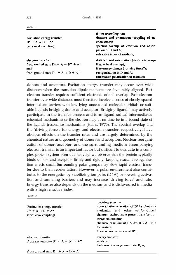
Table 1
donors and acceptors. Excitation energy transfer may occur over widedistances when the transition dipole moments are favourably aligned. Fastelectron transfer requires sufficient electronic orbital overlap. Fast electrontransfer over wide distances must therefore involve a series of closely spacedintermediate carriers with low lying unoccupied molecular orbitals or suit-able ligands bridging donor and acceptor. Bridging ligands may activelyparticipate in the transfer process and form ligand radical intermediates(chemical mechanism) or the electron may at no time be in a bound state ofthe ligands (resonance mechanism) (Hains, 19’75). The spectral overlap andthe ‘driving force’, for energy and electron transfer, respectively, haveobvious effects on the transfer rates and are largely determined by thechemical nature and geometry of donors and acceptors. Nuclear reorgani-zation of donor, acceptor, and the surrounding medium accompanyingelectron transfer is an important factor but difficult to evaluate in a com-plex protein system even qualitatively; we observe that the protein typicallybinds donors and acceptors firmly and rigidly, keeping reactant reorganiza-tion effects small. Surrounding polar groups may slow rapid electron trans-fer due to their reorientation. However, a polar environment also contri-butes to the energetics by stabilizing ion pairs (D + A-) or lowering activa-tion and tunneling barriers and may increase ‘driving force’ and rate.Energy transfer also depends on the medium and is disfavoured in mediawith a high refractive index.Table 2
R. Huber 579
Processes competing with productive energy and electron transfer fromexcited states ‘lurk’ everywere (Table 2). Quite generally, they are minimizedby high transfer rates and conformational rigidity of the cofactors imposedby the protein.
I will discuss these factors in relation to the biological structures later on.
2. The role of cofactorsThe naturally occurring amino acids are transparent to visible light andseem also to be unsuitable as single electron carriers with the exception oftyrosine. Tyrosyl radicals have been identified in PS II as and interme-diates, which are involved in electron transfer from the water splittingmanganese protein complex to the photooxidized P680+ (for reviews, seeBarber, 1987; Prince, 1988). Their identification has been assisted by theobservation that Tyr L162 lies in the electron transfer path from the
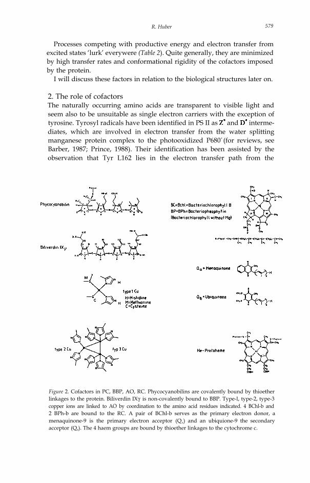
Figure 2. Cofactors in PC, BBP, AO, RC. Phycocyanobilins are covalently bound by thioetherlinkages to the protein. Biliverdin IXγ is non-covalently bound to BBP. Type-l, type-2, type-3copper ions are linked to AO by coordination to the amino acid residues indicated. 4 BChl-b and2 BPh-b are bound to the RC. A pair of BChl-b serves as the primary electron donor, amenaquinone-9 is the primary electron acceptor (QA) and an ubiquione-9 the secondaryacceptor (QB). The 4 haem groups are bound by thioether linkages to the cytochrome c.

580 Chemistry 1988
cytochrome to BCLP in the bacterial RC (Deisenhofer et al., 1985) (see3.2.2.2 and Figure 10c). A tyrosyl radical is not generated in the bacterialsystem, because the redox potential of P960+ is insufficient.
Generally therefore cofactors, pigments and metal ions, serve as lightenergy acceptors and redox active elements in biological materials.
Figure 2 is a gallery of the pigments and metals clusters which will bediscussed further on, namely the bile pigments, phycocyanobilin and biliver-din in the light harvesting complexes, the BChl-b, BP-b, and quinonesin the purple bacterial RC and the copper centres in the blue oxidases.
The physical chemical properties of these cofactors determine the coarsefeatures of the protein pigment complexes, but the protein part exerts adecisive influence on the spectral and redox properties.
3. The role of the proteinThe role of the protein follows a hierarchy in determining the properties ofthe functional protein cofactor complexes shown in Table 3. These interac-tions are different for the various systems and shall be described separately,except point 1, as there are common features in the action of the protein asa polydentate ligand ascribed to a ‘rack mechanism’.
Table 3. Hierarchy of protein cofactor interactions
1. Influence on configuration and conformation of the cofactors by the nature and geometry ofligands (the protein as a polydentate ligand).
2. Determination of the spatial arrangements of arrays of cofactors (the protein as a scaffold ).3. The protein as the medium.4. Mediation of the interaction with other components in the supramolecular biological system.
3.1. The protein as a polydentate ligandThe ‘rack mechanism’ was introduced by Lumry and Eyring (Lumry &Eyring, 1954) and Gray and Malmström (Gray & Malmström, 1983) toexplain unusual reactivities, spectral and redox properties of amino acidsand cofactors by the distortion enforced by the protein.
A comparison of isolated and protein-bound bile pigments gives a cleardemonstration of this effect. Isolated bile pigments in solution and in thecrystalline state prefer a macrocyclic helical geometry with configurationZZZ and conformation syn, syn, syn and show weak absorption in the visiblerange and low fluorescence quantum yield (Scharnagl et al., 1983; Huber etal., 1987a,b). When bound as cofactors to light harvesting phycocyaninsthey have strong absorption in the visible range and high fluorescence yield(Figure 3). The auxochromic shift, essential for the light harvesting func-tions, is due to a strained conformation of the chromophore, which hasconfiguration ZZZ and conformation anti, syn, anti stabilized by tight polarinteractions with the protein (Schirmer et. al., 1985, 1986, 1987) (Figure 4).Particularly noteworthy is an aspartate residue (A87 here) bound to thecentral pyrrole nitrogens and conserved in all pigment sites. It influencesprotonation, charge, and spectral properties of the tetrapyrrole systems.

R. Huber 581
Figure 3. Tetrapyrrole structures in PC and BBP and the associated optical andcircular dichroism spectra (Schirmer et al. 1987; Huber et al., 1987 b).
Tight binding is also effective against deexcitation by conformationalchanges. The structure shown in Figure 3 as representative of the freepigment is in fact observed in a bilin binding protein from insects (Huber etal., 1987a,b). This protein serves a different function and prefers the lowenergy conformer. The open chain tetrapyrrole bilins are conformationallyadaptable, a property, which makes them appropriate cofactors for differ-ent purposes.
The cyclic BChl in the RC is conformationally restrained but responds tothe environment by twisting and bending of the macrocycle. This may beone cause for the different electron transfer properties of the two pigmentbranches in the RC as will be discussed later. A more profound influence of

582 Chemistry 1988
Figure 4. Stereo drawing of phycocyanobilin A84 (thick bonds) and its protein environment(thin bonds). All polar groups of the bilin except those of the terminal D pyrrole ring are boundby hydrogen bonds and salt links to protein groups (Schirmer et al., 1987).
the protein on the RC pigment system is seen in the absorption spectra,which differ from the composite spectra of the individual components(Figure 5). The protein binds a pair of BChl-b (BCP) so that the two BChl-binteract strongly between their pyrrole rings I including the acetyl substitu-ents and the central magnesium ions (Deisenhofer et al., 1984). Alignmentof the transition dipole moments and close approach cause excitonic coup-ling which partially explains the long wavelength absorption band P960(Knapp et al., 1985).
The optical spectra are even more perturbed in blue copper proteinscompared with cupric ions in normal tetragonal coordination (Figure 6 ).The redox potential is also raised to about 300 - 500 mV vs 150 mV forCu2+ (aq) (Gray & Solomon, 1981). These effects are caused by the distort-ed tetraedral coordination of the type-l copper (a strained conformationstabilizing the cuprous state) and a charge transfer transition from a ligandcysteine S- Cu2 + (Blair et al., 1985; Gray & Malmström, 1983).
The examples presented demonstrate the influence of the protein on thecofactors by various mechanisms, stabilization of unstable conformers andstrained ligand geometries and the generation of contacts between pig-ments leading to strong electronic interaction.
The fixation of the relative arrangements of systems of cofactors is thebasis of the energy and charge transfer properties in each system.

R. Huber 583
Figure 5. Stereodrawing of the special pair BCP in the RC (Deisenhofer et al. 1984) mainlyresponsible for the spectral alterations and the long wavelength absorption of the RC of Rps.viridis (- - -) compared with the spectra of BChl-b in ether solution (-) (spectra from Parsonet al.. 1985).
Figure 6. The type-l copper and its ligands in AO in stereo. The coordination of the copper isto His A446, His A513, Met A518, Cys A508 (Messerschmidt et al., 1989). The opticalabsorption spectra of “blue” copper in copper proteins (-) are compared with normal tetra-gonal copper (- - -) (spectrum from Gray & Solomon, 1981).

584 Chemistry I988
3.2. Protein as a scaffold3.2.1. Light harvesting by phycobilisomesThe limited number of pigment molecules associated with RC would absorbonly a small portion of incident sunlight. The RC are therefore associatedwith LHC, which may be located within the photosynthetic membrane, orform layers or antenna-like organelles in association with the photosyntheticmembrane. Cyanobacteria have particularly intricate light harvesting sys-tems, the PBS organelles peripheral to the thylakoid membrane. Theyabsorb light of shorter wavelengths than do PS I and II, so that a widespectral range of sunlight is used (Figure 7). The PBS are assembled fromcomponents with finely tuned spectral properties such that the light energyis channeled along an energy gradient to PS II.
3.2.1.1. MorphologyPBS consist of biliproteins and linker polypeptides. Biochemical and elec-tron microscopy studies (Gantt et al., 1976; Mörschel et al., 1977; Bryant etal., 1979; Nies & Wehrmeyer, 1981) lead to the model representative of ahemidiscoidal PBS in Figure 7. Accordingly PBS rods are assembled in apolar way from PE or PEC and PC, which is attached to a central core ofAPC. APC is next to the photosynthetic membrane and close to PS II (for areview see, MacColl & Guard-Friar, 1987). The PC component consists of α−and P-protein subunits, which are arranged as disc-like aggregates withdimensions 120Å x 60Å (for reviews, see Scheer, 1982; Cohen-Bazire &Briant, 1982; Glazer, 1985; Zilinskas & Greenwald, 1986; Zuber, 1985,1986).
From crystallographic analyses, a detailed picture of PC and PEC compo-nents has emerged (Schirmer et al. 1985, 1986, 1987; Duerring, 1988;Duerring et al., 1989). Amino acid sequence homology suggests that allcomponents have similar structures.
R. Huber 585
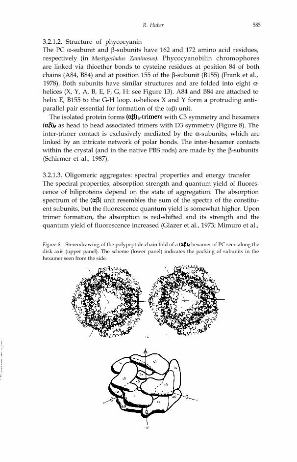
3.2.1.2. Structure of phycocyaninThe PC α-subunit and β-subunits have 162 and 172 amino acid residues,respectively (in Mastigocladus Zaminosus). Phycocyanobilin chromophoresare linked via thioether bonds to cysteine residues at position 84 of bothchains (A84, B84) and at position 155 of the β-subunit (B155) (Frank et al.,1978). Both subunits have similar structures and are folded into eight α-helices (X, Y, A, B, E, F, G, H: see Figure 13). A84 and B84 are attached tohelix E, B155 to the G-H loop. α-helices X and Y form a protruding anti-parallel pair essential for formation of the (αβ) unit.
The isolated protein forms with C3 symmetry and hexamers as head to head associated trimers with D3 symmetry (Figure 8). The
inter-trimer contact is exclusively mediated by the α-subunits, which arelinked by an intricate network of polar bonds. The inter-hexamer contactswithin the crystal (and in the native PBS rods) are made by the β-subunits(Schirmer et al., 1987).
3.2.1.3. Oligomeric aggregates: spectral properties and energy transferThe spectral properties, absorption strength and quantum yield of fluores-cence of biliproteins depend on the state of aggregation. The absorptionspectrum of the unit resembles the sum of the spectra of the constitu-ent subunits, but the fluorescence quantum yield is somewhat higher. Upontrimer formation, the absorption is red-shifted and its strength and thequantum yield of fluorescence increased (Glazer et al., 1973; Mimuro et al.,
Figure 8. Stereodrawing of the polypeptide chain fold of a hexamer of PC seen along thedisk axis (upper panel). The scheme (lower panel) indicates the packing of subunits in thehexamer seen from the side.

586 Chemistry 1988
Figure 9a. Arrangement of chromophores and preferred energy transfer pathways in trimers, hexamers and stacked hexamers based on Table 10 in Schirmer et al.. (1987). Forthe trimer the detailed structures of the chromophores are drawn, otherwise their approximatetransition dipole directions are indicated. For the trimer and hexamer the view is along the diskaxis; for the stacked hexamers it is perpendicular. In the stacked hexamers only the inter-hexamer transfers are indicated. The strength of coupling is indicated by the thickness of theconnecting lines. Transfer paths within and between the trimers are represented by full andbroken lines, respectively.

R. Huber 587
1986; for a review, see Glazer, 1985). In the complexes, thefluorescence is further increased and the absorption spectrum furtheraltered (Lundell et al., 1981).
These observations can be rationalized by the structure of the aggregates.Formation of units causes little change in the environment of thechromophores. They remain quite separated with distances > 36Å (Figure9a). Upon trimer formation, the environment of chromophore A84 changesprofoundly by approach of chromophore B84 of a related unit (Figure 9a,upper panel). In the hexamer (Figure 9a, middle panel) the A84 and B155chromophores interact pairwise strongly across the trimer interface. Alsothe molecular structures become more rigid with increasing size of theaggregates as seen in the crystals of the trimeric and hexameric aggregates(Schirmer et al., 1986, 1987). Rigidity hinders excitation relaxation byisomerization and thus increases the fluorescence quantum yield.
The chromophores can be divided into subsets of s (sensitizing) and f(fluorescing) chromophores (Teale & Dale, 1970; Zickendraht-Wendelstadtet al., 1980). The s-chromophores absorb at the blue edge of the absorptionband and transfer the excitation energy rapidly to the f-chromophores. Thistransfer is accompanied by depolarization (Hefferle et al., 1983). Excitationat the red absorption edge (f-chromophores), however, results in littledepolarization, suggesting that the energy is transferred along stacks ofsimilarly oriented f-chromophores (Gillbro et al., 1985). The assignment ofthe chromophores to s and f was made by steady-state spectroscopy ondifferent aggregates (Mimuro et al., 1986), by chemical modification guidedby the spatial structure (Siebzehnriibl et al., 1987) and conclusively bymeasurement of linear dichroism and polarized fluorescence in single cry-stals (Schirmer & Vincent, 1987). Accordingly B155 is the s-, B84 the f-, andA84 the intermediate chromophore.
Light energy is transferred rapidly within 50 to 100 psec from the tips ofthe PBS to the core (for a review, see e.g. Glazer, 1985; Porter et al., 1978;Searle et al., 1978; Wendler et al., 1984; Yamazaki et al., 1984; Gillbro etal., 1985; Holzwarth, 1986). The transfer times from the periphery to thebase are several orders of magnitude faster than the intrinsic fluorescencelife-times of the isolated components (Porter et al., 1978; Hefferle et al.,1983). The distances between the chromophores within and between thehexamers are too large for strong (excitonic) coupling, but efficient energytransfer by inductive resonance occurs. A Förster radius of about 50Å hasbeen suggested by Grabowski & Gantt (1978). The relative orientations anddistances of the chromophores as obtained by Schirmer et al. (1987) werethe basis for the calculation of the energy transfer rates in Figure 9a. Itshows the preferred energy transfer pathways in units, trimers,
hexamers and stacked disks as models for native antenna rods. There isvery weak coupling of the chromophores in the units. Some energytransfer takes place, however, as indicated by steady-state polarizationmeasurements (Switalski & Sauer, 1984; Mimuro et al., 1986) probablybetween B155 and B84. Trimer formation generates strong coupling be-


R. Huber 589
tween A84 and B84, but B155 is integrated only weakly. In the hexamermany additional transfer pathways are opened and B155 is efficiently coup-led. Hexamers are obviously the functional units, as the energy can bedistributed and concentrated on the central f-chromophores, which couplethe stacks of hexamers. Kinetic studies (Glazer et al., 1985; Gillbro et al.,1985; Holzwarth, 1985; Mimuro et al., 1986) have confirmed the picture ofenergy transfer along the rods as a random walk (trap or diffusion limited)along a one-dimensional array of f-chromophores. Sauer et al. (1987) havesuccessfully simulated the observed energy transfer kinetics in PC aggre-gates on the basis of the structures using Förster’s mechanism. The PECcomponent at the tips of PBS rods is extremely similar to PC (Duerring,1989; Duerring et al., 1989). Its short wavelength absorbing chromophoreA84 is located at the periphery (Figure 10a) as are the additional chromo-phores in PE which is also a tip component (Figure 9b).
The phycobilisome rods act as light collectors and energy concentratorsfrom the peripheral onto the central chromophores, that is, as excitationenergy funnels from the periphery to centre and from the tip to the bottom.
We may expect functional modulations by the linker polypeptides. Someof them are believed to be located in the central channel of the hexamers,where they may interact with B84.
3.2.2. Electron transfer in the reaction centre*3.2.2.1. Reaction centre, composition**The RC of Rps. viridis is a complex of four protein subunits, C, L, M, H andcofactors arranged as in Figure 10a. As shown by the amino acid sequencethey consist of 336, 273, 323, and 258 residues, respectively (Michel et al.,1985; Michel et al., 1986a; Weyer et al., 1987). The c-type cytochrome hasfour haem groups covalently bound via thioether linkages. The cofactors
are four BChl-b (BCMP, BCLP, BCLA, two BPh-b (BPM, BPI), onemenaquinone-9 (QA), and a ferrous iron involved in electron transfer. Asecond quinone (ubiquinone-9) (QB), which is a component of the function-al complex, is partially lost during preparation and crystallization of the RC.
3.2.2.2. Chromophore arrangement and electron transferThe chromophores are arranged in L- and M-branches related by an axis ofapproximately two-fold symmetry which meet at BCP (Deisenhofer et al.,1984). This axis is normal to the plane of the membrane.
While many of the optical properties of the pigment system are ratherwell understood on the basis of the spatial structure (Knapp et al., 1985),electron transfer is less well understood. The excited BCP is quenched byelectron transfer to BPL in 3 psec and further on to the primary acceptorQA in about 200 psec, driven by the redox potential gradient between*
A historical background of the development of concepts and key features of the purplebacterial reaction centre is given by Parson (1978).**
The arrangement of the reaction centre in the thylakoid membranes of Rps. viridis asobtained by electron microscopy is described by Stark et al. (1984).

590 Chemistry 1988
P*/P+ (about - 760 mV) and (about - 110 mV). The redox poten-tial of BP- is intermediate with about - 400 mV (Cogdell and Crofts, 1972;Carithers and Parson, 1975; Prince et al., 1976; Netzel et al., 1977; Bolton,1978; Holten et al., 1978; Woodbury et al., 1985; Breton et al., 1986).These functional data are summarized in Figure 10a. General factors con-trolling the transfer rates have been summarized in Table 1 and are detailedfor the RC here:
Fast electron transfer requires effective overlap of the molecular orbitals.The orbital interaction decreases exponentially with the edge to edge dis-tance of donor and acceptor and is insignificant at distances larger thanabout 10Å (Kavarnos and Turro, 1986; McLendon, 1988). In the RC thedistance between BCp and QA is far too large to allow fast direct electrontransfer; instead the electron migrates via BPL. BP L
- is a spectroscopicallyand kinetically well defined intermediate. Although located between BCP
and BPL, BCLA- is not an intermediate but is probably involved in electron
transfer by a “superexchange” mechanism mediating a strong quantummechanical coupling (Fleming et al., 1988; for a review see Barber, 1988).The distance between BPL and QA seems large for a fast transfer. Indeed the

R.Huber 591
Figure 10b. Stereo drawing of the arrangement of BPL, Trp M250 and QA in the L-branch of theRC pigment system.
gap is bridged by the aromatic side chain of Trp M250 in the L branch ofthe pigment system (Figure 10b) (Deisenhofer et al., 1985; Michel et al.,1986b), which might mediate coupling via appropriate orbitals. In addition,the isoprenoid side chain of QA is close to BPL. Electron transfer via longconnecting chains by through-bond coupling of donor and acceptor orbitalshas been observed (Pasman et al., 1982; Moore et al., 1984; Kavarnos &Turro, 1986) but there is only Van der Waals contact here.
A second important factor for electron transfer is the free energy change(∆ G), which is governed by the chemical nature of the components, bygeometrical factors, and by the environment (solvent polarity). It dependson the ionization potential of the donor in its excited state, the electronaffinity of the acceptor, and on the coulombic interaction of the radical ionpair, which is probably small as donor and acceptor (BCP and QA in the RC)are far apart. The effect of the environment may be substantial by stabiliz-ing the radical ion pair by ionic interactions and hydrogen bonds. ∆ G is adeterminant of the activation energy of electron transfer. Another is nucle-ar rearrangements of the reactants and the environment. As the charge ondonor and acceptor develops the nuclear configurations change. Thesechanges are likely to be small in the RC as the BChl-b macrocycles arerelatively rigid and tightly packed in the protein and the charge is distribut-ed over the extended aromatic electron systems. Reorientable dipolargroups (peptide groups and side chains) may contribute strongly to theenergy barrier of electron transfer. A matrix with high electronic polariza-bility on the other hand stabilizes the developing charge in the transitionstate of the reaction and reduces the activation energy. An alternativepicture is that the potential energy barrier to electron tunnelling is de-creased. Aromatic compounds which are concentrated in the vicinity of theelectron carriers in the RC have these characteristics (see Trp M250).
The electron transfer from P* to QA occurs with very low activationenergy (Arnold and Clayton, 1960; Parson, 1974; Parson and Cogdell,1975; Carithers and Parson, 1975; Bolton, 1978; Kirmaier et al., 1985;Woodbury et al., 1985) and proceeds readily at 1°K. Thermally activated

592 Chemistry 1988
processes, nuclear motions, and collisions are therefore not important forthe initial very fast charge separation steps. There is even a slight increase inrate with temperature decrease either due to a closer approach of thepigments at low temperature, or to changes of the vibrational levels whichmay lead to a more favourable Franck-Condon factor.
The electron transfer between primary and secondary quinone acceptors,QA and QB is rather different from the previous processes, because it ismuch slower (about 6 µs at pH7, derived from Carithers and Parson, 1975)and has a substantial activation energy of about 8 kcal mol-1. In Rps. viridisQA is a menaquinone-9 and QB a ubiquinone-9, which differ in their redoxpotentials in solution by about 100 mV. In other purple bacteria both QA
and QB are ubiquinones. The redox potential difference required forefficient electron transfer in these cases is generated by the asymmetricprotein matrix. The protein matrix is also responsible for the quite differentfunctional properties of QA and QB. QA accepts only one electron (leadingto a semiquinone anion), which is transferred to QB before the next electrontransfer can occur. QB, however, accepts two electrons and is protonated toform a hydroquinone, which diffuses from the RC (two-electron gate(Wraight, 1982)). QB is close to Glu L212, which opens a path to the H-subunit and may protonate QB. The path between QA and QB is verydifferent from the environment of the primary electron transfer compo-nents. The line connecting QA and QB (the QB binding site has been inferredfrom the binding mode of competitive inhibitors and ubiquinone-1 in Rps.viridis crystals (Deisenhofer et al., 1985)) is occupied by the iron and its fivecoordinating ligands, four histidine (M217, M264, L190, L230) and aglutamic acid (M232) residue. His M 217 forms a hydrogen bond to QA. HisL 190 is close to QB. QAand QB have an edge to edge distance of about 15Å which might explain the slow transfer. If electron transfer and protonationare coupled, the observed pH dependence of the electron transfer rate ofQA to QB (Kleinfeld et al., 1985) could be explained and nuclear motionsrequired for proton transfer may generate the observed activation energybarrier. The role of the charged Fe-His4-Glu complex in the QA to QB
electron transfer is poorly understood at present, as it also occurs in theabsence of the iron (Debus et al., 1986). Its role seems to be predominantlystructural.
The cycle of electron transfer is closed by rereduction of the BCp* fromthe cytochrome bridging a distance of about 11Å between pyrrole ring I ofhaem 3 and pyrrole ring II of BCLP. The transfer time is 270 ns (Holten etal., 1978; elder measurements in Case et al., 1970), considerably slowerthan the initial processes. Tyr L162, which is located midway (Figure 10c)may facilitate electron transfer by mediating electronic coupling betweenthe widely spaced donor and acceptor. The biphasic temperature depen-dance indicates a complex mechanism in which at high temperatures nucle-ar motions play a role (for a review see, Dutton & Prince, 1978; DeVault &Chance, 1966).
The favourable rate controlling factors discussed are a necessary, but notsufficient condition for electron transfer, which competes with other
R. Huber 593
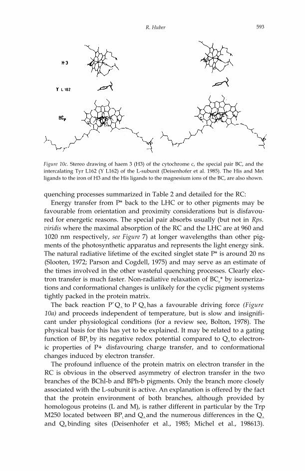
Figure 10c. Stereo drawing of haem 3 (H3) of the cytochrome c, the special pair BC, and theintercalating Tyr L162 (Y L162) of the L-subunit (Deisenhofer et al. 1985). The His and Metligands to the iron of H3 and the His ligands to the magnesium ions of the BC, are also shown.
quenching processes summarized in Table 2 and detailed for the RC:Energy transfer from P* back to the LHC or to other pigments may be
favourable from orientation and proximity considerations but is disfavou-red for energetic reasons. The special pair absorbs usually (but not in Rps.viridis where the maximal absorption of the RC and the LHC are at 960 and1020 nm respectively, see Figure 7) at longer wavelengths than other pig-ments of the photosynthetic apparatus and represents the light energy sink.The natural radiative lifetime of the excited singlet state P* is around 20 ns(Slooten, 1972; Parson and Cogdell, 1975) and may serve as an estimate ofthe times involved in the other wasteful quenching processes. Clearly elec-tron transfer is much faster. Non-radiative relaxation of BCp* by isomeriza-tions and conformational changes is unlikely for the cyclic pigment systemstightly packed in the protein matrix.
The back reaction P+ QA- to P QA has a favourable driving force (Figure
10a) and proceeds independent of temperature, but is slow and insignifi-cant under physiological conditions (for a review see, Bolton, 1978). Thephysical basis for this has yet to be explained. It may be related to a gatingfunction of BPL by its negative redox potential compared to QA to electron-ic properties of P+ disfavouring charge transfer, and to conformationalchanges induced by electron transfer.
The profound influence of the protein matrix on electron transfer in theRC is obvious in the observed asymmetry of electron transfer in the twobranches of the BChl-b and BPh-b pigments. Only the branch more closelyassociated with the L-subunit is active. An explanation is offered by the factthat the protein environment of both branches, although provided byhomologous proteins (L and M), is rather different in particular by the TrpM250 located between BPL and QA and the numerous differences in the QA
and QB binding sites (Deisenhofer et al., 1985; Michel et al., 198613).

594 Chemistry 1988
Asymmetry is observed in the BC, due to different distortions and hydrogenbonding of the macrocycles and in the slightly different spatial arrange-ments of the BCA and BP. It is suggested to facilitate electron release intothe L-branch (Michel-Beyerle et al.; 1988). The M-branch may have influ-ence as a pendant group though.
The protein matrix also serves to dissipate the excess energy of about 650mV (Prince et al., 1976) of the excited special pair (P* QA) over the radicalion pair P+QA
-. These processes are probably very fast.In summary, the very fast electron transfer from BCp* to QA occurs
between closely spaced aromatic macrocyles with matched redox potentials.The protein matrix in which the pigments are tightly held is lined predomi-nantly with apolar amino acid side chains with a high proportion of aromaticresidues. The electron path is removed from bulk water.
3.2.3. The blue oxidasesOxidases catalyse the reduction of dioxygen in single electron transfersfrom substrates. Dioxygen requires 4 electrons and 4 protons to be reducedto two water molecules. Oxidases must provide recognition sites for the twosubstrates, a storage site for electrons and/or means to stabilize reactivepartially reduced oxygen intermediates (Malmström, 1978, 1982; Farver &Pecht, 1984)
The ‘blue’ oxidases are classified corresponding to distinct spectroscopicproperties of the three types of copper which they contain: Type-l Cu++ isresponsible for the deep blue color of these proteins; type-2 or normalCu++ has undetectable optical absorption; type-l and type-2 cupric ionsare paramagnetic; type-3 copper has a strong absorption around 330 nmand is antiferromagnetic, indicating coupling of a pair of cupric ions. Thecharacteristic optical and electron paramagnetic resonance spectra disap-pear upon reduction.
Studies of the catalytic and redox properties of the ‘blue’ oxidases arewell documented in several recent reviews (e.g. for lactase, Reinhammar,1984; for ascorbate oxidase, Mondovi & Avigliano, 1984; for ceruloplas-min, Rydén, 1984). Basically type-l Cu++ is reduced by electron transferfrom the substrate. The electron is transferred on to the type-3 and type-2copper ions: The second substrate, dioxygen, is associated with the type-3and/or type-2 copper ions.
3.2.3.1. Ascorbate oxidase, composition and copper arrangementAscorbate oxidase is a polypeptide of 553 amino acid residues folded intothree tightly associated domains (Messerschmidt et al., 1989). It is a dimerin solution, but the functional unit is the monomer. It belongs to the groupof ‘blue’ oxidases together with laccase and ceruloplasmin (Malkin & Malm-ström, 1970).
Structures of copper proteins containing only one of the different coppertypes are known: Plastocyanin has a ‘blue’ type-l copper, which is coordi-nated to two histidine residues and the sulfur atoms of cysteine and methio-

R. Huber 595
nine as a distorted tetrahedron (Guss & Freeman, 1983). Cu-Zn-superoxidedismutase contains a type-2 copper, which has 4 histidine ligands withslightly distorted quadratic coordination (Richardson et al., 1975). Hemo-cyanin of Panulirus interruptus has type-3 copper, a pair of copper ions 3.4Å apart with 6 histidine ligands (Gaykema et al., 1984).
In domain 3 of ascorbate oxidase (see section 4.4.) a copper ion is foundin a strongly distorted tetrahedral (approaching trigonal pyramidal geome-try) coordination by the ligands His, Cys, His, Met as had been shown inFigure 6. It resembles the blue type-l copper in plastocyanin. Betweendomain 1 and domain 3, a trinuclear copper site is enclosed and shown inFigure 11a. Four (-His-X-His-) amino acid sequences provide the eighthistidine ligands. The trinuclear copper site is subdivided in a pair ofcoppers (Cu31, Cu32) with 2x3 histidyl (A108, A451, A507; A64, A106,A509) ligands forming a trigonal prism. It represents the type-3 copperpair, as a comparable arrangement is observed in hemocyanin. The remain-ing copper (Cu 2) has two histidyl ligands (A62, A449). It is type-2 copper.The trinuclear copper cluster is the site where dioxygen binds, but thestructural details including the presence of additional non-protein ligandsrequire clarification. The close spatial association of the three copper ionsin the cluster suggests facile electron exchange. It may function as anelectron storage site and cooperative three-electron donor to dioxygen, toirreversibly break the O - O bond.
3.2.3.2. Intramolecular electron transfer in ascorbate oxidaseElectrons are transferred from the type-l copper to the trinuclear site. Theshortest pathway is via Cys-A508 and His-A507 or His-A509. The (His-X-His-) segment links electron donor and acceptor bridging a distance of 12Å
Figure 11a. Stereo drawing of the trinuclear copper site in AO. Coordination bonds betweenthe copper ions and the protein residues are marked (- - - -) (Messerschmidt et al., 1989).

596 Chemistry 1988
Figure 11b. Stereo drawing of the tridentate peptide ligand (-His 507-Cys 508-His 509-) bridg-ing type-l copper (Cu 1) and the trinuclear cluster (Cu 31, Cu 32, Cu 2) (Messerschmidt et al.1989).
(Figure 11b). The cysteine sulfur and the imidazole components of thebridging ligand have low lying unoccupied molecular orbitals and mayfavour a chemical mechanism of electron transfer but the interveningaliphatic and peptide chains are unlikely to form transient radicals and mayparticipate by resonance. The optical absorption of the blue copper as-signed to a cysteine S charge transfer transition supports thesuggested electron pathway.
The putative electron path branches at the atom of Cys A508. Modelcompounds have shown inequivalence and faster transfer in the N-C direc-tion of amide linking groups (Schmidt et al., 1988). This may apply also tothe blue oxidases and cause preferred transfer to A507.
The redox potential differences between the type-l copper and the type-3copper are -40 mV in ascorbate oxidase. Unfortunately, there are nodirect measurements of the intramolecular electron transfer rates available.The turnover number serves as a lower limit and is 7.5 X 10³ sec-1 in AO(Dawson, 1966; Get-win et al., 1974) indicating a quite rapid transfer despitethe long distance and small driving force. The electron pathway is intramo-lecular and removed from bulk water.
The characteristic distribution of redox centres as mono- and trinuclearsites in the blue oxidases may be found also in the most complex oxidase,cytochrome oxidase (see the hypothetical model of Holm et al., 1987) and inthe water-splitting manganese protein complex of PS II, which carries outthe reverse reaction of the oxidases. For its (Mn)4 cofactor either twobinuclear or a tetranuclear metal centre is favoured (Babcock, 1987), butmono- and trinuclear arrangements can not be excluded.

R. Huber 597
3.3. The protein as mediumThe boundary between the action of the protein as ligand and as medium isfluid. The protein medium is microscopically extremely complex in struc-ture, polarity and polarisability, which may influence energy and electrontransfer. There is no obvious common structural scheme in the proteinsystems discussed except a high proportion of aromatic residues (particular-ly tryptophans) bordering the electron transfer paths in RC and AO andtheir wide separation from bulk water by internal location within the pro-tein and the hydrocarbon bilayer (in RC). These effects have been men-tioned in sections 1.3 and 3.2.2.2.
4. Structural relationships and internal repeatsAll four protein systems mentioned show internal repetition of structuralmotifs or similarities to other proteins of known folding patterns. This is aquite common phenomenon and not confined to energy and electrontransfer proteins. It is also not uncommon that these relationships oftenremained undetected on the basis of the amino acid sequences, ultimately areflection of our ignorance about the sequence structure relationships. Ananalysis of structural relationships will shed light on evolution and functionof the protein systems and is thus appropriate here.
4.1. Retinol and bilin binding proteinsThe simplest case is shown in Figure 12, where BBP (Huber et al., 1987) iscompared to RBP (Newcomer et al., 1984). The structural similarity isobvious for the bottom of the P-barrel structure, while the upper part whichis involved in binding of the pigments, biliverdin and retinol, differs greatly.The molecule is apparently divided in framework and hypervariable seg-ments which determine binding specificity in analogy to the immunoglobu-lins (Huber, 1984). The relationship suggests carrier functions for BBP asfor RBP, although it serves also for pigmentation in butterflys.
Figure 12 . Comparison of the polypeptide chain folds of BBP and RBP with bound cofactors.

598 Chemistry 1988
4.2. PhycocyaninThe PC consist of two polypeptide chains α and β which are clearly relatedin structure (Figure 13) and originate probably from a common precursor.
The α-subunit is shorter in the .GH turn and lacks the s-chromophoreB155 (see section 3.2.1.3.). The loss or acquisition of chromophores duringevolution may be less important than differentiation of the and subun-its, which occupy non-equivalent positions in the trimer, so that thehomologous chromophores A84 and B84 are non-equivalent with B84 lyingon the inner wall of the disk. In addition the α and β subunits play verydifferent roles in the formation of the hexamer as had been shown inFigure 8. Symmetrical precursor hexamers might have existed and could
Figure 13. Polypeptide chain folds of the a- and P-subunits of phycocyanin (Schirmer et al.1987) (lower part, left and right) and comparison of the arrangements of a-helices in myoglobinand phycocyanin (upper part, left and right).

R. Huber 599
have formed stacks, but would lack the differentiation of the chromo-phores, in particular the inequivalence and close interaction of A84 andB84 in the trimer. Functional improvement has probably driven divergentevolution of the and β-subunits. .
A most surprising similarity was discovered between the PC subunits andthe globins shown in Figure 23. The globular helical assemblies A to H showsimilar topology. The N-terminal X,Y forming a U-shaped exten-sion in PC is essential for formation of the substructure. The amino acidsequence comparison after structural superposition reveals some homologysuggesting divergent evolution of phycobiliproteins and globins (Schirmeret al., 1987), however, what function a precursor of light harvesting andoxygen binding proteins might have had remains mysterious.
4.3. Reaction centreThe RC lacks symmetry across the membrane plane, not surprising for acomplex, which catalyses a vectorial process across the membrane. How-ever, there is quasi-symmetry relating the L- and M subunits and thepigment system. Structural similarity and amino acid sequence homology
Figure 14. Stereo drawing of the polypeptide fold of the RC subunits and the cofactor system.The membrane-spanning α-helices of the L- and M-subunits (A, B, C, D, E in sequential and A,B, C, E, D in spatial order) and the H-subunit (H) are labelled (Deisenhofer et al., 1985).

600 Chemistry 1988
between the L- and M-subunits suggest a common evolutionary origin. Thisrelationship is extended to the PS II components Dl and D2 on the basis ofsequence homology and conservation of residues involved in cofactor bind-ing (for reviews, see Trebst, 1986;. Michel & Deisenhofer, 1988). Theputative precursor was a symmetrical dimer with identical electron transferpathways. The interaction with the H subunit introduces asymmetry, par-ticularly noteworthy at the N-terminal transmembrane α-helix of the H-subunit (H), which is close to the E transmembrane α-helix of the M-subunitand the L-branch of the pigment system and QA (Figure 14). The improve-ment of the interaction with the H-subunit, which appears to play a role inthe electron transfer from QA to QB and in protonation of QB might havedriven divergent evolution of the L- and M-subunits at the expense of theinactivation of the M pigment branch. However, the electron transfer fromBC, to QA is extremely fast and not rate-limiting for the overall reaction.The evolutionary conservation of the M branch of pigments may be offunctional significance in light harvesting and electron transfer as a pendantgroup. There are also structural reasons, as its deletion would generate voidspace.
The cytochrome subunit adds to the asymmetry of the L-M complex andshows itself an internal duplication (Deisenhofer et al., 1985). All four hemegroups are associated with a helix-turn-helix motif, but the turns are shortfor haem groups 1 and 3 and long for 2 and 4.
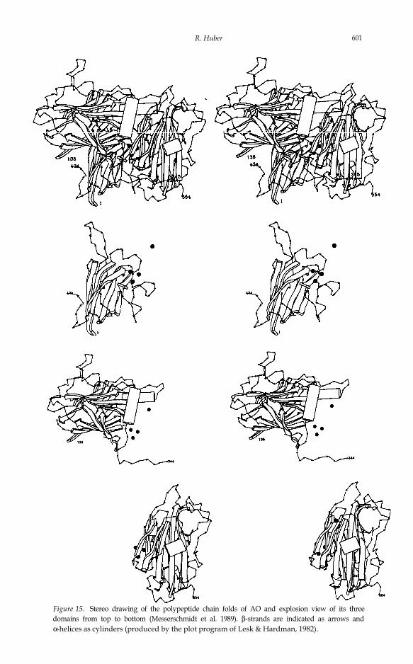
4.4. Blue oxidasesGene multiplication and divergent evolution is most evident in the blueoxidase, ascorbate oxidase. Figure 15 shows the polypeptide chain of 553amino acid residues folded into 3 closely associated domains of similartopology (Messerschmidt et al., 1989). Although nearly twice as large, theyresemble the simple, small copper protein plastocyanin (Guss & Freeman,1983) (Figure 16). In the blue oxidase domains I and III enclose thetrinuclear copper cluster in a quasi-symmetrical fashion, but only domainIII contains-the type 1 copper, the electron donor to the trinuclear site. Apotential electron transfer pathway in domain I is not realized, reminiscentof the M-branch of pigments in the RC. Similar to the H subunit in the RC,the linking domain II introduces asymmetry in AO, which might have drivenevolutionary divergence of domains I and III.
The proteins plastocyanin, ascorbate oxidase, laccase, and ceruloplasminare members of a family of copper proteins as indicated by structuralrelations and sequence homology (Messerschmidt et al., 1989; Ohkawa etal., 1988; Germann et al., 1988; Takahashi et al., 1984). They provide arecord from which an evolutionary tree may be proposed (Figure 17). Thesimplest molecule is plastocyanin containing only a type-l copper. A dimerof plastocyanin-like molecules could provide the 2x4 histidyl ligands for thetrinuclear copper cluster, representing a symmetrical oxidase. From thishypothetical precursor the modern blue oxidases and ceruloplasmin mighthave evolved following different paths of gene (domain) insertion and loss
R. Huber 601
Figure 15. Stereo drawing of the polypeptide chain folds of AO and explosion view of its threedomains from top to bottom (Messerschmidt et al. 1989). β-strands are indicated as arrows andα-helices as cylinders (produced by the plot program of Lesk & Hardman, 1982).

Chemistry 1988
Figure 16. Stereo drawing and superposition of domain III of AO (thin lines) and PCY (thicklines). The trinuclear copper site in AO is buried between domain I (not shown) and domain III.
Figure 17. Homologous domains in plasto-cyanin, ascorbate oxidase, laccase and ceru-loplasmin. The mono- and trinuclear coppersites are indicated.

R. Huber 603
or aquisition of coppers. In both the arrangement of the N- and C-terminaldomains, which contain the functional copper cluster has been preserved.Recombinant DNA technology has the tools to reconstruct the hypotheticalprecursor oxidase. This is under investigation.
5. Implications from the structure of the reaction centre for membraneproteins in generalThe structures of water soluble proteins show a seemingly unlimited diversi-ty, although they are built from only a few defined secondary structuralelements as helices, β-sheets and turns and despite their construction fromdomains and recurring structural motifs. The proteins discussed provideample evidence. That there seems to be a limited set of basic folds may berelated to the evolution of proteins from a basic set of structures and/or toconstraints by protein stability and rates of folding. These basic foldingmotifs do not represent rigid building blocks, however, but adapt to se-quence changes and respond to the environment and association with otherstructural elements. Adaptability and plasticity (which is not to be confusedwith flexibility) is related to the fact that the entire protein and solventsystem must attain the global minimum, not its individual components.Water is a good hydrogen bond donor and acceptor and is thus able tosaturate polar surface exposed peptide groups nearly as well as intraproteinhydrogen bonds do (except for entropic effects).
Membrane proteins face the inert hydrocarbon part of the phospholipidbilayer and must satisfy their hydrogen bonds intramolecularly. Only twosecondary structures form closed hydrogen bonding arrangements of theirmain chains, which satisfy this condition, namely the helix and the β-barrel.For assemblies of α-helices packing rules have been derived which predictcertain preferred angles between the helix axes although with a broaddistribution. Similarly, the arrangement of strands in β-sheets and β-barrelsfollows defined rules (Chothia, 1984).
5.1. Structure of the membrane associated parts of the RCThe structure of the RC may support some conclusions about membraneproteins in general, of which the RC structure was the first to be deter-mined at atomic resolution after the low resolution structure of bacterio-rhodopsin which has some common features (Henderson & Unwin, 1975).The RC has 11 transmembrane α-helices, which consist of 26 residues (H-subunit) or 24-30 residues (L- and M-subunits) appropriate lengths tospan the membrane. The amino acid sequences of these segments aredevoid of charged residues (Figure 18). Few charged residues occur close tothe ends of the α-helices. Glycine residues initiate and terminate almost allα-helical segments, both the transmembrane and the connecting a-helices.It is well known from soluble proteins that glycine residues are abundant inturns and often associated with flexible regions of proteins (Bennett andHuber 1984). They may be important for the insertion into the membraneby allowing rearrangements. The angles between the axes of the contacting

Chemistry 1988
Figure 18. Stereo drawing of the polypeptide chains of the L- and M-subunits of the RC inribbon representation. The N-terminal residues of the membrane-spanning α-helices are la-belled (including the prefix M and L) and the tetra-helical motif of the D and E α-helices ismarked by shading and lines. The side chains of charged residues are drawn. Asp, Glu, andcarboxy-termini as negatively charged, and Lys, Arg and amino-termini are counted as positivelycharged and added for the cytoplasmic and periplasmic sides (Deisenhofer el al., 1985, 1988).
α-helices of the L- and M-complex are inclined by 20º to 30º, a preferredangular range for the packing of the α-helices in soluble proteins. They havefeatures in common with buried α-helices in large globular proteins, whichare also characterized by the absence of charged residues and the prefer-ence of glycines and prolines at the termini (Loebermann et al., 1984;Remington et al., 1982). In addition, the D E α-helices of the L- and M-subunits (Figure 18) find counterparts in soluble proteins. They are associat-ed around the local diad axis and form the centre of the LM module, whichbinds the iron and the BC,. The four D and E α-helices of the L- and M-subunits are arranged as a bundle tied together by the iron ion and splay outtowards the cytoplasmic side to accommodate the large special pair. Thismotif is quite common in soluble electron transfer proteins (Weber andSalemme, 1980). I will resume this discussion later and suggest appropriatesubstructures of soluble proteins as models for pore forming membraneproteins.
5.2. Membrane insertionThe structure of the RC is similarly important for our views of the mecha-nism of integration of membrane proteins into the phospholipid bilayer.The RC is composed of components which are quite differently arrangedwith respect to the membrane: The C-subunit is located on the periplasmicside. The H-subunit is folded into two parts: a globular part located on thecytoplasmic side and a transmembrane α-helix. The L- and M-subunits are

R. Huber 605
incorporated into the phospholipid bilayer. Consequently, C has to becompletely translocated across the membrane from its intracellular site ofsynthesis. In the H-, L-, and M-subunits the transmembrane α-helices areembedded in the bilayer. Only the N-terminal segment of H and the C-termini and connecting segments of the α-helices located at the periplasmicside of L and M (A - B, C - D) require transfer.
It is interesting to note that only the cytochrome gene possesses a prokar-yotic signal sequence, as indicated by the sequence of the gene (Weyer et al.,1987). Transfer of the large hydrophilic C-subunit may require a complextranslocation system and a signal sequence, while H, L, and M may sponta-neously insert into the bilayer due to the affinity of the contiguous hydro-phobic segments with the phospholipids (for a review of this and relatedproblems see Rapoport, 1986). A “simple” dissolution still requires transferacross the membrane of those charged residues which are located at theperiplasmic side (Deisenhofer et al., 1985; Michel et al, 1986b). Theincreasingly favourable protein lipid interaction which develops with inser-tion may assist in this process. M and L have considerably more chargedresidues at the cytoplasmic side (41) than at the periplasmic side (24),providing a lower activation energy barrier for correct insertion. The netcharge distribution of the LM complex is asymmetric with 6 positive chargesat the cytoplasmic side and 8 negative charges at the periplasmic side. As theintracellular membrane potential is negative, the observed orientation ofthe LM complex is energetically favoured (Figure 18).
The H-subunit has a very polar amino acid sequence at the C-terminus ofthe transmembrane α-helix with a stretch of 7 consecutive charged residues(H33 - H39) (Deisenhofer et al., 1985; Michel et al., 1985) which mayefficiently stop membrane insertion. Similarly, there are 3 to 11 chargedresidues in each of the connecting segments of the α-helices at the cytoplas-mic side of the L- and M-subunits, which might stop the transfer of α-helicesor α-helical pairs (Engelman et al., 1986). As an alternative to sequentialinsertion the L-, M-subunits may be inserted into the membrane as assem-bled protein pigment complexes, because they cohere tightly by proteinprotein and protein cofactor interactions.
5.3. Models of pore forming proteinsIt is not obvious whether the structural principles observed in the RC applyalso to ‘pore’ or ‘channel’ forming α-helical proteins. These could, inprinciple, elaborate quite complex structures within the aqueous channel(Lodish, 1988), but available evidence at low resolution for gap junctionproteins (Milks et al., 1988) indicates in this specific case a simple hexamericarrangement of membrane spanning amphiphilic α-helices, whose polarsides face the aqueous channel.
Guided by the observation that rules for structure and packing of helices derived for soluble proteins apply also to the RC, we may derivemodels for membrane pore forming proteins from appropriate solubleprotein substructures. The penta-helical pore seen at high resolution in the

606 Chemistry 1988
icosaedral multi-enzyme complex riboflavin synthase seems to be a suitablemodel (Ladenstein et al., 1988) (Figure 19). 5 amphiphilic of 23residues each are nearly perpendicular to the capsid surface. The coiled coilof has a right-handed twist and forms a pore for the putativeimport of substrates and export of products. They pack with their apolarsides against the central 4-stranded β-sheet of the protein, which mimics thehydrocarbon part of a phospholipid bilayer and project charged residuesinto the aqueous channel.
Similar modelling of membrane protein structures may be extended toanother class of membrane proteins which have β-structures spanning theouter membrane, the bacterial pot-ins (Kleffel et al., 1985). In solubleproteins β-barrels observed have 4 to 8 or more strands. The lower limit isdetermined by the distortion of regular hydrogen bonds. An upper limitmay be given by the possible sizes of stable protein domains. A four-stranded β-barrel with 4 parallel strands duplicated head to head withsymmetry D4 is seen in the ovomucoid octamer (Weber et al., 1981). The strands lean against the hydrophobic core of the molecule and project their(short) polar residues into the channel (which is extremely narrow here).
6. Some thoughts on the future of protein crystallographyThirty years after the elucidation of the first protein crystal structures byPerutz and Kendrew and after steady development, protein crystallographyis undergoing a revolution. Recent technical and methodical developmentsenable us to analyse large functional protein complexes like the RC (Deisen-hofer et al., 1985; Allen et al., 1987), large virus structures (to mention onlyHarrison et al., 1978; Rossmann et al., 1985; Hogle et al., 1985), proteinDNA complexes (to mention only Ollis et al., 1985), and multi-enzymecomplexes like riboflavin synthase (Ladenstein et al., 1988).
The significance of these studies for understanding biological functions isobvious and has excited the interest of the scientific community in general.
Figure 19. Penta-helical pore in heavy riboflavin synthase in stereo (Ladenstein et al., 1988).
R. Huber 607
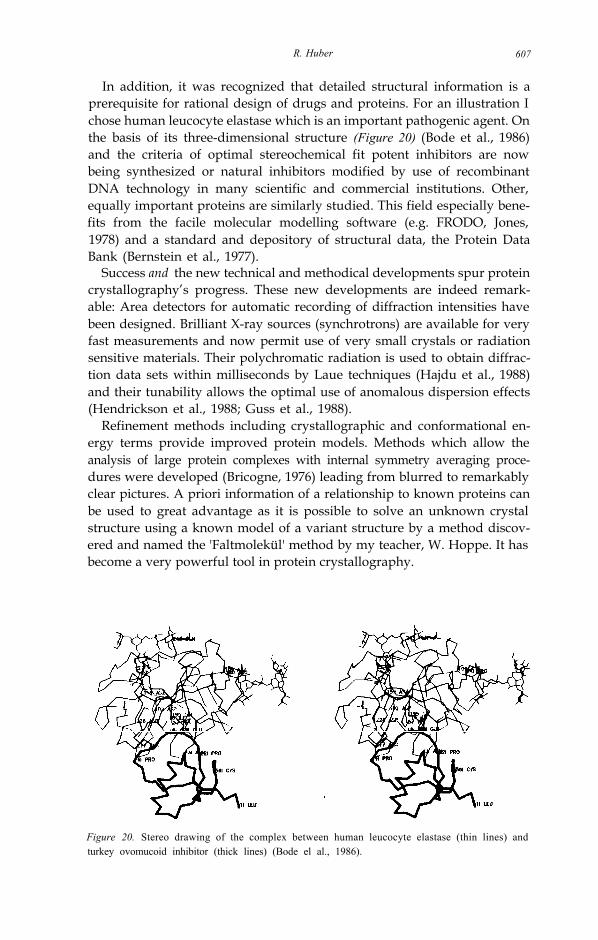
In addition, it was recognized that detailed structural information is aprerequisite for rational design of drugs and proteins. For an illustration Ichose human leucocyte elastase which is an important pathogenic agent. Onthe basis of its three-dimensional structure (Figure 20) (Bode et al., 1986)and the criteria of optimal stereochemical fit potent inhibitors are nowbeing synthesized or natural inhibitors modified by use of recombinantDNA technology in many scientific and commercial institutions. Other,equally important proteins are similarly studied. This field especially bene-fits from the facile molecular modelling software (e.g. FRODO, Jones,1978) and a standard and depository of structural data, the Protein DataBank (Bernstein et al., 1977).
Success and the new technical and methodical developments spur proteincrystallography’s progress. These new developments are indeed remark-able: Area detectors for automatic recording of diffraction intensities havebeen designed. Brilliant X-ray sources (synchrotrons) are available for veryfast measurements and now permit use of very small crystals or radiationsensitive materials. Their polychromatic radiation is used to obtain diffrac-tion data sets within milliseconds by Laue techniques (Hajdu et al., 1988)and their tunability allows the optimal use of anomalous dispersion effects(Hendrickson et al., 1988; Guss et al., 1988).
Refinement methods including crystallographic and conformational en-ergy terms provide improved protein models. Methods which allow theanalysis of large protein complexes with internal symmetry averaging proce-dures were developed (Bricogne, 1976) leading from blurred to remarkablyclear pictures. A priori information of a relationship to known proteins canbe used to great advantage as it is possible to solve an unknown crystalstructure using a known model of a variant structure by a method discov-ered and named the 'Faltmolekül' method by my teacher, W. Hoppe. It hasbecome a very powerful tool in protein crystallography.
Figure 20. Stereo drawing of the complex between human leucocyte elastase (thin lines) andturkey ovomucoid inhibitor (thick lines) (Bode el al., 1986).
608 Chemistry 1988
With this last paragraph I wish to pay tribute to W. Hoppe who in 1957laid the foundation to Patterson search methods by discovering that thePatterson function (the Fourier transform of the diffraction intensities) ofmolecular crystals can be decomposed into sums of intra- and intermolecu-lar vector sets (Hoppe, 1957) from which orientation and translation of themolecules can be derived when their approximate structure is known (Fi-gure 21 ). Hoppe’s method was profoundly elaborated, computerized, andreformulated (Rossmann & Blow, 1962; Huber, 1965; Crowther & Blow,1967). It provided a short-cut to the crystal structure of the RC of Rb .sphaeroides which was solved on the basis of the molecular structure of theRC of Rps. viridis and subsequently refined (Allen et al., 1986, 1987). Themolecular architectures are very similar although the Rb. sphaeroides RClacks the permanently bound cytochrome. The structure solution was inde-pendently confirmed using similar methods by Chang et al., (1986). Withthe Faltmolekül method the orientation and location of a molecule in acrystal cell can be determined. The detailed molecular structure and itsdeviations from the parent model have to be worked out by crystallographicrefinement, to which W. Steigemann and J. Deisenhofer (in his thesis work)laid a foundation in my laboratory (Huber et al., 1974; Deisenhofer &Steigemann, 1975).
Recently, NMR techniques (nuclear magnetic resonance) have demon-strated their capability to determine three-dimensional structure of smallproteins in solution. In one case, a detailed comparison between crystal andsolution structure has shown very good correspondance (Kline et al., 1986;Pflugrath et al., 1986) but future developments will be needed to extend thepower of the method to larger protein structures.
Protein crystallography is the only tool to unravel in detail the architec-ture of the large protein complexes described here and will continue in theforeseeable future to be the only experimental method that provides atomicresolution data on atom-atom and molecule-molecule interactions. It is thesuccessful analytic method E. Fischer addressed in his 9th Faraday Lectureby pointing out that ‘the precise nature of the assimilation process. . . willonly be accomplished when biological research, aided by improved analyt-ical methods, has succeeded in following the changes which take place in theactual chlorophyll granules’ (Fischer, 1907). Yet an ultimate goal for whichwe all struggle is the solution of the folding problem. The growing numberof known protein structures and the design of single residue variants byrecombinant DNA technology and their analysis by protein crystallographyhas brought us nearer to this goal. We are able to study contributions ofindividual residue to rates of folding, structure, stability, and function. Alsotheoretical analysis of protein structures has progressed (to mention onlyLevitt & Sharon, 1988) but a clue to the code relating sequence andstructure is not in sight (Jaenicke, 1988). Like Carl von Linné who 250 yearsago created a system of plants on the basis of morphology (Genera plan-tarum, Leiden 1737), we classify proteins by their shapes and structures.Whether this may lead to a solution of the folding problem is unclear, but it

R. Huber 609
Figure 21. Faltmolekül construct A) (a) and (El) (E2) (b)are the intra- and intermolecular vector sets of a triangular structure respectively. Their sumrepresents the Patterson function. The intramolecular vector set can be constructed from themolecular structure, is located at the origin, and permits the determination of the orientation.From the intermolecular vector set the translation component relative to the mirror line can bederived. In (b) the intermolecular vector sets corresponding to two different orientations of are shown (Huber, 1985).
B)Drawing of the main chain of the M-, L-, H-subunits and the cofactors which served as searchmodel to solve the phase problem for the Rb. sphaeroides RC crystal structure. For the calcula-tion all homologous main chain and side chain atoms were included (Allen et al., 1986).

610 Chemistry 1988
is certain that the end of protein crystallography will only come throughprotein crystallography.
ACKNOWLEDGEMENTJ. Deisenhofer’s and my interest in structural studies of the photosyntheticreaction centre of Rps. viridis was raised by the establishment of D. Oester-helt’s department in Martinsried in 1980; he brought with him H. Michel,with whom a fruitful collaboration on the analysis of the crystal structure ofthis large protein complex began. Later other members of my group, O.Epp and K. Miki, became involved. We had been studying enzymes, pro-teases and their natural inhibitors, immunoglobulins and had developedmethods to improve data collection, electron density map interpretationand crystallographic refinement. The tools were available to attack a prob-lem which was and still is the largest asymmetric protein analysed at atomicresolution today.
The “heureka” moment of protein crystallography is at the very endwhen one sees for the first time a new macromolecule with the eyes of adiscoverer of unknown territories. To reach this moment much, sometimestedious, work has to be done with the ever present possibility of failure. Iam deeply grateful to my collaborators, those who are with me and thosewho had left, for their dedicated and patient work over many years. Imention by name those involved in the studies of the light harvestingcyanobacterial proteins and the blue oxidases: W. Bode, M. Duerring, R.Ladenstein, A. Messerschmidt, T. Schirmer. These projects were collabora-tive undertakings with biochemists in Switzerland (H. Zuber, W. Sidler),USA (M. L. Hackert) and Italy (M. Bolognesi, A. Marchesini, A. Finazzi-Agro.
Scientific work needs a stimulating environment which was provided atthe Max-Planck-Institut für Biochemie and it needs steady financial sup-port, which was provided by the Max-Planck-Gesellschaft and the DeutscheForschungsgemeinschaft.
I thank R. Engh, S. Knof, R. Ladenstein, M. Duerring, E. Meyer for theirhelpful comments on this manuscript.
LITERATURE
Allen, J. P., Feher, G., Yeates, T. O., Rees, D. C., Deisenhofer, J., Michel, H.,Huber, R. (1986) Proc. Natl. Acad. Sci. USA 83, 8589-8593.
Allen, J. P., Feher, G., Yeates, T. O., Kemiya, H., Rees, D. C. (1987) Proc. Natl.Acad. Sci. USA 84, 6162-6166.
Arnold, W., and Clayton, R. D. (1960), Proc. Natl. Acad. Sci. USA 46, 769-776.Babcock, G. T. (1987) Oxygen-Evolving Process in Photosynthesis (J. Amesz, ed.)
Elsevier Science Publ.Barber, J. (1987) Trends Biochem. Sci. 12, 321-326.Barber, J. (1988) Nature 333, 114.Barltrop, J. A. and Coyle, J. D. (1978) Principles of Photochemistry, John Wiley and
Sons, Chichester.Bernstein, F. C., Koetzle, T. F., Williams, G. J. B., Meyer, Jr., E. F., Brice, M. D.,

R. Hubcr 611
Rodgers, J. R., Kennard, O., Shimonouchi, T., Tasumi, M (1977) J. Mol. Biol.112, 535-542.
Bennett, W. S. and Huber, R. (1984), CRC Critical Reviews in Biochemistry 15,2 9 1 - 3 8 4 .
Blair, D. F., Campbell, G. W., Schoonover, J. R., Chan, S. I., Gray, H. B., Malm-ström, B. G., Pecht, I., Swanson, B. F., Woodneff, W. H., Cho, W. K., English,A. R., Fry, A. H., Lum, V., and Norton, K. A. (1985) J. Amer. Chem. Soc. 10,5755-5766.
Bode, W., Wei, A., Huber, R., Meyer, E., Travis, P., Neumann, S. (1986) EMBO J. 5,2453-2458.
Bolton, J. R. (1978) in: The Photosynthetic Bacteria, (R. K. Clayton and W. R.Sistrom, eds.), Plenum Press, New York and London, pp. 419-442.
Boltzmann, L. (1886) Der Zweite Hauptsatz der mechanischen Wärmetheorie (Essayin Populire Schriften) L. Boltzmann-Gesamtausgabe Bd. 7 (1919) Akad. Druck-und Verlagsanstalt Vieweg, Wiesbaden, pp 25-46 (English translation fromArnon, D. L. (1961) in: Light and Life (W. D. McElroy, B. Glass, eds.) The JohnsHopkins Press, Baltimore, pp. 489-569.
Breton, J. (1985) Biochim. Biophys. Acta 810, 235-245.Breton, J., Farkas, D. L., and Parson, W. W. (1985) Biochim. Biophys. Acta 808,
4 2 1 - 4 2 7 .Breton, J., Martin, J.-L., Migus, A., Antonetti, A., and Orszag, A. (1986) Proc. Natl.
Acad. Sci. USA 83, 5121-5125.Bricogne, G. (1976) Acta Crystallogr. A 32, 832-847.Bryant, D. A., Guglielmi, G., Tandeau de Marsac, N., Castets, A.-M. & Cohen-
Bazire, G. (1979) Arch. Microbial. 123, 113-127.Burkert, U., Allinger, N. L. (1982) Molecular Mechanics, American Chemical
Society.Calvin, M. and Bassham, J. A. (1962) in: The Photosynthesis of Carbon Compounds,
Benjamin, New York, pp. 1-127.Carithers, R. P., and Parson, W. W. (1975) Biochim. Biophys. Acta 387, 194-211.Case, G. D., Parson, W. W., and Thomber, J. P. (1970) Biochim. Biophys. Acta 223,
122-128 .Chang, C.-H., Tiede, D., Tang, J., Smith, U., Norris, J., Schiffer, M. (1986) FEBS
Lett. 205, 82-86.Chothia, C. (1984) Ann. Rev. Biochem. 53, 537-572.Cogdell, R. J. and Crofts, A. R. (1972) FEBS Lett. 27,176-178.Cohen-Bazire, G. & Briant, D. A. (1982) in: The Biology of Cyanobacteria (Cat-r, N.
G. & Whitton, B. eds.), Blackwell, London, pp. 143-189.Crowther, R. A. & Blow, D. M. (1967) Acta Cryst. 23, 544-548.Cramer, W. A., and Crofts, A. R. (1982) in: Electron and Proton Transport in
Photosynthesis: Energy Conversion by Plants and Bacteria Vol 1, Academic PressInc., pp. 387.
Dawson, C. R. (1966) in: The Biochemistry of Copper (Peisach, J., Aison, P. andBlumberg, W. E., eds.), Academic Press, New York, pp. 305-337.
Debus, R. J., Feher, G., and Okamura, M. Y. (1986) Biochemistry 25, 2276-2287.Deisenhofer, J., Huber, R., Michel, H. (1986) Nachr. Chem. Tech. Lab. 34, 416-
422.Deisenhofer, J., Steigemann, W. (1975) Acta Cryst. B 31, 238-280.Deisenhofer, J., Epp, O., Miki, K., Huber, R., and Michel, H., (1984) J. Mol: Biol.
180 ,385-398 .Deisenhofer, J., Epp, O.. Miki, K., Huber, R., and Michel, H., (1985) Nature 318,
618-624 .Deisenhofer, J., Michel, H., and Huber, R. (1985a) Trends Biochem. Sci. 10, 243-
248.

612 Chemistry 1988
Deisenhofer, J., Huber, R., Michel, H. (1989) in: Prediction of Protein Structureand the Principles of Protein Conformation (G. D. Fasman, ed.) Plenum Pub.Corp., New York, (in press.).
DeVault D., and Chance, B. (1966) Biophys. J. 6, 825-847.Duerring, M. (1988) Thesis, Technical University, Miinchen.Duerring, M., Bode, W., Huber, R., Ruembeli, R., Zuber, H. (1989) to be submit-
ted.Duerring, M., Huber, R. and Bode W. (1988) FEBS Letters, 236, 167- 170.Dutton, P. L., and Prince, R. C. (1978) in: The Photosynthetic Bacteria (R. K. Clay-
ton and W. R. Sistrom, eds.), Plenum Press, New York and London, pp. 525-565.
Eberson, L. (1982) Adv. Phys. Org. Chem. 18, 79-185.Engelman, D. M., Steitz, T. A., and Goldman, A. (1986) Ann. Rev. Biophys. Chem.
15, 321-353.Farver, O., Pecht, I. (1984) in: Copper Proteins and Copper Enzymes (R. Lontie,
ed.) 1, CRC Press, Inc., Boca Raton, Florida, pp. 183-214.Fischer, E. (1907) J. Chem. Soc. 91, 1749-1765.Fleming, G. R., Marti, J. L. and Breton, J. (1988) Nature 333, 190-192.Forster, T. (1948) Ann. Physik 2, 55-75.Förster, T. (1967) in: Comprehensive Biochemistry (M. Florkin, E. H. Stotz, eds.),
Vol 22, pp. 61-80, Elsevier, Amsterdam.Frank, G., Sidler, W., Widmer, H. and Zuber, H. (1978) Hoppe-Seyler’s Z. Physiol.
Chem. 359, 1491-1507.Frommherz, P. and Reinbold, G. (1988) Thin Solid Films 160, 347 - 353.Gaykema, W. P. J., Hol, W. G. J., Verijken, J. M., Soeter, N. M., Bak, H. J., and
Beintema, J. J. (1984) Nature 309, 23-29.Gantt, E., Lipschultz, C.A., Zilinskas, B. (1976) Biochim. Biophys. Acta 430, 375-
388.Germann, U. A., Müller, G., Hunziker, P. E., Lerch, K. (1988) J. Biol. Chem. 263,
885-896 .Gillbro, T., Sandstrom, A., Sundstrom, V., Wendler, J. and Holzwarth, A. R. (1985)
Biochem. Biophys. Acta 808, 52-65.Gerwin, B., Burstein, S. R., and Westley, J. (1974) J. Biol. Chem. 249, 2005-2008.Glazer, A. N., Fang, S. and Brown, D. M. (1973) J. Biol. Chem. 16, 5679-5685.Glazer, A. N. (1985) Ann. Rev. Biophys. Chem. 19, 47-77.Grabowski, J. and Gantt, E. (1978) Photochem. Photobiol. 28, 39-45.Gray, H. B., and Solomon, E. I. (1981) in: Copper proteins (T. G. Spiro, ed.) J. Wiley
& Sons, New York, pp. l-39.Gray, H. B., and Malmström, B. G. (1983) Comments Inorg. Chem. 2, 203-209.Gray, H.B. (1986) Chem. Soc. Rev. 15, 17-30.Gust, D., Moore, T. A., Lidell, P. A., Nemeth, G. A., Makings, L. R., Moore, A. L.,
Barrett, D., Pessiki, P. J., Bensasson, R.V., Rougée, M., Chachaty, C., DeSchryver, F. C., Van der Anweraer, M., Holzwarth, A. R., and Connolly, J. S.(1987) J. Amer. Chem. Soc. 109, 846-856.
Guss, J. M., Freeman, H. C. (1983) J. Mol. Biol. 169, 521-563.Guss, J. M., Merritt, E.A., Phizackerly, R. P., Hedman, B., Murata M., Hodgson,
K. O., Freeman, H. C. (1988) Science 241, 806-811.Hajdu, J., Acharya, K. R., Stuart, D.A., Barford, D., and Johnson, L. (1988) Trends
Biochem. Sci. 13, 104-109.Hains, A. (1975) Acc. Chem. Res. 8, 264-272.Harrison, S. C., Olsson, A. J., Schutt, C. E., Winkler, F. K., Bricogne, G. (1978)
Nature (London) 276, 368-373.Hendrickson, W. A., Smith, J. L., Phizackerly, R. P., Merritt, E. A. (1988) Proteins 4,
7 7 - 8 8 .

R. Huber 613
Hefferle, P., Nies, M., Wehrmeyer, W. and Schneider, S. (1983) Photobiochem.Photobiophys. 5, 41-51.
Henderson, R. and Unwin, P. N. T. (1975) Nature 257, 28-32.Higuchi, Y., Kusunoki, M., Matsuura, Y., Yasuoka, N., Kakudo, M. (1984) J. Mol.
Biol. 172, 109-139.Hogle, J. M., Chow, M., Filman, D. J. (1985) Science 229, 1358-1365.Holm, L., Saraste, M., and Wikström, M. (1987) EMBO J. 6, 2819-2823.Holten, D., Windsor, M. W., Parson, W. W. and Thomber, J. P. (1978) Biochem.
Biophys. Acta 501, 112-126.Holzwarth, A. R. (1985) in: Antennas and Reaction Centers of Photosynthetic Bacte-
ria (Michel-Beyerle, M. E., ed.), Springer-Verlag, Berlin, pp. 45-52.Holzwarth, A. R. (1986) Photochem. Photobiol. 43, 707-725.Hopfield, J. J. (1974) Proc. Natl. Acad. Sci. USA 71, 3640-3644.Hoppe, W. (1957) Acta Cryst. 10, 750-751.Huber, R. (1965) Acta Cryst. 19, 353-356.Huber, R. (1985) in: Molecular Replacement. Proceedings of the Daresbury Study
Weekend (P. Machin, ed.) Daresbury Laboratory, pp. 58-61.Huber, R., Kukla, D., Bode, W., Schwager, P., Bartels, K., Deisenhofer, J., Steige-
mann, W. (1974) J. Mol. Biol. 89, 70-101.Huber, R. (1984) in: Behring Institute Mitteilungen 76, 1-14 (Gronski, P., Seiler,
F. R., eds.), Die Medizinische Verlagsgesellschaft mbH, Marburg.Huber, R., Schneider, M., Epp, O., Mayr, I., Messerschmidt, A., Pflugrath, J. and
Kayser, H. (1987a) J. Mol. Biol. 195, 423-434.Huber, R., Schneider, M., Mayr, I., Müller, R., Deutzmann, R., Suter, F., Zuber,
H., Falk, H. and Kayser, H. (1987b) J. Mol.Biol. 198, 499-513.Huber, R. (1988) Angew. Chem. Int. Ed. Engl. 27, 79-88.Isied, S., Vassilian, A., Magnuson, R., and Schwarz, H. (1985) J. Am. Chem. Soc.
107, 7432-7438.Jaenicke, R. (1988) in: 39. Mosbacher Kolloquium (E.-L. Winnacker and R. Huber,
eds.) pp. 16 - 36, Springer-Verlag, Berlin Heidelberg.Jones, A. T. (1978) J. Appl. Cryst. 11, 268-272.Karplus, M., McCammon, J. A. (1981) CRC Crit. Rev. Biochem. 9, 293-349.Kavarnos, G. J. and Turro, N. J. (1986) Chem. Rev. 86, 401-449.Kebarle, P., and Chowdhury, S. (1987) Chem. Rev. 87, 513-534.Kirmaier, Ch., Holten, D., and Parson, W. W. (1985) Biochim. Biophys.Acta 810,
3 3 - 4 8 .Kirmaier, Ch., Holton, D., and Parson, W. W. (1985). Biochem. Biophys. Acta 810,
4 9 - 6 1 .Kleffel, B., Garavito, R. M., Baumeister, N., Rosenbusch, J. P. (1985) EMBO J. 4,
1589-1592.Kleinfeld, D., Okamura, M. Y., Feher, G. (1985) Biochem. Biophys.Acta 809, 291-
310.Kline, A. D., Braun, W., Wüthrich, K. (1986) J. Mol. Biol. 189, 377-382.Knapp, E. W., Fischer, S. F., Zinth, W., Sander, M., Kaiser, W., Deisenhofer, J., and
Michel, H. (1985) Proc. Natl. Acad. Sci. USA 82, 8463-8467.Kuhn, H. (1970) J.Chem. Phys. 53, 101-108.Ladenstein, R., Schneider, M., Huber, R., Bartunik, H.-D., Wilson, K., Schott, K.,
Bacher, A. (1988) J. Mol. Biol. 203, 1045-1070.Levitt, M., Sharon, R. (1988) Proc. Natl. Acad. Sci. USA 85, 7557-7561.Lesk, A. M. and Hardman, K. D. (1982) Science 216, 539-540.Lodish, H. F. (1988) Trends Biochem. Sci. 13, 332-334.Loebermann, H., Tokuoka, R., Deisenhofer, J., Huber, R. (1984) J. Mol. Biol. 177,
531-556 .Lumry, R., Eyring, H. (1954) J. Phys. Chem. US 58, 110-2.
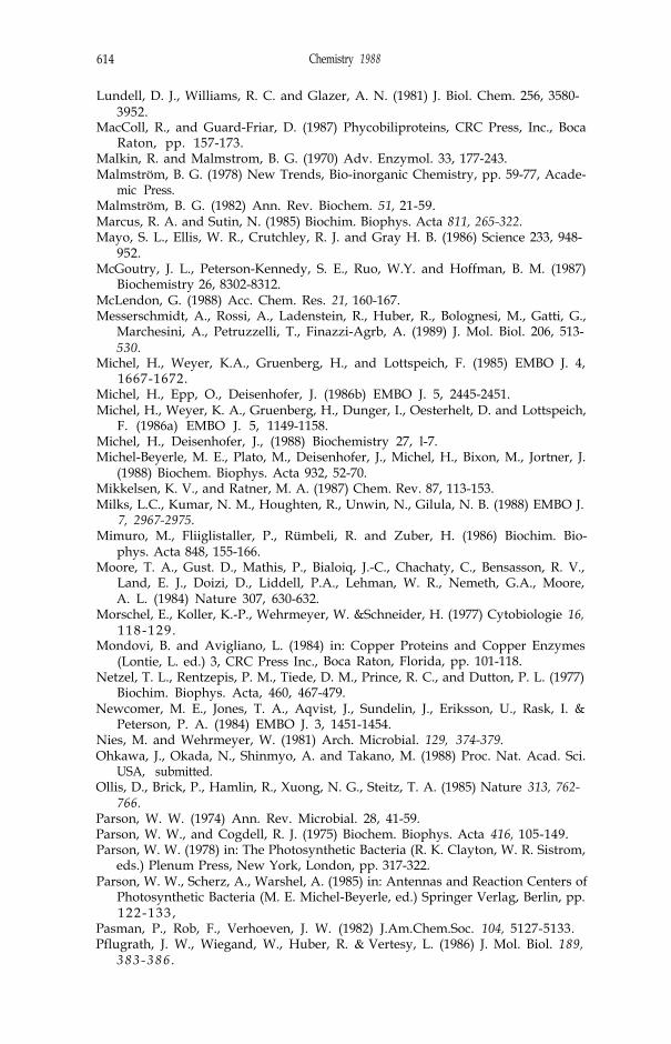
614 Chemistry 1988
Lundell, D. J., Williams, R. C. and Glazer, A. N. (1981) J. Biol. Chem. 256, 3580-3952.
MacColl, R., and Guard-Friar, D. (1987) Phycobiliproteins, CRC Press, Inc., BocaRaton, pp. 157-173.
Malkin, R. and Malmstrom, B. G. (1970) Adv. Enzymol. 33, 177-243.Malmström, B. G. (1978) New Trends, Bio-inorganic Chemistry, pp. 59-77, Acade-
mic Press.Malmström, B. G. (1982) Ann. Rev. Biochem. 51, 21-59.Marcus, R. A. and Sutin, N. (1985) Biochim. Biophys. Acta 811, 265-322.Mayo, S. L., Ellis, W. R., Crutchley, R. J. and Gray H. B. (1986) Science 233, 948-
952.McGoutry, J. L., Peterson-Kennedy, S. E., Ruo, W.Y. and Hoffman, B. M. (1987)
Biochemistry 26, 8302-8312.McLendon, G. (1988) Acc. Chem. Res. 21, 160-167.Messerschmidt, A., Rossi, A., Ladenstein, R., Huber, R., Bolognesi, M., Gatti, G.,
Marchesini, A., Petruzzelli, T., Finazzi-Agrb, A. (1989) J. Mol. Biol. 206, 513-530.
Michel, H., Weyer, K.A., Gruenberg, H., and Lottspeich, F. (1985) EMBO J. 4,1667-1672.
Michel, H., Epp, O., Deisenhofer, J. (1986b) EMBO J. 5, 2445-2451.Michel, H., Weyer, K. A., Gruenberg, H., Dunger, I., Oesterhelt, D. and Lottspeich,
F. (1986a) EMBO J. 5, 1149-1158.Michel, H., Deisenhofer, J., (1988) Biochemistry 27, l-7.Michel-Beyerle, M. E., Plato, M., Deisenhofer, J., Michel, H., Bixon, M., Jortner, J.
(1988) Biochem. Biophys. Acta 932, 52-70.Mikkelsen, K. V., and Ratner, M. A. (1987) Chem. Rev. 87, 113-153.Milks, L.C., Kumar, N. M., Houghten, R., Unwin, N., Gilula, N. B. (1988) EMBO J.
7, 2967-2975.Mimuro, M., Fliiglistaller, P., Rümbeli, R. and Zuber, H. (1986) Biochim. Bio-
phys. Acta 848, 155-166.Moore, T. A., Gust. D., Mathis, P., Bialoiq, J.-C., Chachaty, C., Bensasson, R. V.,
Land, E. J., Doizi, D., Liddell, P.A., Lehman, W. R., Nemeth, G.A., Moore,A. L. (1984) Nature 307, 630-632.
Morschel, E., Koller, K.-P., Wehrmeyer, W. &Schneider, H. (1977) Cytobiologie 16,118-129 .
Mondovi, B. and Avigliano, L. (1984) in: Copper Proteins and Copper Enzymes(Lontie, L. ed.) 3, CRC Press Inc., Boca Raton, Florida, pp. 101-118.
Netzel, T. L., Rentzepis, P. M., Tiede, D. M., Prince, R. C., and Dutton, P. L. (1977)Biochim. Biophys. Acta, 460, 467-479.
Newcomer, M. E., Jones, T. A., Aqvist, J., Sundelin, J., Eriksson, U., Rask, I. &Peterson, P. A. (1984) EMBO J. 3, 1451-1454.
Nies, M. and Wehrmeyer, W. (1981) Arch. Microbial. 129, 374-379.Ohkawa, J., Okada, N., Shinmyo, A. and Takano, M. (1988) Proc. Nat. Acad. Sci.
USA, submitted.Ollis, D., Brick, P., Hamlin, R., Xuong, N. G., Steitz, T. A. (1985) Nature 313, 762-
766.Parson, W. W. (1974) Ann. Rev. Microbial. 28, 41-59.Parson, W. W., and Cogdell, R. J. (1975) Biochem. Biophys. Acta 416, 105-149.Parson, W. W. (1978) in: The Photosynthetic Bacteria (R. K. Clayton, W. R. Sistrom,
eds.) Plenum Press, New York, London, pp. 317-322.Parson, W. W., Scherz, A., Warshel, A. (1985) in: Antennas and Reaction Centers of
Photosynthetic Bacteria (M. E. Michel-Beyerle, ed.) Springer Verlag, Berlin, pp.122-133 ,
Pasman, P., Rob, F., Verhoeven, J. W. (1982) J.Am.Chem.Soc. 104, 5127-5133.Pflugrath, J. W., Wiegand, W., Huber, R. & Vertesy, L. (1986) J. Mol. Biol. 189,
383-386 .

R. Huber 615
Pierrot, M., Haser, R., Frey, M., Payan, F. & Astier, J.P. (1982) J. Biol. Chem. 257,14341-14348.
Porter, G., Tredwell, C. J., Searle, G. F. W. and Barber, J. (1978) Biochim. Biophys.Acta 501, 232-245.
Prince, R. C., Leigh, J. S., and Dutton, P. L. (1976) Biochim. Biophys. Acta 440,622-636 .
Prince, R. C. (1988) Trends Biochem. Sci. 13, 286-288.Rapoport, T. A. (1986) CRC Critical Rev. Biochem. 20, 73-137.Reinhammar, B. (1984) in: Copper Proteins and Copper Enzymes (Lontie, L. ed.)
Vol. 3, pp. 1-35, CRC Press Inc., Boca Raton, Florida.Remington, S., Wiegand, G., Huber, R. (1982) J. Mol. Biol. 158, 111-152.Richardson, J. S., Thomas, K. A., Rubin, B. H., and Richardson, D. C. (1975) Proc.
Natl. Acad. Sci. USA 72, 1349-1353.Rossmann, M. G. and Blow, D. M. (1962) Acta Cryst. 15, 24-31.Rossmann, M. G., Arnold, E., Erickson, J. W., Frankenberger, E. A., Griffith, J. P.,
Hecht, H.-J., Johnson, J. E., Kamer, G., Luo, M., Mosser, A. G., Rueckert, R.,Sherry, B., and Vriand, G. Nature (London) 317, 145-153.
Ryden, L. (1984) in: Copper Proteins and Copper Enzymes (Lontie, L. ed.) 3,CRC Press Inc., Boca Raton, Florida, pp. 34-100.Sauer, K., Scheer, H., and Sauer, P. (1987) Photochem. Photobiol. 46, 427-440.Scharnagl, C., Köst-Reyes, E., Schneider, S., Köst, H.-P., Scheer, H. (1983) Z.
Naturforsch. 38c, 951-959.Scheer, H. (1982) in: Light Reaction Path of Photosynthesis (Fong, F. K., ed.)
Springer, Berlin, pp. 7-45.Schirmer, T., Bode, W., Huber, R., Sidler, W. and Zuber, H. (1985) J. Mol. Biol.
184, 257-277.Schirmer, T., Huber, R., Schneider, M., Bode, W., Miller, M. and Hackert, M. L.
(1986) J. Mol. Biol. 188, 651-676.Schirmer, T., Bode, W., and Huber, R. (1987) J. Mol. Biol. 196, 677-695.Schirmer, T. & Vincent M. G. (1987) Biochem. Biophys. Acta 893, 379-385.Schmidt, J. A., McIntosh, A. R., Weedon, A. C., Bolton, J. R., Connolly, J. S.,
Huxley, J. K., and Wasielewski, M. R. (1988) J. Amer. Chem. Soc. 110, 1733-1740.
Searle, G. F. W., Barber, J., Porter, G., Tredwell, C. J. (1978) Biochem. Biophys.Acta 501, 246-256.
Siebzehnrübl, S., Fischer, R., and Scheer, H. (1987) Z. Naturforsch. 42i, 258-262.Slooten, L. (1972) Biochim. Biophys. Acta 256, 452-466.Stark, N., Kuhlbrandt, W., Wildhaber, I., Wehrli, E. & Mühlethaler, K. (1984)
EMBO J. 3, 777-783.Switalski, S. C., and Sauer, J. (1984) Photochem. Photobiol. 40, 423-427.Takahashi, N., Ortel, T. L., Putnam, F. W. (1984) Proc. Natl. Acad. Sci. 81, 390-
394.Taube, H., and Gould, E. S. (1969) Acc. Chem. Res. 2, 321-329.Teale, F. W. J. and Dale, R. E. (1970) Biochem. J. 116, 161-169.Trebst, A. (1986) Z. Naturforsch. 41c, 240-245.Tronrud, D. E., Schmid, M. F., and Matthews, B. W. (1986) J. Mol. Biol. 188, 443-
454.Weber, P. C., and Salemme, F. R. (1980) Nature (London) 287, 82-84.Weber, E., Papamokos, E., Bode, W., Huber, R., Kato, F., Laskowski, M. (1981) J.
Mol. Biol. 149, 109-123.Wendler, J., Holzwarth, A. R., Wehrmeyer, W. (1984) Biochem. Biophys. Acta 765,
5 8 - 6 7 .Weyer, K. A., Lottspeich, F., Gruenberg, H., Lang, F., Oesterhelt, D., and Michel,
H. (1987) EMBO J. 6, 2197-2202.
616 Chemistry 1988
Woodbury, N. W., Becker, M., Middendorf, D., and Parson, W. W. (1985) Biochem-istry 24, 7516-7521.
Wraight, C. A. (1982) in: Function of Quinones in Energy Conserving Systems (B. L.Trumpower, ed.) Academic Press, London, pp, 181-197.
Yamazaki, I., Mimuro, M., Murao, T., Yamazaki, T., Yoshihara, K., Fujita, Y. (1984)Photochem. Photobiol. 39, 233-240
Zickendraht-Wendelstadt, B., Friedrich, J., and Rüdiger, W. (1980) Photochem.Photobiol. 31, 367-376.
Zilinskas, B. A. and Greenwald, L. S. (1986) Photosynth. Res. 10, 7-35.Zuber, H. (1985) Photochem. Photobiol. 42, 821-844.Zuber, H. (1986) Trends Biochem. Sci. 11, 414-419.