a simple and straightforward mechanochemical synthesis of
TRANSCRIPT

RSC Advances
PAPER
Ope
n A
cces
s A
rtic
le. P
ublis
hed
on 0
8 Ju
ne 2
015.
Dow
nloa
ded
on 1
0/30
/201
8 9:
58:3
5 A
M.
Thi
s ar
ticle
is li
cens
ed u
nder
a C
reat
ive
Com
mon
s A
ttrib
utio
n 3.
0 U
npor
ted
Lic
ence
.
View Article OnlineView Journal | View Issue
A simple and stra
aInstitute of Nanotechnology, Kar
Hermann-von-Helmholtz-Platz 1, 76344 Egg
[email protected]; [email protected]
721-60828929bInstitute of Geotechnics, Slovak Academy o
SlovakiacInstitute for Chemistry and Technology of
(NAWI Graz), Stremayrgasse 9, 8010 Graz, AdInstitute of Physics, Pavol Jozef Safarik Uni
SlovakiaeInstitute of Physical Chemistry and Electro
Callinstr., 3-3a, 30167 Hannover, GermanyfCenter for Solid State Chemistry and N
Hannover, Callinstr., 3-3a, 30167 HannovergDepartment of Physics, State University of
Maringa, Brazil
Cite this: RSC Adv., 2015, 5, 54321
Received 15th May 2015Accepted 8th June 2015
DOI: 10.1039/c5ra09098a
www.rsc.org/advances
This journal is © The Royal Society of C
ightforward mechanochemicalsynthesis of the far-from-equilibrium zincaluminate, ZnAl2O4, and its response to thermaltreatment
Martin Fabian,*ab Patrick Bottke,c Vladimır Girman,d Andre Duvel,ef
Klebson Lucenildo Da Silva,ag Martin Wilkening,c Horst Hahn,a Paul Heitjansef
and Vladimır Sepelak*abf
Zinc aluminate (ZnAl2O4) nanoparticles with an average size of about 10 nm are synthesized via one-step
mechanochemical processing of the ZnO : g-Al2O3 stoichiometric mixture at ambient temperature. The
mechanochemically induced formation of the phase is followed by XRD and 27Al MAS NMR. High-
resolution TEM studies reveal a non-uniform nanostructure of mechanosynthesized aluminate consisting
of ordered grains surrounded or separated by disordered surface and interfacial regions. Due to the
capability of 27Al MAS NMR to probe the local environment of the Al cations, valuable insights into the
short-range structure of ZnAl2O4 on the Angstrom length scale are provided. It is demonstrated that the
as-prepared aluminate possesses a partly inverse spinel structure with a far-from equilibrium
arrangement of cations and distorted polyhedra, which are spatially confined to the surface and
interfacial regions with a volume fraction of ca. 50% and a thickness of ca. 1 nm. The response of the
nanostructured ZnAl2O4 to subsequent thermal treatment is further investigated. It turned out that the
thermally induced grain growth is accompanied by a release of microstrain, by a shrinkage of the lattice
parameter, as well as by a variation in the oxygen parameter and metal–oxygen bond lengths. Evidence
is given of the thermally induced redistribution of cations approaching their equilibrium positions. Upon
heating above 1100 K, mechanosynthesized ZnAl2O4 relaxes towards a structural state that is similar to
the bulk one.
1. Introduction
The ability of spinels to redistribute their cations over crystal-lographically nonequivalent positions has attracted consider-able interest from many scientists. The cubic spinel structure(space group Fd�3m) is characterized by close-packed arrays of
lsruhe Institute of Technology,
enstein-Leopoldshafen, Germany. E-mail:
du; Fax: +49-721-60826368; Tel: +49-
f Sciences, Watsonova 45, 04001 Kosice,
Materials, Graz University of Technology
ustria
versity, Park Angelinum 9, 04154 Kosice,
chemistry, Leibniz University Hannover,
ew Materials (ZFM), Leibniz University
, Germany
Maringa, Av. Colombo 5790, 87020-900
hemistry 2015
oxygen atoms with one eighth of the tetrahedral and one half ofthe octahedral sites occupied by heterovalent cations (Fig. 1). Toemphasize the site occupancy on the atomic level, the structuralformula of 2–3 spinels of the type M12+M22
3+O4 (where 2–3 referto the valences of the M1 and M2 cations) may be written as(M11�lM2l)[M1lM22�l]O4, where parentheses and squarebrackets enclose cations that are either tetrahedrally (A) oroctahedrally [B] coordinated by oxygen anions, respectively. lrepresents the so-called degree of inversion that is dened asthe fraction of the (A) sites occupied by trivalent (M2) cations.Spinel compounds with l ¼ 0 are denoted as normal spinels,whereas those with l ¼ 1 are called fully inverse spinels. Thevalue of lrd ¼ 2/3 corresponds to a random distribution ofcations over the (A) and [B] positions.1 It is well recognized thatphysico-chemical properties of spinels are determined to a largeextent by their degree of inversion.2–4 Thus, a detailed under-standing of the functional behavior of spinels relies on carefulcharacterization of their cation distribution.
In its equilibrium state, zinc aluminate (ZnAl2O4, gahnite)possesses the structure of a normal spinel (lc ¼ 0) with thefollowing crystal chemical formula: (Zn)[Al2]O4.5 Considerable
RSC Adv., 2015, 5, 54321–54328 | 54321

Fig. 1 Crystal structure of normal spinel ZnAl2O4 (space group Fd3�m).Zn2+ and Al3+ cations are distributed over the tetrahedrally (A) andoctahedrally [B] coordinated sites.
RSC Advances Paper
Ope
n A
cces
s A
rtic
le. P
ublis
hed
on 0
8 Ju
ne 2
015.
Dow
nloa
ded
on 1
0/30
/201
8 9:
58:3
5 A
M.
Thi
s ar
ticle
is li
cens
ed u
nder
a C
reat
ive
Com
mon
s A
ttrib
utio
n 3.
0 U
npor
ted
Lic
ence
.View Article Online
attention has been paid to several of its multifunctional appli-cations such as catalyst and catalyst support, UV-transparentsupport conductor, sensor, dielectric and optical material.6–8
The conventional solid state, i.e., ceramic, synthesis ofZnAl2O4 requires long periods of calcination of the reactionprecursors at considerably high temperatures.9 In many cases,this causes the loss of zinc due to its high volatility and,consequently, it results in the formation of multiphase productsand the degradation of microstructural and functional proper-ties of the aluminate. Various wet chemistry-based routes,including, e.g., hydrothermal,10 sol–gel,11 combustion,12 co-precipitation,9 complexation,13 solvothermal6 and sonochem-ical14 methods, have also been developed to synthesize nano-sized ZnAl2O4 powders. Most of the solution chemistry-basedroutes, however, still require calcination steps, at relativelylow temperatures. Non-conventional mechanochemicalsynthesis (mechanosynthesis) has been recognized as an alter-native low-temperature route; in general, it provides an efficientone-step and facile access to nanomaterials.15 In this context,the present work focuses on the one-step synthesis of nano-crystalline ZnAl2O4 viamechanochemical processing of a ZnO +g-Al2O3 mixture at ambient temperature. Although the mecha-nosynthesis of nanocrystalline ZnAl2O4 has already beenreported in a few papers,9,16 to the best of our knowledge there isno report in the literature focusing on the defect state or thedisordered local structure of ZnAl2O4 prepared by non-conventional mechanochemical routes.
Mechanosynthesized complex oxides are oen inherentlyunstable because of their small constituent sizes, disorderedstructural state, and high chemical activity.17 To gain insightinto thermal stability and relaxation of structural disorder, thepresent experimental work also deals with the study of theresponse of mechanosynthesized ZnAl2O4 when exposed tohigher temperatures. For a comprehensive characterization ofstructural relaxation paths of the non-equilibrium product, wesimultaneously apply X-ray diffraction (XRD), which is sensitive
54322 | RSC Adv., 2015, 5, 54321–54328
to medium- and long-range structural order, and 27Al magicangle spinning (MAS) nuclear magnetic resonance (NMR),which reveals local magnetic and electronic structures. More-over, the thermally induced evolution of the aluminatesynthesized is systematically monitored with Fourier transforminfrared (FTIR) spectroscopy and transmission electronmicroscopy (TEM).
2. Experimental
Solid precursors, zinc oxide (ZnO, 99.9% purity; Aldrich) andaluminium oxide (g-Al2O3, 99% purity; Aldrich), were used forthe mechanosynthesis of ZnAl2O4. 5 g of the ZnO : g-Al2O3
mixture was milled for various times (up to 2 h) in a high-energyplanetary ball mill (Pulverisette 7 Premium line (Fritsch)). Agrinding chamber (80 cm3 in volume) and balls (10 mm indiameter) made of tungsten carbide were used. The ball-to-powder weight ratio was 40 : 1. Milling experiments were per-formed in ambient atmosphere at 600 rpm. To investigate thethermally induced structural relaxation of mechanosynthesizedZnAl2O4, the material was subsequently annealed at varioustemperatures up to 1273 K in air for 4 hours.
In addition, polycrystalline ZnAl2O4 (with the average crys-tallite size ca. 105 nm) was synthesized from the mixture of ZnOand g-Al2O3 precursors following a conventional ceramicprocess. This sample served as reference material. Note that anexcess of ZnO (5 wt%) with respect to the stoichiometric ratiowas used to avoid the formation of a multiphase product. In thiscase, powdered reactants were hand-milled, pressed into pelletsand sintered at 1273 K for 24 hours. This process was repeatedfour times, reaching the nal time of sintering of 120 hours.
The XRD patterns were collected using a D8 Advancediffractometer (Bruker) operating with Cu Ka radiation inBragg–Brentano conguration. The generator was set up at40 kV and 40 mA. The divergence and receiving slits were 0.3�
and 0.1 mm, respectively. The patterns were recorded in therange of 20� to 105� 2qwith a step of 0.02� and ameasuring timeof 20 s. The JCPDS PDF database18 was utilized for phaseidentication. Rietveld renements of XRD data of the as-prepared and subsequently annealed samples were performedusing the Fullprof computer program19 utilizing regularThompson–Cox–Hastings pseudo-Voigt prole parameters. Inorder to obtain proper geometry set-up and to eliminateinstrumental broadening the instrumental resolution functionwas determined by the renement of the LaB6 standard spec-imen. The cubic spinel structure of ZnAl2O4 was visualizedusing the Diamond program.20
The morphology of powders was studied using a combinedeld-emission (scanning) transmission electron microscope(S)TEM (JEOL JEM-2100F). Prior to the TEM investigations, thepowders were crushed in a mortar, dispersed in ethanol, andxed on a copper-supported carbon grid.
27Al MAS NMR measurements were performed using anAvance III 500 MHz spectrometer (Bruker) connected to an11.4 T magnet corresponding to a Larmor frequency of130.29 MHz for 27Al. Some of the samples were measured withan Avance III 600 MHz spectrometer (14.1 T, 156.4 MHz Larmor
This journal is © The Royal Society of Chemistry 2015
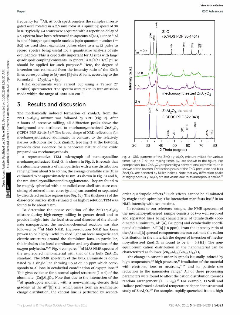
Fig. 2 XRD patterns of the ZnO : g-Al2O3 mixture milled for varioustimes (up to 2 h); the milling times, tm, are shown in the figure. Forcomparison, bulk ZnAl2O4 prepared by a conventional ceramic route isshown at the bottom. Diffraction peaks of the ZnO precursor and bulkZnAl2O4 are denoted by Miller indices. Note that any diffraction peaksof highly porous g-Al2O3 are not visible due to its amorphous nature.22
Paper RSC Advances
Ope
n A
cces
s A
rtic
le. P
ublis
hed
on 0
8 Ju
ne 2
015.
Dow
nloa
ded
on 1
0/30
/201
8 9:
58:3
5 A
M.
Thi
s ar
ticle
is li
cens
ed u
nder
a C
reat
ive
Com
mon
s A
ttrib
utio
n 3.
0 U
npor
ted
Lic
ence
.View Article Online
frequency for 27Al). At both spectrometers the samples investi-gated were rotated in a 2.5 mm rotor at a spinning speed of 30kHz. Typically, 64 scans were acquired with a repetition delay of5 s. Spectra have been referenced to aqueous Al(NO3). Since
27Alis a half-integer quadrupole nucleus (spin-quantum number I ¼5/2) we used short excitation pulses close to a p/12 pulse torecord spectra being useful for a quantitative analysis of siteoccupancies. This is especially important for Al sites with largequadrupole coupling constants. In general, a p/[4(I + 1/2)] pulseshould be applied for such purpose.21 Here, the degree ofinversion was estimated from the intensity ratio of the NMRlines corresponding to (A)- and [B]-site Al ions, according to theformula l ¼ 2I(A)/(I(A) + I[B]).
FTIR experiments were carried out using a Tensor 27(Bruker) spectrometer. The spectra were taken in transmissionmode within the range of 1200–380 cm�1.
3. Results and discussion
The mechanically induced formation of ZnAl2O4 from theZnO : g-Al2O3 mixture was followed by XRD (Fig. 2). Aer2 hours of intensive milling, all diffraction peaks above thebackground are attributed to mechanosynthesized ZnAl2O4
(JCPDS PDF 82-1043).18 The broad shape of XRD reections formechanosynthesized aluminate, in contrast to the relativelynarrow reections for bulk ZnAl2O4 (see Fig. 2 at the bottom),provides clear evidence for a nanoscale nature of the oxideprepared via mechanosynthesis.
A representative TEM micrograph of nanocrystallinemechanosynthesized ZnAl2O4 is shown in Fig. 3. It reveals thatthe aluminate consists of nanoparticles with a size distributionranging from about 5 to 40 nm; the average crystallite size (D) isestimated to be approximately 10 nm. As shown in Fig. 3a and b,the nm-sized crystallites tend to agglomerate. They are found tobe roughly spherical with a so-called core–shell structure con-sisting of ordered inner cores (grains) surrounded or separatedby disordered surface regions (see Fig. 3c). The thickness t of thedisordered surface shell estimated via high-resolution TEM wasfound to be about 1 nm.
To determine the phase evolution of the ZnO : g-Al2O3
mixture during high-energy milling in greater detail and toprovide insight into the local structural disorder of the alumi-nate nanoparticles, the mechanochemical reaction was alsofollowed by 27Al MAS NMR. High-resolution NMR has beenproven to be highly useful to shed light on local magnetic andelectric structures around the aluminium ions. In particular,this includes also local coordination and any distortions of theoxygen polyhedra.15,23 Fig. 4 compares 27Al MAS NMR spectra ofthe as-prepared nanomaterial with that of the bulk ZnAl2O4
standard. The NMR spectrum of the bulk aluminate is domi-nated by a single line showing up at ca. 15 ppm which corre-sponds to Al ions in octahedral coordination of oxygen ions.24
This gives evidence for a normal spinel structure (l ¼ 0) of thealuminate, (Zn)[Al2]O4. Note that due to the interaction of the27Al quadrupole moment with a non-vanishing electric eldgradient at the Al3+[B] site, which arises from an asymmetriccharge distribution, the central line is perturbed by second-
This journal is © The Royal Society of Chemistry 2015
order quadrupole effects.5 Such effects cannot be eliminatedby magic angle spinning. The interaction manifests itself in anNMR intensity with two maxima.
In contrast to our reference sample, the NMR spectrum ofthe mechanosynthesized sample consists of two well resolvedand separated lines being characteristic of tetrahedrally coor-dinated aluminium, Al3+(A), (70 ppm) and octahedrally coordi-nated aluminium, Al3+[B] (18 ppm). From the intensity ratio ofthe (A) and [B] spectral components one can estimate the cationdistribution in the material; the degree of inversion of mecha-nosynthesized ZnAl2O4 is found to be l ¼ 0.31(2). The non-equilibrium cation distribution in the nanomaterial can becharacterized as follows: (Zn0.7Al0.3)[Zn0.3Al1.7]O4.
The change in cationic order in spinels is usually induced byhigh temperature,25 high pressure,26 irradiation of the materialwith electrons, ions or neutrons,27,28 and its particle sizereduction to the nanometer range.5 All of these processingparameters were found to affect the cation distribution towardsrandom arrangement (l / lrd).15 For example, O'Neill andDollase performed a detailed temperature-dependent structuralstudy of ZnAl2O4.29 For samples rapidly quenched from a high
RSC Adv., 2015, 5, 54321–54328 | 54323
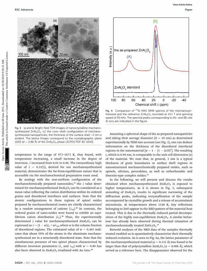
Fig. 3 (a and b) Bright-field TEM images of nanocrystalline mechano-synthesized ZnAl2O4, (c) the core–shell configuration of mechano-synthesized nanoparticles; the thickness of the surface shell �1 nm isevident. The lattice fringes correspond to the crystallographic plane(220) (d ¼ 2.86 A) of the ZnAl2O4 phase (JCPDS PDF 82-1043).
Fig. 4 Comparison of 27Al MAS NMR spectra of the mechanosyn-thesized and the reference ZnAl2O4 recorded at 14.1 T and spinningspeed of 30 kHz. The spectral peaks corresponding to (A)- and [B]-siteAl ions are indicated in the figure.
RSC Advances Paper
Ope
n A
cces
s A
rtic
le. P
ublis
hed
on 0
8 Ju
ne 2
015.
Dow
nloa
ded
on 1
0/30
/201
8 9:
58:3
5 A
M.
Thi
s ar
ticle
is li
cens
ed u
nder
a C
reat
ive
Com
mon
s A
ttrib
utio
n 3.
0 U
npor
ted
Lic
ence
.View Article Online
temperature in the range of 973–1673 K, they found, withtemperature increasing, a small increase in the degree ofinversion. l increased from 0.01 to 0.06. The extraordinary highvalue of l ¼ 0.31(2), derived for our mechanosynthesizedmaterial, demonstrates the far-from-equilibrium nature that isaccessible via the mechanochemical preparation route used.
By analogy with the non-uniform conguration of themechanochemically prepared nanooxides,15 the l value deter-mined for mechanosynthesized ZnAl2O4 can be considered as amean value reecting the cation distribution within its orderedgrains and disordered interfaces and surfaces. Note that theatomic congurations in these regions of spinel oxidesprepared by mechanochemical routes are chiey characterizedby a random arrangement of cations (lrd).30 In contrast, theordered grains of nano-oxides were found to exhibit an equi-librium cation distribution (lc).30 Thus, the experimentallydetermined l value for mechanosynthesized ZnAl2O4 can beexpressed as l¼ (1� w)lc + wlrd, where w is the volume fractionof disordered regions. The estimated value of w ¼ 0.465 indi-cates that about 50% of the atoms in the aluminate mechano-synthesized are in a structurally disordered state. Note that thesimultaneous presence of two spinel phases characterized bydifferent inversion parameters (lc and lrd) with w ¼ 0.80 hasalso been observed in ZnAl2O4 irradiated with Au ions.28
54324 | RSC Adv., 2015, 5, 54321–54328
Assuming a spherical shape of the as-prepared nanoparticlesand taking their average diameter (D ¼ 10 nm) as determinedexperimentally by TEM into account (see Fig. 3), one can deduceinformation on the thickness of the disordered interfacialregions in the nanomaterial [w ¼ 1 � (1 � 2t/D)3]. The resultingt, which is 0.94 nm, is comparable to the unit cell dimension (a)of the material. We note that, in general, 1 nm is a typicalthickness of grain boundaries or surface shell regions innanostructured mechanochemically prepared oxides, such asspinels, olivines, perovskites, as well as orthorhombic andilmenite-type complex oxides.15
In the following, we will present and discuss the resultsobtained when mechanosynthesized ZnAl2O4 is exposed tohigher temperatures. As it is shown in Fig. 5, subsequentannealing of ZnAl2O4 results in signicant narrowing of thediffraction peaks, indicating recrystallization of the sampleaccompanied by crystallite growth and a release of accumulatedmicrostrain. At temperatures above 1148 K, tiny reectionsbelonging to ZnO appear in the XRD pattern of thematerial heattreated. This is due to the thermally induced partial decompo-sition of the highly non-equilibrium ZnAl2O4. A similar behav-iour has already been observed during thermal relaxation ofmechanochemically treated ZnFe2O4.31
Rietveld analyses of the XRD data of the samples thermallytreated enabled us to quantitatively characterize their thermallyinduced evolution. As is seen in Fig. 6a, the lattice parameter ofthe mechanosynthesized material (a ¼ 8.121 A) was found to belarger than that of polycrystalline ZnAl2O4 (a ¼ 8.088 A), whichserved as a reference here. The disappearance observed for the
This journal is © The Royal Society of Chemistry 2015
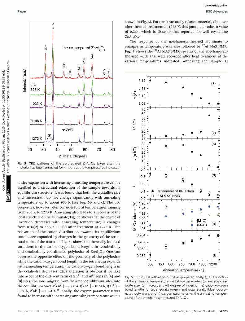
Fig. 5 XRD patterns of the as-prepared ZnAl2O4 taken after thematerial has been annealed for 4 hours at the temperatures indicated.
Fig. 6 Structural relaxation of the as-prepared ZnAl2O4 as a functionof the annealing temperature. (a) Lattice parameter, (b) average crys-tallite size, (c) microstrain, (d) degree of inversion (e) cation–oxygenbond lengths for tetrahedrally (green) and octahedrally (blue) coordi-nated polyhedra, and (f) oxygen parameter vs. the annealing temper-ature of the mechanosynthesized ZnAl2O4.
Paper RSC Advances
Ope
n A
cces
s A
rtic
le. P
ublis
hed
on 0
8 Ju
ne 2
015.
Dow
nloa
ded
on 1
0/30
/201
8 9:
58:3
5 A
M.
Thi
s ar
ticle
is li
cens
ed u
nder
a C
reat
ive
Com
mon
s A
ttrib
utio
n 3.
0 U
npor
ted
Lic
ence
.View Article Online
lattice expansion with increasing annealing temperature can beascribed to a structural relaxation of the sample towards itsequilibrium structure. It was found that both the crystallite sizeand microstrain do not change signicantly with annealingtemperature up to about 900 K (see Fig. 6b and c). The twoproperties, however, alter considerably at temperatures rangingfrom 900 K to 1273 K. Annealing also leads to a recovery of thelocal structure of the aluminate; Fig. 6d shows that the degree ofinversion decreases with annealing temperature; l changesfrom 0.34(2) to about 0.02(2) aer treatment at 1273 K. Therelaxation of the cation distribution towards its equilibriumstate is accompanied by changes in the geometry of the struc-tural units of the material. Fig. 6e shows the thermally inducedvariations in the cation–oxygen bond lengths in tetrahedrallyand octahedrally coordinated polyhedra of ZnAl2O4. One canobserve the opposite effect on the geometry of the polyhedra;while the cation–oxygen bond length in the tetrahedra expandswith annealing temperature, the cation–oxygen bond length inthe octahedra decreases. This alteration is obvious if we takeinto account the different radii of Zn2+ and Al3+ ions in (A) and[B] sites; the ions migrate from their nonequilibrium sites intothe equilibrium ones; r(Zn2+)¼ 0.60 A, r[Zn2+]¼ 0.74 A, r(Al3+)¼0.39 A, r[Al3+] ¼ 0.54 A.32 Finally, the oxygen parameter u wasfound to increase with increasing annealing temperature as it is
This journal is © The Royal Society of Chemistry 2015
shown in Fig. 6f. For the structurally relaxed material, obtainedaer thermal treatment at 1273 K, this parameter takes a valueof 0.264, which is close to that reported for well crystallineZnAl2O4.33
The response of the mechanosynthesized aluminate tochanges in temperature was also followed by 27Al MAS NMR.Fig. 7 shows the 27Al MAS NMR spectra of the mechanosyn-thesized oxide that were recorded aer heat treatment at thevarious temperatures indicated. Annealing the sample at
RSC Adv., 2015, 5, 54321–54328 | 54325
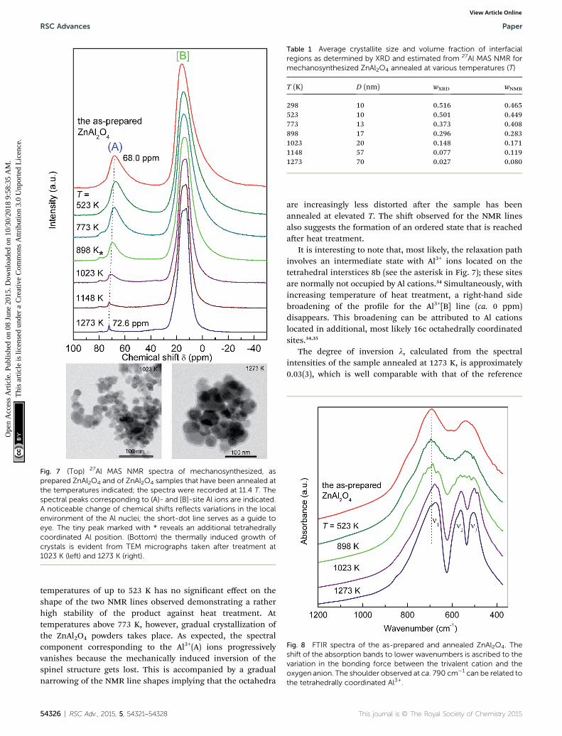
Fig. 7 (Top) 27Al MAS NMR spectra of mechanosynthesized, asprepared ZnAl2O4 and of ZnAl2O4 samples that have been annealed atthe temperatures indicated; the spectra were recorded at 11.4 T. Thespectral peaks corresponding to (A)- and [B]-site Al ions are indicated.A noticeable change of chemical shifts reflects variations in the localenvironment of the Al nuclei; the short-dot line serves as a guide toeye. The tiny peak marked with * reveals an additional tetrahedrallycoordinated Al position. (Bottom) the thermally induced growth ofcrystals is evident from TEM micrographs taken after treatment at1023 K (left) and 1273 K (right).
Table 1 Average crystallite size and volume fraction of interfacialregions as determined by XRD and estimated from 27Al MAS NMR formechanosynthesized ZnAl2O4 annealed at various temperatures (T)
T (K) D (nm) wXRD wNMR
298 10 0.516 0.465523 10 0.501 0.449773 13 0.373 0.408898 17 0.296 0.2831023 20 0.148 0.1711148 57 0.077 0.1191273 70 0.027 0.080
Fig. 8 FTIR spectra of the as-prepared and annealed ZnAl2O4. Theshift of the absorption bands to lower wavenumbers is ascribed to thevariation in the bonding force between the trivalent cation and theoxygen anion. The shoulder observed at ca. 790 cm�1 can be related tothe tetrahedrally coordinated Al3+.
RSC Advances Paper
Ope
n A
cces
s A
rtic
le. P
ublis
hed
on 0
8 Ju
ne 2
015.
Dow
nloa
ded
on 1
0/30
/201
8 9:
58:3
5 A
M.
Thi
s ar
ticle
is li
cens
ed u
nder
a C
reat
ive
Com
mon
s A
ttrib
utio
n 3.
0 U
npor
ted
Lic
ence
.View Article Online
temperatures of up to 523 K has no signicant effect on theshape of the two NMR lines observed demonstrating a ratherhigh stability of the product against heat treatment. Attemperatures above 773 K, however, gradual crystallization ofthe ZnAl2O4 powders takes place. As expected, the spectralcomponent corresponding to the Al3+(A) ions progressivelyvanishes because the mechanically induced inversion of thespinel structure gets lost. This is accompanied by a gradualnarrowing of the NMR line shapes implying that the octahedra
54326 | RSC Adv., 2015, 5, 54321–54328
are increasingly less distorted aer the sample has beenannealed at elevated T. The shi observed for the NMR linesalso suggests the formation of an ordered state that is reachedaer heat treatment.
It is interesting to note that, most likely, the relaxation pathinvolves an intermediate state with Al3+ ions located on thetetrahedral interstices 8b (see the asterisk in Fig. 7); these sitesare normally not occupied by Al cations.34 Simultaneously, withincreasing temperature of heat treatment, a right-hand sidebroadening of the prole for the Al3+[B] line (ca. 0 ppm)disappears. This broadening can be attributed to Al cationslocated in additional, most likely 16c octahedrally coordinatedsites.34,35
The degree of inversion l, calculated from the spectralintensities of the sample annealed at 1273 K, is approximately0.03(3), which is well comparable with that of the reference
This journal is © The Royal Society of Chemistry 2015
Paper RSC Advances
Ope
n A
cces
s A
rtic
le. P
ublis
hed
on 0
8 Ju
ne 2
015.
Dow
nloa
ded
on 1
0/30
/201
8 9:
58:3
5 A
M.
Thi
s ar
ticle
is li
cens
ed u
nder
a C
reat
ive
Com
mon
s A
ttrib
utio
n 3.
0 U
npor
ted
Lic
ence
.View Article Online
material (l ¼ 0.01(3)). In detail, the results on the relaxationprocess of far-from-equilibrium ZnAl2O4 are listed in Table 1.
Furthermore, FTIR spectroscopy was employed to provideinformation on the relaxation process. As shown in Fig. 8, thespectrum of the as-prepared aluminate is dominated by twobroadened bands centred at about 695 and 537 cm�1. Theycan be assigned to stretching vibrations in the oxide. Ashoulder at about 790 cm�1 can be related to the vibrations ofAl3+(A) ions.36,37 With increasing annealing temperature theabsorption bands become sharper and the peak centred at537 cm�1 splits into two absorption maxima at n2 ¼ 564 cm�1
and n3 ¼ 504 cm�1. This can be ascribed to the relaxation ofthe geometry of distorted polyhedra in ZnAl2O4. Since theposition of vibrational modes is rather sensitive to thechemical nature of trivalent cations, i.e., to the bonding forcebetween a trivalent cation and an oxygen anion,38 the observedred shi indicates the redistribution of cations from theirnonequilibrium sites towards the equilibrium ones. The latteris accompanied by the gradual disappearance of the shoulderat 790 cm�1.
4. Conclusions
The present study demonstrates that nanostructured ZnAl2O4
with an average crystallite size of about 10 nm in diameter canbe prepared via simple and straightforward mechanochem-ical synthesis starting with a stoichiometric mixture ofZnO : g-Al2O3. The synthesis was carried out at ambienttemperature and the reaction time was relatively short (2 h). Ithas been found that the as-prepared, nanostructured alumi-nate consists of ordered crystalline grains surrounded orseparated by disordered interfacial regions characterized by avolume fraction of about 50%. 27Al MAS NMR spectroscopydemonstrates that the nano-aluminate is characterized bydistorted polyhedra; moreover, the oxide shows a far-fromequilibrium arrangement of cations characterized by degreeof inversion of l ¼ 0.31(2). Fortunately, the range of thermalstability of the mechanosynthesized product extends up to ca.523 K. Upon annealing at T > 773 K, the nonequilibriumcation distribution relaxes towards the equilibrium congu-ration. Simultaneously, the crystallites grow and the accu-mulated microstrain releases during annealing. Thisrelaxation process is accompanied by a disappearance of thelattice expansion and variations in the cation–oxygen bondlengths. Thus, during heating, mechanosynthesized ZnAl2O4
relaxes towards a structural state that is similar to that of thebulk oxide.
Acknowledgements
The support by the DFG within the framework of the PriorityProgram “Crystalline Nonequilibrium Phases” (SPP 1415, grantsno.: WI 3600 5-2 and HE 1574 11-2) and the VEGA (projects 2/0097/13 and 2/0097/14) is gratefully acknowledged. M.F.thanks “Action Austria-Slovakia” cooperation programme forsupport of his work at TU Graz. V.S. acknowledges additionalsupport by the APVV (0528-11).
This journal is © The Royal Society of Chemistry 2015
Notes and references
1 V. Sepelak, S. M. Becker, I. Bergmann, S. Indris,M. Scheuermann, A. Feldhoff, C. Kubel, M. Bruns,N. Sturzl, A. S. Ulrich, M. Ghafari, H. Hahn, C. P. Grey,K. D. Becker and P. Heitjans, J. Mater. Chem., 2012, 22,3117.
2 V. Sepelak, I. Bergmann, D. Menzel, A. Feldhoff, P. Heitjans,F. J. Litterst and K. D. Becker, J. Magn. Magn. Mater., 2007,316, 764.
3 J. Wu, Z. Huang, W. Zhou, C. Ouyang, Y. Hou, Y. Gao,R. Chen and J. Chu, J. Appl. Phys., 2014, 115, 113703.
4 A. Kan, T. Moriyama, S. Takhashi and H. Ogawa, Jpn. J. Appl.Phys., 2013, 52, 09KH01.
5 V. Sepelak, I. Bergmann, S. Indris, A. Feldhoff, H. Hahn,K. D. Becker, C. P. Grey and P. Heitjans, J. Mater. Chem.,2011, 21, 8332.
6 W. Staszak, M. Zawadzki and J. Okal, J. Alloys Compd., 2010,492, 500.
7 N. J. van der Laag, M. D. Snel, P. C. M. M. Magusin and G. deWith, J. Eur. Ceram. Soc., 2004, 24, 2417.
8 X. Y. Chen, C. Ma, Z. J. Zhang and B. N. Wang, Mater. Sci.Eng., B, 2008, 151, 224.
9 A. D. Ballarini, S. A. Bocanegra, A. A. Castro, S. R. de Miqueland O. A. Scelza, Catal. Lett., 2009, 129, 293.
10 K. Sakoda and M. Hirano, Ceram. Int., 2014, 40, 15841;H. Zhao, Y. Dong, P. Jiang, G. Wang, J. Zhang andC. Zhang, Chem. Eng. J., 2015, 260, 623; M. Y. Guan,D. M. Xu, Y. F. Song and Y. Guo, Sens. Actuators, B, 2013,188, 1148; Z. Chen, E. Shi, W. Li, Y. Zheng, N. Wu andW. Zhong, J. Am. Ceram. Soc., 2002, 85, 2949.
11 R. K. Sharma and G. Ranjana, Ceram. Int., 2014, 40, 3209;M. S. Zulfakar, H. Abdullah, W. N. W. Jalal, S. Shaari andZ. Zainuddin, Adv. Mater. Res., 2014, 895, 63; X. Duan,D. Yuan, X. Wang and H. Xu, J. Sol-Gel Sci. Technol., 2005,35, 221.
12 R. Ianos, S. Borcanescu and R. Lazau, Chem. Eng. J., 2014,240, 260.
13 C. G. Anchieta, D. Sallet, E. L. Foletto, S. S. da Silva,O. Chiavone-Filho and C. A. O. do Nascimento, Ceram. Int.,2014, 40, 4173.
14 D. P. Dutta, R. Ghildiyal and A. K. Tyagi, J. Phys. Chem. C,2009, 113, 16954.
15 V. Sepelak, A. Duvel, M. Wilkening, K.-D. Becker andP. Heitjans, Chem. Soc. Rev., 2013, 42, 7507.
16 M. V. Zdujic, O. B. Milosevic and L. C. Karanovic,Mater. Lett.,1992, 13, 125.
17 K. L. Da Silva, D. Menzel, A. Feldhoff, C. Kubel, M. Bruns,A. Paesano Jr, A. Duvel, M. Wilkening, M. Ghafari,H. Hahn, F. J. Litterst, P. Heitjans, K. D. Becker andV. Sepelak, J. Phys. Chem. C, 2011, 115, 7209.
18 Joint Committee on Powder Diffraction, Standards (JCPDS)Powder Diffraction File (PDF), International Centre forDiffraction Data, Newtown Square, PA, 2004.
19 J. Rodriguez-Carvajal, Fullprof Program, Version 2.4.2, ILLGrenoble, Grenoble, France, 1993.
RSC Adv., 2015, 5, 54321–54328 | 54327
RSC Advances Paper
Ope
n A
cces
s A
rtic
le. P
ublis
hed
on 0
8 Ju
ne 2
015.
Dow
nloa
ded
on 1
0/30
/201
8 9:
58:3
5 A
M.
Thi
s ar
ticle
is li
cens
ed u
nder
a C
reat
ive
Com
mon
s A
ttrib
utio
n 3.
0 U
npor
ted
Lic
ence
.View Article Online
20 K. Brandenburg and H. Putz, Diamond—Crystal andMolecular Structure Visualization Soware, Version 3.0a,Crystal Impact GbR, Bonn, Germany, 2004.
21 D. Fenzke, D. Freude, T. Frohlich and J. Haase, Chem. Phys.Lett., 1984, 111, 171; D. Massiot, C. Bessada, J. P. Couturesand F. Taullele, J. Magn. Reson., 1990, 90, 231.
22 L. Samain, A. Jaworski, M. Eden, D. M. Ladd, D.-K. Seo,F. J. Garcia-Garcia and U. Haussermann, J. Solid StateChem., 2014, 217, 1.
23 A. Duvel, E. Romanova, M. Shari, D. Freude, M. Wark,P. Heitjans andM.Wilkening, J. Phys. Chem. C, 2011, 115, 22770.
24 R. L. Millard, R. C. Peterson and B. K. Hunter, Am. Mineral.,1992, 77, 44.
25 S. A. T. Redfern, R. J. Harrison, H. S. C. O'Neill andD. R. R. Wood, Am. Mineral., 1999, 84, 299.
26 Z. Wang, P. Lazor, S. K. Saxena and G. Artioli, J. Solid StateChem., 2002, 165, 165.
27 T. Soeda, S. Matsumura, C. Kinoshita and N. J. Zaluzec, J.Nucl. Mater., 2000, 283–287, 952.
28 G. Baldinozzi, D. Simeone, D. Gosset, M. Dolle, L. Thomeand L. Mazerolles, Nucl. Instrum. Methods Phys. Res., Sect.B, 2006, 250, 119.
54328 | RSC Adv., 2015, 5, 54321–54328
29 H. S. C. O'Neill and W. A. Dollase, Phys. Chem. Miner., 1994,20, 541.
30 V. Sepelak, I. Bergmann, A. Feldhoff, P. Heitjans,F. Krumeich, D. Menzel, F. J. Litterst, S. J. Campbell andK. D. Becker, J. Phys. Chem. C, 2007, 111, 5026.
31 V. Sepelak, L. Wilde, U. Steinike and K. D. Becker,Mater. Sci.Eng., A, 2004, 375–377, 865.
32 R. D. Shannon, Acta Crystallogr. A, 1976, 32, 751.33 M. Ardit, G. Cruciani and M. Dondi, Am. Mineral., 2012, 97,
1394.34 K. E. Sickafus, J. M. Wills and N. W. Grimes, J. Am. Ceram.
Soc., 1999, 82, 3279.35 V. Sreeja, T. S. Smitha, D. Nand, T. G. Ajithkumar and
P. A. Joy, J. Phys. Chem. C, 2008, 112, 14737; H. Maekawa,S. Kato, K. Kawamura and T. Yokokawa, Am. Mineral.,1997, 82, 1125.
36 A. A. Da Silva, A. D. S. Conçalves and M. R. Davolos, J. Sol-GelSci. Technol., 2009, 49, 101.
37 P. Tarte, Spectrochim. Acta, 1967, 23, 2127.38 J. Preudhomnei and P. Tarte, Spectrochim. Acta, 1971, 27,
1817.
This journal is © The Royal Society of Chemistry 2015