a review of the tragal pointer: anatomy and its importance
TRANSCRIPT

Folia Morphol. Vol. 71, No. 2, pp. 59–64
Copyright © 2012 Via MedicaISSN 0015–5659
www.fm.viamedica.plR E V I E W A R T I C L E
59
Address for correspondence: M. Loukas, MD, PhD, Professor and Chair, Department of Anatomical Sciences, St. George’s University,School of Medicine, Grenada, West Indies, tel: 473 444 4175 x2005, fax: 473 444 2887, e-mail: [email protected]
A review of the tragal pointer: anatomyand its importance as a landmarkin surgical proceduresM.A. Muhleman1, C.T. Wartmann2, R. Hage1, P. Matusz3, M.M. Shoja4, 5,R.S. Tubbs6, M. Loukas1, 7
1Department of Anatomical Sciences, School of Medicine, St. George’s University, Grenada, West Indies2Department of Otolaryngology/Head and Neck Surgery, University of Maryland Medical Centre, MD, USA3Department of Anatomy, Victor Babes University of Medicine and Pharmacy, Timisoara, Romania4Division of Neurosurgery, University of Alabama at Birmingham, AL, USA5Tuberculosis and Lung Disease Institute, Tabriz University of Medical Sciences, Tabriz, Iran6Paediatric Neurosurgery, Children’s Hospital, Birmingham, AL, USA7Department of Anatomy, Medical School Varmia and Mazuria, Olsztyn, Poland
[Received 21 February 2012; Accepted 15 April 2012]
The tragal pointer has long been used as a surgical landmark for the identifica-tion of the facial nerve trunk and the maxillary artery in such procedures asparotidectomy, internal fixation of subcondylar and condylar fractures, man-dibular osteotomy, temporomandibular joint arthroplasty, and percutaneousblocks of branches of the trigeminal nerve and pterygopalatine ganglion. Asidefrom its use as an external landmark, it has also been implicated as a contribu-tor to crease formation in the presence of peripheral arterial disease. This arti-cle will review the available literature on the tragal pointer’s use as an externallandmark. (Folia Morphol 2012; 71, 2: 59–64)
Key words: tragal pointer, tragus, facial nerve trunk, maxillaryartery, anterior tragal crease, landmarks, peripheral arterial disease
INTRODUCTIONIn every surgical procedure, the location and
identification of certain anatomic structures is cru-cial. Surgeons rely on the use of anatomical land-marks for identifying the location of various struc-tures. Good landmarks should be easy to identify,remain in a relatively constant position throughoutthe procedure, and be easy to palpate. These fea-tures allow fast and safe identification of the ana-tomical structures of importance [11, 13].
The tragal pointer has become an important land-mark for locating the facial nerve trunk and maxil-lary artery intraoperatively. This review discusses theanatomic location of the tragal pointer and its as-sociation with surrounding structures such as the
facial nerve trunk and the maxillary artery, as wellas using it to diagnose peripheral arterial disease[11, 13].
ANATOMYThe external ear is made up of two structures,
the auricle or pinna and the external acoustic me-atus. The lateral surface of the auricle faces slightlyanteriorly and has an irregular concavity to it alongwith multiple depressions and projections. The he-lix is a prominent curved rim that most often hasa small tubercle posterosuperiorly: Darwin’s tuber-cle. Anterior and parallel to the helix is the antihelix,which superiorly divides into two crura, creatinga triangular fossa. Between the helix and the anti-
60
Folia Morphol., 2012, Vol. 71, No. 2
helix is a depression called the scaphoid fossa. Theconcha of the auricle is encircled by the antihelixposteriorly and divided by the anterior end of thehelix. The depression between the anterior helix andthe inferior crus of the antihelix is the cymba con-chae. The cymba concha lies over the suprameataltriangle of the temporal bone and mastoid antrum[5, 14, 15].
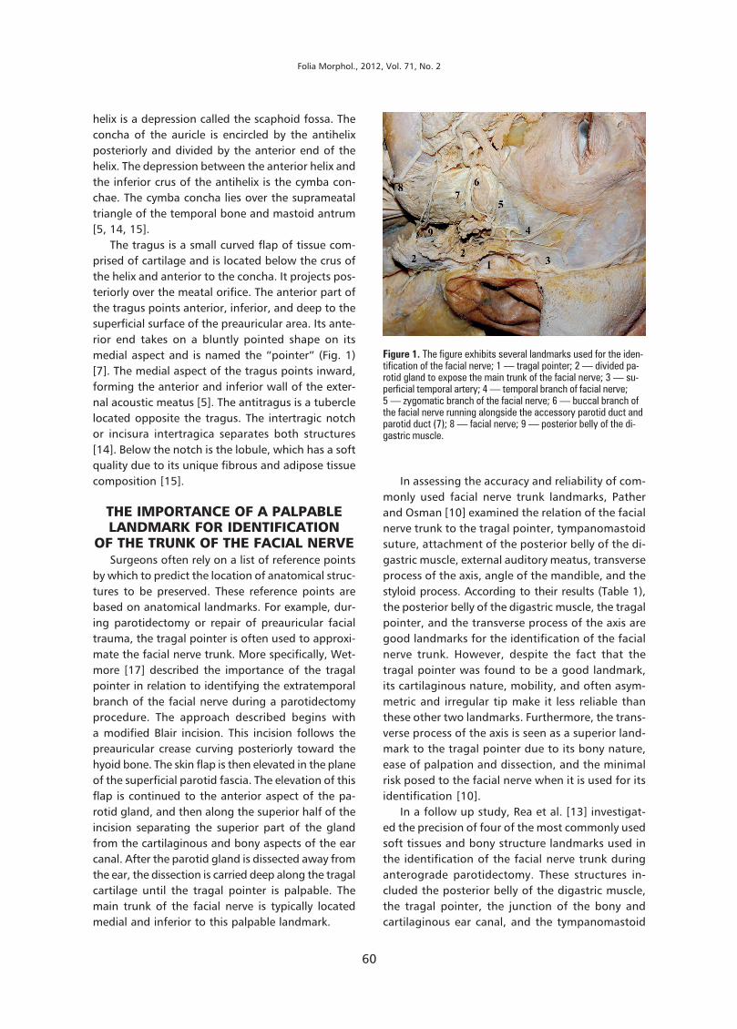
The tragus is a small curved flap of tissue com-prised of cartilage and is located below the crus ofthe helix and anterior to the concha. It projects pos-teriorly over the meatal orifice. The anterior part ofthe tragus points anterior, inferior, and deep to thesuperficial surface of the preauricular area. Its ante-rior end takes on a bluntly pointed shape on itsmedial aspect and is named the “pointer” (Fig. 1)[7]. The medial aspect of the tragus points inward,forming the anterior and inferior wall of the exter-nal acoustic meatus [5]. The antitragus is a tuberclelocated opposite the tragus. The intertragic notchor incisura intertragica separates both structures[14]. Below the notch is the lobule, which has a softquality due to its unique fibrous and adipose tissuecomposition [15].
THE IMPORTANCE OF A PALPABLELANDMARK FOR IDENTIFICATION
OF THE TRUNK OF THE FACIAL NERVESurgeons often rely on a list of reference points
by which to predict the location of anatomical struc-tures to be preserved. These reference points arebased on anatomical landmarks. For example, dur-ing parotidectomy or repair of preauricular facialtrauma, the tragal pointer is often used to approxi-mate the facial nerve trunk. More specifically, Wet-more [17] described the importance of the tragalpointer in relation to identifying the extratemporalbranch of the facial nerve during a parotidectomyprocedure. The approach described begins witha modified Blair incision. This incision follows thepreauricular crease curving posteriorly toward thehyoid bone. The skin flap is then elevated in the planeof the superficial parotid fascia. The elevation of thisflap is continued to the anterior aspect of the pa-rotid gland, and then along the superior half of theincision separating the superior part of the glandfrom the cartilaginous and bony aspects of the earcanal. After the parotid gland is dissected away fromthe ear, the dissection is carried deep along the tragalcartilage until the tragal pointer is palpable. Themain trunk of the facial nerve is typically locatedmedial and inferior to this palpable landmark.
In assessing the accuracy and reliability of com-monly used facial nerve trunk landmarks, Patherand Osman [10] examined the relation of the facialnerve trunk to the tragal pointer, tympanomastoidsuture, attachment of the posterior belly of the di-gastric muscle, external auditory meatus, transverseprocess of the axis, angle of the mandible, and thestyloid process. According to their results (Table 1),the posterior belly of the digastric muscle, the tragalpointer, and the transverse process of the axis aregood landmarks for the identification of the facialnerve trunk. However, despite the fact that thetragal pointer was found to be a good landmark,its cartilaginous nature, mobility, and often asym-metric and irregular tip make it less reliable thanthese other two landmarks. Furthermore, the trans-verse process of the axis is seen as a superior land-mark to the tragal pointer due to its bony nature,ease of palpation and dissection, and the minimalrisk posed to the facial nerve when it is used for itsidentification [10].
In a follow up study, Rea et al. [13] investigat-ed the precision of four of the most commonly usedsoft tissues and bony structure landmarks used inthe identification of the facial nerve trunk duringanterograde parotidectomy. These structures in-cluded the posterior belly of the digastric muscle,the tragal pointer, the junction of the bony andcartilaginous ear canal, and the tympanomastoid
Figure 1. The figure exhibits several landmarks used for the iden-tification of the facial nerve; 1 — tragal pointer; 2 — divided pa-rotid gland to expose the main trunk of the facial nerve; 3 — su-perficial temporal artery; 4 — temporal branch of facial nerve;5 — zygomatic branch of the facial nerve; 6 — buccal branch ofthe facial nerve running alongside the accessory parotid duct andparotid duct (7); 8 — facial nerve; 9 — posterior belly of the di-gastric muscle.

61
M.A. Muhleman et al., A review of the tragal pointer: anatomy and its importance as a landmark in surgical procedures
llary artery to the tragal pointer, Frankfort hori-zontal plane, and tip of the condyle (Table 3).These selected anatomic landmarks are importantduring surgical procedures such as bilateralcondylectomies, gap arthroplasties, and internalfixation of bilateral subcondylar fractures in orderto avoid iatrogenic injuries to the maxillary artery.The focus of the study was to obtain measure-ments of the distance between these structuresand the maxillary artery in order to create a cata-logue for intraoperative identification and loca-tion of the maxillary artery. From the results theauthors [9] observed that there was substantialvariation in the course (even in the same cadaver)and depth of the maxillary artery and that themost vulnerable segment of the maxillary arteryis that which courses close to the medial borderof the subcondylar region.
ASSOCIATION WITH PERIPHERALARTERIAL DISEASE
Recently, imaging studies have shown that themaxillary artery is affected by atherosclerotic changes[2, 3, 16]. Interestingly, artherosclerotic plaque build-up in the maxillary artery causes distention of itswalls. With this distension the overlying structure,
suture. The results of the measurements found thatthe closest structure to the main trunk of the fa-cial nerve was the tympanomastoid suture, whilethe farthest landmark was the cartilaginous ex-ternal canal (Table 2). With regard to the tragalpointer, the investigators observed considerablevariability in the size, shape, and direction in whichit “pointed”, which was not always in the direc-tion of the facial nerve trunk. Consequently, thetympanomastoid suture was found to be a supe-rior landmark for the identification of the facialnerve trunk. Notwithstanding its superiority, thetympanomastoid suture did, however, displaya statistical difference in the distance between sex-es. This finding may be due to larger skull, deepermandibular ramus, larger mastoid process, andlarger nuchal crest rigidity and rugose muscle at-tachments in males [8, 18].
ASSOCIATION OFTHE TRAGAL POINTER WITH
THE MAXILLARY ARTERYThe maxillary artery is the largest terminal
branch of the external carotid artery. Its locationnear the neck of the mandible predisposes it toiatrogenic injury from surgical procedures (e.g. in-ternal fixation of subcondylar and condylar frac-tures, mandibular osteotomies, temporomandib-ular joint arthroplasties, and percutaneous blocksof the branches of the trigeminal nerve or ptery-gopalatine ganglion) in this area. Thus, this ar-tery may sustain puncture or severing injuries thatcan lead to significant haemorrhage. In order toavoid such complications, the tragal pointer hasbeen used as a landmark.
Orbay et al. [9] in their study examined the dis-tance of the first and second portions of the maxi-
Table 1. Distance of the facial never trunk to sevencommonly used anatomical landmarks
Structure Distance (mean) [mm]
Tragal pointer 24.3–49.2 (34)
External auditory canal 7.3–21.9 (13.4)
Posterior belly of digastric 9.7–24.3 (14.6)
Tympanomastoid suture 4.9–18.6 (10.0)
Styloid process 4.3–18.6 (9.8)
Transverse process of the axis 9.7–36.8 (16.9)
Angle of the mandible 25.3–48.69 (38.1)
Table 2. Average distance of the facial nerve trunkto four commonly used landmarks in anterogradeparotidectomy
Structure Average distance [mm]
Posterior belly of the 5.5 ± 2.1digastric muscle
Tragal pointer 6.9 ± 1.8
Cartilaginous ear canal 10.9 ± 1.7
Tympanomastoid suture 2.5 ± 0.4
Table 3. Distance of the maxillary artery branch point tosurrounding structures
Structure Mean distance [mm]
Medial boarder of the subcondylar 6.8portion of the mandible
Tragal pointer 16.2 (anterior)and 21.4 (deep)
Frankfort horizontal plane 25.7
Tip of the condyle in the vertical plane 22.4

62
Folia Morphol., 2012, Vol. 71, No. 2
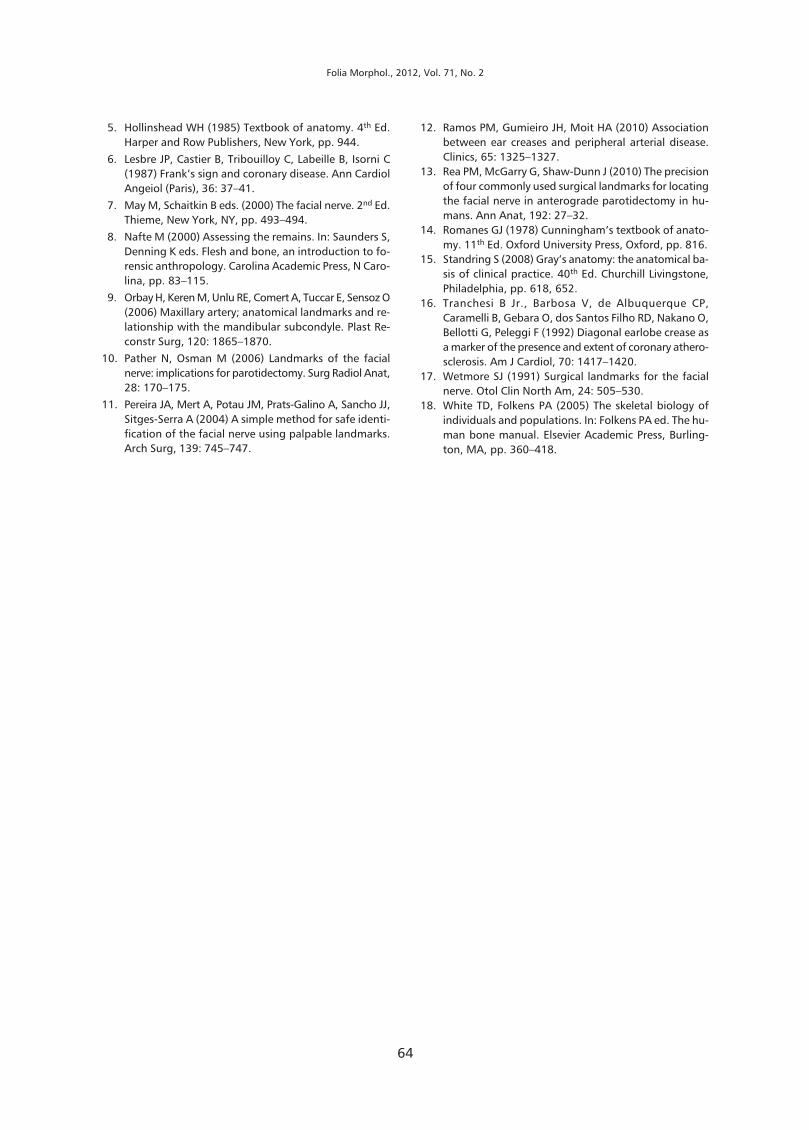
the parotid gland, is pushed superficially. The pos-terior aspect of the superior half of the parotid glandis fixed by dense fibrous tissue at the point of thetragal pointer. The anterior tragal crease is createdby the superficial displacement of the parotid glandand the downward angle of the tragus (Fig. 2). Thepresence of this crease has been proposed to be ofclinical use to detect the presence of asymptomaticperipheral arterial disease and consequently coro-nary artery disease [12].
Prior to the anterior tragal crease another ex-ternal indicator, Frank’s sign, was proposed as clini-cally important for the detection of asymptomaticperipheral arterial disease. Frank’s sign, or diago-nal ear lobe crease, is defined as a crease that ex-tends diagonally from the tragus across the ear lobeto the posterior edge of the auricle [4]. Lesbre etal. [6] studied the sensitivity and the predictivevalue of Frank’s sign to detect coronary artery dis-ease. The criteria selected for a positive diagnosisof coronary artery disease was stenosis of 75% orhigher in one of the three main coronary arteries.Frank’s sign was found to have sensitivity, speci-ficity, and positive predictive values of 75%, 57.5%and 80.3%, respectively. From the results no corre-lation was found between Frank’s sign and the se-verity of coronary artery disease. It was the opi-nion of the author that Frank’s sign is a marker forcoronary artery disease, independent of known riskfactors. Interestingly, the presence of Frank’s signis found in 66% of those with asymptomatic coro-nary artery disease. However, the absence of Frank’ssign cannot exclude the diagnosis of coronary ar-tery disease.
In a similar study of the diagonal earlobe crease,another Davis et al. [1] compared the speci-ficity, sensitivity, and positive predictive value ofthe diagonal earlobe crease in cases of coronaryartery disease and diabetic retinopathy. It wasfound that the presence of a diagonal earlobecrease was higher in those with coronary arterydisease than it was in those without. From theresults it was found that the diagonal earlobecrease has a sensitivity and specificity of 60% and48%, respectively, in regards to asymptomaticcoronary artery disease. The sensitivity and speci-ficity of the diagonal earlobe crease to detect dia-betic retinopathy was 61% and 47%, respective-ly. It is the opinion of this author that a diagonalear lobe crease has no value in predicting the pres-ence of asymptomatic coronary artery disease ordiabetic retinopathy [1].
Evrengul et al. [3] studied the association ofbilateral diagonal ear lobe crease and its predic-tive value in patients with asymptomatic coronaryartery disease. Four hundred and fifteen patientswere examined for the presence of bilateral diago-nal ear lobe crease and underwent an angiogra-phy looking for the presence of coronary arterydisease. The results of the study found bilateraldiagonal ear lobe crease to be a significant inde-pendent variable as well as a positive predictivefactor for those with coronary artery disease, hy-pertension and a family history of coronary arterydisease. Bilateral diagonal ear lobe crease wasfound to have a sensitivity of 51.3%, specificity of84.8%, positive predictive value of 89.4%, andnegative predictive value of 41.2%. It is the opin-ion of the author that the presence of bilateraldiagonal ear lobe crease was a significant risk fac-tor for coronary artery disease.
Ramos et al. [12] compared and investigatedthe correlation of the diagonal earlobe crease andanterior tragal crease among patients with docu-mented peripheral artery disease of the lowerlimbs to those without documented peripheralarterial disease. The results of the study showeda higher percentage of diagonal ear lobe crease andanterior tragal crease among those with periphe-
Figure 2. This figure demonstrates an anterior tragal crease. Thiscrease is typically is created by the superficial displacement ofthe parotid gland and the downward angle of the tragus.

63
M.A. Muhleman et al., A review of the tragal pointer: anatomy and its importance as a landmark in surgical procedures
ral arterial disease than those without. The per-centage of a diagonal ear lobe crease was 73%vs. 25% while that of anterior tragal crease was80% vs. 43% in the peripheral arterial diseasegroup and the control group, respectively. Asa result, the diagonal ear lobe crease and the ante-rior tragal crease were independently associatedwith peripheral arterial disease and could be usedas an external indicator for identification ofasymptomatic peripheral arterial disease and con-sequently coronary artery disease.
DISCUSSIONThe four commonly used landmarks in identi-
fication of the facial nerve trunk during surgicalprocedures are: the tragal pointer, the posteriorbelly of digastric muscle, the junction of the bonyand cartilaginous ear canal, and the tympano-mastoid suture. Multiple cadaveric studies havebeen conducted to determine the reliability ofthese landmarks in a large series of specimens.From the multiple studies conducted a generalconsensus has not been formulated on whichexternal landmark is the most consistent and su-perior in identifying and locating the facial nervetrunk. In reference to the tragal pointer, the stud-ies have shown it to fulfil the criteria as a consis-tent landmark for locating the facial nerve trunk,but when compared to the other commonly usedlandmarks it has been shown to be inferior. Dueto the lack of consensus on which single land-mark is the best to use, it is now becominga standard of care to utilise more than one ofthe common external landmarks when identify-ing and locating the facial nerve trunk prior tosurgical procedures.
The location of the tragal pointer has not onlybeen used as a landmark for identifying and lo-cating the facial nerve trunk but also the maxil-lary artery. As with the investigations concern-ing its precision as a landmark for the identifica-tion of the facial nerve trunk, there have also beenstudies of its accuracy in identifying and locat-ing the maxillary artery. These studies comparedthe tragal pointer to other commonly used land-marks such as the condyle, the subcondyle, andthe sigmoid notch and revealed that there wassignificant variation in the course and the depthof the maxillary artery. Due to the significant vari-ations, locating the maxillary artery preoperative-ly is still difficult even with the use of the mostconsistent landmarks.
Although the reliability of the tragal pointeras an external landmark has come into question,it may have another important clinical use. Thelocation of the tragal pointer to the maxillary ar-tery has an effect on the topographical anatomyin the preauricular area. In the development of ath-erosclerosis plaques in the maxillary artery, theartery distends. As the artery distends it pushesthe overlying parotid gland superficially. As theparotid gland is pushed superficially the posteri-or attachment, which is fixed to the tragal point-er by dense fibrous tissue, causes a pivot point inwhich a crease develops. The presence of an ante-rior tragal crease is proposed as an external signfor asymptomatic peripheral arterial disease, andby extension, coronary artery disease. As shownin this review there are significant variations tothe size, shape, and the distance of the tragalpointer to other proximal structures such as themaxillary artery. Due to these variations, the de-velopment of the anterior tragal crease may notbe as reliable as once thought.
CONCLUSIONSThe four commonly used landmarks in identifi-
cation of the facial nerve trunk during surgical pro-cedures are: the tragal pointer, the posterior bellyof digastric muscle, the junction of the bony andcartilaginous ear canal, and the tympanomastoidsuture. Multiple cadaveric studies have been con-ducted to determine the reliability of these land-marks. Based on our review of the literature regard-ing the tragal pointer as a landmark, a more reliablebony landmark or multiple landmarks should beconsidered for locating the facial nerve trunk andmaxillary artery.
REFERENCES1. Davis TM, Balme M, Jackson D, Stuccio G, Bruce DG
(2000). The diagonal ear lobe crease (Frank’s sign) isnot associated with coronary artery disease or retino-pathy in type 2 diabetes: the Fremantle Diabetes Study.Aust N Z J Med, 30: 573–577.
2. Edston E (2006) The earlobe crease, coronary artery dis-ease, and sudden cardiac death: an autopsy study of520 individuals. Am J Forensic Med Pathol, 27: 129––133.
3. Evrengül H, Dursunoðlu D, Kaftan A, Zoghi M, Tanri-verdi H, Zungur M, Kilic M (2004) Bilateral diagonalearlobe crease and coronary artery disease: a signifi-cant association. Dermatology (Basel), 209: 271–275.
4. Frank ST (1973) Aural sign of coronary-artery disease.N Engl J Med, 289: 327–328.
64
Folia Morphol., 2012, Vol. 71, No. 2
5. Hollinshead WH (1985) Textbook of anatomy. 4th Ed.Harper and Row Publishers, New York, pp. 944.
6. Lesbre JP, Castier B, Tribouilloy C, Labeille B, Isorni C(1987) Frank’s sign and coronary disease. Ann CardiolAngeiol (Paris), 36: 37–41.
7. May M, Schaitkin B eds. (2000) The facial nerve. 2nd Ed.Thieme, New York, NY, pp. 493–494.
8. Nafte M (2000) Assessing the remains. In: Saunders S,Denning K eds. Flesh and bone, an introduction to fo-rensic anthropology. Carolina Academic Press, N Caro-lina, pp. 83–115.
9. Orbay H, Keren M, Unlu RE, Comert A, Tuccar E, Sensoz O(2006) Maxillary artery; anatomical landmarks and re-lationship with the mandibular subcondyle. Plast Re-constr Surg, 120: 1865–1870.
10. Pather N, Osman M (2006) Landmarks of the facialnerve: implications for parotidectomy. Surg Radiol Anat,28: 170–175.
11. Pereira JA, Mert A, Potau JM, Prats-Galino A, Sancho JJ,Sitges-Serra A (2004) A simple method for safe identi-fication of the facial nerve using palpable landmarks.Arch Surg, 139: 745–747.
12. Ramos PM, Gumieiro JH, Moit HA (2010) Associationbetween ear creases and peripheral arterial disease.Clinics, 65: 1325–1327.
13. Rea PM, McGarry G, Shaw-Dunn J (2010) The precisionof four commonly used surgical landmarks for locatingthe facial nerve in anterograde parotidectomy in hu-mans. Ann Anat, 192: 27–32.
14. Romanes GJ (1978) Cunningham’s textbook of anato-my. 11th Ed. Oxford University Press, Oxford, pp. 816.
15. Standring S (2008) Gray’s anatomy: the anatomical ba-sis of clinical practice. 40th Ed. Churchill Livingstone,Philadelphia, pp. 618, 652.
16. Tranchesi B Jr., Barbosa V, de Albuquerque CP,Caramelli B, Gebara O, dos Santos Filho RD, Nakano O,Bellotti G, Peleggi F (1992) Diagonal earlobe crease asa marker of the presence and extent of coronary athero-sclerosis. Am J Cardiol, 70: 1417–1420.
17. Wetmore SJ (1991) Surgical landmarks for the facialnerve. Otol Clin North Am, 24: 505–530.
18. White TD, Folkens PA (2005) The skeletal biology ofindividuals and populations. In: Folkens PA ed. The hu-man bone manual. Elsevier Academic Press, Burling-ton, MA, pp. 360–418.