a review of equine muscle disorders - home page |...
TRANSCRIPT
www.elsevier.com/locate/nmd
Neuromuscular Disorders 18 (2008) 277–287
Review
A review of equine muscle disorders
M. Aleman *
Department of Medicine and Epidemiology, Tupper Hall 2108, One Shields Avenue, School of Veterinary Medicine,
University of California, Davis, CA 95616, USA
Received 8 September 2007; received in revised form 17 December 2007; accepted 6 January 2008
Abstract
Muscle disorders are a common cause of disability in horses. For many years, clinical manifestations such as muscle pain, exerciseintolerance, weakness, and stiffness were believed to be caused by a single syndrome. However, in the past years a broad spectrum ofmuscle disorders have been recognized including glycogen and polysaccharide storage myopathies, malignant hyperthermia, mitochon-drial myopathy, hyperkalemic periodic paralysis and others. For some, a specific mutation has been identified. Recognition of the myo-pathic clinical phenotype and thorough clinical, electrodiagnostic, and histological evaluations are essential to further our understandingof equine myopathies. Advances in understanding equine myopathies may potentially benefit other species including humans.� 2008 Elsevier B.V. All rights reserved.
Keywords: Equine; Exertional; Myopathy; Myotonia; Rhabdomyolysis
1. Introduction
Muscle disorders are a common cause of disability inaffected horses, and in the past, have been known by sev-eral names including tying up, Monday morning disease,azoturia, equine rhabdomyolysis, and equine myoglobinu-ria. Although originally thought to be a single clinical syn-drome, it is now clear that these clinical manifestations arecommon to several different muscle disorders with differentetiologies [1]. In 1992 the first hereditary muscle disease,hyperkalemic periodic paralysis in Quarter Horses, wasreported [2,3] and a genetic test is available for diagnosis.Recently metabolic, inflammatory, dystrophic and otherinherited muscle diseases have been described in horses[4–10]. A specific genetic defect and mode of inheritancehave only been identified in hyperkalemic periodic paraly-sis [3], glycogen branching enzyme deficiency [7], andmalignant hyperthermia [8].
Horses have a number of muscle disorders which sharesimilar clinical, histopathological and in some cases molecu-lar features with humans. Thus the horse can be considered
0960-8966/$ - see front matter � 2008 Elsevier B.V. All rights reserved.
doi:10.1016/j.nmd.2008.01.001
* Tel.: +1 530 752 0290; fax: +1 530 752 9815.E-mail address: [email protected]
as an animal model for human muscle diseases. Disordersthat affect horses and man include equine motor neuron dis-ease [11] with many similarities to human amyotrophic lat-eral sclerosis [12], malignant hyperthermia in QuarterHorses with a mutation in the calcium release channel ofthe skeletal muscle sarcoplasmic reticulum, RyR1 gene [8],hyperkalemic periodic paralysis in Quarter Horses causedby a mutation in the a-subunit of the skeletal muscle sodiumchannel, SCN4A gene [2], and glycogen branching enzyme 1deficiency in Quarter Horse and Paint foals due to a muta-tion in the glycogen branching enzyme 1, GBE1 gene [7].With increased recognition of the myopathic phenotype inhorses by veterinarians, and use of state-of-the-art histolog-ical, biochemical and molecular techniques, the spectrum ofmyopathies affecting the horse will be greatly expanded.
Known causes of equine myopathies are shown in Table1. This classification separates myopathies into non-exer-tional and exertional categories. These categories are fur-ther divided into whether or not they are associated withrhabdomyolysis. A final category covers diseases associatedwith altered membrane conduction. This review highlightsthe most important recognized muscle disorders in horseswith an emphasis on inherited, metabolic, toxic, andinflammatory myopathies.
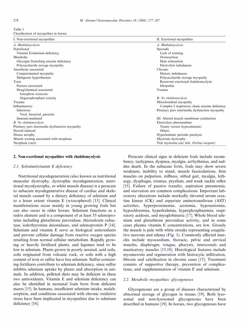
Table 1Classification of myopathies in horses
I. Non-exertional myopathies II. Exertional myopathies
A. Rhabdomyolysis A. Rhabdomyolysis
Nutritional SporadicVitamin E/selenium deficiency Lack of training
Metabolic OverexertionGlycogen branching enzyme deficiency Heat exhaustionPolysaccharide storage myopathy Electrolyte imbalances
Anesthesia associated ChronicCompartmental myopathy Dietary imbalancesMalignant hyperthermia Polysaccharide storage myopathy
Toxic Recurrent exertional rhabdomyolysisPasture associated IdiopathicDrug/chemical associated Trauma
Ionophore toxicosisOrganophosphate toxicity B. No rhabdomyolysis
Trauma Mitochondrial myopathyInflammatory Complex I respiratory chain enzyme deficiency
Infectious Pituitary pars intermedia dysfunction myopathyViral, bacterial, parasitic
Immune-mediated III. Altered muscle membrane conductionB. No rhabdomyolysis Electrolyte abnormalitiesPituitary pars intermedia dysfunction myopathy Tetany (severe hypocalcemia)Steroid induced OthersDisuse atrophy Hyperkalemic periodic paralysisMuscle wasting associated with neoplasia Myotonic dystrophyNeoplasia (rare) Tick myotonia (ear tick: Otobius megnini)
278 M. Aleman / Neuromuscular Disorders 18 (2008) 277–287
2. Non-exertional myopathies with rhabdomyolysis
2.1. Selenium/vitamin E deficiency
Nutritional myodegeneration (also known as nutritionalmuscular dystrophy, dystrophic myodegeneration, nutri-tional myodystrophy, or white muscle disease) is a peracuteto subacute myodegenerative disease of cardiac and skele-tal muscle caused by a dietary deficiency of selenium andto a lesser extent vitamin E (a-tocopherol) [13]. Clinicalmanifestations occur mainly in young growing foals butcan also occur in older horses. Selenium functions as aredox element and is a component of at least 35 selenopro-teins including glutathione peroxidase, thioredoxin reduc-tase, iodothyronine deionidases, and selenoprotein P [14].Selenium and vitamin E serve as biological antioxidantsand prevent cellular damage from reactive oxygen speciesresulting from normal cellular metabolism. Rapidly grow-ing or heavily fertilized plants, and legumes tend to below in selenium. Plants grown in poorly aerated, acid soils,soils originated from volcanic rock, or soils with a highcontent of iron or sulfur have less selenium. Sulfur-contain-ing fertilizers contribute to selenium deficiency, since sulfurinhibits selenium uptake by plants and absorption in ani-mals. In addition, pelleted diets may be deficient in thesetwo antioxidants. Vitamin E and selenium deficiency canalso be identified in neonatal foals born from deficientmares [15]. In humans, insufficient selenium intake, malab-sorption, and conditions associated with chronic oxidativestress have been implicated in myopathies due to seleniumdeficiency [16].
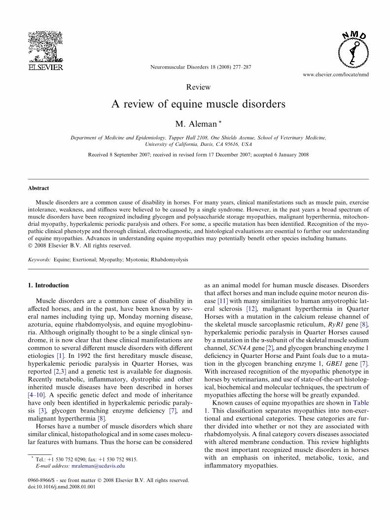
Peracute clinical signs in deficient foals include recum-bency, tachypnea, dyspnea, myalgia, arrhythmias, and sud-den death. In the subacute form, foals may show severeweakness, inability to stand, muscle fasciculations, firmmuscles on palpation, stiffness, stilted gait, myalgia, leth-argy, dysphagia, trismus, ptyalism, and weak suckle reflex[15]. Failure of passive transfer, aspiration pneumonia,and starvation are common complications. Important lab-oratory alterations include markedly elevated serum crea-tine kinase (CK) and aspartate aminotransferase (AST)activities, hyperproteinemia, azotemia, hyponatremia,hypochloremia, hyperkalemia, hyperphosphatemia, respi-ratory acidosis, and myoglobinuria [17]. Whole blood sele-nium and glutathione peroxidase activity, and in somecases plasma vitamin E concentrations, are low. Grosslythe muscle is pale with white streaks representing coagula-tive necrosis and edema (Fig. 1). Commonly affected mus-cles include myocardium, thoracic, pelvic and cervicalmuscles, diaphragm, tongue, pharynx, intercostals andmasticatory muscles [15,18]. Histological features includemyonecrosis and regeneration with histiocytic infiltration,fibrosis and calcification in chronic cases [15]. Treatmentconsists of supportive therapy, prevention of complica-tions, and supplementation of vitamin E and selenium.
2.2. Metabolic myopathies: glycogenoses
Glycogenoses are a group of diseases characterized byabnormal storage of glycogen in tissues [19]. Both lyso-somal and non-lysosomal glycogenoses have beendescribed in humans [19]. In horses, two glycogenoses have
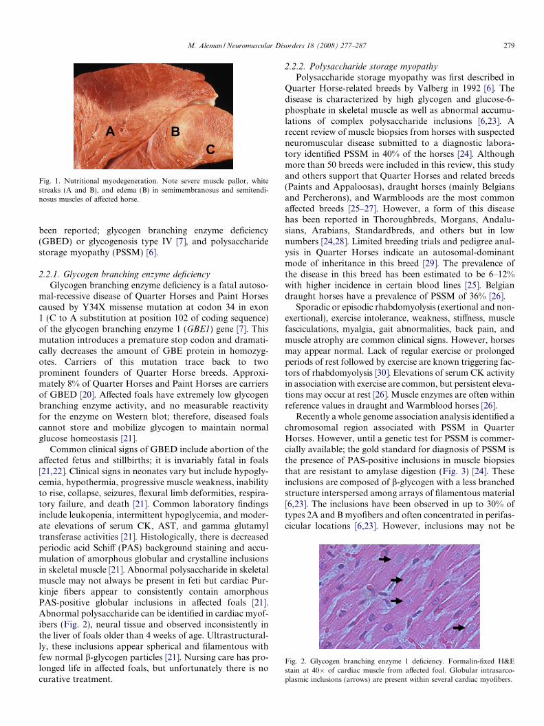
Fig. 1. Nutritional myodegeneration. Note severe muscle pallor, whitestreaks (A and B), and edema (B) in semimembranosus and semitendi-nosus muscles of affected horse.
Fig. 2. Glycogen branching enzyme 1 deficiency. Formalin-fixed H&Estain at 40� of cardiac muscle from affected foal. Globular intrasarco-plasmic inclusions (arrows) are present within several cardiac myofibers.
M. Aleman / Neuromuscular Disorders 18 (2008) 277–287 279
been reported; glycogen branching enzyme deficiency(GBED) or glycogenosis type IV [7], and polysaccharidestorage myopathy (PSSM) [6].
2.2.1. Glycogen branching enzyme deficiency
Glycogen branching enzyme deficiency is a fatal autoso-mal-recessive disease of Quarter Horses and Paint Horsescaused by Y34X missense mutation at codon 34 in exon1 (C to A substitution at position 102 of coding sequence)of the glycogen branching enzyme 1 (GBE1) gene [7]. Thismutation introduces a premature stop codon and dramati-cally decreases the amount of GBE protein in homozyg-otes. Carriers of this mutation trace back to twoprominent founders of Quarter Horse breeds. Approxi-mately 8% of Quarter Horses and Paint Horses are carriersof GBED [20]. Affected foals have extremely low glycogenbranching enzyme activity, and no measurable reactivityfor the enzyme on Western blot; therefore, diseased foalscannot store and mobilize glycogen to maintain normalglucose homeostasis [21].
Common clinical signs of GBED include abortion of theaffected fetus and stillbirths; it is invariably fatal in foals[21,22]. Clinical signs in neonates vary but include hypogly-cemia, hypothermia, progressive muscle weakness, inabilityto rise, collapse, seizures, flexural limb deformities, respira-tory failure, and death [21]. Common laboratory findingsinclude leukopenia, intermittent hypoglycemia, and moder-ate elevations of serum CK, AST, and gamma glutamyltransferase activities [21]. Histologically, there is decreasedperiodic acid Schiff (PAS) background staining and accu-mulation of amorphous globular and crystalline inclusionsin skeletal muscle [21]. Abnormal polysaccharide in skeletalmuscle may not always be present in feti but cardiac Pur-kinje fibers appear to consistently contain amorphousPAS-positive globular inclusions in affected foals [21].Abnormal polysaccharide can be identified in cardiac myof-ibers (Fig. 2), neural tissue and observed inconsistently inthe liver of foals older than 4 weeks of age. Ultrastructural-ly, these inclusions appear spherical and filamentous withfew normal b-glycogen particles [21]. Nursing care has pro-longed life in affected foals, but unfortunately there is nocurative treatment.
2.2.2. Polysaccharide storage myopathy
Polysaccharide storage myopathy was first described inQuarter Horse-related breeds by Valberg in 1992 [6]. Thedisease is characterized by high glycogen and glucose-6-phosphate in skeletal muscle as well as abnormal accumu-lations of complex polysaccharide inclusions [6,23]. Arecent review of muscle biopsies from horses with suspectedneuromuscular disease submitted to a diagnostic labora-tory identified PSSM in 40% of the horses [24]. Althoughmore than 50 breeds were included in this review, this studyand others support that Quarter Horses and related breeds(Paints and Appaloosas), draught horses (mainly Belgiansand Percherons), and Warmbloods are the most commonaffected breeds [25–27]. However, a form of this diseasehas been reported in Thoroughbreds, Morgans, Andalu-sians, Arabians, Standardbreds, and others but in lownumbers [24,28]. Limited breeding trials and pedigree anal-ysis in Quarter Horses indicate an autosomal-dominantmode of inheritance in this breed [29]. The prevalence ofthe disease in this breed has been estimated to be 6–12%with higher incidence in certain blood lines [25]. Belgiandraught horses have a prevalence of PSSM of 36% [26].
Sporadic or episodic rhabdomyolysis (exertional and non-exertional), exercise intolerance, weakness, stiffness, musclefasciculations, myalgia, gait abnormalities, back pain, andmuscle atrophy are common clinical signs. However, horsesmay appear normal. Lack of regular exercise or prolongedperiods of rest followed by exercise are known triggering fac-tors of rhabdomyolysis [30]. Elevations of serum CK activityin association with exercise are common, but persistent eleva-tions may occur at rest [26]. Muscle enzymes are often withinreference values in draught and Warmblood horses [26].
Recently a whole genome association analysis identified achromosomal region associated with PSSM in QuarterHorses. However, until a genetic test for PSSM is commer-cially available; the gold standard for diagnosis of PSSM isthe presence of PAS-positive inclusions in muscle biopsiesthat are resistant to amylase digestion (Fig. 3) [24]. Theseinclusions are composed of b-glycogen with a less branchedstructure interspersed among arrays of filamentous material[6,23]. The inclusions have been observed in up to 30% oftypes 2A and B myofibers and often concentrated in perifas-cicular locations [6,23]. However, inclusions may not be
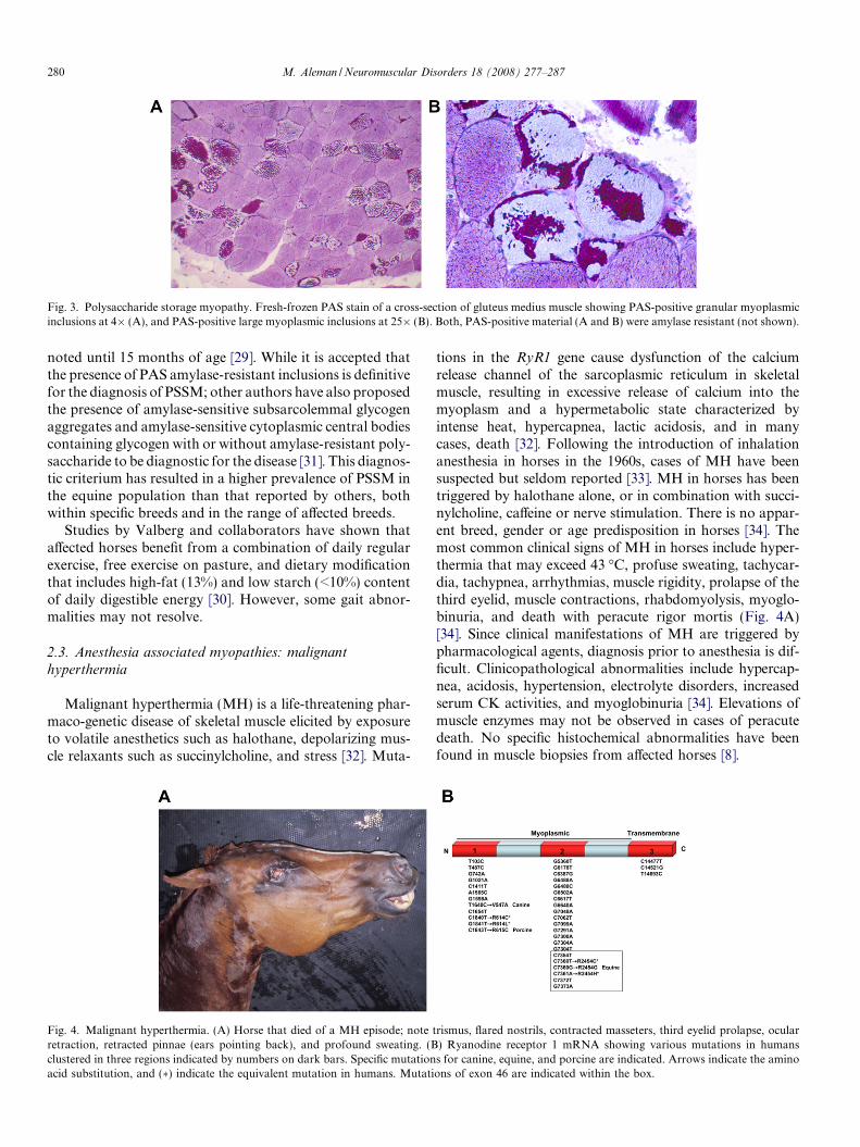
Fig. 3. Polysaccharide storage myopathy. Fresh-frozen PAS stain of a cross-section of gluteus medius muscle showing PAS-positive granular myoplasmicinclusions at 4� (A), and PAS-positive large myoplasmic inclusions at 25� (B). Both, PAS-positive material (A and B) were amylase resistant (not shown).
280 M. Aleman / Neuromuscular Disorders 18 (2008) 277–287
noted until 15 months of age [29]. While it is accepted thatthe presence of PAS amylase-resistant inclusions is definitivefor the diagnosis of PSSM; other authors have also proposedthe presence of amylase-sensitive subsarcolemmal glycogenaggregates and amylase-sensitive cytoplasmic central bodiescontaining glycogen with or without amylase-resistant poly-saccharide to be diagnostic for the disease [31]. This diagnos-tic criterium has resulted in a higher prevalence of PSSM inthe equine population than that reported by others, bothwithin specific breeds and in the range of affected breeds.
Studies by Valberg and collaborators have shown thataffected horses benefit from a combination of daily regularexercise, free exercise on pasture, and dietary modificationthat includes high-fat (13%) and low starch (<10%) contentof daily digestible energy [30]. However, some gait abnor-malities may not resolve.
2.3. Anesthesia associated myopathies: malignant
hyperthermia
Malignant hyperthermia (MH) is a life-threatening phar-maco-genetic disease of skeletal muscle elicited by exposureto volatile anesthetics such as halothane, depolarizing mus-cle relaxants such as succinylcholine, and stress [32]. Muta-
Fig. 4. Malignant hyperthermia. (A) Horse that died of a MH episode; noteretraction, retracted pinnae (ears pointing back), and profound sweating. (Bclustered in three regions indicated by numbers on dark bars. Specific mutationacid substitution, and (�) indicate the equivalent mutation in humans. Mutati
tions in the RyR1 gene cause dysfunction of the calciumrelease channel of the sarcoplasmic reticulum in skeletalmuscle, resulting in excessive release of calcium into themyoplasm and a hypermetabolic state characterized byintense heat, hypercapnea, lactic acidosis, and in manycases, death [32]. Following the introduction of inhalationanesthesia in horses in the 1960s, cases of MH have beensuspected but seldom reported [33]. MH in horses has beentriggered by halothane alone, or in combination with succi-nylcholine, caffeine or nerve stimulation. There is no appar-ent breed, gender or age predisposition in horses [34]. Themost common clinical signs of MH in horses include hyper-thermia that may exceed 43 �C, profuse sweating, tachycar-dia, tachypnea, arrhythmias, muscle rigidity, prolapse of thethird eyelid, muscle contractions, rhabdomyolysis, myoglo-binuria, and death with peracute rigor mortis (Fig. 4A)[34]. Since clinical manifestations of MH are triggered bypharmacological agents, diagnosis prior to anesthesia is dif-ficult. Clinicopathological abnormalities include hypercap-nea, acidosis, hypertension, electrolyte disorders, increasedserum CK activities, and myoglobinuria [34]. Elevations ofmuscle enzymes may not be observed in cases of peracutedeath. No specific histochemical abnormalities have beenfound in muscle biopsies from affected horses [8].
trismus, flared nostrils, contracted masseters, third eyelid prolapse, ocular) Ryanodine receptor 1 mRNA showing various mutations in humanss for canine, equine, and porcine are indicated. Arrows indicate the aminoons of exon 46 are indicated within the box.

M. Aleman / Neuromuscular Disorders 18 (2008) 277–287 281
Six distinct loci for malignant hyperthermia susceptibil-ity (MHS) in humans have been identified (MHS1 toMHS6) [35]. Mutations in RyR1 gene causing or associatedwith the disease have also been reported in pigs [36], dogs[37], and horses [8] (Fig. 4B). In humans and dogs, themutations have an autosomal-dominant pattern of inheri-tance, while in pigs MH is an autosomal-recessive trait.Genetic evaluation of some of the parents of affectedhorses, and development of clinical signs under anesthesiasuggest a dominant trait [8]. In three Quarter Horses, aC7360G missense mutation that generates a R2454Gamino acid substitution was found [8].
Mutations in the RyR1 gene have been implicated incentral core disease and multiminicore disease [38]. Thereis one report of a 10-month-old pony foal with stiff gait,flexural limb deformities, and moderate hypotonia sincebirth that showed central cores devoid of oxidative activityin muscle biopsies; resembling those of central core diseasein humans [39]. Its association with RyR1 gene is unknown.
2.4. Toxic myopathies
Atypical myopathy or myoglobinuria of unknown etiol-ogy is a sporadic frequently fatal myopathy that occurs ingrazing horses and ponies. The first report of the diseasewas made in 1939 in the North of Wales, Great Britain [40].Since then, reports of outbreaks have been made in severalEuropean countries [41,42] with similar reports in Australia,Canada and United States [43–45]. This myopathy seems tohave a seasonal occurrence with most cases observed in thefall [41,42]. Inclement weather including cold, humidity,and rain prior to the onset of clinical signs is a common fea-ture [41,43]. Most horses have been at pasture for weeks tomonths. Isolated cases or several horses in a group can beaffected. Young horses, months to few years of age (medianage of 2 years), are predominantly affected [41,42].
Affected horses are generally in good body condition atthe onset of clinical signs. Clinical signs include non-exer-cise associated sudden onset of weakness, tachycardia,muscle fasciculations, pain, myoglobinuria, sweating, fullbladder on rectal palpation, reluctance to move and stiff-ness that rapidly progresses to recumbency and deathwithin 3–72 h [41–43]. Dyspnea, tachypnea and respiratorydistress are often reported and thought to be due to severeperacute myodegeneration of intercostal muscles, cardiacmuscle and diaphragm.
Characteristic clinical laboratory abnormalities includeprofound elevations of serum CK (>100,000 IU/L), aspar-tate aminotransferase and lactate dehydrogenase, andmyoglobinuria [41,42]. Large areas of muscle necrosis moresevere in respiratory, postural and cardiac muscles areobserved at necropsy [41]. Zenker degeneration and myo-necrosis affecting predominantly type 1 fibers are the mostcommon myopathic alterations in muscle biopsies [41]. Inaddition, alterations in mitochondria and sarcoplasmic lip-idosis are characteristic ultrastructural features [41]. Themortality rate has been estimated to be 89% despite therapy
due to respiratory failure and cardiac damage. Only a fewsurviving horses will fully recover and regain previous lev-els of physical activity. A similar seasonal pasture myopa-thy with a high mortality was reported in the MidwesternUnited States [45].
2.5. Inflammatory and necrotizing myopathies
The most common inflammatory myopathies (IM) inhumans include immune-mediated dermatomyositis, poly-myositis, and inclusion body myositis [46]. In horses, IMsare a result of immune-mediated and infectious causes.Based on clinical presentation and immunohistochemicalevaluation, there are three distinct immune-mediated myo-pathies in horses that are characterized by acute severerhabdomyolysis, severe vasculitis with infarction, and rapidmuscle atrophy [5,47,48]. Infectious causes include clostrid-ial, streptococcal, and parasitic myositis [48,49].
2.5.1. Streptococcal acute severe rhabdomyolysis
Severe acute rhabdomyolysis is a rare but often fatalcomplication following upper respiratory infection withStreptococcus equi equi, a b-hemolytic Lancefield group Cstreptococcus. Myalgia, myoglobinuria, stiffness, stiltedgait most notably of the pelvic limbs, severe swelling andpitting edema of the epaxial and gluteal muscles, andrecumbency are common clinical signs [47]. Clinical signsprogress rapidly and may result in death despite aggressiveantimicrobial, anti-inflammatory, fluid, and supportivetherapy. The severity and rapid progression of illness inthese horses resembles streptococcal toxic shock syndromein humans, characterized by deep-tissue infection, bactere-mia, sepsis, vascular collapse, and organ failure [50].Affected horses have leukocytosis with neutrophilia, andhyperfibrinogemia. The serum CK activity is markedly ele-vated (>100,000 IU/L). Myonecrosis of up to 75% of myof-ibers with histiocytic infiltration is the most commonpathological alteration in muscle biopsy samples [47]. Scat-tered lymphocytes may also be observed [47]. Staining withLancefield group C carbohydrate-specific and S. equi Mprotein (SeM) specific antisera have revealed numerouscocci in skeletal muscle [47]. In addition, affected horseshave elevated serum antibody titers to S. equi myosin bind-ing protein [47]. The pathophysiology of the disease is notfully understood but the presence of streptococcal superan-tigens may play an important role as described in humanswith streptococcal necrotizing myopathy [51].
2.5.2. Infarctive purpura hemorrhagica
Purpura hemorrhagica is a non-contagious disease ofhorses characterized by vasculitis leading to extensiveedema and hemorrhage of the mucosa and subcutaneoustissue. The disease has been recognized as a sequela toinfection or exposure to Streptococcus equi equi, Strepococ-
cus equi zooepidemicus, Rhodococcus equi, Corynebacteriumpseudotuberculosis and vaccination against S. equi equi [52].An infarctive form of purpura hemorrhagica occurs in
282 M. Aleman / Neuromuscular Disorders 18 (2008) 277–287
horses that resembles Henoch-Schonlein purpura inhumans [53]. Young to middle-aged horses are commonlyaffected [52]. Clinical signs develop acutely within 2–4weeks following a respiratory infection. The most commonmyopathic signs include muscle swelling and stiffness withelevated muscle enzyme activities (CK > 50,000 IU/L andAST > 1000 IU/L) [48,52]. In addition, there is well demar-cated subcutaneous edema of all four limbs, lethargy, anor-exia, hemorrhages on mucous membranes, fever andtachycardia [52]. Neutrophilia with left shift, hypoalbumi-nemia, and abnormal clotting parameters are commonalterations. Horses have high titers for antibodies againstS. equi M protein by ELISA. Histologically, there is severe,multifocal coagulative necrosis of skeletal muscle. Inflam-matory cells consist primarily of degenerate neutrophils,lymphocytes, plasma cells, and macrophages. Several othertissues (lungs, liver, intestines) show hemorrhages and leuk-oclastic vasculitis that progress to infarction [48]. The mor-tality rate for horses with purpura hemorrhagica wasreported to be 7.5% in a retrospective study [52]. However,fatalities are high in horses with the infarctive form [48].
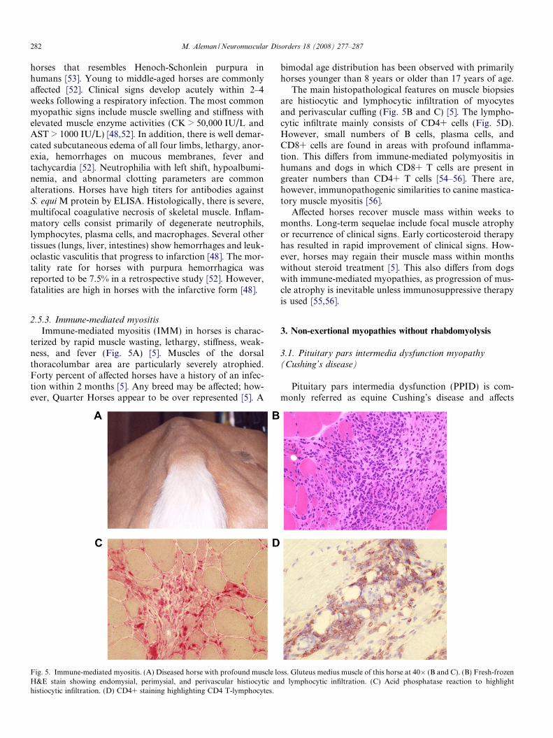
2.5.3. Immune-mediated myositis
Immune-mediated myositis (IMM) in horses is charac-terized by rapid muscle wasting, lethargy, stiffness, weak-ness, and fever (Fig. 5A) [5]. Muscles of the dorsalthoracolumbar area are particularly severely atrophied.Forty percent of affected horses have a history of an infec-tion within 2 months [5]. Any breed may be affected; how-ever, Quarter Horses appear to be over represented [5]. A
Fig. 5. Immune-mediated myositis. (A) Diseased horse with profound muscle loH&E stain showing endomysial, perimysial, and perivascular histiocytic anhistiocytic infiltration. (D) CD4+ staining highlighting CD4 T-lymphocytes.
bimodal age distribution has been observed with primarilyhorses younger than 8 years or older than 17 years of age.
The main histopathological features on muscle biopsiesare histiocytic and lymphocytic infiltration of myocytesand perivascular cuffing (Fig. 5B and C) [5]. The lympho-cytic infiltrate mainly consists of CD4+ cells (Fig. 5D).However, small numbers of B cells, plasma cells, andCD8+ cells are found in areas with profound inflamma-tion. This differs from immune-mediated polymyositis inhumans and dogs in which CD8+ T cells are present ingreater numbers than CD4+ T cells [54–56]. There are,however, immunopathogenic similarities to canine mastica-tory muscle myositis [56].
Affected horses recover muscle mass within weeks tomonths. Long-term sequelae include focal muscle atrophyor recurrence of clinical signs. Early corticosteroid therapyhas resulted in rapid improvement of clinical signs. How-ever, horses may regain their muscle mass within monthswithout steroid treatment [5]. This also differs from dogswith immune-mediated myopathies, as progression of mus-cle atrophy is inevitable unless immunosuppressive therapyis used [55,56].
3. Non-exertional myopathies without rhabdomyolysis
3.1. Pituitary pars intermedia dysfunction myopathy
(Cushing’s disease)
Pituitary pars intermedia dysfunction (PPID) is com-monly referred as equine Cushing’s disease and affects
ss. Gluteus medius muscle of this horse at 40� (B and C). (B) Fresh-frozend lymphocytic infiltration. (C) Acid phosphatase reaction to highlight

M. Aleman / Neuromuscular Disorders 18 (2008) 277–287 283
horses of all breeds but Morgans and ponies appear to beat greater risk [57]. The disease is the most common endo-crinopathy in late middle-aged and geriatric horses [57].However, horses as young as 7 years of age have been diag-nosed with the disease. The disease is a primary dopami-nergic neurodegenerative hypothalamic disease thatresults in loss of dopaminergic inhibition of the pituitarypars intermedia leading to increased melanotroph functionand subsequent excessive production of proopiomelano-cortin derived peptides which include a-melanocyte stimu-lating hormone (a-MSH), b-endorphin (b-END),corticotrophin-like intermediate lobe peptide (CLIP), andadrenocorticotropin (ACTH) [58]. Diseased horses develophypertrophy, hyperplasia or functional adenomas of thepituitary pars intermedia.
Common clinical signs include hirsutism, considered thehallmark of the disease, hyperhidrosis, polydipsia, poly-uria, muscle wasting, laminitis, and chronic or recurrentinfections [57]. Muscle wasting has been reported to varyfrom 35% to 88% of the cases; along with abnormal fat dis-tribution reported to be 9–67% of cases (Fig. 6A) [57].Lethargy, weakness, poor performance and/or decreasedphysical activity are commonly observed features and asso-ciated with decreased muscle mass and strength [4].
Laboratory work may be normal or may include a non-inflammatory leukogram, hyperglycemia and occasionalelevations of liver enzymes. Serum muscle enzyme activitiesare within reference values [4]. There are no specific electro-myographic abnormalities [4]. Horses with PPID developmild non-inflammatory myopathic alterations with themost prominent feature being atrophy of type 2A and 2Bmyofibers (Fig. 6B) [4]. Atrophic type 2 fibers have adecreased cross-sectional area, and may appear anguloidto angular in shape [4]. In addition, there is loss of type2B myofibers, increased type 1 to type 2 myofiber ratiocompared to age-matched control horses, and increasedcoefficient of variability indicating abnormal myofiber sizevariation. Lipid accumulation is observed at bothinter- and intramyofiber spaces [4]. Subsarcolemmalaccumulation of swollen mitochondria is observed at theultrastructural level [4].
Fig. 6. Pituitary pars intermedia dysfunction. (A) Horse with PPID myopathyMyofibrillar ATPase activity at pH 9.8 (25�) showing type 2 atrophy in glute
4. Exertional myopathies with rhabdomyolysis
4.1. Recurrent exertional rhabdomyolysis
Recurrent exertional rhabdomyolysis (RER) is the mostprevalent muscle disease in Thoroughbred horses. It hasbeen estimated that 5–10% of racing Thoroughbredsdevelop exertional rhabdomyolysis during a racing seasonwith recurrences of up to 17% [59,60]. This disorder hasan autosomal-dominant mode of inheritance with variableexpression [9]. RER is believed to be caused by an abnor-mality in intracellular calcium regulation, and not by lacticacidosis [61–63]. However, important genes involved in theregulation of myoplasmic calcium such as ryanodine recep-tor 1 (RyR1), sarcoplasmic reticulum calcium ATPase(ATP2A1), and dihydropyridine receptor-voltage sensor(CACNA1S) genes were excluded from linkage to RER[64]. The disease has also been observed in Standardbreds[65].
Clinical signs include muscle cramping, sttiffness, shift-ing lameness, sweating, reluctance to move, tachypnea,and colic-like signs. Episodes are observed more frequentlyafter horses reach a level of fitness then are restrained to aslower pace [59,60]. Episodes after racing occur infre-quently. Exertional rhabdomyolysis has also been observedin Thoroughbreds performing other physical activities suchas polo, steeplechase, and the cross-country phase of a 3-day event. Risk factors associated with RER include ayoung age, female, high strung, having rested for morethan 1 day prior to exercise, gallop during exercise, dietsconsisting of more than 4.5 kg of grain per day, and con-current lameness [59].
A presumptive diagnosis of RER can be established byclinical signs, risk factors, an increased serum CK activity4–6 h postexercise, and myopathic features in musclebiopsy specimens. These features include centrally locatednuclei in mature type 2A and type 2B muscle fibers,increased subsarcolemmal glycogen, and variable amountsof necrosis and regeneration [1,66]. In addition, RERaffected horses have an abnormal in vitro contractureresponse to potassium, caffeine and/or halothane
; note horse with hirsutism, muscle wasting, and pendulous abdomen. (B)us medius muscle. Myofiber types are indicated with Arabic numbers.
284 M. Aleman / Neuromuscular Disorders 18 (2008) 277–287
[61,62,66]. Treatment consists of supportive care, anxietyand muscle pain relief, replacement of fluid and electrolytelosses, rest while recovering from rhabdomyolysis, andenvironmental and dietary modifications [67,68].
5. Exertional myopathies without rhabdomyolysis
5.1. Mitochondrial myopathy
Mitochondrial respiratory chain abnormalities are animportant cause of neuromuscular disease in humans,and may be due to defects of the mitochondrial or nucleargenome [69]. A 3-year-old Arabian mare with normal mus-cle mass, stiffness and exhaustion upon only a few minutesof light exercise was diagnosed with mitochondrial myopa-thy caused by a deficiency of Complex I in the respiratorychain [10]. Upon exercise tolerance testing, the mare devel-oped shifting lameness, short stride, myalgia, profusesweating, and came to a standstill [10]. Blood gases werewithin reference ranges; however a marked lactic acidosisand increased hematocrit developed. Serum CK and ASTactivities were within reference values. On histochemicalanalysis, type 2A and 2B fibers stained intensively withNADH-TR and Gomori trichrome [10]. In addition, fewfibers had a ‘‘ragged red” appearance. Extensive accumula-tion of enlarged mitochondria underneath the sarcolemmaand between myofibrils was evident on electron microscopy[10]. The cristae were distorted and arranged concentri-cally. Lipid droplets were also prominent in the musclefibers [10].
6. Altered muscle membrane conduction
6.1. Hyperkalemic periodic paralysis
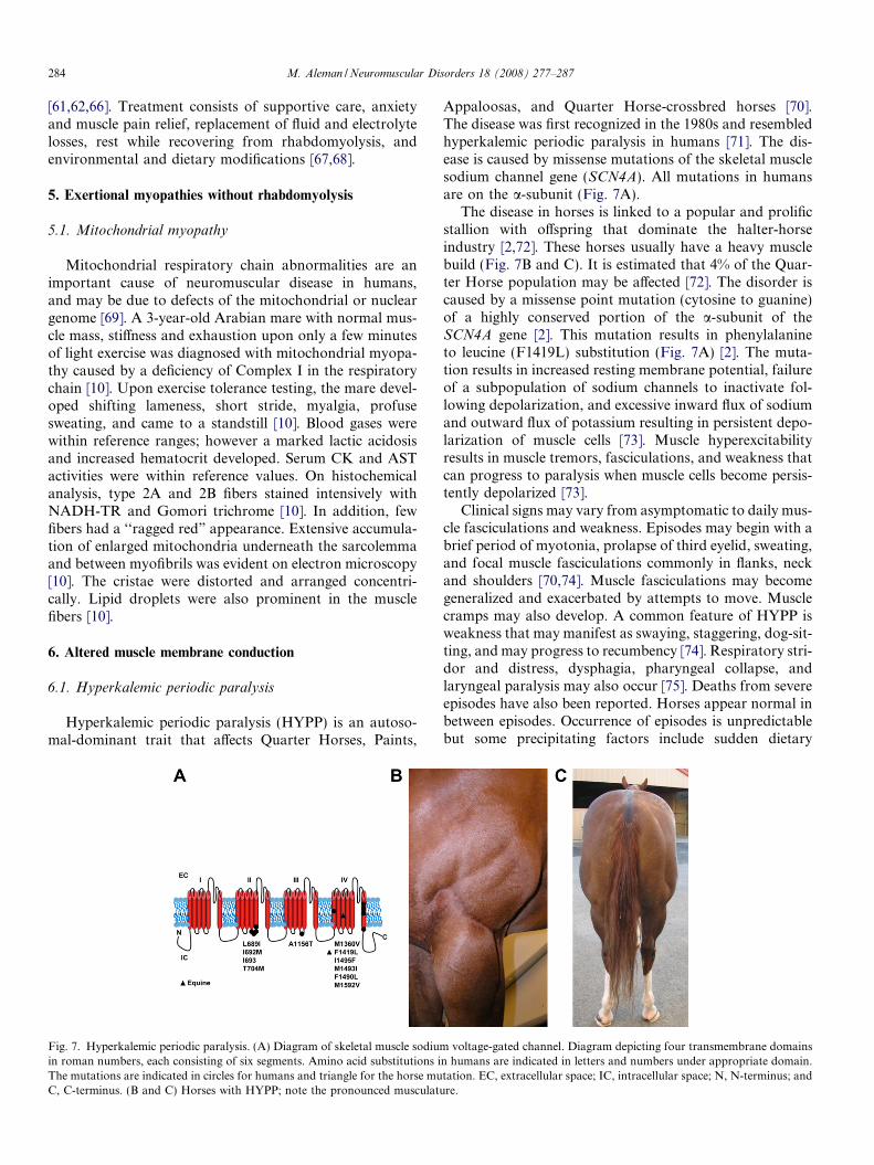
Hyperkalemic periodic paralysis (HYPP) is an autoso-mal-dominant trait that affects Quarter Horses, Paints,
Fig. 7. Hyperkalemic periodic paralysis. (A) Diagram of skeletal muscle sodiumin roman numbers, each consisting of six segments. Amino acid substitutions iThe mutations are indicated in circles for humans and triangle for the horse muC, C-terminus. (B and C) Horses with HYPP; note the pronounced musculatu
Appaloosas, and Quarter Horse-crossbred horses [70].The disease was first recognized in the 1980s and resembledhyperkalemic periodic paralysis in humans [71]. The dis-ease is caused by missense mutations of the skeletal musclesodium channel gene (SCN4A). All mutations in humansare on the a-subunit (Fig. 7A).
The disease in horses is linked to a popular and prolificstallion with offspring that dominate the halter-horseindustry [2,72]. These horses usually have a heavy musclebuild (Fig. 7B and C). It is estimated that 4% of the Quar-ter Horse population may be affected [72]. The disorder iscaused by a missense point mutation (cytosine to guanine)of a highly conserved portion of the a-subunit of theSCN4A gene [2]. This mutation results in phenylalanineto leucine (F1419L) substitution (Fig. 7A) [2]. The muta-tion results in increased resting membrane potential, failureof a subpopulation of sodium channels to inactivate fol-lowing depolarization, and excessive inward flux of sodiumand outward flux of potassium resulting in persistent depo-larization of muscle cells [73]. Muscle hyperexcitabilityresults in muscle tremors, fasciculations, and weakness thatcan progress to paralysis when muscle cells become persis-tently depolarized [73].
Clinical signs may vary from asymptomatic to daily mus-cle fasciculations and weakness. Episodes may begin with abrief period of myotonia, prolapse of third eyelid, sweating,and focal muscle fasciculations commonly in flanks, neckand shoulders [70,74]. Muscle fasciculations may becomegeneralized and exacerbated by attempts to move. Musclecramps may also develop. A common feature of HYPP isweakness that may manifest as swaying, staggering, dog-sit-ting, and may progress to recumbency [74]. Respiratory stri-dor and distress, dysphagia, pharyngeal collapse, andlaryngeal paralysis may also occur [75]. Deaths from severeepisodes have also been reported. Horses appear normal inbetween episodes. Occurrence of episodes is unpredictablebut some precipitating factors include sudden dietary
voltage-gated channel. Diagram depicting four transmembrane domainsn humans are indicated in letters and numbers under appropriate domain.tation. EC, extracellular space; IC, intracellular space; N, N-terminus; andre.
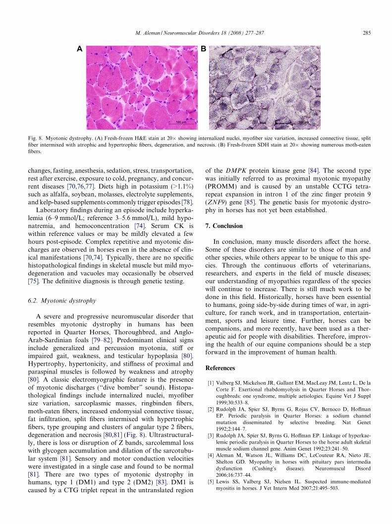
Fig. 8. Myotonic dystrophy. (A) Fresh-frozen H&E stain at 20� showing internalized nuclei, myofiber size variation, increased connective tissue, splitfiber intermixed with atrophic and hypertrophic fibers, degeneration, and necrosis. (B) Fresh-frozen SDH stain at 20� showing numerous moth-eatenfibers.
M. Aleman / Neuromuscular Disorders 18 (2008) 277–287 285
changes, fasting, anesthesia, sedation, stress, transportation,rest after exercise, exposure to cold, pregnancy, and concur-rent diseases [70,76,77]. Diets high in potassium (>1.1%)such as alfalfa, soybean, molasses, electrolyte supplements,and kelp-based supplements commonly trigger episodes [78].
Laboratory findings during an episode include hyperka-lemia (6–9 mmol/L; reference 3–5.6 mmol/L), mild hypo-natremia, and hemoconcentration [74]. Serum CK iswithin reference values or may be mildly elevated a fewhours post-episode. Complex repetitive and myotonic dis-charges are observed in horses even in the absence of clin-ical manifestations [70,74]. Typically, there are no specifichistopathological findings in skeletal muscle but mild myo-degeneration and vacuoles may occasionally be observed[75]. The definitive diagnosis is through genetic testing.
6.2. Myotonic dystrophy
A severe and progressive neuromuscular disorder thatresembles myotonic dystrophy in humans has beenreported in Quarter Horses, Thoroughbred, and Anglo-Arab-Sardinian foals [79–82]. Predominant clinical signsinclude generalized and percussion myotonia, stiff orimpaired gait, weakness, and testicular hypoplasia [80].Hypertrophy, hypertonicity, and stiffness of proximal andparaspinal muscles is followed by weakness and atrophy[80]. A classic electromyographic feature is the presenceof myotonic discharges (‘‘dive bomber” sound). Histopa-thological findings include internalized nuclei, myofibersize variation, sarcoplasmic masses, ringbinden fibers,moth-eaten fibers, increased endomysial connective tissue,fat infiltration, split fibers intermixed with hypertrophicfibers, type grouping and clusters of angular type 2 fibers,degeneration and necrosis [80,81] (Fig. 8). Ultrastructural-ly, there is loss or disruption of Z bands, sarcolemmal losswith glycogen accumulation and dilation of the sarcotubu-lar system [81]. Sensory and motor conduction velocitieswere investigated in a single case and found to be normal[81]. There are two types of myotonic dystrophy inhumans, type 1 (DM1) and type 2 (DM2) [83]. DM1 iscaused by a CTG triplet repeat in the untranslated region
of the DMPK protein kinase gene [84]. The second typewas initially referred to as proximal myotonic myopathy(PROMM) and is caused by an unstable CCTG tetra-repeat expansion in intron 1 of the zinc finger protein 9(ZNF9) gene [85]. The genetic basis for myotonic dystro-phy in horses has not yet been established.
7. Conclusion
In conclusion, many muscle disorders affect the horse.Some of these disorders are similar to those of man andother species, while others appear to be unique to this spe-cies. Through the continuous efforts of veterinarians,researchers, and experts in the field of muscle diseases;our understanding of myopathies regardless of the specieswill continue to increase. There is still much work to bedone in this field. Historically, horses have been essentialto humans, going side-by-side during times of war, in agri-culture, for ranch work, and in transportation, entertain-ment, sports and leisure time. Further, horses can becompanions, and more recently, have been used as a ther-apeutic aid for people with disabilities. Therefore, improv-ing the health of our equine companions should be a stepforward in the improvement of human health.
References
[1] Valberg SJ, Mickelson JR, Gallant EM, MacLeay JM, Lentz L, De laCorte F. Exertional rhabdomyolysis in Quarter Horses and Thor-oughbreds: one syndrome, multiple aetiologies. Equine Vet J Suppl1999;30:533–8.
[2] Rudolph JA, Spier SJ, Byrns G, Rojas CV, Bernoco D, HoffmanEP. Periodic paralysis in Quarter Horses: a sodium channelmutation disseminated by selective breeding. Nat Genet1992;2:144–7.
[3] Rudolph JA, Spier SJ, Byrns G, Hoffman EP. Linkage of hyperkae-lemic periodic paralysis in Quarter Horses to the horse adult skeletalmuscle sodium channel gene. Anim Genet 1992;23:241–50.
[4] Aleman M, Watson JL, Williams DC, LeCouteur RA, Nieto JE,Shelton GD. Myopathy in horses with pituitary pars intermediadysfunction (Cushing’s disease). Neuromuscul Disord2006;16:737–44.
[5] Lewis SS, Valberg SJ, Nielsen IL. Suspected immune-mediatedmyositis in horses. J Vet Intern Med 2007;21:495–503.
286 M. Aleman / Neuromuscular Disorders 18 (2008) 277–287
[6] Valberg SJ, Cardinet 3rd GH, Carlson GP, DiMauro S. Polysaccha-ride storage myopathy associated with recurrent exertional rhabdo-myolysis in horses. Neuromuscul Disord 1992;2:351–9.
[7] Ward TL, Valberg SJ, Adelson DL, Abbey CA, Binns MM,Mickelson JR. Glycogen branching enzyme (GBE1) mutation caus-ing equine glycogen storage disease IV. Mamm Genome2004;15:570–7.
[8] Aleman M, Riehl J, Aldridge BM, Lecouteur RA, Stott JL, PessahIN. Association of a mutation in the ryanodine receptor 1 gene withequine malignant hyperthermia. Muscle Nerve 2004;30:356–65.
[9] Dranchak PK, Valberg SJ, Onan GW, et al. Inheritance of recurrentexertional rhabdomyolysis in Thoroughbreds. J Am Vet Med Assoc2005;227:762–7.
[10] Valberg SJ, Carlson GP, Cardinet 3rd GH, et al. Skeletal musclemitochondrial myopathy as a cause of exercise intolerance in a horse.Muscle Nerve 1994;17:305–12.
[11] Divers TJ, Cummings JE, de Lahunta A, Hintz HF, Mohammed HO.Evaluation of the risk of motor neuron disease in horses fed a dietlow in vitamin E and high in copper and iron. Am J Vet Res2006;67:120–6.
[12] Eisen A. Recent considerations in the etiopathogenesis of ALS. SupplClin Neurophysiol 2004;57:187–90.
[13] Higuchi T, Ichijo S, Osame S, Ohishi H. Studies on serum seleniumand tocopherol in white muscle disease of foal. Nippon JuigakuZasshi 1989;51:52–9.
[14] Rayman MP. The importance of selenium to human health. Lancet2000;356:233–41.
[15] Lofstedt J. White muscle disease of foals. Vet Clin North Am EquinePract 1997;13:169–85.
[16] Chariot P, Bignani O. Skeletal muscle disorders associated withselenium deficiency in humans. Muscle Nerve 2003;27:662–8.
[17] Perkins G, Valberg SJ, Madigan JM, Carlson GP, Jones SL.Electrolyte disturbances in foals with severe rhabdomyolysis. J VetIntern Med 1998;12:173–7.
[18] Pearson EG, Snyder SP, Saulez MN. Masseter myodegeneration as acause of trismus or dysphagia in adult horses. Vet Rec2005;156:642–6.
[19] DiMauro S, Lamperti C. Muscle glycogenoses. Muscle Nerve2001;24:984–99.
[20] Wagner ML, Valberg SJ, Ames EG, et al. Allele frequency and likelyimpact of the glycogen branching enzyme deficiency gene in QuarterHorse and Paint Horse populations. J Vet Intern Med2006;20:1207–11.
[21] Valberg SJ, Ward TL, Rush B, et al. Glycogen branching enzymedeficiency in Quarter Horse foals. J Vet Intern Med 2001;15:572–80.
[22] Render JA, Common RS, Kennedy FA, Jones MZ, Fyfe JC.Amylopectinosis in fetal and neonatal Quarter Horses. Vet Pathol1999;36:157–60.
[23] Annandale EJ, Valberg SJ, Mickelson JR, Seaquist ER. Insulinsensitivity and skeletal muscle glucose transport in horses with equinepolysaccharide storage myopathy. Neuromuscul Disord2004;14:666–74.
[24] McCue ME, Ribeiro WP, Valberg SJ. Prevalence of polysaccharidestorage myopathy in horses with neuromuscular disorders. EquineVet J Suppl 2006;36:340–4.
[25] McCue ME, Valberg SJ. Estimated prevalence of polysaccharidestorage myopathy among overtly healthy Quarter Horses in theUnited States. J Am Vet Med Assoc 2007;231:746–50.
[26] Firshman AM, Baird JD, Valberg SJ. Prevalences and clinical signsof polysaccharide storage myopathy and shivers in Belgian drafthorses. J Am Vet Med Assoc 2005;227:1958–64.
[27] Valentine BA, Credille KM, Lavoie JP, et al. Severe polysaccharidestorage myopathy in Belgian and Percheron draught horses. EquineVet J 1997;29:220–5.
[28] Quiroz-Rothe E, Novales M, Aguilera-Tejero E, Rivero JL. Poly-saccharide storage myopathy in the M. longissimus lumborum ofshowjumpers and dressage horses with back pain. Equine Vet J2002;34:171–6.
[29] De La Corte FD, Valberg SJ, MacLeay JM, Mickelson JR.Developmental onset of polysaccharide storage myopathy in 4Quarter Horse foals. J Vet Intern Med 2002;16:581–7.
[30] Firshman AM, Valberg SJ, Bender JB, Finno CJ. Epidemiologiccharacteristics and management of polysaccharide storage myopathyin Quarter Horses. Am J Vet Res 2003;64:1319–27.
[31] Valentine BA, Cooper BJ. Incidence of polysaccharide storagemyopathy: necropsy study of 225 horses. Vet Pathol 2005;42:823–7.
[32] McCarthy EJ. Malignant hyperthermia: pathophysiology, clinicalpresentation, and treatment. AACN Clin Issues 2004;15:231–7.
[33] Klein L. A hot horse. Vet Anesth 1975;2:41–2.[34] Aleman M, Brosnan RJ, Williams DC, et al. Malignant hyperthermia
in a horse anesthetized with halothane. J Vet Intern Med2005;19:363–7.
[35] Greenbaum I, Weigl Y, Pras E. The genetic basis of malignanthyperthermia. Isr Med Assoc J 2007;9:39–41.
[36] Fujii J, Otsu K, Zorzato F, et al. Identification of a mutation inporcine ryanodine receptor associated with malignant hyperthermia.Science 1991;253:448–51.
[37] Roberts MC, Mickelson JR, Patterson EE, et al. Autosomaldominant canine malignant hyperthermia is caused by a mutationin the gene encoding the skeletal muscle calcium release channel(RYR1). Anesthesiology 2001;95:716–25.
[38] McCarthy TV, Quane KA, Lynch PJ. Ryanodine receptor mutationsin malignant hyperthermia and central core disease. Hum Mutat2000;15:410–7.
[39] Paciello O, Pasolini MP, Navas L, Russo V, Papparella S. Myopathywith central cores in a foal. Vet Pathol 2006;43:579–83.
[40] Bowen JN, Craig JF. Myoglobinuria in horses. Vet Rec 1942;35:354.[41] Cassart D, Baise E, Cherel Y, et al. Morphological alterations in
oxidative muscles and mitochondrial structure associated with equineatypical myopathy. Equine Vet J 2007;39:26–32.
[42] Votion DM, Linden A, Saegerman C, et al. History and clinicalfeatures of atypical myopathy in horses in Belgium (2000–2005). J VetIntern Med 2007;21:1380–91.
[43] Whitwell KE, Harris P, Farrington PG. Atypical myoglobinuria: anacute myopathy in grazing horses. Equine Vet J 1988;20:357–63.
[44] Pope DC, Heslop CH. An outbreak of myoglobinuria in light horses.Can Vet J 1960;1:171–4.
[45] Finno CJ, Valberg SJ, Wunschmann A, Murphy MJ. Seasonalpasture myopathy in horses in the midwestern United States: 14 cases(1998–2005). J Am Vet Med Assoc 2006;229:1134–41.
[46] Briani C, Doria A, Sarzi-Puttini P, Dalakas MC. Update onidiopathic inflammatory myopathies. Autoimmunity 2006;39:161–70.
[47] Sponseller BT, Valberg SJ, Tennent-Brown BS, Foreman JH, KumarP, Timoney JF. Severe acute rhabdomyolysis associated withStreptococcus equi infection in four horses. J Am Vet Med Assoc2005;227:1800–7, [753–4].
[48] Kaese HJ, Valberg SJ, Hayden DW, et al. Infarctive purpurahemorrhagica in five horses. J Am Vet Med Assoc 2005;226:1893–8.
[49] Peek SF, Semrad SD, Perkins GA. Clostridial myonecrosis in horses(37 cases 1985–2000). Equine Vet J 2003;35:86–92.
[50] Brown EJ. The molecular basis of streptococcal toxic shocksyndrome. N Engl J Med 2004;350:2093–4.
[51] Alouf JE, Muller-Alouf H. Staphylococcal and Streptococcal super-antigens: molecular, biological and clinical aspects. Int J MedMicrobiol 2003;292:429–40.
[52] Pusterla N, Watson JL, Affolter VK, Magdesian KG, Wilson WD,Carlson GP. Purpura haemorrhagica in 53 horses. Vet Rec2003;153:118–21.
[53] van der Boon F, Groeneweg M. Acute abdominal pain as the firstsign of Henoch-Schonlein purpura: a hidden diagnosis in the absenceof purpura. Ned Tijdschr Geneeskd 2005;149:2522–6.
[54] Figarella-Branger D, Civatte M, Bartoli C, Pellissier JF. Cytokines,chemokines, and cell adhesion molecules in inflammatory myopa-thies. Muscle Nerve 2003;28:659–82.

M. Aleman / Neuromuscular Disorders 18 (2008) 277–287 287
[55] Evans J, Levesque D, Shelton GD. Canine inflammatory myopathies:a clinicopathologic review of 200 cases. J Vet Intern Med2004;18:679–6791.
[56] Pumarola M, Moore PF, Shelton GD. Canine inflammatory myop-athy: analysis of cellular infiltrates. Muscle Nerve 2004;29:782–9.
[57] Schott II HC. Pituitary pars intermedia dysfunction: equine Cush-ing’s disease. Vet Clin North Am Equine Pract 2002;18:237–70.
[58] McFarlane D. Advantages and limitations of the equine disease,pituitary pars intermedia dysfunction as a model of spontaneousdopaminergic neurodegenerative disease. Ageing Res Rev2007;6:54–63.
[59] MacLeay JM, Sorum SA, Valberg SJ, Marsh WE, Sorum MD.Epidemiologic analysis of factors influencing exertional rhabdomy-olysis in Thoroughbreds. Am J Vet Res 1999;60:1562–6.
[60] McGowan CM, Fordham T, Christley RM. Incidence and riskfactors for exertional rhabdomyolysis in Thoroughbred racehorses inthe United Kingdom. Vet Rec 2002;151:623–6.
[61] Ward TL, Valberg SJ, Gallant EM, Mickelson JR. Calciumregulation by skeletal muscle membranes of horses with recurrentexertional rhabdomyolysis. Am J Vet Res 2000;61:242–7.
[62] Lentz LR, Valberg SJ, Herold LV, Onan GW, Mickelson JR, GallantEM. Myoplasmic calcium regulation in myotubes from horses withrecurrent exertional rhabdomyolysis. Am J Vet Res 2002;63:1724–31.
[63] Mlekoday JA, Mickelson JR, Valberg SJ, Horton JH, Gallant EM,Thompson LV. Calcium sensitivity of force production and myofibr-illar ATPase activity in muscles from Thoroughbreds with recurrentexertional rhabdomyolysis. Am J Vet Res 2001;62:1647–52.
[64] Dranchak PK, Valberg SJ, Onan GW, et al. Exclusion of linkage ofthe RYR1, CACNA1S, and ATP2A1 genes to recurrent exertionalrhabdomyolysis in Thoroughbreds. Am J Vet Res 2006;67:1395–400.
[65] Collinder E, Lindholm A, Rasmuson M. Genetic markers inStandardbred trotters susceptible to the rhabdomyolysis syndrome.Equine Vet J 1997;29:117–20.
[66] Lentz LR, Valberg SJ, Balog EM, Mickelson JR, Gallant EM.Abnormal regulation of muscle contraction in horses with recurrentexertional rhabdomyolysis. Am J Vet Res 1999;60:992–9.
[67] McKenzie EC, Valberg SJ, Godden SM, et al. Plasma and urineelectrolyte and mineral concentrations in Thoroughbred horses withrecurrent exertional rhabdomyolysis after consumption of dietsvarying in cation–anion balance. Am J Vet Res 2002;63:1053–60.
[68] McKenzie EC, Valberg SJ, Godden SM, et al. Effect of dietary starch,fat, and bicarbonate content on exercise responses and serum creatinekinase activity in equine recurrent exertional rhabdomyolysis. J VetIntern Med 2003;17:693–701.
[69] Taylor RW, Schaefer AM, Barron MJ, McFarland R, Turnbull DM.The diagnosis of mitochondrial muscle disease. Neuromuscul Disord2004;14:237–45.
[70] Spier SJ, Carlson GP, Holliday TA, Cardinet 3rd GH, Pickar JG.Hyperkalemic periodic paralysis in horses. J Am Vet Med Assoc1990;197:1009–17.
[71] Cox JH, De Bowes RM. Episodic weakness caused by hyperkalemicperiodic paralysis in horses. Compend Contin Educ Pract Vet1990;12:83–9.
[72] Bowling AT, Byrns G, Spier S. Evidence for a single pedigree sourceof the hyperkalemic periodic paralysis susceptibility gene in QuarterHorses. Anim Genet 1996;27:279–81.
[73] Pickar JG, Spier SJ, Snyder JR, Carlsen RC. Altered ionic perme-ability in skeletal muscle from horses with hyperkalemic periodicparalysis. Am J Physiol 1991;260:C926–33.
[74] Meyer TS, Fedde MR, Cox JH, Erickson HH. Hyperkalaemicperiodic paralysis in horses: a review. Equine Vet J 1999;31:362–7.
[75] Carr EA, Spier SJ, Kortz GD, Hoffman EP. Laryngeal andpharyngeal dysfunction in horses homozygous for hyperkalemicperiodic paralysis. J Am Vet Med Assoc 1996;209:798–803.
[76] Waldridge B, Lin H, Purohit R. Anesthetic management of horseswith hyperkalemic periodic paralysis. Compend Contin Educ PractVet 1996;18:1030–8.
[77] Sah RL, Tsushima RG, Backx PH. Effects of local anesthetics onNa+ channels containing the equine hyperkalemic periodic paralysismutation. Am J Physiol 1998;275:C389–400.
[78] Reynolds AJ, Potter GD, Greene LW, et al. Genetic-diet interactionsin the hyperkalemic periodic paralysis syndrome in Quarter Horsesfed varying amounts of potassium: III. The relationship betweenplasma potassium concentration and HYPP symptoms. J Equine VetSci 1998;18:731–5.
[79] Hegreberg GA, Reed SM. Skeletal muscle changes associated withequine myotonic dystrophy. Acta Neuropathol 1990;80:426–31.
[80] Reed SM, Hegreberg GA, Bayly WM, Brown CM, Paradis MR,Clemmons RM. Progressive myotonia in foals resembling humandystrophia myotonica. Muscle Nerve 1988;11:291–6.
[81] Montagna P, Liguori R, Monari L, et al. Equine muscular dystrophywith myotonia. Clin Neurophysiol 2001;112:294–9.
[82] Shirakawa T, Ide M, Taniyama H, et al. Muscular dystrophy-likedisease in a Thoroughbred foal. J Comp Pathol 1989;100:287–94.
[83] Logigian EL, Ciafaloni E, Quinn LC, et al. Severity, type, anddistribution of myotonic discharges are different in type 1 and type 2myotonic dystrophy. Muscle Nerve 2007;35:479–85.
[84] Brook JD, McCurrach ME, Harley HG, et al. Molecular basis ofmyotonic dystrophy: expansion of a trinucleiotide (CTG) repeat atthe 30 end of a transcript encoding a protein kinase family member.Cell 1992;68:799–808.
[85] Liquori CL, Ricker K, Moseley ML, et al. Myotonic dystrophy type2 caused by a CCTG expansion in intron 1 of ZNF9. Science2001;293:864–7.