a phase i/ii study of carboplatin combined with hyperfractionated radiotherapy for brainstem gliomas
TRANSCRIPT

A Phase I/II Study of Carboplatin Combined withHyperfractionated Radiotherapy for BrainstemGliomas
Jeffrey Allen, M.D.1,3
Joao Siffert, M.D.1
Bernadine Donahue, M.D.4
Anita Nirenberg, R.N.3
Regina Jakacki, M.D.5
Patricia Robertson, M.D.6
Robert DaRosso, M.D.1
Louisa Thoron, M.D.3
Mark Rosovsky, M.D.4
Richard Pinto, M.D.2
1 Department of Neurology, Beth Israel MedicalCenter, New York, New York.
2 Department of Radiology, Beth Israel MedicalCenter, New York, New York.
3 Department of Neurology, New York UniversityMedical Center, New York, New York.
4 Department of Radiology, New York UniversityMedical Center, New York, New York.
5 Department of Pediatrics, Riley Childrens Hospi-tal, Indianapolis, Indiana.
6 Department of Neurology, University of MichiganMedical Center, Ann Arbor, Michigan.
Address for reprints: Jeffrey Allen, M.D., Beth Is-rael Medical Center—Singer, 170 East End Ave-nue, New York, NY 10128.
Received October 13, 1998; revision received Feb-ruary 8, 1999; accepted March 22, 1999.
BACKGROUND. Brainstem gliomas often respond to radiotherapy but long term
disease control is exceptional. The concomitant administration of a chemotherapy
agent with radiosensitizing properties such as carboplatin may increase the effi-
cacy of radiotherapy.
METHODS. A dose escalation schedule of carboplatin was devised to determine the
maximum tolerated dose (MTD) of intravenous carboplatin when given on a
twice-weekly schedule during a course of hyperfractionated, involved field radio-
therapy (100 centigrays [cGy] twice daily to 7200 cGy). The starting dose was 20
mg/m2 and the dose was increased by 15 mg/m2 after every 3 patients provided no
Grade 3 or 4 (according to the National Institutes of Health Common Toxicity
Criteria) toxicity occurred. Magnetic resonance imaging (MRI) scans (brain and
spine) were obtained before treatment and at the time of disease progression.
Clinical entry criteria included an MRI scan demonstrating a diffuse intrinsic
pontine tumor and a typical 2–3-month history of evolving cranial neuropathies
and a gait disorder. Biopsy-confirmed evidence of a high grade glioma was re-
quired for nonpontine brain stem tumors.
RESULTS. A total of 34 patients were enrolled. The median age of the patients was
7.8 years (range, 3.6 –15.4 years) and the median prodrome duration was 1.5
months (range, 0.25–36 months). The MTD was 110 mg/m2 or a total cumulative
dose of 1540 mg/m2 over 7 weeks. The dose-limiting toxicity was hematologic. The
median progression free survival was 8 months (range, 0 –1041 months) and the
overall survival was 12 months (range, 5–1041 months). At last follow-up there
were 5 long term survivors (15%) who remained in continuous remission after a
mean follow-up period of 79 months (range, 46 –104 months). Fifteen of the 29
patients (52%) with recurrence and or disease progression developed leptomenin-
geal/intraaxial tumor spread beyond the local radiation field.
CONCLUSIONS. The cumulative MTD for carboplatin is 1540 mg/m2 when admin-
istered concomitantly with involved field, hyperfractionated radiotherapy in a
twice-weekly schedule for 7 weeks. Subsequent Phase II and III clinical trials can be
conducted safely at this level. Cancer 1999;86:1064 –9.
© 1999 American Cancer Society.
KEYWORDS: brainstem glioma, radiosensitizers, radiotherapy, chemotherapy, clin-ical trial, Phase I/II trials.
Children with intrinsic tumors of the pons have been reported tohave a dismal prognosis after either daily conventional involved
field radiotherapy, various hyperfractionated radiotherapy doseschedules, or other protocols combining adjuvant or neoadjuvantchemotherapy and radiotherapy.1–3 Three-year survival in the major-ity of large series is ,10% and there is an urgent need for innovative
1064
© 1999 American Cancer Society

therapies. Although the majority of children die withlocal recurrences, it is becoming apparent that thesechildren are at risk for developing leptomeningeal me-tastases as well.4 Interest in the use of platinum ana-logues as radiosensitizers has emerged from preclini-cal studies5,6 and several clinical trials have beencompleted in adults with head and neck carcinomaand nonsmall cell lung carcinoma. Carboplatin or cis-platin has been given concomitantly with radiother-apy in controlled prospective clinical trials in whichthe chemotherapy is distributed over the course ofradiotherapy. A higher objective response rate of theprimary disease and improved survival has been ob-served with acceptable toxicity in nonsmall cell lungcarcinoma.7
A multimodality protocol was devised combiningcarboplatin and hyperfractionated radiotherapy forchildren with newly diagnosed brainstem gliomas. In-travenous carboplatin was administered twice weeklyfor the 7 weeks of radiotherapy. Because the centralnervous system (CNS) and hematologic toxicity of thisinteraction are unknown, a dose escalation scheduleof carboplatin was utilized as in a dose-searchingPhase I trial. All consecutively accessioned patientswith clinically diagnosed intrinsic pontine tumors orbiopsy-confirmed high grade gliomas of the medullaor midbrain seen at the participating medical centersduring the study period (1992–1995) were entered onthis protocol. The goals of this study were to deter-mine the maximum tolerable dose (MTD) of carbopla-tin and to monitor event free and total survival andpatterns of recurrence or progression.
METHODSPatient SelectionPatients with pontine tumors were eligible withouthistologic confirmation if they were age .3 years and,21 years at the time of radiologic diagnosis with aclinical syndrome comprised of a brief history (,3months) of progressive brainstem dysfunction (char-acterized by diplopia, facial weakness, and/or dyspha-gia) combined with ataxia or hemiparesis. Patientswith an atypical history, neurofibromatosis, or a pri-mary brainstem tumor arising outside the pons re-quired histologic confirmation of a high grade glioma.
Neuroradiologic Eligibility and Monitoring CriteriaFor clinically diagnosed patients, magnetic resonanceimaging (MRI) must identify an intrinsic, diffusebrainstem tumor with an epicenter in the pons. Therecould be local extension to the medulla, cerebellarpeduncle and midbrain, variable contrast enhance-ment, and exophytic components in the fourth ven-tricle or prepontine cistern. The T2-weighted se-
quence must reveal a diffuse signal abnormalityinvolving at least 50% of the pons. If the epicenter ofthe tumor involved either the midbrain or medulla,histologic confirmation of a high grade glioma wasrequired. A baseline gadolinium-enhanced spinal MRIwas recommended for all patients prior to therapy.Routine cerebrospinal fluid cytologic analysis was notrequired. The assessment of response was comprisedof comparing the pretreatment MRI with the post-treatment MRI which showed either maximum re-sponse or disease progression. The response criteriawere: complete response (a normal appearance of thebrainstem); partial response (a $50% reduction in sizeof the T2-weighted abnormality or a 50% reduction incontrast enhancement on the T1-weighted images);and stable disease (,25% change). Recurrence or pro-gression constituted at least a 25% increase in thetumor size as measured by the product of the 2 largestperpendicular dimensions in either the T1 gadoliniumimage if the tumor enhanced or the T2 image. If clin-ical or radiographic recurrence/disease progressionwas observed, a spinal MRI also was recommended.
ChemotherapyInformed consent was obtained prior to the initiationof therapy. Thereafter, an indwelling central venousline was recommended. A baseline complete bloodcount, chemistry profile, creatinine clearance, and au-diometry were obtained prior to chemotherapy ad-ministration. Carboplatin was administered twiceweekly, usually on Monday and Wednesday, through-out the 7 weeks of radiotherapy (Fig. 1). A standarddose escalation scheme was followed for carboplatin.The first 3 patients were treated with an initial dose of20 mg/m2 for 14 doses. The dose was incremented by15 mg/m2 after every 3 patients as long as 2 patientsexperienced , Grade 3 or 4 hematologic or other
FIGURE 1. Protocol schema for combination chemotherapy and radiotherapy
in the treatment of children with brainstem gliomas.
Chemoradiotherapy for Brainstem Gliomas/Allen et al. 1065
organ toxicity. A modification of the Common ToxicityCriteria (National Institutes of Health) was used.Grade 3 neutropenia was comprised of an absoluteneutrophil count (ANC) .500/mm3 but ,1000/mm3
and Grade 4 neutropenia was comprised of an ANC,500/mm3. Grade 3 thrombocytopenia was ,50,000/mm3 but .30,000 platelets/mm3 and Grade 4 throm-bocytopenia was ,30,000 mm3. Complete bloodcounts and a serum chemistry panel were obtainedweekly. The MTD of carboplatin was defined as thedose below the level at which two patients developedGrade 3 or 4 toxicity. If Grade 3 or 4 toxicity wasencountered in any given patient during radiotherapy,chemotherapy was deferred until complete hemato-logic recovery but radiotherapy was continued. Noadjuvant chemotherapy was administered.
RadiotherapyInvolved field radiotherapy was administered to allpatients. The “involved field” was defined as the T2abnormality on MRI plus a 2-cm margin. Treatmentwas delivered through lateral parallel opposed portals.The energy was 6-megavolt photon therapy. The treat-ment was administered in 100-centigray (cGy) frac-tions, given twice daily, separated by at least 6 hours toa total dose of 7200 cGy delivered over 7 weeks. Thedose to the cervical spine, optic chiasm, and pituitaryfossa were limited to 5000 cGy. For uncooperativepatients, the patient was sedated for each radiother-apy treatment under the supervision of an anesthesi-ologist. Patients were examined and weighed weeklyand if they lost .10% of body weight, total parenteralor enteral nutrition was administered. Radiation tox-icity was assessed according to the Childrens CancerGroup (CCG) Radiotherapy Toxicity Criteria. The ra-diotherapy toxicity was evaluated most reliably in thecohort of 18 patients treated at New York University(NYU) Medical Center. Patients also were followedclosely for the development of any significant skinreaction. The patients usually were maintained oncorticosteroids for the first 2 weeks of the radiotherapyand then the corticosteroids were tapered slowly.
SurveillanceOne month after the completion of radiotherapy, anenhanced brain MRI was obtained and a neurologicexamination was performed. These procedures wererepeated every 4 months or sooner if an adverse neu-rologic event occurred. Any signs or symptoms of amyelopathy prompted the performance of spinal MRIimaging. If disease recurrence or progression was sus-pected a head and spinal MRI were obtained.
SurvivalAfter radiotherapy, an attempt was made to wean allpatients off corticosteroids. Both event free and totalsurvival were calculated from the time of the diagnos-tic MRI scan to the time of the scan that demonstrateda significant change. This usually meant either a $25%increase in the area of contrast enhancement or T2-weighted abnormality in the brainstem or a new dis-tant or contiguous lesion. The occurrence of a new orprogressive neurologic symptom alone without a sig-nificant radiologic change did not in and of itself con-stitute an adverse event.
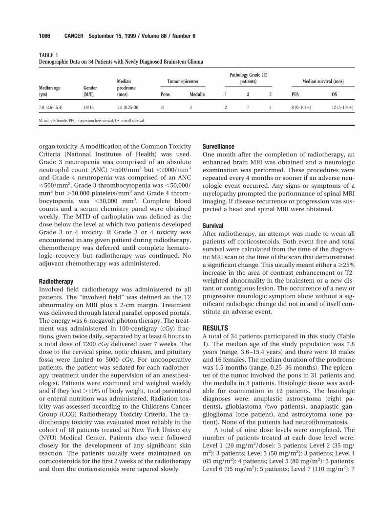
RESULTSA total of 34 patients participated in this study (Table1). The median age of the study population was 7.8years (range, 3.6 –15.4 years) and there were 18 malesand 16 females. The median duration of the prodromewas 1.5 months (range, 0.25–36 months). The epicen-ter of the tumor involved the pons in 31 patients andthe medulla in 3 patients. Histologic tissue was avail-able for examination in 12 patients. The histologicdiagnoses were: anaplastic astrocytoma (eight pa-tients), glioblastoma (two patients), anaplastic gan-glioglioma (one patient), and astrocytoma (one pa-tient). None of the patients had neurofibromatosis.
A total of nine dose levels were completed. Thenumber of patients treated at each dose level were:Level 1 (20 mg/m2/dose): 3 patients; Level 2 (35 mg/m2): 3 patients; Level 3 (50 mg/m2): 3 patients; Level 4(65 mg/m2): 4 patients; Level 5 (80 mg/m2): 3 patients;Level 6 (95 mg/m2): 5 patients; Level 7 (110 mg/m2): 7
TABLE 1Demographic Data on 34 Patients with Newly Diagnosed Brainstem Glioma
Median age(yrs)
Gender(M/F)
Medianprodrome(mos)
Tumor epicenterPathology Grade (12
patients) Median survival (mos)
Pons Medulla 1 2 3 PFS OS
7.8 (3.6–15.4) 18/16 1.5 (0.25–36) 31 3 2 7 3 8 (0–1041) 12 (5–1041)
M: male; F: female; PFS: progression free survival; OS: overall survival.
1066 CANCER September 15, 1999 / Volume 86 / Number 6

patients; Level 8 (125 mg/m2): 3 patients; and Level 9(140 mg/m2): 3 patients. The MTD was Level 7 (i.e.,110 mg/m2 dose or a cumulative dose of 1540 mg over7 weeks). The incidence of Grade 3– 4 toxicity per doselevel was: Level 7: 1 patient; Level 8: 2 patients; andLevel 9: 2 patients. The major toxicity was hematologicand was comprised of thrombocytopenia. Three pa-tients were enrolled prematurely on Level 9 beforedelayed thrombocytopenia was appreciated in two ofthe three patients at Level 8. Not all children receivedthe prescribed 14 courses of chemotherapy over the7-week period of radiotherapy; 8 courses were given to1 patient, 10 courses were given to 2 patients, 11courses were given to 1 patient, 12 courses were givento 1 patient, 13 courses were given to 5 patients, and14 courses were given to 24 patients.
Response was evaluable in 29 of the 34 patientsbased on a comparison of the pretreatment and post-treatment MRI scans. An objective (complete or par-tial) response was observed in 15 patients (52%) andthese responses were observed at all dose levels. Thespecific response patterns were: complete: 1 patient;partial: 14 patients; stable disease: 8 patients; anddisease progression: 6 patients. With regard to theradiotherapy toxicity in the NYU Medical Center co-hort, all 18 patients experienced Grade 4 alopecia. Theskin toxicity was: Grade 3: 5 patients; Grade 2: 12patients; and Grade 1: 1 patient. There was no consis-tent dose of carboplatin that produced the five in-stances of Grade 3 toxicity. Three children developedsymptomatic external otitis. The majority of the chil-dren developed only mild erythema of the external earand canal. No child required interruption of the radio-therapy because of treatment-related toxicity. Audi-tory acuity was not monitored consistently in this trialbut one patient with long term survival developedsevere sensorineural hearing impairment and at lastfollow-up required hearing aids.
The median progression free survival was 8months (range, 0 –1041 months) and the medianoverall survival was 12 months (range, 5–1041months). At last follow-up a total of 29 patients haddeveloped disease progression and died and the re-maining 5 patients were alive in continuous responseafter a mean period of follow-up of 79 months (range,46 –104 months). These five long term survivors weretreated at different dose levels of carboplatin and allreceived the same radiotherapy treatment plan perprotocol. Three of the long term survivors had typicaldiffuse pontine tumors at diagnosis; one had a com-plete response. Another long term survivor had thelongest prodrome (36 months). His tumor epicenterwas in the medulla and the histology was an anaplas-tic ganglioglioma. The fifth survivor had an anaplastic
astrocytoma partially resected from the medulla. Thesites of recurrence in the 29 patients who died were:local only: 14 patients; local plus diffuse leptomenin-geal metastases (LM): 13 patients; local plus LM plusdistant intraaxial: 1 patient; and LM plus metastases tosubcutaneous tissues adjacent to the surgical incision:1 patient. Thus, 15 of the 29 patients (52%) with dis-ease recurrence had metastases beyond the primarytumor site and the involved radiotherapy treatmentfield. There were no clinical or treatment factors suchas the performance of a surgical procedure, age, orhistology that distinguished the group of patients whodied with LM compared with those who died of localrecurrence alone. For example, tumor resections wereattempted in 6 of the 15 patients who died with LM(40%) compared with 6 of the 14 patients who died oflocal recurrences (42%).
DISCUSSIONWe have determined that the MTD of carboplatinwhen given simultaneously with hyperfractionated ra-diotherapy in a twice-weekly schedule is 110 mg/m2/dose or a total cumulative dose of 1540 mg given over7 weeks. The dose-limiting toxicity was thrombocyto-penia and no unexpected toxicity related to the hyper-fractionated radiotherapy was observed. No signifi-cant impact on progression free or overall survival wasobserved although 5 children (15%) experienced longterm survival. The treatment protocol may have al-tered the pattern of recurrence given the high fre-quency (52%) of metastatic disease outside the radio-therapy field at the time of clinical and radiographicrecurrence/disease progression.
The rationale for the concomitant use of carbo-platin and radiotherapy comes from several preclin-ical and clinical studies involving both cisplatin andcarboplatin. Carboplatin alone has relatively lowactivity in patients with recurrent brainstem glio-mas.8,9 However, objective responses have been ob-served using 2 different dose schedules: 4 timesweekly at a dose of 175 mg/m2 and 560 mg/m2 every4 weeks.9,10 Carboplatin also has been given concur-rently with radiation in an attempt to enhance theresponse to radiation, both through additive andsupraadditive (synergistic) interactions. The mech-anisms by which carboplatin enhances the efficacyof radiotherapy include arresting tumor cells in theradiosensitive phase of the cell cycle,5 inhibition ofrepair of radiation-induced damage,11 and in-creased induction of chromosomal aberrations.12 Toour knowledge the optimal dose and dose schedulefor maximizing this synergy is not known.
In several solid tumor trials in adults, the concur-rent use of platinum and radiation therapy has re-
Chemoradiotherapy for Brainstem Gliomas/Allen et al. 1067

sulted in improved survival and local disease controlcompared with radiation therapy alone7,13 or sequen-tial chemoradiotherapy.14 To our knowledge the opti-mal dose and schedule of administering platinumagents with radiation has not been established, al-though the highest therapeutic gain has been ob-served when platinum is given daily during fraction-ated irradiation.7,15 We selected a twice-weekly doseschedule primarily for patient convenience, althoughbased on these studies, daily dosing would have beenpreferable. The cytotoxicity of the platinums and theirradiosensitizing properties are a function of theamount of unbound drug. We chose carboplatinrather than cisplatin because of the much longer free-platinum half-life of carboplatin (6 hours),16 whichallows a greater proportion of platinum to penetratethe CNS.17
Although our study identified the MTD of twice-weekly carboplatin, we cannot draw any specific con-clusions regarding the degree to which chemothera-py/radiotherapy synergism occurred or whether therewas any beneficial effect of this interaction. Hyper-fractionated radiotherapy was selected at the concep-tion of this study because of its use in the cooperativegroup settings. It was considered a safer way of esca-lating the total dose.18 Furthermore, there theoreti-cally would be twice as many opportunities for syner-gism to occur. Hyperfractionation radiotherapy alonedoes not provide any prolongation of progression freeor overall survival compared with daily fractionationschedules.
Although our study was not designed to evaluateefficacy, given the lack of a randomized control pop-ulation and the fact that each cohort of three to fourchildren received a different dose of carboplatin, it isuseful to survey any trends in the patterns of diseaserecurrence and long term survival. Our patient popu-lation was comprised of the typical poor risk group ofchildren with diffuse pontine tumors or histologicallydocumented high grade medullary astrocytomas. Infact, the median progression free and overall survivalsof 8 and 12 months, respectively, are typical of otherlarger contemporary experiences.19 This outcomeconfirms the reliability of clinical and radiologic crite-ria in identifying a poor risk group of patients withintrinsic brainstem tumors.20
At last follow-up there were 5 long term survivorswho remained in continuous remission for a mean of79 months. Three of the patients had typical diffuseintrinsic pontine gliomas and the other two had ana-plastic gliomas of the medulla. The frequency (15 of 29[52%]) of LM at disease recurrence either in associa-tion with local recurrence (14 patients) or alone (1patient) in the patients who developed disease recur-
rence or progression is the highest we were able todocument in the contemporary clinical or postmor-tem brainstem glioma series. Although the LM maynot have had a major impact on the duration of sur-vival, given the fact that the majority of patients diedof symptoms related to local recurrence of their pon-tine glioma, the LM certainly caused considerablemorbidity and suffering. Spinal imaging performed atthe first clinical signs of recurrence most likely in-creased our awareness of early metastatic disease.Clearly the salvage therapy employed after the recog-nition of recurrence was of little benefit because themedian duration from disease progression to deathwas only 4 months.
How may this experience help with the formula-tion of new protocols? First, we strongly would urgespinal imaging to be included at the time of diagnosisand at regular intervals after therapy. Although iso-lated LM at recurrence was uncommon (1 of 29), thehigh prevalence of CNS metastases at some time in thecourse of disease suggests that some form of CNSprophylaxis or maintenance therapy should be con-sidered. Clearly, local control measures are the mostcritical concerns of any new protocol. Carboplatin orother radiosensitizing agents could be given daily orcontinuously during radiotherapy to determine theirultimate synergistic potential. A drug to enhance pen-etration of the blood-brain barrier could be addedsimultaneously such as RMP-7,21 as proposed in aCCG brainstem glioma pilot study. Third, other radio-sensitizing, nonchemotherapeutic drugs could be ex-plored in a similar paradigm.
REFERENCES1. Hibi T, Shitara N, Genka S, Fuchinoue T, Hayakawa I,
Tsuchida T, et al. Radiotherapy for pediatric brain stemglioma: radiation dose, response, and survival. Neurosurgery1992;31:643–50.
2. Freeman C. Hyperfractionated radiotherapy for diffuse in-trinsic brain stem tumors in children. Pediatr Neurosurg1996;24:103–10.
3. Jenkin DT, Boesel C, Ertel I, Evans A, Hittle R, Ortega J, et al.Brain-stem tumors in childhood: a prospective trial of irra-diation with and without adjuvant CCNU, vincristine andprednisone. A report of the Children’s Cancer Study Group.J Neurosurg 1987;66:227–33.
4. Donahue B, Allen J, Siffert J, Rosovsky M, Pinto R. Patterns ofrecurrence in brain stem gliomas: evidence for craniospinaldissemination. Int J Radiat Oncol Biol Phys 1998;40:677– 80.
5. Skov K, MacPhail S. Interaction of platinum drugs withclinically relevant x-ray doses in mammalian cells: a com-parison of cisplatin, carboplatin, iproplatin and tetraplatin.Int J Radiat Oncol Biol Phys 1991;20:221–5.
6. Schwachoffer J, Crooijmans R, Hoogenhout J, Kal H, Theeu-wes A. Effectiveness in inhibition of recovery of cell survivalby cisplatin and carboplatin. Influence of treatment se-quence. Int J Radiat Oncol Biol Phys 1991;20:1235– 41.
1068 CANCER September 15, 1999 / Volume 86 / Number 6
7. Turrisi A, Kyungmann K, Blum R, Sause W, Livingston R,Komaki R, et al. Twice-daily compared with once daily tho-racic radiotherapy in limited small-cell lung cancer treatedconcurrently with cisplatin and etoposide. N Engl J Med1999;340:265–71.
8. Zeltzer P, Epport K, Nelson M Jr., Huff K, Gaynon P. Pro-longed response to carboplatin in an infant with brain stemglioma. Cancer 1991;67:43–7.
9. Allen J, Walker R, Luks E, Jennings M, Barfoot S, Tan C.Carboplatin and recurrent childhood brain tumors. J ClinOncol 1987;5:459 – 63.
10. Gaynon P, Ettinger L, Baum E, Siegel S, Krailo M, HammondG. Carboplatin in childhood brain tumors. A Children’s Can-cer Study Group phase II trial. Cancer 1990;66:2465–9.
11. Douple E, Richmond R, O’Hara J, Coughlin C. Carboplatin isa potentiator of radiation therapy. Cancer Treat Rev 1985;12:111–24.
12. Dewit L. Combined treatment of radiation and cis-diam-mine-dichloroplatinum. A review of experimental and clin-ical data. Int J Radiat Oncol Biol Phys 1987;13:403–26.
13. Jeremic B, Shibamoto Y, Acimovic L, Milisavljectiv S. Hyper-fractionated radiation therapy with or without concurrentlow-dose daily carboplatin/etoposide for stage III non-smallcell lung cancer. J Clin Oncol 1996;14:1065–70.
14. Volling P, Staar S, Achterrath W, Muller R. Phase I/II study ofsimultaneous carboplatin and radiotherapy in unresectablesquamous cell carcinoma of the head and neck. Semin On-col 1992;19:66 –71.
15. Bartelin H, Kallman R, Rapacchietta D, Hart A. Therapeuticenhancement in mice by clinically relevant dose and frac-tionation schedules of cis-diaminedichloroplatinum and ir-radiation. Radiother Oncol 1986;6:61–74.
16. Alberts D, Mason-Liddil N. Carboplatin in the treatment ofovarian cancer. Semin Oncol 1989;16:19 –26.
17. Patel M, Godwin K, McCully C, Adamson P, Balis F. Plasmaand cerebrospinal fluid pharmacokinetics of carboplatinand cisplatin. Proc AACR 1986;37:403– 4.
18. Freeman C, Bourgouin P, Sanford R, Cohen M, Friedman H,Kun L. Long term survivors of childhood brain stem gliomastreated with hyperfractionated radiotherapy. Clinical char-acteristics and treatment related toxicities. The PediatricOncology Group. Cancer 1996;77:555– 62.
19. Packer R, Boyett J, Zimmerman R, Albright A, Kaplan A,Rorke L, et al. Outcome of children with brain stem gliomasafter treatment with 7800 cGy of hyperfractionated radio-therapy. A Childrens Cancer Group phase I/II trial. Cancer1994;74:1827–34.
20. Barkovich A, Krischer J, Kun L, Packer R, Zimmerman R,Freeman C, et al. Brain stem gliomas: a classification systembased on magnetic resonance imaging. Pediatr Neurosurg1990;16:73– 83.
21. Matsukado K, Inamura T, Nakano S, Fukui M, Bartus R,Black K. Enhanced tumor uptake of carboplatin and survivalin glioma-bearing rats by intracarotid infusion of bradykininanalog, RMP-7. Neurosurgery 1996;39:125–33.
Chemoradiotherapy for Brainstem Gliomas/Allen et al. 1069