a pcb-based electronic elisa system for rapid, portable...
TRANSCRIPT

A PCB-Based Electronic ELISA System For Rapid,Portable Infectious Disease Diagnosis
Konstantinos I. Papadimitriou, Daniel Evans, Hywel Morgan and Themistoklis ProdromakisNanoelectronics & Nanotechnology Research Group, School of Electronics and Computer Science
University of Southampton, Southampton, SO17 1BJ, UKEmail: {kp1y13, dje1r15, hm, tp1f12}@ecs.soton.ac.uk
Abstract—In this paper an amperometric electrochemicaldetection method is demonstrated and implemented usingexclusively Printed Circuit Board (PCB)-based technologies.A portable, reconfigurable, multichannel amperometric data-acquisition board has been designed and fabricated, dedicatedto the measurement of current-input signals delivered by thePCB-based biosensor. The electronic read-out circuit is able toprovide constant biasing voltages to the amperometric sensor,measure in real-time the sensor’s output currents, digitise themusing high-accuracy Analog-to-Digital Converters (ADCs) andsend the binary data to the user either through a USB2.0 interfaceor via an on-board TFT touch-screen. In order to validatethe robustness and accuracy of the combined system, proof-of-concept amperometric experiments have taken place using ourcustom-made PCB-based system and standard electrochemicalsubstrates. The results obtained have been cross-validated bymeans of standard colorimetric analysis and their differenceshave been highlighted and analysed.
Keywords—Amperometry, Analog-to-Digital Conversion, DataAcquisition, Electrochemical Assay, ELISA, PCB-Based Sensor,Point-Of-Care Diagnostics, TMB
I. INTRODUCTION
The modern era has seen extraordinary advances in medicalscience and analytical technologies and our understanding ofthe precise biomolecular epidemiology that underlies patho-logical processes has improved considerably [1]. Diagnosticand prognostic biomarkers for many common diseases arebeing revealed in much greater detail, driven by significantadvances in detection technology and sensitivity. As a result,diagnostic systems are required to identify biomarkers atvery low concentrations, e.g. pg/mL. Furthermore, increasedbiomolecular analysis as part of the diagnostic process requiresa higher level of diagnostic efficiency and portability. For thesereasons traditional “gold standard” techniques such as theEnzyme Linked Immuno-Sorbent Assay (ELISA) are evolvingtowards full automation and more efficient methodologiesof signal resolution. Amperometry has been shown to bean effective conduit for ELISA signal detection due to theelectrochemically active nature of many existing colorimetricand fluorimetric reporter compounds [2], [3].
We present the natural progression of amperometric re-search technology into a lightweight, versatile, low-cost assaysystem manufactured fully on PCBs. Assay technology hasbeen engineered to occur directly at the PCB surface andamperometric detection of a common colorimetric product isdemonstrated at higher sensitivity than is allowed by colori-metric protocols. Thus, the system can be used to significantly
increase the sensitivity of most industry standard diagnosticELISA kits and improve their portability, due to the exclu-sion of bulky high-sensitivity spectrometric apparatus. Withthis technology we can quantify biochemical reporters usingstandard electronic techniques, both in analog and in digitaldomain, without the need to convert the optical signal into acurrent or voltage.
In Point-of-Care (POC) applications, a handheld, electronicELISA (“eELISA”) has long been viewed as the next stepin future diagnostic technology. In order to provide accurateelectrical colorimetrically analogous results from an “eELISA”,an electronic read-out circuit is required. In this paper, wepropose a complete, proof-of-concept system, including amulti-channel data acquisition board and a passive PCB-basedsensor, where the molecular “eELISA” is taking place. Thepreliminary results obtained with the described technology,allow us to proceed with an embedded system, where the read-out electronics and biosensors would communicate in a fully-integrated user-friendly manner within an architecture that per-mits multiplexed detection across a range of interchangeableassay chips [4], [5].
We discuss initially the concept of the “eELISA” andpresent the data-acquisition board in detail. Subsequently, weshow preliminary amperometric analyses with comparison toexisting colorimetric systems. Finally, we offer some conclu-sions and future prospects for the presented technology.
II. THE “eELISA” DETECTION APPROACH
The amperometric detection method utilised in this analysisis often described as second generation amperometry [6].Unlike other amperometric approaches this system does notrely on reactions occurring at the electrode but measurescharge carrier concentration directly in the form of distinctcurrent levels. In order to integrate amperometric detection intostandard immunodiagnostic assay systems it is necessary onlythat the reporter component deliver a measurable change incharge carriage capable species.
A reporter system commonly employed in colorimet-ric ELISAs is the enzymatic conversion of 3, 3′, 5, 5′-Tetramethylbenzidine (TMB) to the corresponding diimine bythe horseradish peroxidase (HRP) enzyme (TMB is colour-less, while the diimine product is bright blue) [7]. Hydrogenperoxide (H2O2) is consumed as a co-factor in this process,providing free hydroxyl ions to the solution. Both the TMBdiimine product and hydroxyl ions are charge carriage capableand therefore electrochemically detectable, thus, theoretically,

Fig. 1. The proposed “eELISA” system. In (A) and (B) the top and bottomside of the boinstrumentation board can be seen, respectively. On the top layerthe ADC for the first eight channels is located, while on the bottom layer thesecond ADC for the remaining eight channels can be seen. In (C) the PCB-based sensor is shown with its 285 sensor plates, while in (D) a schematicrepresentation of a cross-section of the sensor’s architecture is provided.
allowing any standard TMB assay to be assessed amperometri-cally. Our intention includes the improved sensitivity of exist-ing immunodiagnostic assay systems through the replacementof standard colorimetry with amperometric detection.
A. The PCB-Based Amperometric Sensor
The fabricated PCB-based sensing system can be seen inFigure 1(C). The sensor surface is comprised of three goldelectrodes on a PCB forming working, counter and referenceelectrodes. Gold provides a stable amperometric sensor surfaceand allows thiol linkage to single chain cysteine modified anti-body fragment (Fab′) so that diagnostic assay chemistry can beperformed directly at the sensor. All sensor surfaces measure2.4x1.2mm and are commercially fabricated by deposition ofa thin layer of gold over a standard thickness copper contactpad. Wells were fabricated from PDMS and fixed to the PCB toallow introduction of samples to the electrode surfaces underconditions through which the current path in the electrolytecould be controlled. A schematic representation of a cross-section of the sensor’s architecture is shown in Figure 1(D).
B. The Multichannel Amperometric Bioinstrumentation Board
An electronic multi-channel, reconfigurable, portable bioin-strumentation platform has been designed, fabricated and pro-grammed for the appropriate interface with the aforementionedPCB-based sensors. A view of the top and bottom layersof the fabricated board can be found in Figure 1(A) and(B), where all its distinct compartments are marked-up. Acommercially available development board which includes anATxmega128A1 microcontroller for the control of the variousIntegrated Circuits (ICs) and a 2.8-inch TFT touch-screen has
been connected to the bottom layer of our custom-made PCBusing standard board-to-board connectors.
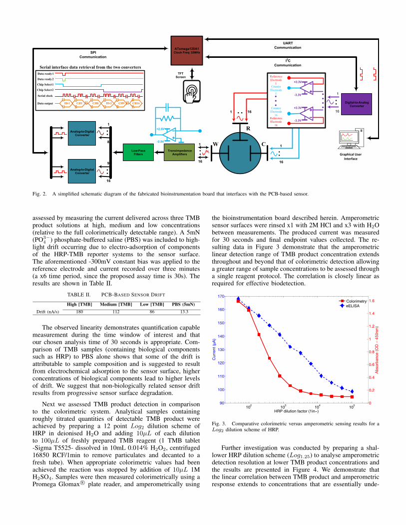
Two, fully-differential eight channel, 16-bit resolutionvoltage-input ADCs have been employed for the digitisationof the converted current values received from the front-endlow-noise transimpedance amplifiers (TIAs) LTC2055. All16 TIAs include 0.1% tolerance resistors for precise andmatched current readings and appropriate value capacitorsfor the filtering of high-frequency noise. The ADCs operatewith bipolar power supply (±2.5V ) and thus, are able todetect positive and negative reaction currents. The overalldesign of the board revolves around the idea of immediatedigitisation of the analog biosensor signals, process them bymeans of standard DSP techniques and finally send them tothe user in digital or in analog form [8]. The system can bepowered-up via a USB-connection or by a single 5V battery,ensuring its portability. A simplified schematic diagram of thecomplete electronic platform is shown in Figure 2, includingthe data retrieval timings for the Serial Peripheral Interface(SPI) communication protocol been used for the two ADCs.
With the use of the 12-bit, single-output Digital-to-AnalogConverter (DAC) AD5321 and an appropriate circuitry, thebioinstrumentation board can provide bipolar biasing voltagesto the sensor from -3.3V up to +3.3V with a resolution of∼1.6mV, using the standard three-electrode potentiostat archi-tecture (see Figure 2). The electronic system is able to read-outsimultaneously from 16-channels in a real-time manner anddynamically change the gain of the internal ProgrammableGain Amplifiers (PGAs) of the ADCs, if the readings arebelow certain threshold values. Moreover, through the on-board DAC, the user is able to perform a Cyclic Voltammetry(CV) calibration before every measurement, if necessary. TableI summarises the characteristics of the fabricated board.
TABLE I. BIOINSTRUMENTATION BOARD’S CHARACTERISTICS
Input current range (dB) 90.3Input current resolution (nA) 122ADCs resolution (bit) 16ADCs data rate (kSPS) 0.125 - 8Board dimensions (mm) 83 x 93DSP unit ATxmega128A1 - 32MHzTotal current required forthe fabricated PCB of Figure 1 (mA) ∼ 45Power supply 5V (Battery or USB)
III. ELECTROCHEMICAL RESULTS
We require a system that shows clear definition betweenmeasured sample currents but we also wish to minimise thebiasing voltage to reduce damage observed at the gold sensorsurface following repeated measurements. Performance was as-sessed through standard CV under different biasing voltages toestablish effective sensor calibration. Negative bias values from0 to -400mV with 50mV steps were investigated. Analytesof various TMB product concentration were produced as de-scribed below and three distinct samples corresponding to low,medium and high colorimetric levels were measured at eachbiasing voltage. CV showed -300mV to provide maximumsignal elevation and thus, optimum system sensitivity, withoutshowing excessive deterioration of sensor surface integrityunder extended or repeated analysis.
Subsequently, a number of experiments were conducted toverify the performance of our system. The sensor’s drift was
R
CW1
16
1
16
1 16
ReferenceElectrode
1CounterElectrode
1
CounterElectrode16
ReferenceElectrode16
S3U3V
u3U3V
S3U3V
u3U3V
TransimpedanceFAmplifiers
DigitalutouAnalogConverter
1
16
LowuPassFilters
AnalogutouDigitalConverter
1
8
9
16
ATxmega128A1ClockFFreq:F32MHzSPI
CommunicationI2C
Communication
UARTCommunication
TFTFScreen
S2U5V
u2U5V
AnalogutouDigitalConverter
Serialpinterfacepdatapretrievalpfromptheptwopconverters
ID-1 CH1 CH8 ID-2 CH9 CH16
Datapreadyp1
Datapreadyp2
ChippSelectp1
Serialpclock
Datapoutput
ChippSelectp2
Graphical User Interface
Fig. 2. A simplified schematic diagram of the fabricated bioinstrumentation board that interfaces with the PCB-based sensor.
assessed by measuring the current delivered across three TMBproduct solutions at high, medium and low concentrations(relative to the full colorimetrically detectable range). A 5mN(PO3−
4 ) phosphate-buffered saline (PBS) was included to high-light drift occurring due to electro-adsorption of componentsof the HRP-TMB reporter systems to the sensor surface.The aforementioned -300mV constant bias was applied to thereference electrode and current recorded over three minutes(a x6 time period, since the proposed assay time is 30s). Theresults are shown in Table II.
TABLE II. PCB-BASED SENSOR DRIFT
High [TMB] Medium [TMB] Low [TMB] PBS (5mN)Drift (nA/s) 180 112 86 13.3
The observed linearity demonstrates quantification capablemeasurement during the time window of interest and thatour chosen analysis time of 30 seconds is appropriate. Com-parison of TMB samples (containing biological componentssuch as HRP) to PBS alone shows that some of the drift isattributable to sample composition and is suggested to resultfrom electrochemical adsorption to the sensor surface, higherconcentrations of biological components lead to higher levelsof drift. We suggest that non-biologically related sensor driftresults from progressive sensor surface degradation.
Next we assessed TMB product detection in comparisonto the colorimetric system. Analytical samples containingroughly titrated quantities of detectable TMB product wereachieved by preparing a 12 point Log2 dilution scheme ofHRP in deionised H2O and adding 10µL of each dilutionto 100µL of freshly prepared TMB reagent (1 TMB tablet-Sigma T5525- dissolved in 10mL 0.014% H2O2, centrifuged16850 RCF/1min to remove particulates and decanted to afresh tube). When appropriate colorimetric values had beenachieved the reaction was stopped by addition of 10µL 1MH2SO4. Samples were then measured colorimetrically using aPromega Glomax R© plate reader, and amperometrically using
the bioinstrumentation board described herein. Amperometricsensor surfaces were rinsed x1 with 2M HCl and x3 with H2Obetween measurements. The produced current was measuredfor 30 seconds and final endpoint values collected. The re-sulting data in Figure 3 demonstrate that the amperometriclinear detection range of TMB product concentration extendsthroughout and beyond that of colorimetric detection allowinga greater range of sample concentrations to be assessed througha single reagent protocol. The correlation is closely linear asrequired for effective biodetection.
102
103
104
105
90
100
110
120
130
140
150
160
170
HRPIdilutionIfactorIm1in−y
Cur
rent
ImµA
y
eELISA
0
0.2
0.4
0.6
0.8
1
1.2
1.4
1.6
Abs
orba
nceI
mOD
I−I4
50nm
y
Colorimetry
Fig. 3. Comparative colorimetric versus amperometric sensing results for aLog2 dilution scheme of HRP.
Further investigation was conducted by preparing a shal-lower HRP dilution scheme (Log1.25) to analyse amperometricdetection resolution at lower TMB product concentrations andthe results are presented in Figure 4. We demonstrate thatthe linear correlation between TMB product and amperometricresponse extends to concentrations that are essentially unde-

tectable by colorimetry. This discovery implies that the systemwill naturally allow the higher assay sensitivity required inmodern medical biomarker analyses. In many colorimetricELISAs, assay sensitivity is limited by the resolution in opticaldetection of the assay product. Here, we have shown that TMBproduct concentrations that are undetectable by optical densitycan be discriminated by amperometry thus, allowing improvedassay sensitivity without changing any molecular aspects of theassay or protocol.
We further analysed our low concentration detection resultsof Figure 4 by expressing the difference between sequentialpoints as a percentage increase and plotting the results inFigure 5. A broadly linear increase is seen for the wholeamperometric dataset, while the colorimetric data indicatethat progressive absorbance in the low concentration analysisonly occurs at the highest two points. Thus, highlightingthe improved detection sensitivity provided by the describedamperometric system.
102
103
24
26
28
30
32
34
36
38
40
42
44
HRP.dilution.factor.m1in−y
Cur
rent
.mµA
y
eELISA
0.09
0.1
0.11
0.12
0.13
0.14
0.15
0.16
0.17
Abs
orba
nce.
mOD
.−.4
50nm
y
Colorimetry
Fig. 4. Colorimetric versus amperometric sensing results for a Log1.25dilution scheme of HRP allowing measurement of lower TMB productconcentrations, compared to Figure 3. (The final TMB product concentrationdepends on both HRP dilution and development time, thus, cross-comparisonof Figure 3 and Figure 4 is achieved by colorimetric values rather than HRPdilution factor.)
IV. CONCLUSION
This paper presents an electrochemical detection platform,developed using commercially available PCB technologies.The proposed system comprises the custom-made sensor anda multi-channel bioinstrumentation board, built entirely fromdiscrete “off-the-shelf ” components. The electronic board canbe powered-up by a battery or via USB connection, dependingwhether a lab- or POC-based application is required. Thepresented experimental results demonstrate that the behaviourof the “eELISA” approach is similar to the standard col-orimetric ELISA protocol and more specifically, in lowerconcentrations the “eELISA” exhibits higher sensitivity thanthe colorimetric method. These encouraging results allow usto proceed to the development of a final POC system version,which will combine a similar electronic board with a PCB-based sensing platform, including an embedded PCB-basedmicrofluidic network [9].
Seq
uent
ial8D
evia
tion8
Fy)
HRP8dilution8factor8F1in−)0 125 156 195 244 305 381 477 596 745 1000
−10
−5
0
5
10
15
20
25
30AmperometryColorimetryAmperometry8Linear8FitColorimetry8Linear8Fit
Fig. 5. Percentage difference within the obtained amperometric and col-orimetric results. The “eELISA” method demonstrates a roughly linear signalincrease in response to TMB product concentration at levels producing noconsistent colorimetric readings.
ACKNOWLEDGMENT
The authors acknowledge the financial support of the En-gineering and Physical Sciences Research Council (EPSRC),EP/L020920/1 research grant. The authors also thank theirindustrial partner Newbury Electronics Ltd. for the fabricationof the custom-made PCB-based biosensors.
REFERENCES
[1] R. McNerney and P. Daley, “Towards a point-of-care test for activetuberculosis: obstacles and opportunities,” Nature Reviews Microbiology,vol. 9, no. 3, pp. 204–213, 2011.
[2] A. Bhimji, A. A. Zaragoza, L. S. Live, and S. O. Kelley, “Electrochemicalenzyme-linked immunosorbent assay featuring proximal reagent genera-tion: Detection of human immunodeficiency virus antibodies in clinicalsamples,” Analytical chemistry, vol. 85, no. 14, pp. 6813–6819, 2013.
[3] C. Hu, I. Zeimpekis, K. Sun, S. Anderson, P. Ashburn, and H. Morgan,“Low-cost nanoribbon sensors for protein analysis in human serum usinga miniature bead-based enzyme-linked immunosorbent assay,” Analyticalchemistry, vol. 88, no. 9, pp. 4872–4878, 2016.
[4] T. Prodromakis, Y. Liu, J. Yang, D. Hollinghurst, and C. Toumazou,“A novel design approach for developing chemical sensing platformsusing inexpensive technologies,” in Biomedical Circuits and SystemsConference (BioCAS), 2011 IEEE, Nov 2011, pp. 369–372.
[5] T. Prodromakis, Y. Liu, and C. Toumazou, “A low-cost disposablechemical sensing platform based on discrete components,” ElectronDevice Letters, IEEE, vol. 32, no. 3, pp. 417–419, 2011.
[6] C. Chen, Q. Xie, D. Yang, H. Xiao, Y. Fu, Y. Tan, and S. Yao, “Recentadvances in electrochemical glucose biosensors: a review,” Rsc Advances,vol. 3, no. 14, pp. 4473–4491, 2013.
[7] P. Fanjul-Bolado, M. B. Gonzalez-Garcıa, and A. Costa-Garcıa, “Amper-ometric detection in tmb/hrp-based assays,” Analytical and Bioanalyticalchemistry, vol. 382, no. 2, pp. 297–302, 2005.
[8] K. I. Papadimitriou, C. Wang, M. L. Rogers, S. A. Gowers, C. L.Leong, M. G. Boutelle, and E. M. Drakakis, “High-performance bioin-strumentation for real-time neuroelectrochemical traumatic brain injurymonitoring,” Frontiers in Human Neuroscience, vol. 10, p. 212, 2016.
[9] N. Vasilakis, D. Moschou, D. Carta, H. Morgan, and T. Prodromakis,“Long-lasting fr-4 surface hydrophilisation towards commercial pcbpassive microfluidics,” Applied Surface Science, 2015.