a palatal speech bulb—a case study
TRANSCRIPT

Hardwick, C. J., & Puryer, J. (2019). A Palatal Speech Bulb: A CaseStudy. Reports, 2(1), [5]. https://doi.org/10.3390/reports2010005
Publisher's PDF, also known as Version of recordLicense (if available):CC BYLink to published version (if available):10.3390/reports2010005
Link to publication record in Explore Bristol ResearchPDF-document
This is the final published version of the article (version of record). It first appeared online via MDPI athttps://www.mdpi.com/2571-841X/2/1/5. Please refer to any applicable terms of use of the publisher.
University of Bristol - Explore Bristol ResearchGeneral rights
This document is made available in accordance with publisher policies. Please cite only thepublished version using the reference above. Full terms of use are available:http://www.bristol.ac.uk/red/research-policy/pure/user-guides/ebr-terms/

Case Report
A Palatal Speech Bulb—A Case Study
Constance Hardwick 1 and James Puryer 2,*1 Bristol Dental Hospital, Lower Maudlin Street, Bristol BS1 2LY, UK; [email protected] Bristol Dental School, Lower Maudlin Street, Bristol BS1 2LY, UK* Correspondence: [email protected]; Tel.: +44-0117-342-4425
Received: 4 January 2019; Accepted: 29 January 2019; Published: 30 January 2019�����������������
Abstract: Palatal defects of the oral cavity can be either congenital or acquired following trauma orsurgical excision of malignant disease. Palatal defects can greatly affect function and subsequentquality of life. Rehabilitation using a removable obturator can be a preferable treatment optionas it allows regular review post-surgery. This case study reports on the design and constructionof a removable “speech bulb” obturator. A 50-year-old female patient presented complaining ofnasal regurgitation and looseness of her current palatal obturator. She had previously undergonewide surgical excision of her soft palate under general anaesthesia due to adenoid cystic carcinoma.Treatment consisted of the provision of a new removable obturator, paying careful attention to thedesign of the “speech bulb” itself. The design of the “speech bulb” is crucial to optimise function,and the method of prosthesis fabrication is fully described. This case highlights the impact ofobturator fit on a patient’s quality of life and will be of benefit to clinicians from many disciplinesincluding dentists, oral and maxillofacial surgeons, Ear, Nose & Throat (ENT) surgeons and speechand language therapists.
Keywords: prosthesis; obturator; rehabilitation; design; carcinoma
1. Introduction
Palatal defects can be either congenital, i.e., cleft palate, or acquired following trauma or surgicalexcision of malignant disease. Palatal defects can lead to nasal regurgitation of fluid and food,hypernasality of speech and difficulty in swallowing and whistling which can all affect a patient’sphysical and mental wellbeing and their quality of life [1]. The two treatment options available toocclude such defects are either surgical reconstruction (common in congenital cases), or the provisionof a dental prosthesis. A removable dental prosthesis is generally preferred after malignancy or tumourremoval to allow for regular review of the surgical site, facilitating identification of recurrence [2].In addition, surgical correction may be contraindicated by systemic or local factors.
Normal physiological functions are regulated by the velopharyngeal valve which helps to separatethe oral and nasal cavities during swallowing and speech [3]. This valve comprises the soft palate,lateral pharyngeal wall and posterior pharyngeal wall which directs air flow and sound into the oraland nasal cavities. When there is a defect or impairment in this mechanism, for example, after surgicalexcision, trauma or a congenital defect, the valve does not fully close. A dental prosthesis is designedto not only obturate the palatal defect but to restore function and to re-establish velopharyngeal valveclosure [4]. A pharyngeal obturator or “speech bulb” is a removable maxillary prosthesis with anextension protruding into the pharynx. This protrusion separates the oropharynx and the nasopharynxduring speaking and swallowing, aiming to improve function, speech and ultimately quality of life forthe patient [5].
When designing an obturator prosthesis, it is important not to overlook fundamental principlesthat are applicable to all removable prostheses [6]:
Reports 2019, 2, 5; doi:10.3390/reports2010005 www.mdpi.com/journal/reports

Reports 2019, 2, 5 2 of 6
• Support• Retention• Stability• Aesthetics
This case study describes the design and construction of a definitive removable prosthesis fora patient following the wide surgical excision of her soft palate due to an adenoid cystic carcinoma.It highlights the importance of careful fabrication of the “speech bulb” itself to restore optimal functionand improve patient quality of life.
2. Case Presentation Section
The presenting complaint of the female patient (CH) aged 50-years was one of nasal regurgitationand looseness of her current palatal speech bulb prosthesis which had been constructed two yearspreviously. The patient suffered from myalgic encephalomyelitis and rheumatoid arthritis and wasallergic to penicillin and non-steroidal anti-inflammatories. Her medications included paracetamol,as needed, and she was a non-smoker.
CH was diagnosed with an adenoid cystic carcinoma of her soft palate in 2012 and underwent awide local excision under general anaesthesia. The extent of the surgical excision of the soft palate canbe seen in Figure 1.
Reports 2019, 2, x FOR PEER REVIEW 2 of 6
• Retention • Stability • Aesthetics
This case study describes the design and construction of a definitive removable prosthesis for a patient following the wide surgical excision of her soft palate due to an adenoid cystic carcinoma. It highlights the importance of careful fabrication of the “speech bulb” itself to restore optimal function and improve patient quality of life.
2. Case Presentation Section
The presenting complaint of the female patient (CH) aged 50-years was one of nasal regurgitation and looseness of her current palatal speech bulb prosthesis which had been constructed two years previously. The patient suffered from myalgic encephalomyelitis and rheumatoid arthritis and was allergic to penicillin and non-steroidal anti-inflammatories. Her medications included paracetamol, as needed, and she was a non-smoker.
CH was diagnosed with an adenoid cystic carcinoma of her soft palate in 2012 and underwent a wide local excision under general anaesthesia. The extent of the surgical excision of the soft palate can be seen in Figure 1.
Figure 1. Intra-oral view showing the healed tissues following wide surgical excision of the soft
palate.
A temporary palatal speech bulb was fitted at the time of initial surgery. CH underwent a further general anaesthetic approximately one month later where further impressions were taken for the construction of a second interim prosthesis. Although the surgical site had healed well, CH had struggled with this second interim prosthesis, particularly in relation to swallowing solids. Approximately two months after the initial surgery, impressions were taken for a definitive cobalt-chrome prosthesis incorporating an acrylic palatal speech bulb and this prosthesis was duly constructed and fitted. Throughout this period following surgery, CH had regular appointments with a speech and language therapist to aid her adaptation to speaking whilst wearing these various prostheses.
CH had regular reviews, and approximately 18 months after the provision of this last prosthesis, she started to struggle with retention of the prosthesis and nasal regurgitation of liquids. On re-presentation, all extra-oral and intra-oral tissues appeared healthy.
The current cobalt-chrome prosthesis had poor retention and, whilst not “dropping”, contributed to the patient’s nasal regurgitation of both liquids and solids due to poor posterior and lateral seals associated with the acrylic speech bulb. When the prosthesis was not worn, the patient’s breathing was difficult, with obvious hypernasal speech, although she was still understandable. She was unable to swallow any solids or liquids, including her own saliva when the prosthesis was not worn. As a result, CH was reluctant to have the prosthesis removed from her mouth for any length
Figure 1. Intra-oral view showing the healed tissues following wide surgical excision of the soft palate.
A temporary palatal speech bulb was fitted at the time of initial surgery. CH underwent afurther general anaesthetic approximately one month later where further impressions were takenfor the construction of a second interim prosthesis. Although the surgical site had healed well,CH had struggled with this second interim prosthesis, particularly in relation to swallowingsolids. Approximately two months after the initial surgery, impressions were taken for a definitivecobalt-chrome prosthesis incorporating an acrylic palatal speech bulb and this prosthesis was dulyconstructed and fitted. Throughout this period following surgery, CH had regular appointmentswith a speech and language therapist to aid her adaptation to speaking whilst wearing thesevarious prostheses.
CH had regular reviews, and approximately 18 months after the provision of this lastprosthesis, she started to struggle with retention of the prosthesis and nasal regurgitation of liquids.On re-presentation, all extra-oral and intra-oral tissues appeared healthy.
The current cobalt-chrome prosthesis had poor retention and, whilst not “dropping”, contributedto the patient’s nasal regurgitation of both liquids and solids due to poor posterior and lateral sealsassociated with the acrylic speech bulb. When the prosthesis was not worn, the patient’s breathing wasdifficult, with obvious hypernasal speech, although she was still understandable. She was unable to

Reports 2019, 2, 5 3 of 6
swallow any solids or liquids, including her own saliva when the prosthesis was not worn. As a result,CH was reluctant to have the prosthesis removed from her mouth for any length of time, despite itslimitations. CH wore the prosthesis continually, apart from when it was removed briefly for cleaning.
CH was fully dentate in both arches, apart from the previous extractions of all four third molarteeth. There was no clinical caries seen and no periodontal pockets were found on probing, althoughthere was evidence of plaque accumulation around the gingival margins in all quadrants.
2.1. Diagnoses
1 Generalised chronic gingivitis;2 Loose upper palatal speech bulb obturator.
2.2. Treatment Plan
The treatment aims were to stabilise the periodontal disease, prevent further periodontal diseaseand to provide a more retentive palatal speech bulb prosthesis. These were achieved by:
1 Referral to a dental hygienist;2 Provision of a new cobalt-chrome removable prosthesis incorporating an acrylic palatal
speech bulb.
2.3. Treatment Method
CH was referred to a hygienist where oral hygiene instruction was given with respect toimproving plaque control around all remaining teeth, and denture hygiene alongside dietary advicefor caries prevention.
Upper and lower primary impressions were taken with an alginate impression material andan upper special tray was constructed from the resultant upper cast. An upper master impression wastaken within the special tray using a medium-bodied silicone putty (Extrude, Kerr, Orange, CA, USA).
A cobalt-chrome framework was cast. In this case, the patient was fully dentate, therefore supportfor the prosthesis could be obtained from the upper dentition, using a number of rests within the castcobalt-chrome framework. The options for retention however were limited due to the patient beingfully dentate. Therefore, multiple occlusally-approaching clasps provided retention for the prosthesis.The framework incorporated a cast distal extension which provided support for the acrylic speechbulb. At the time of fitting, the prosthesis was both stable and retentive, and the patient reportedno discomfort. A chair-side modification of the speech bulb was undertaken using cold cure acrylicresin (Tokuso Rebase, Tokuyama, Osaka Japan) to refine the lateral borders to ensure an optimal seal.The final prosthesis can be seen in Figures 2 and 3.
Reports 2019, 2, x FOR PEER REVIEW 3 of 6
of time, despite its limitations. CH wore the prosthesis continually, apart from when it was removed briefly for cleaning.
CH was fully dentate in both arches, apart from the previous extractions of all four third molar teeth. There was no clinical caries seen and no periodontal pockets were found on probing, although there was evidence of plaque accumulation around the gingival margins in all quadrants.
2.1. Diagnoses
1 Generalised chronic gingivitis; 2 Loose upper palatal speech bulb obturator.
2.2. Treatment Plan
The treatment aims were to stabilise the periodontal disease, prevent further periodontal disease and to provide a more retentive palatal speech bulb prosthesis. These were achieved by:
1 Referral to a dental hygienist; 2 Provision of a new cobalt-chrome removable prosthesis incorporating an acrylic palatal
speech bulb.
2.3. Treatment Method
CH was referred to a hygienist where oral hygiene instruction was given with respect to improving plaque control around all remaining teeth, and denture hygiene alongside dietary advice for caries prevention.
Upper and lower primary impressions were taken with an alginate impression material and an upper special tray was constructed from the resultant upper cast. An upper master impression was taken within the special tray using a medium-bodied silicone putty (Extrude, Kerr, Orange, CA, USA).
A cobalt-chrome framework was cast. In this case, the patient was fully dentate, therefore support for the prosthesis could be obtained from the upper dentition, using a number of rests within the cast cobalt-chrome framework. The options for retention however were limited due to the patient being fully dentate. Therefore, multiple occlusally-approaching clasps provided retention for the prosthesis. The framework incorporated a cast distal extension which provided support for the acrylic speech bulb. At the time of fitting, the prosthesis was both stable and retentive, and the patient reported no discomfort. A chair-side modification of the speech bulb was undertaken using cold cure acrylic resin (Tokuso Rebase, Tokuyama, Osaka Japan) to refine the lateral borders to ensure an optimal seal. The final prosthesis can be seen in Figures 2 and 3.
Figure 2. The constructed definitive prosthesis and palatal speech bulb. Figure 2. The constructed definitive prosthesis and palatal speech bulb.
Reports 2019, 2, 5 4 of 6
Reports 2019, 2, x FOR PEER REVIEW 4 of 6
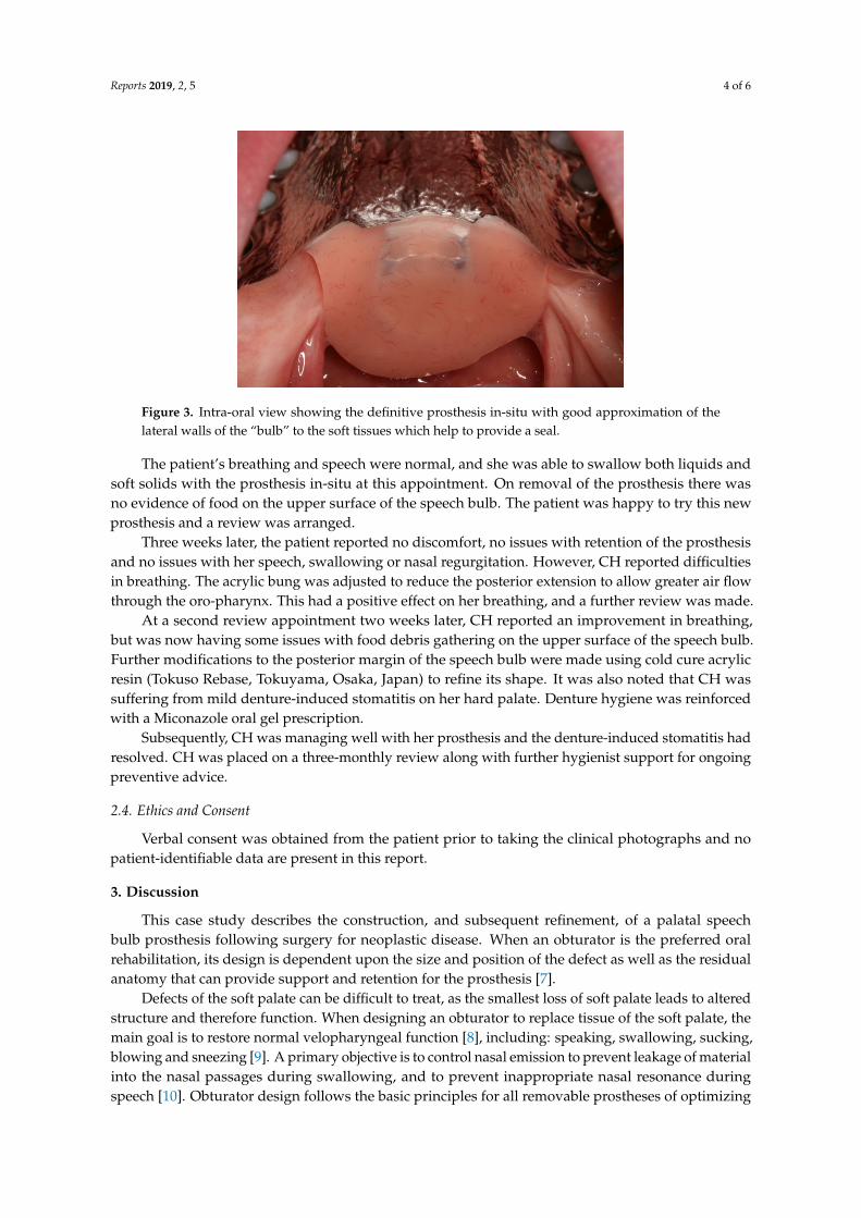
Figure 3. Intra-oral view showing the definitive prosthesis in-situ with good approximation of the lateral walls of the “bulb” to the soft tissues which help to provide a seal.
The patient’s breathing and speech were normal, and she was able to swallow both liquids and soft solids with the prosthesis in-situ at this appointment. On removal of the prosthesis there was no evidence of food on the upper surface of the speech bulb. The patient was happy to try this new prosthesis and a review was arranged.
Three weeks later, the patient reported no discomfort, no issues with retention of the prosthesis and no issues with her speech, swallowing or nasal regurgitation. However, CH reported difficulties in breathing. The acrylic bung was adjusted to reduce the posterior extension to allow greater air flow through the oro-pharynx. This had a positive effect on her breathing, and a further review was made.
At a second review appointment two weeks later, CH reported an improvement in breathing, but was now having some issues with food debris gathering on the upper surface of the speech bulb. Further modifications to the posterior margin of the speech bulb were made using cold cure acrylic resin (Tokuso Rebase, Tokuyama, Osaka, Japan) to refine its shape. It was also noted that CH was suffering from mild denture-induced stomatitis on her hard palate. Denture hygiene was reinforced with a Miconazole oral gel prescription.
Subsequently, CH was managing well with her prosthesis and the denture-induced stomatitis had resolved. CH was placed on a three-monthly review along with further hygienist support for ongoing preventive advice.
2.4. Ethics and Consent
Verbal consent was obtained from the patient prior to taking the clinical photographs and no patient-identifiable data are present in this report.
3. Discussion
This case study describes the construction, and subsequent refinement, of a palatal speech bulb prosthesis following surgery for neoplastic disease. When an obturator is the preferred oral rehabilitation, its design is dependent upon the size and position of the defect as well as the residual anatomy that can provide support and retention for the prosthesis [7].
Defects of the soft palate can be difficult to treat, as the smallest loss of soft palate leads to altered structure and therefore function. When designing an obturator to replace tissue of the soft palate, the main goal is to restore normal velopharyngeal function [8], including: speaking, swallowing, sucking, blowing and sneezing [9]. A primary objective is to control nasal emission to prevent leakage of material into the nasal passages during swallowing, and to prevent inappropriate nasal resonance during speech [10]. Obturator design follows the basic principles for all removable prostheses of optimizing support, stability and retention alongside maintaining good oral health. The major connector was kept free of the anterior palate to help reduce plaque accumulation around the gingival
Figure 3. Intra-oral view showing the definitive prosthesis in-situ with good approximation of thelateral walls of the “bulb” to the soft tissues which help to provide a seal.
The patient’s breathing and speech were normal, and she was able to swallow both liquids andsoft solids with the prosthesis in-situ at this appointment. On removal of the prosthesis there wasno evidence of food on the upper surface of the speech bulb. The patient was happy to try this newprosthesis and a review was arranged.
Three weeks later, the patient reported no discomfort, no issues with retention of the prosthesisand no issues with her speech, swallowing or nasal regurgitation. However, CH reported difficultiesin breathing. The acrylic bung was adjusted to reduce the posterior extension to allow greater air flowthrough the oro-pharynx. This had a positive effect on her breathing, and a further review was made.
At a second review appointment two weeks later, CH reported an improvement in breathing,but was now having some issues with food debris gathering on the upper surface of the speech bulb.Further modifications to the posterior margin of the speech bulb were made using cold cure acrylicresin (Tokuso Rebase, Tokuyama, Osaka, Japan) to refine its shape. It was also noted that CH wassuffering from mild denture-induced stomatitis on her hard palate. Denture hygiene was reinforcedwith a Miconazole oral gel prescription.
Subsequently, CH was managing well with her prosthesis and the denture-induced stomatitis hadresolved. CH was placed on a three-monthly review along with further hygienist support for ongoingpreventive advice.
2.4. Ethics and Consent
Verbal consent was obtained from the patient prior to taking the clinical photographs and nopatient-identifiable data are present in this report.
3. Discussion
This case study describes the construction, and subsequent refinement, of a palatal speechbulb prosthesis following surgery for neoplastic disease. When an obturator is the preferred oralrehabilitation, its design is dependent upon the size and position of the defect as well as the residualanatomy that can provide support and retention for the prosthesis [7].
Defects of the soft palate can be difficult to treat, as the smallest loss of soft palate leads to alteredstructure and therefore function. When designing an obturator to replace tissue of the soft palate, themain goal is to restore normal velopharyngeal function [8], including: speaking, swallowing, sucking,blowing and sneezing [9]. A primary objective is to control nasal emission to prevent leakage of materialinto the nasal passages during swallowing, and to prevent inappropriate nasal resonance duringspeech [10]. Obturator design follows the basic principles for all removable prostheses of optimizing
Reports 2019, 2, 5 5 of 6
support, stability and retention alongside maintaining good oral health. The major connector was keptfree of the anterior palate to help reduce plaque accumulation around the gingival margins and allowthe patient’s tongue to feel some of the natural hard palate [11,12]. An alternative design could havebeen to construct an all-acrylic prosthesis, using Adam’s cribs to gain retention. As with all prostheses,design is a compromise, with better function of the prosthesis often being traded for reduced aesthetics,comfort or oral health [13]. In this case the patient was happy to compromise aesthetics by havingmultiple occlusally-approaching clasps, as function and retention of the prosthesis was her mainpriority. This design also allowed a “fall back” position in that if one of the clasps were to fracture off,the prosthesis could most likely still be worn.
It is essential to ensure that the speech bulb itself is of the correct shape, size and position.Various techniques have been described for the fabrication of the speech bulb [2–4]. Acrylic resinwas used due to its ability to be reduced or relined chair-side, as needed. In this case a chair-sideaddition, using cold-cure acrylic resin was carried out at the fit stage to help provide good lateral seals.Subsequent modification of the bung was undertaken to optimise function and this modification wasbased upon the patient’s verbal feedback.
The patient’s speech was very good whilst wearing the new prosthesis. Several methods of speechevaluation have been described including acoustic spectrogram [14], pressure flow technique [15] andacoustic and aerodynamic techniques [16]. If these techniques are unavailable, it has been suggestedthat a patient’s own perception of speech can be an effective guide [17]. The involvement of a speechand language therapist, as in this case, can be very beneficial at an early stage to help perception ofspeech, especially when these instrumental measures are unavailable [18].
As for all patients with prostheses, oral hygiene and denture hygiene are essential.Plaque accumulation on the prosthesis can increase the risk of dental caries and periodontal disease [19]and poor denture hygiene can lead to denture stomatitis [19]. This risk of denture stomatitis is increasedwhen patients fail to remove their prosthesis at night [20]. In this case, the risk of denture stomatitiswas accepted as CH needed to wear the prosthesis full-time to restore her velopharyngeal function.The application of topical Miconazole gel along with thorough denture hygiene is proving successful.
4. Conclusions
This case describes the successful prosthetic rehabilitation of a patient with a soft palate defectfollowing surgery for an adenoid cystic carcinoma. The design of the obturator followed the basicprinciples of removable prosthesis construction with careful consideration given to the fabrication ofthe speech bulb. From questioning the patient during treatment, there is no doubt that the provision ofthis satisfactory obturator improved her quality of life.
Author Contributions: J.P. treated the patient; C.H. wrote the initial draft manuscript; J.P. reviewed and editedthe manuscript. Both authors have approved the final manuscript and revised the manuscript.
Conflicts of Interest: The authors declare no conflict of interest.
References
1. Kumar, P.; Alvi, H.A.; Roa, J.; Singh, B.P.; Jurel, S.K.; Kumar, L.; Aggarwal, H. Assessment of the quality oflife in maxillectomy patients: A longitudinal study. J. Adv. Prosthodont 2013, 5, 29–35. [CrossRef] [PubMed]
2. Lin, F.H.; Wang, T.C. Prosthodontic Rehabilitation for Edentulous Patients with Palatal Defect: Report of twocases. J. Formos. Med. Assoc. 2011, 110, 120–124. [CrossRef]
3. Fernandez, T.; Harshakumar, K.; Ravichandran, R.; Lyllajam, S. Prosthetic Rehabilitation for a VelopharyngealDefect: A Case Report. J. Dent. Med. Sci. 2015, 14, 1–5.
4. Mohamed, K.; Kumar, V.; Devi, N.; Padmanaban, V. Fabrication of Temporary Speech Bulb Prosthesis:A Clinical Report. J. Indian Prosthodont. Soc. 2010, 10, 71–74. [CrossRef] [PubMed]
5. Ali, M.; Khalifa, N.; Alhajj, N. Quality of life and problems associated with obturators of patients withmaxillectomies. Head Face Med. 2018, 14, 2. [CrossRef] [PubMed]

Reports 2019, 2, 5 6 of 6
6. Davenport, J.; Basker, R.; Health, J.; Ralph, J.; Glantz, P. Removable partial dentures: An introduction.Br. Dent. J. 2000, 189, 646–657. [CrossRef] [PubMed]
7. Ali, R.; Altaie, A.; Nattress, B. Rehabilitation of oncology patients with hard palate defects Part 2: Principlesof obturator design. Dent. Update 2015, 42, 428–434. [CrossRef] [PubMed]
8. Rodenstein, D.; Stanescu, D. The soft palate and breathing. Amer Rev. Resp. Dis. 1986, 134, 311–325. [PubMed]9. Shprintzen, R.; McCall, G.; Skolnick, M.; Lencione, R. Selective movement of the lateral aspects of the
pharyngeal walls during velopharyngeal closure for speech, blowing and whistling in normals. Cleft Palate J.1975, 12, 51–58. [PubMed]
10. Sanders, T.; Oliver, N. A speech-aid prosthesis for anterior maxillary implant-supported prostheses.J. Prosthet. Dent. 1993, 70, 546–547. [CrossRef]
11. Owall, B.; Budtz-Jogensen, E.; Davenport, J.; Mushimoto, E.; Palmqvist, S.; Renner, R.; Sofou, A.; Wostmann, B.Removable partial Dentures Design: A need to focus on hygiene principles? Int. J. Prosthod. 2002, 15, 371–378.
12. Wada, J.; Hideshima, M.; Inukai, S.; Katsuki, A.; Matsuura, H.; Wakabayashi, N. Influence of Oral Morphologyon Speech Production in Subjects wearing maxillary removable partial dentures with major connectors. J. Int.Assoc. Logop. Phoniatr. 2018, 70, 138–148. [CrossRef] [PubMed]
13. Yen-Chen, K.; Yu-Fu, S.; Chiu-Po, C. Extracoronal resilient attachments in distal-extension removable partialdentures. Quintessence Int. 2000, 31, 311–317.
14. Paul, G.; Harlan, B. A supportive type prosthetic speech aid. J. Prosthet. Dent. 1958, 8, 362–369.15. Warren, D. A physiological approach to cleft palate prosthesis. J. Prosthet. Dent. 1965, 15, 770–778. [CrossRef]16. Wood, M.; Warren, D. Effect of cleft palate prostheses on respiratory effort. J. Prosthet. Dent. 1971, 26, 213–218.
[CrossRef]17. Marshell, R.; Jones, R. Effects of palatal lift prosthesis upon speech intelligibility of dysarthric patients.
J. Prosthet. Dent. 1971, 25, 327–333. [CrossRef]18. Beumer, J.; Curtis, T.; Marunick, M. Maxillofacial Rehabilitation: Prosthodontic and Surgical Considerations,
1st ed.; Ishiyaku EuroAmerica: Tokyo, Japan, 1996.19. Puryer, J. Denture Stomatitis—A Clinical Update. Dent. Update 2016, 43, 529–535. [CrossRef] [PubMed]20. Zlataric, D.; Celebic, A.; Valentina, M. The effect of removal partial dentures on periodontal health of
abutment and non-abutment teeth. J. Perio. 2002, 73, 137–144. [CrossRef] [PubMed]
© 2019 by the authors. Licensee MDPI, Basel, Switzerland. This article is an open accessarticle distributed under the terms and conditions of the Creative Commons Attribution(CC BY) license (http://creativecommons.org/licenses/by/4.0/).