a novel pcr method for detecting ace gene insertion
TRANSCRIPT
RESEARCH Open Access
A Novel PCR Method for Detecting ACEGene Insertion/Deletion Polymorphismsand its Clinical ApplicationXue-min Yang1†, Jian-ping Liang1†, Xiao-juan Huang1, Xiang-rong Wang1, Yang Sun3, Chen Dong1, Ya-li Cui1,2* andWen-li Hui1,2*
Abstract
Background: Angiotensin-converting enzyme (ACE) plays a major role in blood pressure regulation andcardiovascular homeostasis. The wide distribution and multifunctional properties of ACE suggest it’s involvement invarious pathophysiological conditions.
Results: In this study, a novel visual detection method for ACE I/D polymorphisms was designed by integratingdirect PCR without the need for DNA extraction using gold magnetic nanoparticles (GMNPs)-based lateral flowassay (LFA) biosensor. The entire detection procedure could enable the genotyping of clinical samples in about 80min. The detection limit was 0.75 ng and results could be obtained in 5 min using the LFA device. Three hundredperipheral blood samples were analyzed using the direct PCR-LFA system and then verified by sequencing todetermine accuracy and repeatability. A clinical preliminary study was then performed to analyze a total of 633clinical samples.
Conclusions: After grouping based on age, we found a significant difference between the genotypes and the ageof patients in the CHD group. The introduction of this method into clinical practice may be helpful for thediagnosis of diseases caused by large fragment gene insertions/deletions.
Keywords: Insertion/deletion polymorphisms, Direct-polymerase chain reaction (direct-PCR), Rapid detection, Goldmagnetic nanoparticle lateral flow assay
BackgroundAt present, most of the detection methods for large frag-ment insertion/deletion (I/D) polymorphism are basedon traditional polymerase chain reaction and electro-phoresis. Rigat et al. were the first to describe a methodfor the detection of I/D polymorphisms using polymer-ase chain reaction (PCR) and agarose gel electrophoresisin 1992 [1, 2]. Other techniques including real-timepolymerase chain reaction (RT-PCR) [3–5], high
resolution melting (HRM) [5, 6], polymerase chainreaction-restriction fragment length polymorphism(PCR-RFLP) [7] and microchip electrophoresis (ME) sys-tem [8] have also been used to detect I/D polymorphism.However, these techniques require tedious experimentalprocedures and expensive and sophisticated instrumentsthat may not be available in clinical institutions. Inaddition, most of these detection systems are based onfluorescence for detection and analysis or have high re-quirements for sample purity. These requirements makeit inconvenient and difficult for the rapid detection of I/D polymorphisms from whole blood. Hence, an easy-to-
© The Author(s). 2021 Open Access This article is licensed under a Creative Commons Attribution 4.0 International License,which permits use, sharing, adaptation, distribution and reproduction in any medium or format, as long as you giveappropriate credit to the original author(s) and the source, provide a link to the Creative Commons licence, and indicate ifchanges were made. The images or other third party material in this article are included in the article's Creative Commonslicence, unless indicated otherwise in a credit line to the material. If material is not included in the article's Creative Commonslicence and your intended use is not permitted by statutory regulation or exceeds the permitted use, you will need to obtainpermission directly from the copyright holder. To view a copy of this licence, visit http://creativecommons.org/licenses/by/4.0/.The Creative Commons Public Domain Dedication waiver (http://creativecommons.org/publicdomain/zero/1.0/) applies to thedata made available in this article, unless otherwise stated in a credit line to the data.
* Correspondence: [email protected]; [email protected]†Xue-min Yang and Jian-ping Liang contributed equally to this work.1The College of Life Science, Northwest University, Xi’an 710069, Shanxi,ChinaFull list of author information is available at the end of the article
Yang et al. Biological Procedures Online (2021) 23:2 https://doi.org/10.1186/s12575-020-00140-6
operate and affordable onsite technique for genotypingwith high efficiency is required.We used ACE I/D polymorphisms as a genotyping
model in this study. We designed a sensitive, rapid, andcost-effective method for I/D polymorphism detection.Angiotensin-converting-enzyme plays an essential rolein two physiological systems, one leading to the produc-tion of angiotensin II and the other for the degradationof bradykinin. ACE metabolizes bradykinin, which is astrong vasodilator, forming the inactive metabolitebradykinin 1–5. ACE can also metabolizes neurokinins,which plays a key role in the transmission of pain, regu-lation of emotions, and alteration of inflammatory andimmune responses. The broad distribution and multi-functionality of these peptides suggest that ACE may beinvolved in the development of several diseases [9]. TheACE gene is located in intron 16 on chromosome 17 inhumans. A 287 bp insertion/deletion (I/D) polymorph-ism [2, 10], results in three genotypes: II (insertionhomozygote), ID (insertion-deletion heterozygote) andDD (deletion homozygote) [11]. Several studies have
demonstrated that this polymorphism is associated withcardiovascular and cerebrovascular diseases [12, 13],while other studies have failed to find an association[14–18]. However, ACE gene polymorphisms has beendemostrated to guide the development of therapeuticdrugs [19, 20]. Hence, the detection of ACE gene poly-morphisms can guide the rational use of drugs in hyper-tensive patients with different ACE genotypes.In our previous study, we established a lateral flow
assay (LFA) assembled with GMNPs that relies on im-mune hybridization reactions for detecting SNPs, suchas MTHFR C677T, which enables the typing of genomicDNA [21, 22]. In this study, we demonstrate a DirectPCR LFA system, which amplifies nucleic acids withoutthe need for DNA extraction and can be used for detect-ing I/D polymorphisms of large fragments. Samples fromfresh whole blood were first treated with NaOH, whichfacilitates DNA release from whole blood for direct amp-lification and eliminates PCR inhibits, such ashemoglobin and enzymes that degrade DNA [23]. Ac-curacy was determined in clinical samples by comparing
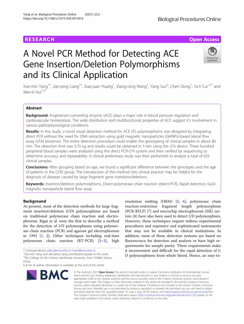
Fig. 1 Schematic diagram of the Direct PCR-LFA system. a Schematic illustration for the GMNP surface modification process. b Structure of thelabeled lateral flow device. c Sample preparation and target amplification. d Results based on visual inspection of bands in the C/T lines
Yang et al. Biological Procedures Online (2021) 23:2 Page 2 of 10

the genotyping results generated from the Direct PCRLFA method with DNA sequencing. The Direct PCRLFA method showed excellent specificity, sensitivity, androbustness for detecting ACE gene I/D polymorphism.
Results and DiscussionPrinciples of the Direct PCR- LFA SystemTo detect ACE I/D polymorphisms, we established theDirect PCR-LFA system. The principles of the DirectPCR-LFA system is schematically illustrated in Fig. 1.GMNPs were synthesized based on the methods de-
scribed previously [24, 25]. The GMNPs were function-alized with cetyltrimethylammonium bromide (CTAB)surfactant, followed by modification of the polyacrylicacid (PAA) and then conjugation of anti-digoxin anti-body through 1-[3-(dimethylamino)propyl]-3-ethylcarbo-diimide hydrochloride chemistry based on a previousmethod [26, 27] (Fig. 1a). The LFA device consisted of astrip composed of five overlapping pads, i.e., sample pad,conjugate pad, nitrocellulose membrane, absorbent pad,and plastic cushion (Fig. 1b). In brief, goat anti-mouseIgG and streptavidin were pre-immobilized in a controlline (C line) and a test line (T line) respectively on a por-ous nitrocellulose membrane using the BioJet HM3010dispenser (BioDot Inc., California, U.S.A.). Then, theprobe solution containing the poly-acrylic acid (PAA)modified gold magnetic nanoparticles (PGMNPs) conju-gated with anti-digoxin antibody was dispensed onto theconjugate pad of the LFA strips. The strips were driedand stored in a sealed aluminum foil bag at roomtemperature until required.Because heparin has an inhibitory effect on PCR [28],
we tested and verified the effect of heparin and EDTAon amplification, and obtained the same results. There-fore, the whole blood sample is required to be collectedin a preservation tube containing EDTA. Fresh wholeblood samples were treated with NaOH solution,followed by PCR amplification using two tubes with thesame prepared blood template (Fig. 1c). After PCR amp-lification, the PCR products in the two tubes were addedonto the sample pad of the LFA strip and the resultsread visually within 5 min (Fig. 1d). For insertion homo-zygous samples (II), a distinct red band was observed onthe T line of the strip from the PCR products of the Mtube but not from the WT tube. For deletion homozy-gous samples (DD), only a red band was observed on thestrip from PCR products of the WT tube but not the Mtube. If red bands with similar intensities are observedon the T lines of both strips, this indicates an insertion/deletion heterozygous sample (I/D).In the whole experiment above, the successful amplifi-
cation of PCR and the normal operation of LFA systemare important steps to obtain correct experimental re-sults. This method is designed to use double tubes and
double channels to complete the deletion and insertiongene amplification at the same time. The T line has adual role. It can read the test results and also serve asthe internal quality control of the PCR system. Inaddition, the C line can judge the effectiveness of theLFA system.Our Direct PCR-GoldMag LFA was faster compared
to existing methods. The pretreatment of the sampleswith NaOH is crucial for this method and makes thewhole testing procedure convenient and rapid. This isbecause, compared to other detection methods, theDNA purification step is eliminated in our assay withthe help of NaOH treatment. This shortens the process-ing time from 1 to 2 h to a mere 5 min. Second, by usingNaOH-treated blood samples for PCR, the problem ofcross-contamination, which exists in traditional bloodDNA purification processes is eliminated. Furthermore,various PCR inhibitors present in the samples such ashemoglobin, IgG, lactoferrin, and proteases are inacti-vated by NaOH treatment [29, 30]. When the directPCR product is loaded onto the sample pad of the LFAstrip, certain impurities such as hemoglobin in the bloodare filtered by the sample pad and conjugate pad, hencethe chromatography is not affected.By introducing mismatches at the penultimate or ante-
penultimate at the 3′ end of the primers, primers speci-ficity was enhanced. We designed several primers todetermine the optimal primers to use. After ARMS-PCR,the LFA device was used to detect the PCR products.With the help of gold magnetic nanoparticles and LFA,accurate and rapid genotyping results could be generatedby visual inspection of colors on the T and C lines. Ittakes only 5 min to obtain the results without the needfor expensive or high-end instruments. We also demon-strated that our method was more convenient and su-perior to the classical agarose gel electrophoresismethod. Hence, this method could be used in medicaland hospital laboratories with limited resources for pur-chasing specialized equipment.
Performance of the Direct PCR- LFA SystemTo determine the optimal assay conditions, optimizationof whole blood direct amplification was performed underdifferent PCR cycling conditions, i.e., annealingtemperature, concentration of the primers, etc. were op-timized using whole blood samples with the three differ-ent ACE genotypes. The magnetic signal at the T lineshowed the best amplification efficiency and specificitywhen the cycle number was 31 (Figure S1A) and the an-nealing temperature was 60 °C (Figure S1B). In addition,the best primer concentration was found to be 2.5 μM(Figure S1C).The specificity of the ACE genotyping was an import-
ant consideration. For specificity analysis, three known
Yang et al. Biological Procedures Online (2021) 23:2 Page 3 of 10

ACE genotypes (II, I/D, DD) were analyzed using theDirect PCR- LFA system (Fig. 2a). We also performed acrossover experiment to validate the primers. Inaddition, ACE genotyping results detected using the lat-eral flow assay (Fig. 2b) was compared with the classicalagarose gel electrophoresis method (Fig. 2c). ACE geneI/D polymorphism is caused by either an insertion or de-letion of the 287 bp Alu repeat. A gene fragment con-taining a repeat sequence cannot be accurately verifiedusing the Sanger method. A homozygous deletion couldbe accurately measured after several rounds of sequen-cing, however, homozygous insertions are more difficultto accurately sequence (Fig. 2d-e).To enhance primer specificity, we designed multiple
primers to determine the optimal primers pairs to use inour assay (Table 1). In addition, we introduced mis-matches in the penultimate or antepenultimate of the 3′end of the primer for PCR. We observed that these mod-ifications had an impact on the specificity of ourmethod, as some allele-specific primers completely lostspecificity upon removal of the mismatches [31]. This isimportant when analyzing clinical samples that requirehigh specificity and accuracy. We then performed PCRand agarose gel electrophoresis using these primers andselected the most optimal primes to use in our assay.The deletion-specific primer sequence was 5′-AACCACATAAAAGTGACTGTATCGG-3′, and had a mismatchat the third of the 3′ end, while the insertion-specificprimer sequence was 5′-TCGAGACCATCCCGGCTA
AAAC-3′. Other primers that were synthesized were ex-cluded due to poor specificity. The list of commonprimers is shown in Table 5. The common forward pri-mer sequence was 5′-AAGGAGAGGAGAGAGACTCAAGCAC-3′.
Performance of the PCR-GoldMag LFATo evaluate the performance of the PCR-GoldMag LFAsystem, the sensitivity of LFA was evaluated using vari-ous amounts of whole blood samples with known ACEgenotypes. Gradient dilutions of whole blood withphysiological saline at 1:1, 1:2, 1:4, 1:5, 1:10, 1:15, 1:20, 1:30, 1:40, 1:60, 1:120 ratio were evaluated. Whole bloodsamples are compared with purified nucleic acid samples(Fig. 3). The LFA typing results were analyzed to deter-mine the sensitivity of the whole blood direct PCRmethod. Simultaneously, we performed blood routinetests obtained from the People’s Hospital of ShaanxiProvince (Table S1). The amount of nucleic acids in awhite blood cell is approximately 5.6 × 10− 9 μg [32]. Theresults demonstrated that the minimum detection sensi-tivity was 0.75 ng.Using our optimized conditions, PCR-GoldMag LFAs
showed high specificity with no false-positive results andhad a higher sensitivity. The detection limit of PCR-LFAcould reach 0.75 ng of DNA, which was comparable to aPCR-DNA microassay commercial kit and had obviousadvantages. As shown in Fig. 3, whole blood sampleswere more sensitive compared to purified DNA. At a
Fig. 2 The specificity of the Direct PCR-LFA system. a Genotyping results of the whole blood Direct PCR-LFA system. M =M tube. WT =WT tube.1: Homozygous insertion-deletion 2: Heterozygous 3: Homozygous deletion. b The genotyping result of PCR-LFA using DNA as the template. cThe genotyping result of agarose gel electrophoresis. d DNA sequencing result of a Homozygous deletion sample. e DNA sequencing result of aHomozygous insertion sample
Yang et al. Biological Procedures Online (2021) 23:2 Page 4 of 10

dilution ratio of 1:60, bands could still be observed usingwhole blood, while from purified DNA, bands werefainter at the same dilution factor. For homozygous dele-tion samples, no bands appeared at a ratio of 1:15. Thismay be due to the loss of white blood cells during DNAextraction, while whole blood eliminated the DNA ex-traction step, and hence had no loss of white blood cells.The sensitivity was higher using whole blood samplescompared to purified DNA. The capacity of antibodyconjugation to nanoparticles is critical for the success ofour assay. In general, the conjugating capacity of nano-particles to antibodies at best is only about 50μgmg− 1.
Because of the novel GoldMag nanoparticle structure(nanoflowers) [24], our antibody conjugation reachedgreater than 100μgmg− 1. This ensured the sensitivityand stability of our established PCR-lateral flow assay[24, 27].
Clinical ApplicationsTo evaluate the reliability of our optimized PCR-GoldMag LFA method, the accuracy was further vali-dated using an additional 300 clinical samples obtainedfrom the Shaanxi Provincial People’s Hospital (Xi’an,China), with informed consent waived. Each sample was
Table 1 Multiple Primer Sequences used for assay optimization
Primer type Primer name Sequence (5′-3′)
Deletion-specific primer F1 TTCTCTAGACCTGCTGCCTATACAG
R1 CCATAACAGGTCTTCATATTTCCGG
F2 TCTCTAGACCTGCTGCCTATACCGT
R2 CCATGCCCATAACAGGTCTTCATAT
F3 AAGGAGAGGAGAGAGACTCAAGCAC
R3 GCGAAACCACATAAAAGTGACTGTATAG
F4 AAGGAGAGGAGAGAGACTCAAGCAC
R4 AACCACATAAAAGTGACTGTATCGG
Insertion-specific primer F1 CTGGAGAGCCACTCCCATCCTTTCT
R1 TCGAGACCATCCCGGCTAAAAC
F2 GCCACTCCCATCCTTTCT
R2 CCTGTAATCCCAGCACTTTG
Fig. 3 Comparison of results generated using gradient dilution of whole blood samples and nucleic acid samples (1:1–1:120). a Homozygousinsertion sample (b) Insert/deletion heterozygous sample (c) Homozygous deletion sample
Yang et al. Biological Procedures Online (2021) 23:2 Page 5 of 10

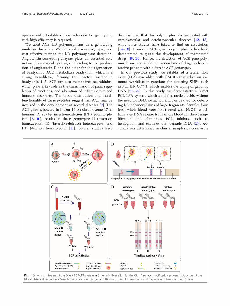
tested using our whole blood Direct PCR-GoldMagLFAs and agarose gel electrophoresis was used to com-pare our results. As shown in Table 2, the genotypingresults were 100% consistent with the results obtainedfrom agarose gel electrophoresis. In addition, we per-formed a Hardy-Weinberg equilibrium test on selectedsamples (Table 2) and analyzed the genotype and allelefrequencies of the 300 cases (Fig. 4). The results wereconsistent with Hardy Weinberg’s law of equilibrium. Itdemonstrated that the selected groups had good grouprepresentation. The genotype frequencies of ACE II, ID,DD types were 42% (127 cases), 44% (132 cases), and14% (41 cases) (Fig. 4a). The allele frequency for the I al-lele was 64% and the D allele was 36% (Fig. 4b). Repre-sentative genotyping results of the two methods areshowed in Figure S2. In addition, we evaluated bloodsamples from cases with high bilirubin levels and lowand high white blood cell counts (Figure S3). Our resultsdemonstrated that the established method had good ac-curacy and reliability. We believe our method has wideapplication value in clinical practice.We established a sensitive, low cost, and easy-to-use
large fragment insertion/deletion polymorphism detectionplatform using whole blood Direct PCR-LFA. This en-abled us to accurately detect insertion/deletion polymor-phisms for specific genetic diseases. With the help of goldmagnetic nanoparticles and LFA, genotyping results couldbe rapidly visualized based on colored T and C lines.Compared to conventional detection methods for I/D
polymorphism, our assay had numerous significant ad-vantages. These include (a) Using the PCR-GoldMagLFA system, it takes only 5 min to obtain results withoutthe need for expensive or high-end instruments. (b)After sample treatment with NaOH, the whole testing
procedure was rapid and convenient. (c) Previousmethods were complex and performed on expensive andsophisticated instruments that may not be available inmany laboratories, however, the PCR-GoldMag LFAmethod is easy-to-operate and affordable for on-sitegenotyping with high efficiency. Hence, this methodcould be run in laboratories that are not fully equippedwith sophisticated instruments.
Case-Control StudyPrevious studies have reported that insertion/deletion (I/D) polymorphisms in the ACE gene were associated withcardiovascular and cerebrovascular diseases. However,the association between ACE gene I/D and cardiovascu-lar and cerebrovascular diseases is controversial. Somestudies have shown that ACE I/D polymorphisms are as-sociated with coronary artery disease (CAD) and cere-bral ischemic stroke (CIS) [14, 16], however, otherstudies have found no association [15, 17]. We per-formed a case-control study and a meta-analysis toevaluate the association between ACE I/D polymor-phisms and coronary atherosclerotic heart disease, aswell as stroke.We analyzed a total of 633 subjects (199 CHD pa-
tients, 207 CIS patients, and 227 control group) for thecase-control study. These samples satisfied the Hardy–Weinberg Law (CHD, c2 = 0.68, p = 0.41 > 0.05; CIS,c2 = 1.58, p = 0.21 > 0.05), which signified a reliable rep-resentative group. Using the chi-square test, no statis-tical differences between these two groups wereobserved (Table 3-4). The ACE genotype frequencies areshown in Fig. 5. Based on statistical analysis, we foundno association between ACE I/D polymorphisms andcoronary heart disease or stroke. However, after
Table 2 ACE genotyping results using whole blood samples between Direct PCR-LFA system and Agarose gel electrophoresis
Genotype Agarose gel electrophoresis Total Agreement
ACE II ACE ID ACE DD
PCR-GoldMag LFA system ACE II 127 0 0 127 100%
ACE ID 0 132 0 132 100%
ACE DD 0 0 41 41 100%
Fig. 4 Pie chart for the genotype and allele frequency of ACE I/D polymorphism. a Genotype frequencies of ACE. b Allele frequencies of I andD alleles
Yang et al. Biological Procedures Online (2021) 23:2 Page 6 of 10

grouping based on age, we observed a significant differ-ence between the genotype and age of the patients in theCHD group (p = 0.02 < 0.05), with no significant differ-ences in the stroke group (p = 0.07 > 0.05) (Table S2).Association studies are influenced by selection bias,
population stratification, confounding factors, and clin-ical criteria used to define patient groups. Future geneticassociation studies should include larger patient cohortsand strict study designs to determine potential associa-tions between genetic susceptibility and cardiovascularand cerebrovascular diseases.
ConclusionsOur genotyping method for determining ACE I/D poly-morphism was established using a combination of DirectPCR and GoldMag-LFA system. This method usedwhole blood samples to directly perform PCR amplifica-tion. It eliminated the need for nucleic acid extractionand reduced the risk of sample cross-contamination dur-ing nucleic acid extraction. The method was rapid andsensitive and did not require expensive instruments. Thewhole PCR reaction took about 80 min and had signifi-cant clinical application. The genotyping results wereobtained within 5min after loading the PCR productsonto the LFA device. The method could be used to de-termine I/D polymorphisms for a variety of genes thatare associated with disease risk.
Materials and MethodsMaterials and ReagentsGoldMag nanoparticles were purchased from Xi’anGoldMag Nanobiotech Co., Ltd. (Xi’an, China). 10 ×HotMaster Taq Buffer and HotMaster Taq DNA Poly-merase were purchased from TIANGEN (Beijing,China). dNTPs and uracil-DNA glycosylase (UDG) were
purchased from Shinegene (Shanghai, China). Water(18.2 MΩ cm) was purified using the Barnstead Nano-pure Water system. MgCl2 (25 mM) was purchased fromThermo Scientific (Shanghai, PRC). 1 × TE (pH = 8) werepurchased from Sangon Biotech. All primers were syn-thesized and purchased from Invitrogen (Shanghai,China).
LFA DeviceStreptavidin and goat anti-mouse IgG were pre-immobilized in a test line (T line) and a control line (Cline) onto a porous nitrocellulose membrane using theBioJet HM3010 dispenser (BioDot Inc., California,U.S.A.). Then, the probe solution containing the poly-acrylic acid (PAA) modified gold magnetic nanoparticles(PGMNPs) conjugated with anti-digoxin antibody wasdispensed onto the conjugate pad of the LFA strips.These strips were dried and stored in a sealed aluminumfoil bag at room temperature until required. The stripswere stable for 12 months.
Primer Design and SynthesisBased on the ARMS-PCR method [33–35], the forwardprimer was designed as the universal primer, and the re-verse primers were the allele-specific primers. Primerspecificity was enhanced by introducing mismatches atthe penultimate or antepenultimate at the 3′ end of theprimers [36, 37]. For convenience, we refer to the wild-type as “WT” and the mutation type as “M”. To geno-type the ACE gene, a set of three specific primers weredesigned using the Primer 5.0 software program (Pri-mer-E Ltd., Plymouth, U.K.). This included the 5′biotin-labeled universal primer, a 5′ digoxin labeled spe-cific primer for WT, and a 5′ digoxin labeled specificprimer for M. Two additional primers were designed for
Table 3 Genotype and allele frequencies of ACE I/D polymorphism in coronary atherosclerotic heart disease and control group
Marker CHD Group (n = 199) Control(n = 227)
χ2 P-Value
ACE I/D Genotype II 80 93 1.27 0.53
ID 88 107
DD 31 27
Allele I 248 293 0.36 0.55
D 150 161
Table 4 Genotype and allele frequencies of ACE I/D polymorphism in the stroke and control group
Marker CIS Group (n = 207) Control (n = 227) χ2 P-Value
ACE I/D Genotype II 81 93 0.37 0.84
ID 104 107
DD 23 27
Allele I 266 293 0.01 0.91
D 150 161
Yang et al. Biological Procedures Online (2021) 23:2 Page 7 of 10
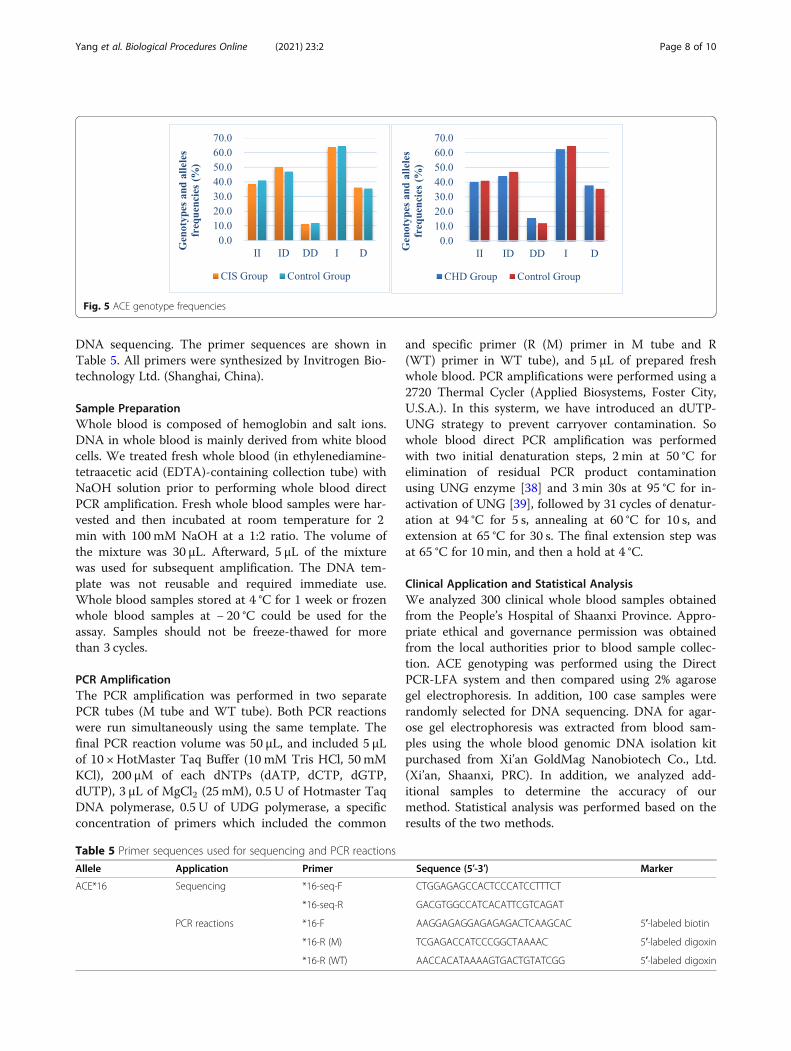
DNA sequencing. The primer sequences are shown inTable 5. All primers were synthesized by Invitrogen Bio-technology Ltd. (Shanghai, China).
Sample PreparationWhole blood is composed of hemoglobin and salt ions.DNA in whole blood is mainly derived from white bloodcells. We treated fresh whole blood (in ethylenediamine-tetraacetic acid (EDTA)-containing collection tube) withNaOH solution prior to performing whole blood directPCR amplification. Fresh whole blood samples were har-vested and then incubated at room temperature for 2min with 100 mM NaOH at a 1:2 ratio. The volume ofthe mixture was 30 μL. Afterward, 5 μL of the mixturewas used for subsequent amplification. The DNA tem-plate was not reusable and required immediate use.Whole blood samples stored at 4 °C for 1 week or frozenwhole blood samples at − 20 °C could be used for theassay. Samples should not be freeze-thawed for morethan 3 cycles.
PCR AmplificationThe PCR amplification was performed in two separatePCR tubes (M tube and WT tube). Both PCR reactionswere run simultaneously using the same template. Thefinal PCR reaction volume was 50 μL, and included 5 μLof 10 × HotMaster Taq Buffer (10 mM Tris HCl, 50 mMKCl), 200 μM of each dNTPs (dATP, dCTP, dGTP,dUTP), 3 μL of MgCl2 (25 mM), 0.5 U of Hotmaster TaqDNA polymerase, 0.5 U of UDG polymerase, a specificconcentration of primers which included the common
and specific primer (R (M) primer in M tube and R(WT) primer in WT tube), and 5 μL of prepared freshwhole blood. PCR amplifications were performed using a2720 Thermal Cycler (Applied Biosystems, Foster City,U.S.A.). In this systerm, we have introduced an dUTP-UNG strategy to prevent carryover contamination. Sowhole blood direct PCR amplification was performedwith two initial denaturation steps, 2 min at 50 °C forelimination of residual PCR product contaminationusing UNG enzyme [38] and 3min 30s at 95 °C for in-activation of UNG [39], followed by 31 cycles of denatur-ation at 94 °C for 5 s, annealing at 60 °C for 10 s, andextension at 65 °C for 30 s. The final extension step wasat 65 °C for 10 min, and then a hold at 4 °C.
Clinical Application and Statistical AnalysisWe analyzed 300 clinical whole blood samples obtainedfrom the People’s Hospital of Shaanxi Province. Appro-priate ethical and governance permission was obtainedfrom the local authorities prior to blood sample collec-tion. ACE genotyping was performed using the DirectPCR-LFA system and then compared using 2% agarosegel electrophoresis. In addition, 100 case samples wererandomly selected for DNA sequencing. DNA for agar-ose gel electrophoresis was extracted from blood sam-ples using the whole blood genomic DNA isolation kitpurchased from Xi’an GoldMag Nanobiotech Co., Ltd.(Xi’an, Shaanxi, PRC). In addition, we analyzed add-itional samples to determine the accuracy of ourmethod. Statistical analysis was performed based on theresults of the two methods.
Fig. 5 ACE genotype frequencies
Table 5 Primer sequences used for sequencing and PCR reactions
Allele Application Primer Sequence (5′-3′) Marker
ACE*16 Sequencing *16-seq-F CTGGAGAGCCACTCCCATCCTTTCT
*16-seq-R GACGTGGCCATCACATTCGTCAGAT
PCR reactions *16-F AAGGAGAGGAGAGAGACTCAAGCAC 5′-labeled biotin
*16-R (M) TCGAGACCATCCCGGCTAAAAC 5′-labeled digoxin
*16-R (WT) AACCACATAAAAGTGACTGTATCGG 5′-labeled digoxin
Yang et al. Biological Procedures Online (2021) 23:2 Page 8 of 10

Supplementary InformationThe online version contains supplementary material available at https://doi.org/10.1186/s12575-020-00140-6.
Additional file 1: Figure S1. Optimization of the detection system. M =M tube. WT = WT tube. 1=II 2=ID 3=DD (A) The cycles of PCRamplification. 31 cycles were the optimal. (B) The annealing temperature.60°C was found to be optimal. (C) The concentration of primers. 2.5 μMprimer was proved to be optimal. (D) The test of the amount of Mg2+
with 3 μL of Mg2+ as the optimum. (E) The amount of the whole bloodtemplate. 5 μL was optimal. Figure S2. Partial genotyping results of thetwo methods. (A) The results of 20 cases of whole blood direct PCR (B)Agarose gel electrophoresis. Figure S3. Test results of special samples. 1-5: High bilirubin sample 6-10: Autoimmune Disease sample 11-15: Lowwhite blood cell concentration sample 16-20: High white blood cell con-centration sample 21-25: High cholesterol sample 26-30: High triglyceridesample 31-35: Hemolysis sample 36-40: Blood disease sample. Table S1.Comparison of nucleic acid quantities and Comparison of nucleic acidquantities. Table S2. Association of ACE (I/D) polymorphism with age ofstudy subjects.
AcknowledgementsNot applicable.
Authors’ ContributionsXue-min Yang and Wen-li Hui put forward this experimental concept. Xue-min Yang and Jian-ping Liang established the methodology. Jian-ping Liangand Yang Sun analyzed the data. Xiao-juan Huang, Xiang-rong Wang andChen Dong completed the verification of clinical data. Ya-li Cui, Wen-li Huiet.al. revised and finally confirmed the manuscript. All authors wrote themanuscript. The authors read and approved the final manuscript.
FundingThis work was funded by the Project of the National Natural ScienceFoundation of China (No. 31771083) and the National Natural ScienceFoundation of China (No. 81772289).
Availability of Data and MaterialsAll data generated or analyzed during this study are included in thispublished article.
Ethics Approval and Consent to ParticipateThe study was approved by the Ethics Committee at the College of LifeSciences at Northwest University (Xi’an, China) and informed consent waswaived.
Consent for PublicationNot applicable.
Competing InterestsAll authors declare that they have no competing interests.
Author details1The College of Life Science, Northwest University, Xi’an 710069, Shanxi,China. 2Shanxi Provincial Engineering Research Center for Nano-BiomedicalDetection, Xi’an 710077, Shanxi, China. 3Data Center of Shaanxi ProvincialPeople’s Hospital, Xi’an 710069, Shanxi, China.
Received: 24 October 2020 Accepted: 15 December 2020
References1. Saracevic A, Simundic AM, Celap I, Luzanic V. Angiotensin-converting
enzyme insertion/deletion polymorphism genotyping error: the cause and apossible solution to the problem. Mol Biol Rep. 2013;40(7):4459–63.
2. Rigat B, Hubert C, Corvol P, Soubrier F. PCR detection of the insertion/deletion polymorphism of the human angiotensin converting enzyme gene(DCP1) (dipeptidyl carboxypeptidase 1). Nucleic Acids Res. 1992;20(6):1433.
3. Djurisic S, Sorensen AE, Hviid TV. A fast and easy real-time PCR genotypingmethod for the HLA-G 14-bp insertion/deletion polymorphism in the 3′untranslated region. Tissue Antigens. 2012;79(3):186–9.
4. Somogyvari F, Szolnoki Z, Marki-Zay J, Fodor L. Real-time PCR assay withfluorescent hybridization probes for exact and rapid genotyping of theangiotensin-converting enzyme gene insertion/deletion polymorphism. ClinChem. 2001;47(9):1728–9.
5. Lin MH, Tseng CH, Tseng CC, Huang CH, Chong CK, Tseng CP. Real-timePCR for rapid genotyping of angiotensin-converting enzyme insertion/deletion polymorphism. Clin Biochem. 2001;34(8):661–6.
6. Nissen PH, Campbell NB, Hojskov CS, Floe A, Hoffmann HJ, Hilberg O, et al.Development of a high-resolution melting genotyping assay for theangiotensin I converting enzyme insertion/deletion polymorphism andestablishment of genotype-specific reference intervals in a Danishpopulation. Ann Clin Biochem. 2015;52(Pt 1):105–12.
7. Hashemi M, Danesh H, Bizhani F, Sattarifard H, Hashemi SM, Bahari G.Detection of a 4-bp insertion/deletion polymorphism within the promoterof EGLN2 using mismatch PCR-RFLP and its association with susceptibilityto breast cancer. Asian Pac J Cancer Prev. 2018;19(4):923–6.
8. Sun Y, Kim SK, Zhang P, Woo N, Kang SH. Fast high-throughput screeningof angiotensin-converting enzyme insertion/deletion polymorphism byvariable programmed electric field strength-based microchipelectrophoresis. J Chromatogr B Anal Technol Biomed Life Sci. 2016;1028:77–85.
9. Sayed-Tabatabaei FA, Oostra BA, Isaacs A, van Duijn CM, Witteman JC. ACEpolymorphisms. Circ Res. 2006;98(9):1123–33.
10. Rigat B, Hubert C, Alhenc-Gelas F, Cambien F, Corvol P, Soubrier F. Aninsertion/deletion polymorphism in the angiotensin I-converting enzymegene accounting for half the variance of serum enzyme levels. J Clin Invest.1990;86(4):1343–6.
11. Soubrier F, Martin S, Alonso A, Visvikis S, Tiret L, Matsuda F, et al. High-resolution genetic mapping of the ACE-linked QTL influencing circulatingACE activity. Eur J Hum. 2002;10(9):553–61.
12. Pinheiro DS, Santos RS, Jardim P, Silva EG, Reis AAS, Pedrino GR, et al. Thecombination of ACE I/D and ACE2 G8790A polymorphisms revelssusceptibility to hypertension: a genetic association study in Brazilianpatients. PLoS One. 2019;14(8):e0221248.
13. Yaqoob I, Tramboo NA, Bhat IA, Pandith A, Beig JR, Hafeez I, et al. Insertion/deletion polymorphism of ACE gene in females with peripartumcardiomyopathy: a case-control study. Indian Heart J. 2018;70(1):66–70.
14. Amara A, Mrad M, Sayeh A, Lahideb D, Layouni S, Haggui A, et al. The effectof ACE I/D polymorphisms alone and with concomitant risk factors oncoronary artery disease. Clin Appl Thromb Hemost. 2018;24(1):157–63.
15. Basol N, Celik A, Karakus N, Ozturk SD, Yigit S. The evaluation ofangiotensin-converting enzyme (ACE) gene I/D and IL-4 gene intron 3 VNTRpolymorphisms in coronary artery disease. In vivo (Athens, Greece). 2014;28(5):983–7.
16. Munshi A, Sultana S, Kaul S, Reddy BP, Alladi S, Jyothy A. Angiotensin-converting enzyme insertion/deletion polymorphism and the risk ofischemic stroke in a south Indian population. J Neurol Sci. 2008;272(1–2):132–5.
17. Domingues-Montanari S, Fernandez-Cadenas I, del Rio-Espinola A,Mendioroz M, Ribo M, Obach V, et al. The I/D polymorphism of the ACE1gene is not associated with ischaemic stroke in Spanish individuals. Eur JNeurol. 2010;17(11):1390–2.
18. Thomas GN, Tomlinson B, Chan JC, Sanderson JE, Cockram CS, Critchley JA.Renin-angiotensin system gene polymorphisms, blood pressure,dyslipidemia, and diabetes in Hong Kong Chinese: a significant associationof tne ACE insertion/deletion polymorphism with type 2 diabetes. DiabetesCare. 2001;24(2):356–61.
19. Stavroulakis GA, Makris TK, Krespi PG, Hatzizacharias AN, Gialeraki AE,Anastasiadis G, et al. Predicting response to chronic antihypertensivetreatment with Fosinopril: the role of angiotensin-converting enzyme genepolymorphism. Cardiovasc Drugs Ther. 2000;14(4):427–32.
20. Heidari F, Vasudevan R, Mohd Ali SZ, Ismail P, Etemad A, Pishva SR, et al.Association of insertion/deletion polymorphism of angiotensin-convertingenzyme gene among Malay male hypertensive subjects in response to ACEinhibitors. J Renin-Angiotensin-Aldosterone Syst. 2015;16(4):872–9.
21. Hui W, Zhang S, Zhang C, Wan Y, Zhu J, Zhao G, et al. A novel lateral flowassay based on GoldMag nanoparticles and its clinical applications forgenotyping of MTHFR C677T polymorphisms. Nanoscale. 2016;8(6):3579–87.
Yang et al. Biological Procedures Online (2021) 23:2 Page 9 of 10

22. Liu X, Zhang C, Liu K, Wang H, Lu C, Li H, et al. Multiple SNPs detectionbased on lateral flow assay for phenylketonuria diagnostic. Anal Chem.2018;90(5):3430–6.
23. Adams DN. Shortcut method for extraction of Staphylococcus aureus DNAfrom blood cultures and conventional cultures for use in real-time PCRassays. J Clin Microbiol. 2005;43(6):2932–3.
24. Hui W, Shi F, Yan K, Peng M, Cheng X, Luo Y, et al. Fe3O4/au/Fe3O4nanoflowers exhibiting tunable saturation magnetization and enhancedbioconjugation. Nanoscale. 2012;4(3):747–51.
25. Cui YHD, Fang Y, Ma J. Preparation and mechanism of Fe3O4/au core/shellsuper-paramagnetic microspheres. Sci China, Ser B: Chem. 2001;4:404–10.
26. Chao X, Guo L, Zhao Y, Hua K, Peng M, Chen C, et al. PEG-modifiedGoldMag nanoparticles (PGMNs) combined with the magnetic field for localdrug delivery. J Drug Target. 2011;19(3):161–70.
27. Yang D, Ma J, Zhang Q, Li N, Yang J, Raju PA, et al. Polyelectrolyte-coatedgold magnetic nanoparticles for immunoassay development: toward pointof care diagnostics for syphilis screening. Anal Chem. 2013;85(14):6688–95.
28. Satsangi J, Jewell DP, Welsh K, Bunce M, Bell JI. Effect of heparin onpolymerase chain reaction. Lancet. 1994;343:1509–10.
29. Connelly CM, Porter LR, TerMaat JR. PCR amplification of a triple-repeatgenetic target directly from whole blood in 15 minutes as a proof-of-principle PCR study for direct sample analysis for a clinically relevant target.BMC Med Genet. 2014;15:130.
30. Monroe C, Grier C, Kemp BM. Evaluating the efficacy of various thermo-stable polymerases against co-extracted PCR inhibitors in ancient DNAsamples. Forensic Sci Int. 2013;228(1–3):142–53.
31. Chen F, Zhao Y, Fan C, Zhao Y. Mismatch extension of DNA polymerasesand high-accuracy single nucleotide polymorphism diagnostics by goldnanoparticle-improved isothermal amplification. Anal Chem. 2015;87(17):8718–23.
32. Smith TJ, Khatcheressian J, Lyman GH, Ozer H, Armitage JO, Balducci L, et al.2006 update of recommendations for the use of white blood cell growthfactors: an evidence-based clinical practice guideline. J Clin Oncol. 2006;24(19):3187–205.
33. Little S. Amplification-refractory mutation system (ARMS) analysis of pointmutations. Curr Protoc Hum Gen. 2001;9:9.8.
34. Newton CR, Graham A, Heptinstall LE, Powell SJ, Summers C, Kalsheker N,et al. Analysis of any point mutation in DNA. The amplification refractorymutation system (ARMS). Nucleic Acids Res. 1989;17(7):2503–16.
35. Liu J, Huang S, Sun M, Liu S, Liu Y, Wang W, et al. An improved allele-specific PCR primer design method for SNP marker analysis and itsapplication. Plant Methods. 2012;8(1):34.
36. Cha RS, Zarbl H, Keohavong P, Thilly WG. Mismatch amplification mutationassay (MAMA): application to the c-H-ras gene. PCR Methods Appl. 1992;2(1):14–20.
37. Li B, Kadura I, Fu DJ, Watson DE. Genotyping with TaqMAMA. Genomics.2004;83(2):311–32.
38. Bacich DJ, Sobek KM, Cummings JL, Atwood AA, O'Keefe DS. False negativeresults from using common PCR reagents. BMC Res Notes. 2011;4:457.
39. Saibal K, Mark HS, James DC. Optimized PCR amplification of influenza avirus RNA using Tth DNA polymerase, incorporating uracil N glycosylase(UNG) in a single tube reaction. J Clin Lab Anal. 1997;11:323–7.
Publisher’s NoteSpringer Nature remains neutral with regard to jurisdictional claims inpublished maps and institutional affiliations.
Yang et al. Biological Procedures Online (2021) 23:2 Page 10 of 10