a novel mathematical model of blood flow ahmad...
TRANSCRIPT

ii
A NOVEL MATHEMATICAL MODEL OF BLOOD FLOW
AHMAD KHUDZAIRI BIN KHALID
A dissertation submitted in partial fulfillment of the
requirements for the award of the degree of
Master of Science (Mathematics)
Faculty of Science
Universiti Teknologi Malaysia
JUNE 2014

iv
Specially dedicated to my beloved father and mother,
Khalid Bin Jusoh and Kalthom Binti Atan,
to my siblings
and
those people who have guided and inspired me throughout my journey of education

v
ACKNOWLEDGEMENTS
All praise to Allah, The Most Gracious The Most Merciful. I hereby placing
all my faith and thankfulness of His greatness all that he piled on me, as I could
afford to complete my task timely.
In order to complete my final year project, I have a lot of assistance from
many people especially my supervisor. In the first place I wish to express my sincere
appreciation to my beloved supervisor, PM Dr Mukheta Isa, for his willingness to
provide me with guidance, knowledge suggestions, valuable comments, constructive
criticisms and encouragement. Without his continued support and interest, this
project would not have been completed in the stated time.
I am also thankful to all my family members especially my loving parents,
Khalid Bin Jusoh and Kalthom Binti Atan, for their patience and support in my
journey of education. They have given me so much comfort, care and love, either
financially or spiritually, of which word could not express and will forever be
remembered in my heart.
Besides, i would like to thank to the Librarian at Faculty of Science also
Librarian at Universiti Teknologi Malaysia who supplies guidance to me in process
of finding relevant books. Last but not least, I would like to thank all my fellow
friends especially Rachel Aswin Bin Jominis and Azmirul Bin Ashaari who had
given me their support and assistance to enable the completion of this research.
Thank You
vi
ABSTRACT
The interface between mathematics and biology has initiated and fostered
new mathematical areas, where the ideas from mathematics and biology are
synergistically applied. Study of fluid dynamics plays a significant role in fluid flow
inside the human body, and modeling of blood flow is an important field in
cardiovascular physics. However, models have been developed so far are very
complex with three dimensional analysis. This project presents a novel and simple
mathematical model of blood flow. The main fluid component of the cardiovascular
system is the body. Assuming blood is a Newtonian fluid which is governed by the
Navier-Stokes equations and continuity equation and with making use of the Navier-
Stokes equation, a simple differential equation called as the Cardiovascular System
equation is derived. Then by applying the logical assumptions on this model, the
general mathematical model of the normal blood flow rate is developed. Using
Poisuelli’s equation, the Cardiovascular System equation is also used to develope a
model for blood pressure. These two models are then analyzed against surface,
pressure gradient and the vessel’s length using MAPLE 13. Our results are in
agreement those obtained by Sanjeev, Chandel and Harjeet (2011) in A
Mathematical Model for Blood Flow and Cross Sectional Area of an Artery.

vii
ABSTRAK
Antara muka antara matematik dan biologi telah memulakan dan membentuk
kawasan matematik baru, di mana idea-idea daripada matematik dan biologi secara
sinergi digunakan. Kajian dinamik bendalir memainkan peranan penting dalam aliran
bendalir di dalam tubuh manusia, dan pemodelan aliran darah adalah tetapan yang
penting dalam fizik kardiovaskular. Walau bagaimanapun, model telah dibangunkan
setakat ini adalah sangat kompleks dengan tiga analisis dimensi. Projek ini
membentangkan model matematik novel dan mudah aliran darah. Komponen utama
cecair sistem kardiovaskular adalah badan. Menganggap darah adalah cecair
Newtonian yang ditadbir oleh persamaan Navier Stoke dan persamaan
kesinambungan dan dengan menggunakan persamaan Navier Stoke, persamaan
perbezaan mudah dipanggil sebagai persamaan sistem kardiovaskular telah
dihasilkan. Kemudian dengan menggunakan andaian logik pada model ini, model
matematik umum kadar aliran darah yang normal dibangunkan. Dengan
menggunakan persamaan Poisuelli ini, persamaan Sistem Kardiovaskular juga
digunakan untuk membangunkan model untuk tekanan darah. Kedua-dua model
kemudiannya dianalisis terhadap permukaan, kecerunan tekanan dan panjang saluran
menggunakan MAPLE 13. Keputusan kami adalah dalam perjanjian yang diperolehi
oleh Sanjeev, Chandel dan Harjeet (2011) di dalam “A Mathematical Model for
Blood Flow and Cross-Sectional Area of an Artery”.

viii
TABLE OF CONTENTS
CHAPTER TITLE PAGE
DECLARATION iii
DEDICATION iv
ACKNOWLEDGEMENT v
ABSTRACT vi
ABSTRAK vii
TABLE OF CONTENTS viii
LIST OF TABLES xi
LIST OF FIGURES xii
LIST OF SYMBOLS xiv
LIST OF TERMINOLOGYS xvi
LIST OF APPENDICES xviii
1 INTRODUCTION 1
1.1 Background of the Study 1
1.2 Basic Concepts 3
1.3 Blood Flow in Cardiovascular System 3
1.4 Arteries of the Cardiovascular System 6
1.5 Vein of the Cardiovascular System 8
1.6 Blood 10
1.7 Cardiac Cycle 11

ix
1.8 Blood Pressure 12
1.9 Viscosity 13
1.10 Newtonian Fluid and non-Newtonian Fluid 14
1.11 Is Blood a Newtonian Fluid? 16
1.12 No Slip Condition 18
1.13 Problem Statement 19
1.14 Objective of the Study 20
1.15 Scopes of the Study 20
1.16 Significance of the Study 21
1.17 Outline of Dissertation 22
1.18 Summary 22
2 LITERATURE REVIEW 23
2.1 Introduction 23
2.2 Navier-Stokes Equation 25
2.3 Continuity Equation 26
2.4 The History of Hagen-Poisuelli’s Law 27
2.5 Properties of Poisuelli’s Law 28
2.6 Conclusion 29
3 RESEARCH METHODOLOGY 30
3.1 Introduction 30
3.2 Derivation of The Navier-Stokes Equation 32

x
3.3 Navier-Stokes Equation in Cartesian Coordinates 41
3.4 Navier-Stokes Equation in Cylindrical
Coordinates 43
3.5 Boundary Conditions 53
3.6 Equation of Continuity 54
3.7 Equation of Poisuelli’s Flow 56
3.8 Conclusion 60
4 RESULTS AND DISCUSSION 61
4.1 Introduction 61
4.2 Mathematical Modeling 62
4.3 Developing the Cardiovascular System
Equation 62
4.4 Blood Flow Rate 85
4.5 Blood Pressure 90
4.6 Conclusion 96
5 CONCLUSION AND RECOMMENDATIONS 97
5.1 Introduction 97
5.2 Conclusion 97
5.3 Recommendations 99
REFERENCES 100
Appendices A-G 104-122

xi
LIST OF TABLES
TABLE NO. TITLE PAGE
Table 2.1 Applications and Contribution of Blood
` Flow Problem 24
xii
LIST OF FIGURES
FIGURE NO. TITLE PAGE
Figure 1.1 The human cardiovascular system. Red
indicates oxygenated blood while blue
indicated deoxygenated blood 5
Figure 1.2 Arteries of the cardiovascular system.
Red indicates oxygenated blood 7
Figure 1.3 Vein of the cardiovascular system.
Blue indicates deoxygenated blood 9
Figure 1.4 Newtonian concept of viscosity 15
Figure 1.5 Typical shear stress rate relationship
for non-Newtonian fluids 18
Figure 3.1 The region of fluid of volume V and bounded
with surface 34
Figure 3.2 Cylindrical Polar Coordinates 43
Figure 3.3 Velocity distribution of a Poisuelli’s flow
across a tube 58

xiii
Figure 4.1 Blood flow rate vesus time for different
increasing cross-sectional area, S (from
0.10 to 0.35 cm2) 87
Figure 4.2 Variation of blood flow rate with vessel radius
using Poisuelli’s equation 88
Figure 4.3 Blood flow rate versus time for different
Increasing pressure gradient (from 50 to
200 mmHg) 89
Figure 4.4 Blood pressure for different increasing cross-
sectional area of vessels (from 0.10 to 0.22 cm2) 92
Figure 4.5 Variation of blood pressure with vessel radius
using Poisuelli’s equation 93
Figure 4.6 Blood pressure for different decreasing length
of vessels (from 5.0 to 1.0 cm) 94
Figure 4.7 Variation of blood pressure with vessel length
using Poisuelli’s equation 95

xiv
LIST OF SYMBOLS
μ - Kinematic viscosity of blood
- Pressure gradient
Q - Volumetric flow rate
P - Pressure
t - Time
F - Force
M - Mass
V - Volume
v - Velocity vector
dS - Element of the surface
ρ - Density of blood
R(z,t) - Radius of blood vessel
L - Length of blood vessel
xv
w(γ,z,t) - The axial velocity component
f(γ,z,t) - The radial velocity component
D
Dt - Substantive derivative
2 - Laplacian operator
, ,s iF F F - A vector body force, its components
- Pi
τ - Shear stress
S - Cross sectional area of the blood vessel

xvi
LIST OF TERMINOLOGYS
Blood - the fluid and its suspended formed elements that are
circulated through the heart, arteries, capillaries, and
veins; blood is the mean by which 1) oxygen and
nutritive materials are transported to the tissues, and
2) carbon dioxide and various metabolic products are
removed for excretion. The blood consists of a pale
yellow or gray-yellow fluid, plasma, in which are
suspended red blood cells (erythrocytes), white blood
cells (leukocytes), and platelets.
Cardiac cycle - the complete round of cardiac systole and diastole
with the intervals between, commencing with any
event in heart’s action and ending when same event is
repeated.
Cardiovascular - relating to the heart and the blood vessels or the
circulation.
Diastole - normal postsystolic dilation of the heart cavities,
during which they fill with blood; diastole of the atria
precedes that of the ventricles; diastole of either
chamber alternates rhythmically with systole or
contraction of that chamber.
Diastolic - relating to diastole.

xvii
Laminar flow - the relative motion of elements of a fluid along
smooth parallel paths, which occurs at lower values of
Reynolds number.
Systole - contraction of the heart, especially of the ventricles,
by which the blood is driven through the aorta and
pulmonary artery to tranverse the systemic and
pulmonary circulations, respectively; its occurrence is
indicated physically by the first sound of the heart
heard on auscultation, by the palpable apex beat, and
by the arterial pulse.
Systolic - relating to, or occuring during cardiac systole.
Vessel - a structure conveying or containing a fluid, especially
a liquid.
Viscous - sticky; marked by high viscosity.

xviii
LIST OF APPENDICES
APPENDIX TITLE PAGE
A MAPLE Programming for Figure 4.1 104
B MAPLE Programming for Figure 4.2 108
C MAPLE Programming for Figure 4.3 110
D MAPLE Programming for Figure 4.4 113
E MAPLE Programming for Figure 4.5 117
F MAPLE Programming for Figure 4.6 119
G MAPLE Programming for Figure 4.7 122

1
CHAPTER 1
INTRODUCTION
1.1 Background of the Study
According to Bender (2000), a mathematical model is a description of a
system using mathematical concept and language. The process of developing a
mathematical model is termed mathematical modeling. Mathematical models is used
not only in the natural science and engineering disciplines, but also in the social
sciences. A model may help to explain a system and to study the effects of different
components and to make predictions about behaviour.
Blood flow is the continuous circulation of blood in the cardiovascular
system. The cardiovascular system in the body consists of three component which is
blood, heart, and blood vessel. This process blood flow in the cardiovasuclar system
ensures the transportation of nutrients, hormones, metabolic wastes, oxygen, and
carbon dioxide through the body to maintain cell, level metabolism, the regulation of
the pH osmotic pressure and temperature of the whole body, and the protection from
microbial and mechanical harms (Gerard and Bryan, 2012). The science dedicated to
describe the physics of blood flow is called hemodynamics.

2
For the basic understanding it is important to be familiar with anatomy of the
cardiovascular system and hydrodynamics. However it is crucial to mention that
blood is the non-Newtonian fluid and blood vessels are not rigid tubes (Fieldman,
Phong, Aubin, and Vinet, 2007). In this study, we considered that the blood is
behaves like a Newtonian fuid which is governed by the Navier-Stokes equations and
the continuity equation (Rahman and Haque, 2012).
In many previous analytical studies in concept with blood flow have been
carried out in the recent past, the non linear velocity profile of pulsatile flow of blood
through the descending aorta have been satisfactorily investigated by Ling and
Atabek (1972) and Imaeda and Goodman (1980) who treated the artery as an
uniformly tapered thick cylindrical tube of isotropic and incompressible material.
Moreover, some of studies have been performed on the measurement of electrical
activity (Faris, Evans and etc, 2003), deformation (Masood, Yang, Pennell and
Firmin, 2000), flow (Kilner, Yang, Wilkes and Mohiaddin, 2000), modeling of the
heart (Shoaib, Haque, and Asaduzzaman, 2010), and computational design of cardiac
activity in the body (Rahman, 2011).
In this research, our aim is to models blood flow of the cardiovascular system,
blood flow rate, and blood pressure. In whole literature, some assumptions have been
considered, which include that although the blood vessels are different in size, they
are all considered being cylindrical shaped and deformable components with circular
cross-sections. They expand as the blood enters into and contract as the blood leaves
it. Although the blood needs the help of the lungs for the supply of oxygen, its
properties remain unchanged by the addition of that oxygen. Another assumption is
required, and that is, the blood has both the radial and axial flow in only one
direction z-direction in a three dimensional system. So, the other two components x
and y-direction are vanished.

3
1.2 Basic Concepts
This project is concerned on how to develop solutions to the governing
equations used to describe incompressible flows. It begins by applying the continuity
and Navier-Stokes equations to analyze steady, laminar flow problems involving a
constant density and constant viscosity fluid. Before discussing the problem to be
discussed in this project, it is necessary to understand a few basic concepts that are
related to blood flow. In the next section will discuss briefly about the blood flow in
cardiovascular system.
1.3 Blood Flow in Cardiovascular System
According to Campbell and Reace (2005), beginning with the pulmonary
(lung) circuit, the right ventricle pumps blood to the lung via the pulmonary arteries.
As the blood flows through capillary beds in the left and right lungs, it loads oxygen
and unloads carbon dioxide. Oxygen rich blood returns from the lung via the
pulmonary veins to left atrium of the heart. Next, the oxygen rich blood flows into
the left ventricle as the ventricle opens and the atrium contracts. The left ventricle
pumps the oxygen rich blood out to body tissues through the systemic circuit.
Blood leaves the left ventricle via the aorta, which conveys blood to arteries
leading throughout the body. The first branches from the aorta are the coronary
arteries, which supply blood to the heart muscle itself. Then, flow branches leading
to capillary beds in the head and arms. The aorta continues in a posterior direction,
supplying oxygen rich blood to arteries leading to arterioles and capillary beds in the
abdominal organs and legs. Within the capillaries, oxygen and carbon dioxide diffuse

4
along their concentration gradients, with oxygen moving from the blood to the
tissues and carbon dioxide produced by cellular respiration diffusing into the
bloodstream.
Capillaries rejoin, forming venules, which convey blood to veins. Oxygen
poor blood from the head, neck and forelimbs is channeled into a large vein called
the anterior or superior vena cava. Another large vein called the posterior or inferior
vena cava drains blood from the trunk and hind limbs. The two venae cavae empty
their blood into the right atrium, from which the oxygen poor blood flows into the
right ventricle.

5
Figure 1.1: The human cardiovascular system. Red indicates oxygenated blood
while blue indicated deoxygenated blood (Wikipedia, 2010).

6
1.4 Arteries of the Cardiovascular System
Internally, the heart is divided into four hollow chambers, two on the left and
two on the right. The upper chambers, called atria, have relatively thin walls and
receive blood returning through the veins. The lower chambers, the ventricles, force
blood out the heart into the arteries to be carried back to the various sites throughout
the body. Arteries are strong, elastic vessels that are adapted for carrying blood away
from heart under relatively high pressure. Arteries divide into progressively thinner
and thinner tubes and eventually become fine branches called arterioles and
capillaries. Arteries parallel the courses taken by veins, which carry the blood back to
the heart, and usually have the same names as their companion veins. For example,
the renal artery parallels the renal veins; the common iliac artery parallels the
common iliac vein, and so forth (Innerbody.com, 1999-2011).
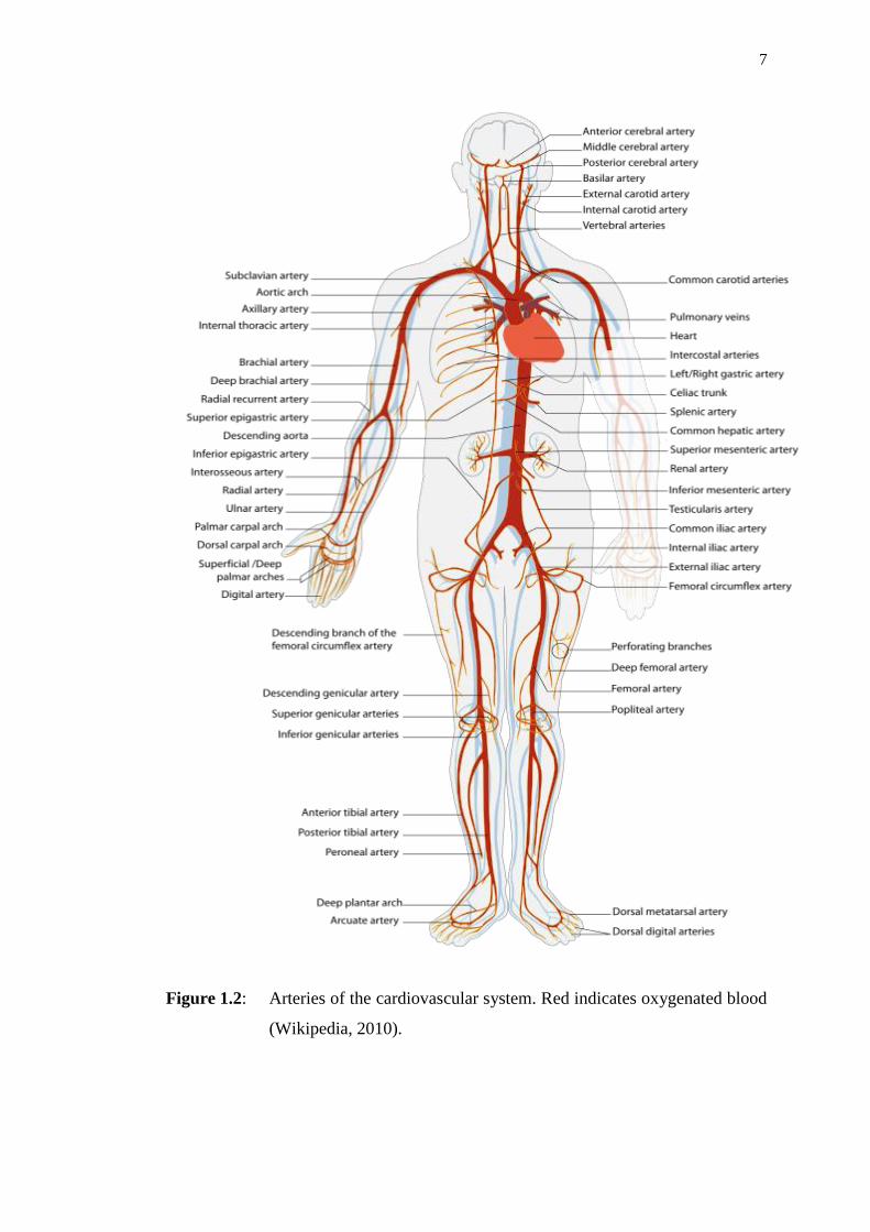
7
Figure 1.2: Arteries of the cardiovascular system. Red indicates oxygenated blood
(Wikipedia, 2010).
8
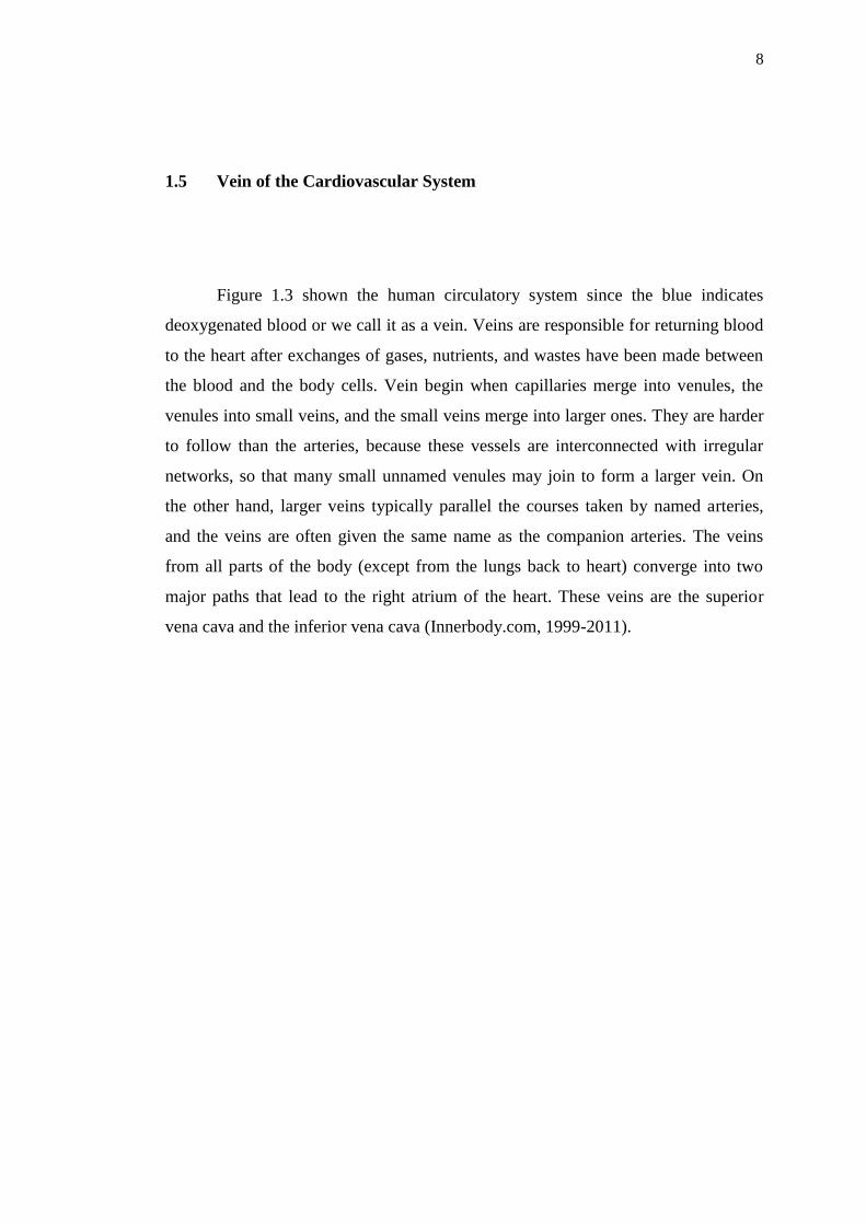
1.5 Vein of the Cardiovascular System
Figure 1.3 shown the human circulatory system since the blue indicates
deoxygenated blood or we call it as a vein. Veins are responsible for returning blood
to the heart after exchanges of gases, nutrients, and wastes have been made between
the blood and the body cells. Vein begin when capillaries merge into venules, the
venules into small veins, and the small veins merge into larger ones. They are harder
to follow than the arteries, because these vessels are interconnected with irregular
networks, so that many small unnamed venules may join to form a larger vein. On
the other hand, larger veins typically parallel the courses taken by named arteries,
and the veins are often given the same name as the companion arteries. The veins
from all parts of the body (except from the lungs back to heart) converge into two
major paths that lead to the right atrium of the heart. These veins are the superior
vena cava and the inferior vena cava (Innerbody.com, 1999-2011).

9
Figure 1.3: Vein of the cardiovascular system. Blue indicates deoxygenated blood
(Wikipedia, 2010).

10
1.6 Blood
Blood is made of four component which are plasma, red blood cells, white
blood cells, and platelets. Plasma is a mixture of water, sugar, fat, protein, potassium,
and calcium salts. It also contains many chemicals that help form blood the clots
necessary to stop bleeding and more than 92% of plasma is water. Red blood cells
contain a special protein called hemoglobin, which carries the oxygen we inhale with
our lungs to all of the parts of our bodies. It then returns carbon dioxides from our
body to our lungs so we can exhale it.
Hemoglobin is also responsible for making red blood cells red. We have so
many red blood cells that our blood itself appears red, even though it contains more
than red blood cells. White blood cells are clear round cells that are bigger than red
blood cells. White blood cells produce proteins called antibodies that help our bodies
fight infections caused by bacteria, viruses, and foreign proteins. Platelets are not
cells, they are just fragments of cells. When we are injured, platelets gather at the site
of the injury and stick to the edges of the wound. They release chemicals that help
start the process of blood clotting so that bleeding will stop.
Blood carries the oxygen that needed around the body for use then returns the
oxygen poor blood back to the lungs to replace the missing oxygen. This process
known as blood flow or blood circulation. Due to complexity of blood rheology, a
mathematical description of blood itself has not yet been completely formulated. In
the systematic circulation, the large vessels are approximated by tubes with thin,
elastic walls, while the blood filling the vessels is considered as a continuum, and
incompressible fluids. According to Rudinger (1970). Blood is known to be an
incompressible viscous fluid in a number of applications such as flow in large blood
vessels. Demiray (2000) studied the contribution of higher order terms to solitary
waves in fluid-filled elastic tubes by employing the modified multiple scale

11
expansion. As far as the biological applications are concerned, in a real life, the
blood is an incompressible and viscous blood.
Blood is also known to be an incompressible non-Newtonian fluid. However,
in the course of flow in arteries, the red cells in the vicinity of arteries move to the
central region of the artery so that the hematocrit ratio becomes quite low near the
arterial wall and the blood viscosity does not change very much with shear rate. Due
to high shear rate near the arterial wall, the viscosity of blood is further reduced.
Therefore, for flow problems in large blood vessels, the blood may be treated as a
Newtonian fluid.
As is well known, it is not easy to deal with the exact equations of motion of
the viscous fluid or also known as Navier-Stokes equation. So, we will make some
simplifying assumptions called hydraulic approximations. By doing this
approximation, it is assumed that the axial velocity is much larger than the radial and
permissible for an averaging procedure with respect to cross-sectional area.
1.7 Cardiac Cycle
According to Guyton and Hall (2006), the cardiac cycle is a term referring to
all or any of the events related to the flow or blood pressure that occurs from the
beginning of one heartbeat to the beginning of the next. The frequency of the cardiac
cycle is described by the heart rate. Each beat of the heart involves five major stages.
The first two stages, often considered together as the “ventricular filling”
stage, involve the movement of blood from the atria into the ventricles. The next
stages involve the movement of blood from the ventricles to the pulmonary artery

12
which is in the case of the right ventricles and the aorta which is in the case of the
left ventricles.
1.8 Blood Pressure
Blood moves through our circulation system because it is under pressure,
caused by the contraction of the heart and by the muscles that surround our blood
vessels. The measure of this force is blood pressure. Blood pressure will always be
highest in the two main arteries, just outside the heart, but, because the pulmonary
circulation is inaccessible, blood pressure is measured in the systemic circulation
only.
According to Razia and Atanu (2013), the blood pressure is the pressure of
blood fluid exerted upon the walls of blood vessels. During each cardiac cycle, the
pressure rises to the maximum level called the systolic blood pressure and as it
relaxes, it falls to the minimum level called the diastolic blood pressure.
Abnormally raised blood pressure is commonly known as hypertension, the
cause of half of all deaths from stroke and heart diseases, worldwide. The World
health statistics (Gupta, 2004) indicates startling finding, i.e., one in three adults
worldwide have hypertension. Cardiovascular diseases are the top most cause of
mortality in India. In India, in year 1990, death due to stroke and 24% of death due to
coronary heart disease in India is linked to hypertension (Gupta and Kasliwal, 2004).

13
1.9 Viscosity
According to Mazumdar (1989), fluids are “sticky” to greater or lesser
extents, and this property is denoted by the term viscosity. Because of viscosity,
when a viscous fluid flows across a wall, the fluid in immediate contact with the wall
is at rest. As we know from watching the flow of streams or rivers, the flow velocity
is greater, the greater is the distance from the riverbank, and it reaches its maximum
in the middle. One reason for this is that there exists a frictional force between
neighboring elements of a viscous fluid. We say that there is a shearing stress
between such elements. Fluids that are not viscous cannot support shearing stresses.
For newtonian fluids, the stress, on a volume element of fluid is proportional to
the rate of deformation or strain rate of volume element, and this constant of
proportionality is called the coefficient of viscosity µ.
The coefficient of viscosity µ in a flowing liquid was defined earlier as the
constant of proportionality between the stress applied τ and the velocity gradient , or
rate of shear, dv dr , of the liquid laminae (Wilmer and Michael, 2005). If the
viscosity is measured in a concentric cylinder viscometer, the rate of shear is
approximately constant throughout the liquid and is measured directly, as is the
applied stress (Cokelet et al., 1963). In order to be able to compare results from the
two methods directly, it is necessary to define stress and rate of shear in terms of the
pressure gradient, the rate of flow and the tube radius. From equation of velocity
gradient which is
1 2.
2
r p pdv
dr L
(1.1)
where v is velocity, r is radius, p is pressure and L is length. We can write

14
1 2
2
r p pdv
dr L
. (1.2)
It is usual in these formulations to use p as the pressure gradient, so that
1 2 /p p p L , and we write
/ 2r p (1.3)
where r is the radius of a given lamina of liquid.
1.10 Newtonian Fluid and Non-Newtonian Fluid
Newton defined the viscosity of a fluid as a lack of slipperiness between the
layers of the fluid, of course, in doing so he implied that there was such a thing as a
“layer of fluid” or “laminae” of fluid and the viscosity arises because of rubbing one
lamina upon the other (Mazumdar, 1989). Suppose we have two laminae, which are
in contact with one another [see Figure 1.4]. Suppose some force F parallel to the x -
axis acts and produces relative motion between the two laminae, such as the top
lamina moves with velocity dv relative to the bottom lamina. Hence, there is a rate
of change of velocity with distance in the y direction (there exists a velocity gradient
dv dy ). It is hypothesized that the force, F is directly proportional to v
Ay
, or

15
,dv
F Ady
(1.4)
where A is the area of contact between the laminae. The proportionality constant is
then defined to be the viscosity of the fluid, and is usually denoted by µ, such as
.dv
F Ady
(1.5)
The dimension of the viscosity is given by
2
2
1.
force MLT M
velocity LT LTLareaLlength
(1.6)
Equation (1.6) is a linear relation, the behavior of the fluid is called
Newtonian. Any fluid for which the relation is non linear is called non-Newtonian.
Figure 1.4: Newtonian concept of viscosity

16
1.11 Is Blood a Newtonian Fluid?
A fluid is said to be Newtonian if the viscous stresses that arise from its flow,
at every point, are proportional to the local strain rate, the rate of change of its
deformation over time (Batchelor, 2000). That is equivalent to saying that those
forces are proportional to the rates of change of the fluid's velocity vector as one
moves away from the point in question in various directions. More precisely, a fluid
is Newtonian if, and only if, the tensors that describe the viscous stress and the strain
rate are related by a constant viscosity tensor that does not depend on the stress state
and velocity of the flow (Kirby, 2010).
Newtonian fluids are the simplest mathematical models of fluids that account
for viscosity. While no real fluid fits the definition perfectly, many common liquids
and gases, such as water and air, can be assumed to be Newtonian for practical
calculations under ordinary conditions.
Poiseuille’s law is also so well established experimentally, that is often used
in order to determine the vicosity coefficient µ of viscous fluids. When blood is
examined in the manner, the viscosity coefficient of blood is found to be about five
times the value for water (µb = 5µw), if the diameter of the tube is relatively large.
Thus at normal physiological temperature of 370C, the viscosity of water µw is
0.007P (P = Poise), and the viscosity of blood µb as determined by Poiseuille’s law in
large tubes is about 0.035P.
The fact that the effective viscosity coefficient of blood according to
Poisuelli’s law depends on the radius of the cube in which it is measured indicates
that blood is not a Newtonian fluid, for which µ is a constant. Rather, blood is said to
behave as non-Newtonian fluid. Fluids, which have elaborate molecular structure, in

17
particular those consisting of long chain molecules, are in general non-Newtonian.
Thus, biological fluids such as cytoplasm can be expected to be non-Newtonian
(Mazumdar, 1989).
In the case of Newtonian fluids
, (1.7)
where τ is the shear stress and γ is shear strain rate. A simple model for non-
Newtonian behavior is the power law model given by
,n (1.8)
where n is the power law index. Non-Newtonian fluids viscosity is not independent
of the applied shear stress. Figure 1.5 shows the relationship between Newtonian and
non-Newtonian fluids.
Most of the biological fluids including blood, lymph and ‘semi fluids’ such as
cytoplasm are in fact non-Newtonian. Atlhough adequate mathematical theory of
such fluids exists at present, its discussion is beyond the scope of this project.
Paterson (1983) in a study of the non-Newtonian behavior of blood observed that the
non-Newtonian fluid flow involved many new features not found in the Newtonian
fluid flow. However for most purposes, blood can be treated theoretically as an
ordinary Newtonian fluid with an appropriate “effective” viscosity coefficient that is
constant.

18
Figure 1.5: Typical shear stress rate relationships for non-Newtonian fluids
(Mazumdar, 1989)
1.12 No-Slip Condition
To solve the governing Navier-Stokes equations and continuity equation,
appropriate boundary and initial conditions must be supplied. Fluid flow is
influenced by the presence of solid boundaries in two manners. First, no fluid can
cross a solid boundary, so that the velocity normal to the surface is zero. Second,
viscous forces make the fluid ‘stick’ to the wall, so that the tangential velocity at the
fluid equal the velocity of the wall that is called the no-slip condition. The tangential
component of boundary velocity UT

19
.T Tu U (1.9)
If a solid surface is not moving, UT = 0, and the no-slip condition becomes
0Tu (Shaughnessy, Katz, and Schaffer, 2005).
1.13 Problem Statement
This study will focus on mathematical modeling of blood blow. Although
blood is the non-Newtonian fluid, in this study will consider the blood as a
Newtonian fluid which is governed by the Navier-Stokes equation for solving blood
flow problems. In this study, we will focus on the derivation of cardiovascular
system equation or called as master equation with the help of continuity equation and
Navier-Stokes equation in order to developed general equation of normal blood flow
and extended normal blood pressure equation. The problems of this study will be
solved using the fluid dynamic assumption assist by differential equation. At the end
of this study, some analysis had been performed to determined the validity of
proposed model by using MAPLE 13.

20
1.14 Objective of the Study
The objectives of the research are:
1. To derive the cardiovascular system equation.
2. To develop the models for blood flow and blood pressure in the body.
3. To analyze the blood flow rate and blood pressure in order to determine
the validity of the proposed model.
1.15 Scopes of the Study
The scope of the research are:
1. Consider the blood flow as Newtonian fluid which governed by Navier-
Stokes and continuity equation.
2. All the vessels are assumed to be same in nature excluding their size,
length and cross-sectional area.
3. Blood has both the radial and axial flow in only one direction, which is z-
direction in a three-dimensional system.
4. Cross-sectional area of blood vessels is constant over time and distance.
5. Pressure gradient is constant over distance.
21
1.16 Significance of the Study
The results of this study will give benefits to the fields of mathematics,
physics, engineering, biology and medical. This research will lead to further
investigation in the mathematical modeling of blood flow theory and method which
is frequently used in mathematics, physics, and mostly in medical field. Besides that,
the result of the research can be a guide line to determine the cardiovascular system
equation, blood flow rate equation and normal blood pressure equation in getting the
strong solution for blood flow problems. This investigation also gives a chance for
readers to know more about benefits of this equations model in solving blood flow
problems.
1.17 Outline of Dissertation
This report divided to five chapters. Chapter 1 is the introduction of the study,
some basic concept of blood flow theory, problem statement, objective of the study,
scopes of the study, significance of the study, and outline of thesis in this report.
Chapter 2 is literature review, will include some introduction and will be
depicts several of applications and contributions that have been done by some
researchers for blood flow problems. Moreover, some introduction of the Navier-
Stokes equation, continuity equation, and poisuelli’s equation will also include in this
chapter.

22
Chapter 3 is about the research methodology of this project. This will include
the derivation of the governing differential equations. In this chapter, we use the
knowledge on the previous chapter to derive differential equations of Navier-Stokes
equation. Next, i will discuss and derive in detail the Navier-Stokes equation in
Cartesian coordinates and Cylindrical coordinate that will be used to solve the
Newtonian fluid of blood flow problem. The problem of steady flow in a Cylindrical
coordinate is known as Poisuelli’s flow. The solution of the Poisuelli’s flow problem
will the produce Poisuelli’s law. The poisuelli’s equation is then applied to develop
the mathematical model of the blood pressure from cardiovascular system equation.
Beside that, the concept of boundary conditions for axial velocity component,
, ,w z t and derivation of continuity equation will also be discussed in this chapter.
Chapter 4 is about results and discussion of this project. I will derive in detail
and solve the mathematical modeling of blood flow from Rahman and Haque (2012).
Moreover, the analisis of blood flow rate and blood pressure for determine the
validity of the proposed model will also be discussed in this chapter.
Finally, the last chapter of this report is conclusion and further research. This
chapter makes a conclusion to the whole investigation and a suggestion for further
research on mathematical modeling of blood flow problem.
1.18 Summary
In this chapter, we discuss about the introduction of this study. We also
explained about some basic concept of blood flow theory. The objective and scopes
of the study is being defined as a guideline of the research. The details of the study
will be discussed in following chapter.
100
REFERENCES
Acheson, D. J. (1990). Elementary Fluid Dynamics. Oxford Applied Mathematics
and Computing Science Series. Oxford University Press.
Batchelor, G. K. (1967). An Introduction to Fluid Dynamics. Cambridge University
Press.
Batchelor, G. K. (2000). An Introduction to Fluid Dynamics. Cambridge
Mathematical Library Series. Cambridge University Press.
Bender, E. A. (2000). An Introduction to Mathematical Modeling. New York: Dover.
Campbell, N. A., Reece, J. B. (2005). Biology. 7th
Edition. Pearson Education, Inc.
Clancy, L. J. (1975). Aerodynamics, Section 3.3. Pitman Publishing Limited,
London.
Cokelet, G. R. (1963). The Rheology of Human Blood Measurement Near and at
Zero Shear Rate. Transaction of the Society Rheology 7, 303-17.
Ehrlish, A., Schroeder, C. L. (2004). “Medical Terminology for Health Professions”.
5th
Edition. Thomson Delman Learning. 131-132.
Faris, O., Evans, F., Ennis, D., Helm, D., Taylor, J., Chesnick, A., Guttman, M.,
Ozturk, C., McVeigh, E. (2003). “Novel Technique for Cardiac
Electromechanical Mapping with Magnetic Resonance Imaging Tagging and
An Epicardial Electrode Sock”. Ann. Biomed. Eng. Vol 31, No. 4. 430-440.
Fieldman, J. S., Phong, D. H., Aubin, Y. S., Vinet, L. (2007). “Rheology”. Biology
and Mechanics of Blood Flows, Part II: Mechanics and Medical Aspects.
Springer. 119-123.

101
Gerard, J. T., Bryan, D. (2012). “The Cardiovascular System: The Blood”. Principles
of Anatomy & Physiology. 13th. John Wiley & Sons.
Gupta, R. (2004). Trends in Hypertension Epidemiology in India. Journal of Human
Hypertension 18: 73-78.
Gupta, R., Kasliwal, R. R. (2004). Understanding Systolic Hypertension in the
Elderly. JAPI 52: 479-485.
Guyton, A. C., Hall, J. E. (2006). Textbook of Medical Physiology. 11th
Edition
Philadelphia: Elsevier Saunder.
Hinghofer- Szalkay, H. G. and Greenleaf, J. E., (1987). “Continuous Monitoring of
Blood Volume Changes in Humans”. Journal of Applied Physiology. Vol. 63:
1003-1007.
Imaeda, K., Goodman, F. O. (1980). “Analysis of Nonlinear Pulsatile Blood Flow in
Arteries”. Journal of Biomechanics 13. 1007-1022.
Innerbody. Com. (1999-2011). Arteries. How To Media, Inc.
Jennifer, S. (2009). Physiological Fluid Mechanics. New York: Oxford University
Press.
Kilner, P., Yang, G., Wilkes, A., Mohiaddin, R. (2000). “Asymmetric Redirection of
Flow Through The Heart”. Nature. Vol. 404. 759-761.
Kirby, B. J. (2010). Micro and Nanoscale Fluid Mechanics: Transport in
Microfluidic Devices. Cambridge University Press.
Lauralee Sherwood. (2005). “Fundamentals of Physiology: a human perspective”.
3rd Edition. Thomson Brooks. 276.
Liu, C. H., Niranjan, S. C., Clark, J. W., San, K. Y., Zwischenberger, J. B., Bidani,
A. (1998). “Airway Mechanics, Gas Exchange, and Blood Flow In a
Nonlinear Model of The Normal Human Lung”. Journal of Applied
Physiology 84: 1447-1469.

102
L. Cromwell, Fred, J. Weibell, and Erich, A. Pfeiffer. (2004). “Biomedical
Instrumentation and Measurement”. 2nd
Edition. Pearson Education:
Singapore. 84-95.
Masood, S., Yang, G., Pennell, D., Firmin, D. (2000). “Investigating Intrinsic
Myocardial Mechanics: The Role of MR Tagging, Velocity Phase Mapping
and Diffusion Imaging”, J. Magn. Reson. Imag. Vol. 12, no. 6. 873-883.
Massey, B. S. (2006). Mechanics of Fluids. 8th
Edition. London & New York: Taylor
& Francis.
Mazumdar, J. (1989). An Introduction to Mathematical Physiology and Biology. New
York: Cambridge University Press.
Paterson, A. R. (1983). A First Course in Fluid Dynamics. New York: Cambridge
University Press.
Pedlosky, Joseph (1987). Geophysical fluid dynamics. Springer. pp.10-13. ISBN
978-0-387-96387-7.
Pfitzner, J. (1976). “Poisuelli’s an his law”. Anaesthesia 31 (2). pp. 273-5.
Rahman, M. S., Haque, M. A., Asaduzzaman, M. (2010). “Mathematical Modeling
of the Heart”. IEEE International Conference on Electrical and Computer
Engineering.626-629.
Rahman, M. S. (2011). “Computational Design of Cardiac Activity”. International
Journal of Medicine and Medical Sciences. Vol. 3(10). 321-330.
Rahman, M. S., Haque, M. A. (2012). Mathematical Modeling of Blood Flow. IEEE
International Conference on Informatics, Electronics & Vision. 672-676.
Razia, S., Atanu, K. P. (2013). “Blood Pressure and Heart Rate Variability and
Diagnosis”. Review Article. Biological Rhythm Research.
Rolf H. Sabersky, Allan J. Acosta, Eward G. Hauptmann. (1989). Fluid Flow, a First
Course In Fluid Mechanics, 3rd
Edition. Macmillian Publishing Company, a
division of Macmillian, Inc New York, United State.

103
Shaughnessy. E. J., Katz, L. M., Schaffer, J. P. (2005). Introduction to Fluid
Mechanics. New York: Oxford University Press.
Sermesant, M., Delingette, H., and Ayache, N. (2006). “An Electromechanical Model
of The Heart for Image Analysis and Simulation”. IEEE Trans. Med. Imag.
Vol. 25: 612-625.
Wikipedia (2010). Cardiovascular System. Wkimedia Foundation, Inc.
Wilmer, W. N., Michael, F. O. (2005). McDonald’s Blood Flow In Arteries. 5th
Edition. London & New York: Hodder Arnold.
Yamaguchi, H. (2008). “Engineering Fluid Mechanics”. Springer.