a novel cold atmospheric pressure air plasma jet for … · a novel cold atmospheric pressure air...
TRANSCRIPT
INTRODUCTION
In recent years, with continuing development and extensive application of dental implant technology, peri-implantitis has become an increasingly frequent concern in dental clinics. Peri-implantitis is a term for inflammatory reactions with loss of supporting bone in surrounding tissues and is a risk factor for implant loss. According to the available clinical data, the incidence of peri-implantitis varies between 28 and 56%1). Thus, peri-implant diseases are unarguably one of the most significant risks for implant failure. As known to all, normal microbial flora of oral cavity is composed of over 700 species of bacteria, and good oral hygiene helps control plaque formation in the mouth2). Moreover, plaque formation is closely related to peri-implantitis. A previous study reported that bacterial colonization occurred within 30 min after the implant placement3). Therefore, poor oral hygiene was considered as a primary risk factor for peri-implantitis4). Of note, Porphyromonas gingivalis appears to be the most significant cause of peri-implantitis5).
At present, the primary techniques used for the treatment of peri-implantitis include surgery, chemotherapy and laser therapy6). Surgery is the most commonly used method to treat peri-implantitis worldwide. Membrane-guided tissue regeneration (MGTR) and bone grafting surgery have achieved good clinical treatment effect. Although surgical treatment
can restore the bone defect caused by inflammation and various kinds of tissue damage, it is likely to cause major trauma and can easily cause postoperative infections. Moreover, systemic drug therapy for the treatment of peri-implantitis will inevitably produce toxic side effects and adverse reactions in the body7,8). Currently, the ability of lasers to kill bacteria makes them highly useful for various dental procedures, such as killing pathogenic bacteria and stimulating bone formation. However, considering the increase in the temperature caused by laser therapy, additional safety evaluation experiments are warranted9). Furthermore, it is an urgent need to identify an effective and safe sterilization technology for peri-implantitis management without any toxic side effects.
In recent years, atmospheric pressure cold plasma technologies, with room-temperature and active chemical properties, have received increasing attention in the field of biomedical applications10,11).
At present, atmospheric pressure cold plasma has demonstrated promising performance in sterilization, blood clotting, skin disease treatment, cancer treatment and oral medicine12-18). In particular, in the area of oral medicine, atmospheric pressure cold plasma has been used for treatment of dental caries, periodontal disease, oral mucosal disease and teeth whitening19). It is known that atmospheric pressure cold plasma includes various agents such as active free radicals (reactive oxygen and nitrogen species [RONS]), ions and ultraviolet (UV). Such agents can interact with the cells and help in inactivation of bacterial and certain physiological
A novel cold atmospheric pressure air plasma jet for peri-implantitis treatment: An in vitro studyYu YANG1, Jinsong GUO2, Xuan ZHOU1, Zhiqiang LIU1, Chenbao WANG1, Kaile WANG3, Jue ZHANG2,3 and Zuomin WANG1
1 Department of Stomatology, Beijing Chao-Yang Hospital, Capital Medical University, Beijing, 100020, China2 College of Engineering, Peking University, Beijing, 100871, China3 Academy for Advanced Interdisciplinary Studies, Peking University, Beijing, 100871, ChinaCorresponding authors, Zuomin WANG; E-mail: [email protected], Jue ZHANG; E-mail: [email protected]
Peri-implantitis is difficult to treat in clinical settings; this is not only because it is a site-specific infectious disease but also because it impedes osseointegration. In this study, a novel cold atmospheric pressure air plasma jet (CAPAJ) was applied to study the treatment of peri-implantitis in vitro. CAPAJ treated the samples for 2, 4 and 6 min, respectively. To evaluate the titanium surface characteristics, the surface elemental composition (X-ray photoelectron spectroscopy [XPS]), roughness and hydrophilicity were evaluated in each group. Concurrently, the sterilization and osseointegration effect of CAPAJ were also examined. Results revealed that after CAPAJ modification, roughness and hydrophilicity of titanium surfaces were significantly increased. Moreover, XPS results demonstrated that the C1s peak was reduced and N1s and O1s peaks were obviously improved. More importantly, CAPAJ showed favorable sterilization and bone formation effects. CAPAJ seemed a simpler and more efficient strategy for the peri-implantitis treatment.
Keywords: Cold atmospheric pressure air plasma (CAPAJ), Optical emission spectroscopy (OES), Surface modification, Sterilization, Osseointegration
Color figures can be viewed in the online issue, which is avail-able at J-STAGE.Received Feb 2, 2017: Accepted May 18, 2017doi:10.4012/dmj.2017-030 JOI JST.JSTAGE/dmj/2017-030
Dental Materials Journal 2017; : –
characteristic changes. Of note, the dose of these agents is constrained by structural characteristics, electrical parameters and the working gas of various atmospheric pressure cold plasma jets20,21). Furthermore, various types of atmospheric pressure plasma jets have emerged22,23), such as traditional planar and cylindrical dielectric barrier discharges (DBDs) jets24,25) and capillary plasma electrode discharge devices26,27). Thus, different atmospheric pressure cold plasma jets appear to have different effects on their biomedical applications.
Specifically, certain studies have reported that cold atmospheric pressure plasma may have potential applications in peri-implantitis treatment28,29). In particular, these researches showed that rare gas plasma devices could damage the membranes of microorganisms and promote osseointegration. Clearly, air, containing abundance of O2 and N2, is freely available, and it has been strongly expected to excite atmospheric pressure cold plasma. However, both O2 and N2 are diatomic molecule gases, whose rotation and vibration loss channels will be offered by the electron energy, and oxygen has a high electronegativity30). Therefore, compared with noble gas plasma jets, only certain types of air plasma jets have been presented31-33). Of note, limited relevant research is available focusing on the use of air plasma jets in peri-implantitis treatment.
In this study, we conducted an independent research and developed an equipment of cold atmospheric pressure air plasma34). Optical emission spectroscopy (OES) was used to detect the major excited reactive species in atmospheric pressure cold plasma. In addition, we evaluated the surface elemental composition, roughness and hydrophilicity in each group. Subsequently, sterilisation effects of cold atmospheric pressure air plasma jet (CAPAJ) on Porphyromonas gingivalis and cell relative growth rate were also studied. Last, we discussed the potential mechanism of CAPAJ action in peri-implantitis treatment.
MATERIALS AND METHODS
Specimen preparationCommercially pure titanium TA2 (Northwest Institute For Non-ferrous Metal Research, Xi’an, China) was used for research purposes, prepared with a diameter of 10 mm and a thickness of 2.0 mm. The chemical compositions (wt%) of TA2 include Fe≤0.30, C≤0.10, N≤0.05, H≤0.015, O≤0.25 and the rest is Ti. Mechanical polishing was performed to even out the surface, and the specimens were ultrasonically cleaned in ethanol, in acetone and in distilled water for 20 min each. Subsequently, the titanium TA2 specimens were sterilised in an autoclave at 121°C under one atmos-phere of pressure (1 ATM) for about 20 min. After sterilization, the specimens were air cooled to room temperature and placed in the oven to dry before being served.
Cell and bacteriaThe human osteoblast-like cell line MG63 (Chinese
Academy of Medical Sciences, 3111C0001CCC000080) and mouse embryonic fibroblast cell line MC3T3-E1 (Chinese Academy of Medical Sciences, 3111C0001CCC000012) were maintained in Dulbecco’s minimum essential medium (DMEM), supplemented with 10% fetal bovine serum (FBS). Subsequently, the cells were cultured in an incubator at 37°C with 5%CO2. The cells were sub-cultured using 0.25% trypsin-EDTA when they were 85–90% confluent. All experiments were performed within 2–3 passages after cell revival from cryopreservation. Cells were harvested and adjusted to a concentration of 1.5×10⁶/mL.
Porphyromonas gingivalis (ATCC 33277) was purchased from American Type Culture Collection (ATCC). Cells were maintained on enriched trypticase soy agar plates containing 3% sheep blood and grown in brain heart infusion (BHI) broth (BD Diagnostics). Bacteria were cultured anaerobically (85% N2, 10% H2 and 5% CO2) and grown aerobically at 37°C until log-phase growth. Subsequently, bacterial concentrations were adjusted to 0.5 McFarland turbidity standards (equivalent to 1.5×10⁸ colony forming units/mL) to perform the experiments described below. The cultivation of Porphyromonas gingival has been described in detail elsewhere35,36).
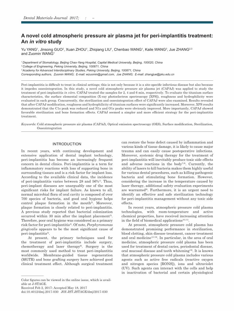
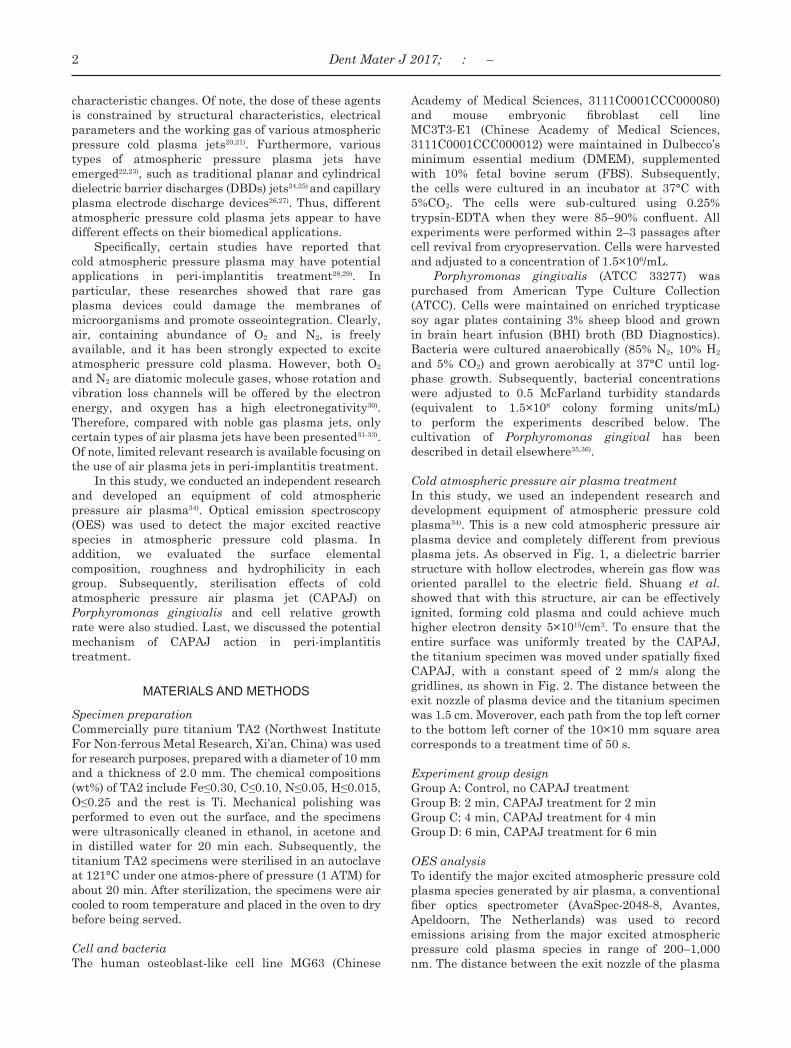
Cold atmospheric pressure air plasma treatmentIn this study, we used an independent research and development equipment of atmospheric pressure cold plasma34). This is a new cold atmospheric pressure air plasma device and completely different from previous plasma jets. As observed in Fig. 1, a dielectric barrier structure with hollow electrodes, wherein gas flow was oriented parallel to the electric field. Shuang et al. showed that with this structure, air can be effectively ignited, forming cold plasma and could achieve much higher electron density 5×10¹⁵/cm3. To ensure that the entire surface was uniformly treated by the CAPAJ, the titanium specimen was moved under spatially fixed CAPAJ, with a constant speed of 2 mm/s along the gridlines, as shown in Fig. 2. The distance between the exit nozzle of plasma device and the titanium specimen was 1.5 cm. Moverover, each path from the top left corner to the bottom left corner of the 10×10 mm square area corresponds to a treatment time of 50 s.
Experiment group designGroup A: Control, no CAPAJ treatmentGroup B: 2 min, CAPAJ treatment for 2 minGroup C: 4 min, CAPAJ treatment for 4 minGroup D: 6 min, CAPAJ treatment for 6 min
OES analysisTo identify the major excited atmospheric pressure cold plasma species generated by air plasma, a conventional fiber optics spectrometer (AvaSpec-2048-8, Avantes, Apeldoorn, The Netherlands) was used to record emissions arising from the major excited atmospheric pressure cold plasma species in range of 200–1,000 nm. The distance between the exit nozzle of the plasma
2 Dent Mater J 2017; : –
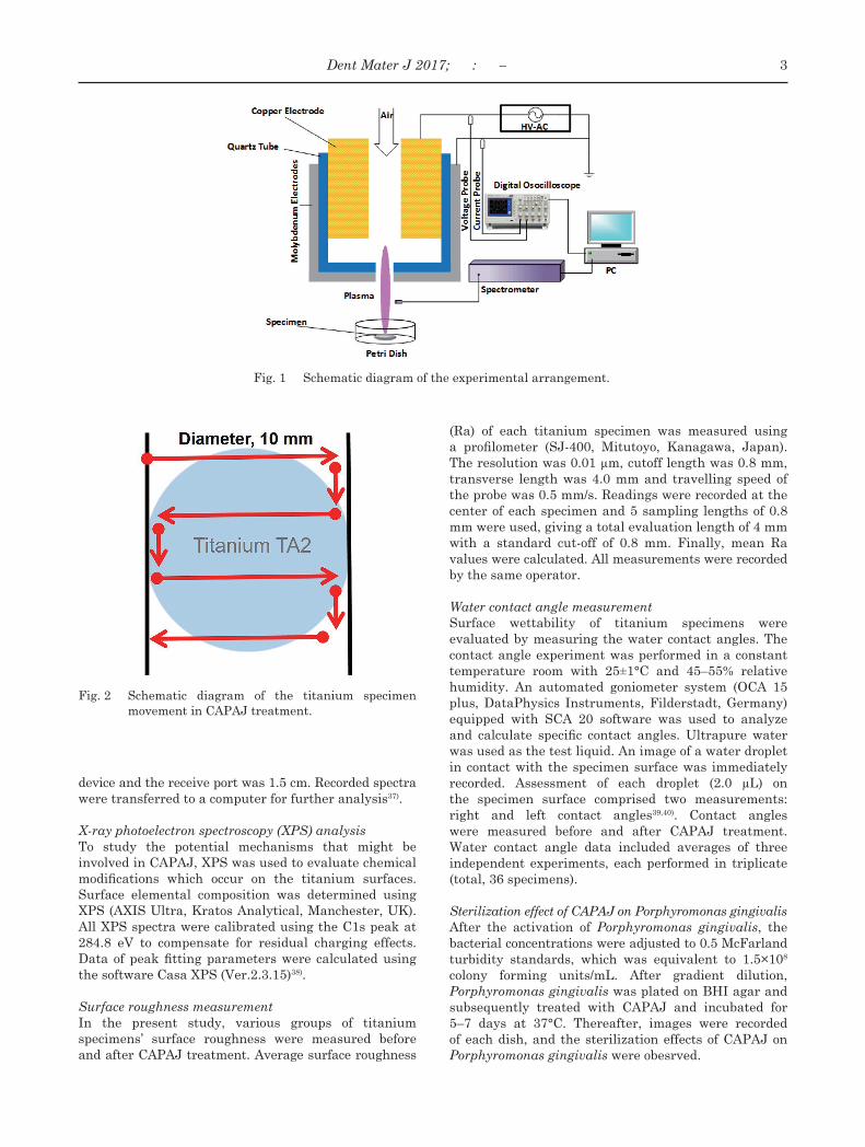
Fig. 1 Schematic diagram of the experimental arrangement.
Fig. 2 Schematic diagram of the titanium specimen movement in CAPAJ treatment.
device and the receive port was 1.5 cm. Recorded spectra were transferred to a computer for further analysis37).
X-ray photoelectron spectroscopy (XPS) analysisTo study the potential mechanisms that might be involved in CAPAJ, XPS was used to evaluate chemical modifications which occur on the titanium surfaces. Surface elemental composition was determined using XPS (AXIS Ultra, Kratos Analytical, Manchester, UK). All XPS spectra were calibrated using the C1s peak at 284.8 eV to compensate for residual charging effects. Data of peak fitting parameters were calculated using the software Casa XPS (Ver.2.3.15)38).
Surface roughness measurementIn the present study, various groups of titanium specimens’ surface roughness were measured before and after CAPAJ treatment. Average surface roughness
(Ra) of each titanium specimen was measured using a profilometer (SJ-400, Mitutoyo, Kanagawa, Japan). The resolution was 0.01 µm, cutoff length was 0.8 mm, transverse length was 4.0 mm and travelling speed of the probe was 0.5 mm/s. Readings were recorded at the center of each specimen and 5 sampling lengths of 0.8 mm were used, giving a total evaluation length of 4 mm with a standard cut-off of 0.8 mm. Finally, mean Ra values were calculated. All measurements were recorded by the same operator.
Water contact angle measurementSurface wettability of titanium specimens were evaluated by measuring the water contact angles. The contact angle experiment was performed in a constant temperature room with 25±1°C and 45–55% relative humidity. An automated goniometer system (OCA 15 plus, DataPhysics Instruments, Filderstadt, Germany) equipped with SCA 20 software was used to analyze and calculate specific contact angles. Ultrapure water was used as the test liquid. An image of a water droplet in contact with the specimen surface was immediately recorded. Assessment of each droplet (2.0 µL) on the specimen surface comprised two measurements: right and left contact angles39,40). Contact angles were measured before and after CAPAJ treatment. Water contact angle data included averages of three independent experiments, each performed in triplicate (total, 36 specimens).
Sterilization effect of CAPAJ on Porphyromonas gingivalisAfter the activation of Porphyromonas gingivalis, the bacterial concentrations were adjusted to 0.5 McFarland turbidity standards, which was equivalent to 1.5×10⁸ colony forming units/mL. After gradient dilution, Porphyromonas gingivalis was plated on BHI agar and subsequently treated with CAPAJ and incubated for 5–7 days at 37°C. Thereafter, images were recorded of each dish, and the sterilization effects of CAPAJ on Porphyromonas gingivalis were obesrved.
3Dent Mater J 2017; : –
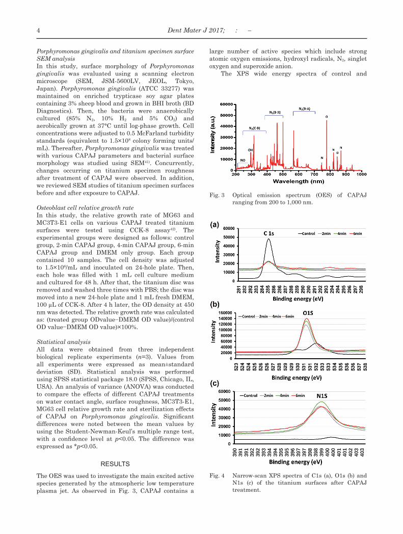
Fig. 3 Optical emission spectrum (OES) of CAPAJ ranging from 200 to 1,000 nm.
Fig. 4 Narrow-scan XPS spectra of C1s (a), O1s (b) and N1s (c) of the titanium surfaces after CAPAJ treatment.
Porphyromonas gingivalis and titanium specimen surface SEM analysisIn this study, surface morphology of Porphyromonas gingivalis was evaluated using a scanning electron microscope (SEM, JSM-5600LV, JEOL, Tokyo, Japan). Porphyromonas gingivalis (ATCC 33277) was maintained on enriched trypticase soy agar plates containing 3% sheep blood and grown in BHI broth (BD Diagnostics). Then, the bacteria were anaerobically cultured (85% N2, 10% H2 and 5% CO2) and aerobically grown at 37°C until log-phase growth. Cell concentrations were adjusted to 0.5 McFarland turbidity standards (equivalent to 1.5×10⁸ colony forming units/mL). Thereafter, Porphyromonas gingivalis was treated with various CAPAJ parameters and bacterial surface morphology was studied using SEM41). Concurrently, changes occurring on titanium specimen roughness after treatment of CAPAJ were observed. In addition, we reviewed SEM studies of titanium specimen surfaces before and after exposure to CAPAJ.
Osteoblast cell relative growth rateIn this study, the relative growth rate of MG63 and MC3T3-E1 cells on various CAPAJ treated titanium surfaces were tested using CCK-8 assay42). The experimental groups were designed as follows: control group, 2-min CAPAJ group, 4-min CAPAJ group, 6-min CAPAJ group and DMEM only group. Each group contained 10 samples. The cell density was adjusted to 1.5×10⁶/mL and inoculated on 24-hole plate. Then, each hole was filled with 1 mL cell culture medium and cultured for 48 h. After that, the titanium disc was removed and washed three times with PBS; the disc was moved into a new 24-hole plate and 1 mL fresh DMEM, 100 µL of CCK-8. After 4 h later, the OD density at 450 nm was detected. The relative growth rate was calculated as: (treated group ODvalue−DMEM OD value)/(control OD value−DMEM OD value)×100%.
Statistical analysisAll data were obtained from three independent biological replicate experiments (n=3). Values from all experiments were expressed as mean±standard deviation (SD). Statistical analysis was performed using SPSS statistical package 18.0 (SPSS, Chicago, IL, USA). An analysis of variance (ANOVA) was conducted to compare the effects of different CAPAJ treatments on water contact angle, surface roughness, MC3T3-E1, MG63 cell relative growth rate and sterilization effects of CAPAJ on Porphyromonas gingivalis. Significant differences were noted between the mean values by using the Student-Newman-Keul’s multiple range test, with a confidence level at p<0.05. The difference was expressed as *p<0.05.
RESULTS
The OES was used to investigate the main excited active species generated by the atmospheric low temperature plasma jet. As observed in Fig. 3, CAPAJ contains a
large number of active species which include strong atomic oxygen emissions, hydroxyl radicals, N2, singlet oxygen and superoxide anion.
The XPS wide energy spectra of control and
4 Dent Mater J 2017; : –
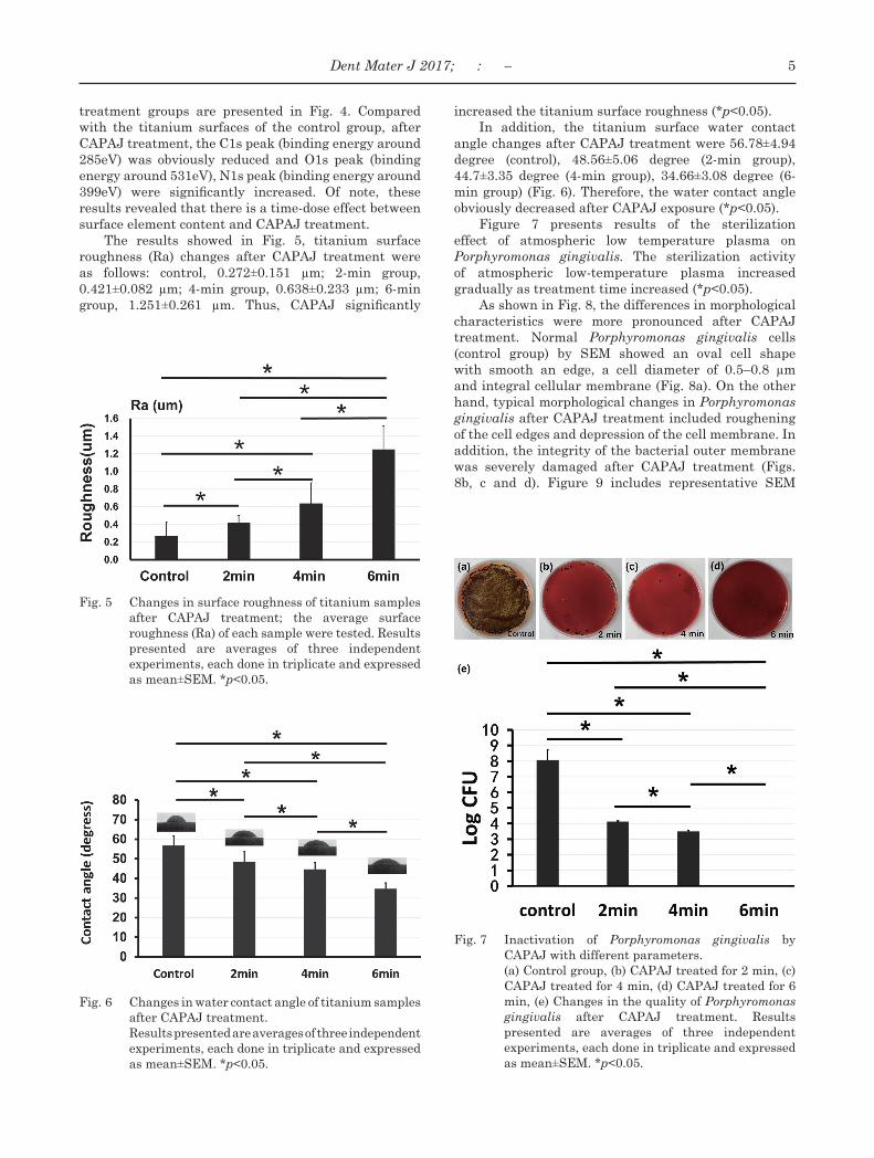
Fig. 5 Changes in surface roughness of titanium samples after CAPAJ treatment; the average surface roughness (Ra) of each sample were tested. Results presented are averages of three independent experiments, each done in triplicate and expressed as mean±SEM. *p<0.05.
Fig. 6 Changes in water contact angle of titanium samples after CAPAJ treatment.
Results presented are averages of three independent experiments, each done in triplicate and expressed as mean±SEM. *p<0.05.
Fig. 7 Inactivation of Porphyromonas gingivalis by CAPAJ with different parameters.
(a) Control group, (b) CAPAJ treated for 2 min, (c) CAPAJ treated for 4 min, (d) CAPAJ treated for 6 min, (e) Changes in the quality of Porphyromonas gingivalis after CAPAJ treatment. Results presented are averages of three independent experiments, each done in triplicate and expressed as mean±SEM. *p<0.05.
treatment groups are presented in Fig. 4. Compared with the titanium surfaces of the control group, after CAPAJ treatment, the C1s peak (binding energy around 285eV) was obviously reduced and O1s peak (binding energy around 531eV), N1s peak (binding energy around 399eV) were significantly increased. Of note, these results revealed that there is a time-dose effect between surface element content and CAPAJ treatment.
The results showed in Fig. 5, titanium surface roughness (Ra) changes after CAPAJ treatment were as follows: control, 0.272±0.151 µm; 2-min group, 0.421±0.082 µm; 4-min group, 0.638±0.233 µm; 6-min group, 1.251±0.261 µm. Thus, CAPAJ significantly
increased the titanium surface roughness (*p<0.05).In addition, the titanium surface water contact
angle changes after CAPAJ treatment were 56.78±4.94 degree (control), 48.56±5.06 degree (2-min group), 44.7±3.35 degree (4-min group), 34.66±3.08 degree (6-min group) (Fig. 6). Therefore, the water contact angle obviously decreased after CAPAJ exposure (*p<0.05).
Figure 7 presents results of the sterilization effect of atmospheric low temperature plasma on Porphyromonas gingivalis. The sterilization activity of atmospheric low-temperature plasma increased gradually as treatment time increased (*p<0.05).
As shown in Fig. 8, the differences in morphological characteristics were more pronounced after CAPAJ treatment. Normal Porphyromonas gingivalis cells (control group) by SEM showed an oval cell shape with smooth an edge, a cell diameter of 0.5–0.8 µm and integral cellular membrane (Fig. 8a). On the other hand, typical morphological changes in Porphyromonas gingivalis after CAPAJ treatment included roughening of the cell edges and depression of the cell membrane. In addition, the integrity of the bacterial outer membrane was severely damaged after CAPAJ treatment (Figs. 8b, c and d). Figure 9 includes representative SEM
5Dent Mater J 2017; : –
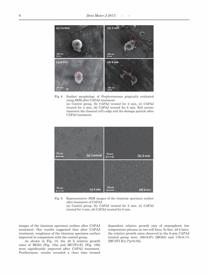
Fig. 8 Surface morphology of Porphyromonas gingivalis evaluated using SEM after CAPAJ treatment.
(a) Control group, (b) CAPAJ treated for 2 min, (c) CAPAJ treated for 4 min, (d) CAPAJ treated for 6 min. Red arrows represent the classical cell’s edge and the damage particle after CAPAJ treatment.
Fig. 9 Representative SEM images of the titanium specimen surface after treatment of CAPAJ.
(a) Control group, (b) CAPAJ treated for 2 min, (c) CAPAJ treated for 4 min, (d) CAPAJ treated for 6 min.
images of the titanium specimen surface after CAPAJ treatment. Our results suggested that after CAPAJ treatment, roughness of the titanium specimen surface improved in comparison with the control group.
As shown in Fig. 10, the 48 h relative growth rates of MG63 (Fig. 10a) and MC3T3-E1 (Fig. 10b) were significantly improved after CAPAJ treatment. Furthermore, results revealed a clear time treated
dependent relative growth rate of atmospheric low temperature plasma on two cell lines. In fact, 48 h later, the relative growth rates observed in the 6-min CAPAJ treated group were 168±9.6% (MG63) and 179±6.1% (MC3T3-E1) (*p<0.05).
6 Dent Mater J 2017; : –
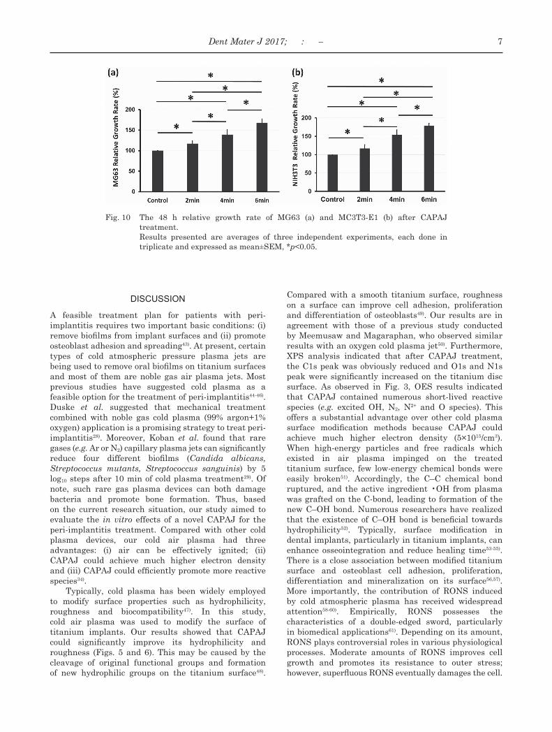
Fig. 10 The 48 h relative growth rate of MG63 (a) and MC3T3-E1 (b) after CAPAJ treatment.
Results presented are averages of three independent experiments, each done in triplicate and expressed as mean±SEM, *p<0.05.
DISCUSSION
A feasible treatment plan for patients with peri-implantitis requires two important basic conditions: (i) remove biofilms from implant surfaces and (ii) promote osteoblast adhesion and spreading43). At present, certain types of cold atmospheric pressure plasma jets are being used to remove oral biofilms on titanium surfaces and most of them are noble gas air plasma jets. Most previous studies have suggested cold plasma as a feasible option for the treatment of peri-implantitis44-46). Duske et al. suggested that mechanical treatment combined with noble gas cold plasma (99% argon+1% oxygen) application is a promising strategy to treat peri-implantitis28). Moreover, Koban et al. found that rare gases (e.g. Ar or N2) capillary plasma jets can significantly reduce four different biofilms (Candida albicans, Streptococcus mutants, Streptococcus sanguinis) by 5 log10 steps after 10 min of cold plasma treatment29). Of note, such rare gas plasma devices can both damage bacteria and promote bone formation. Thus, based on the current research situation, our study aimed to evaluate the in vitro effects of a novel CAPAJ for the peri-implantitis treatment. Compared with other cold plasma devices, our cold air plasma had three advantages: (i) air can be effectively ignited; (ii) CAPAJ could achieve much higher electron density and (iii) CAPAJ could efficiently promote more reactive species34).
Typically, cold plasma has been widely employed to modify surface properties such as hydrophilicity, roughness and biocompatibility47). In this study, cold air plasma was used to modify the surface of titanium implants. Our results showed that CAPAJ could significantly improve its hydrophilicity and roughness (Figs. 5 and 6). This may be caused by the cleavage of original functional groups and formation of new hydrophilic groups on the titanium surface48).
Compared with a smooth titanium surface, roughness on a surface can improve cell adhesion, proliferation and differentiation of osteoblasts49). Our results are in agreement with those of a previous study conducted by Meemusaw and Magaraphan, who observed similar results with an oxygen cold plasma jet50). Furthermore, XPS analysis indicated that after CAPAJ treatment, the C1s peak was obviously reduced and O1s and N1s peak were significantly increased on the titanium disc surface. As observed in Fig. 3, OES results indicated that CAPAJ contained numerous short-lived reactive species (e.g. excited OH, N2, N2+ and O species). This offers a substantial advantage over other cold plasma surface modification methods because CAPAJ could achieve much higher electron density (5×10¹⁵/cm3). When high-energy particles and free radicals which existed in air plasma impinged on the treated titanium surface, few low-energy chemical bonds were easily broken51). Accordingly, the C–C chemical bond ruptured, and the active ingredient •OH from plasma was grafted on the C-bond, leading to formation of the new C–OH bond. Numerous researchers have realized that the existence of C–OH bond is beneficial towards hydrophilicity52). Typically, surface modification in dental implants, particularly in titanium implants, can enhance osseointegration and reduce healing time53-55). There is a close association between modified titanium surface and osteoblast cell adhesion, proliferation, differentiation and mineralization on its surface56,57). More importantly, the contribution of RONS induced by cold atmospheric plasma has received widespread attention58-60). Empirically, RONS possesses the characteristics of a double-edged sword, particularly in biomedical applications61). Depending on its amount, RONS plays controversial roles in various physiological processes. Moderate amounts of RONS improves cell growth and promotes its resistance to outer stress; however, superfluous RONS eventually damages the cell.
7Dent Mater J 2017; : –
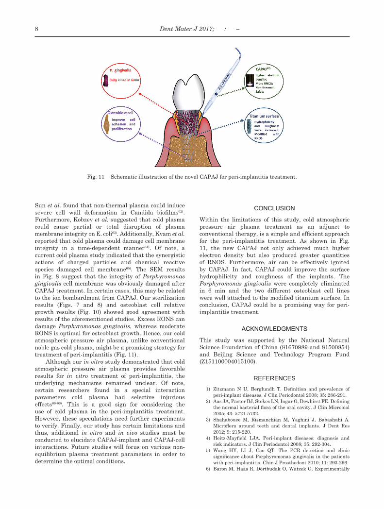
Fig. 11 Schematic illustration of the novel CAPAJ for peri-implantitis treatment.
Sun et al. found that non-thermal plasma could induce severe cell wall deformation in Candida biofilms62). Furthermore, Kobzev et al. suggested that cold plasma could cause partial or total disruption of plasma membrane integrity on E. coli63). Additionally, Kvam et al. reported that cold plasma could damage cell membrane integrity in a time-dependent manner64). Of note, a current cold plasma study indicated that the synergistic actions of charged particles and chemical reactive species damaged cell membrane65). The SEM results in Fig. 8 suggest that the integrity of Porphyromonas gingivalis cell membrane was obviously damaged after CAPAJ treatment. In certain cases, this may be related to the ion bombardment from CAPAJ. Our sterilization results (Figs. 7 and 8) and osteoblast cell relative growth results (Fig. 10) showed good agreement with results of the aforementioned studies. Excess RONS can damage Porphyromonas gingivalis, whereas moderate RONS is optimal for osteoblast growth. Hence, our cold atmospheric pressure air plasma, unlike conventional noble gas cold plasma, might be a promising strategy for treatment of peri-implantitis (Fig. 11).
Although our in vitro study demonstrated that cold atmospheric pressure air plasma provides favorable results for in vitro treatment of peri-implantitis, the underlying mechanisms remained unclear. Of note, certain researchers found in a special interaction parameters cold plasma had selective injurious effects66-68). This is a good sign for considering the use of cold plasma in the peri-implantitis treatment. However, these speculations need further experiments to verify. Finally, our study has certain limitations and thus, additional in vitro and in vivo studies must be conducted to elucidate CAPAJ-implant and CAPAJ-cell interactions. Future studies will focus on various non-equilibrium plasma treatment parameters in order to determine the optimal conditions.
CONCLUSION
Within the limitations of this study, cold atmospheric pressure air plasma treatment as an adjunct to conventional therapy, is a simple and efficient approach for the peri-implantitis treatment. As shown in Fig. 11, the new CAPAJ not only achieved much higher electron density but also produced greater quantities of RNOS. Furthermore, air can be effectively ignited by CAPAJ. In fact, CAPAJ could improve the surface hydrophilicity and roughness of the implants. The Porphyromonas gingivalis were completely eliminated in 6 min and the two different osteoblast cell lines were well attached to the modified titanium surface. In conclusion, CAPAJ could be a promising way for peri-implantitis treatment.
ACKNOWLEDGMENTS
This study was supported by the National Natural Science Foundation of China (81670989 and 81500854) and Beijing Science and Technology Program Fund (Z151100004015100).
REFERENCES
1) Zitzmann N U, Berglundh T. Definition and prevalence of peri-implant diseases. J Clin Periodontol 2008; 35: 286-291.
2) Aas JA, Paster BJ, Stokes LN, Ingar O, Dewhirst FE. Defining the normal bacterial flora of the oral cavity. J Clin Microbiol 2005; 43: 5721-5732.
3) Shahabouee M, Rismanchian M, Yaghini J, Babashahi A. Microflora around teeth and dental implants. J Dent Res 2012; 9: 215-220.
4) Heitz-Mayfield LJA. Peri-implant diseases: diagnosis and risk indicators. J Clin Periodontol 2008; 35: 292-304.
5) Wang HY, LI J, Cao QT. The PCR detection and clinic significance about Porphyromonas gingivalis in the patients with peri-implantitis. Chin J Prosthodont 2010; 11: 293-296.
6) Baron M, Haas R, Dörtbudak O, Watzek G. Experimentally
8 Dent Mater J 2017; : –
induced peri-implantitis: a review of different treatment methods described in the literature. Int J Oral Maxillofac Implants 2000; 15: 533-544.
7) Renvert S, Polyzois I, Claffey N. Surgical therapy for the control of peri-implantitis. Clin Oral Implants Res 2012; 23: 84-94.
8) Bassetti M, Schär D, Wicki B, Eick S, Ramseier CA, Arweiler NB, Salvi GE. Anti-infective therapy of peri-implantitis with adjunctive local drug delivery or photodynamic therapy: 12-month outcomes of a randomized controlled clinical trial. Clin Oral Implants Res 2014; 25: 279-287.
9) Hur Y, Ogata Y. Insufficient evidence to support the use of laser therapy for peri-implantitis. J Am Dent Assoc 2016; 147: 369-371.
10) Laroussi M, Lu X. Room-temperature atmospheric pressure plasma plume for biomedical applications. Appl Phys Lett 2005; 87: 113902.
11) Laroussi M. Low-temperature plasmas for medicine? IEEE T Plasma Sci 2009; 37: 714-725.
12) Lee MH, Park BJ, Jin SC, Kim D, Han IJ, Hyun SO, Chung KH, Park JC. Removal and sterilization of biofilms and planktonic bacteria by microwave-induced argon plasma at atmospheric pressure. New J Phys 2009; 11: 1-11.
13) Chen CY, Fan HW, Kuo SP, Chang J. Blood clotting by low-temperature air plasma. IEEE T Plasma Sci 2009; 37: 993-999.
14) Haddow DB, Macneil S, Short RD. A cell therapy for chronic wounds based upon a plasma polymer delivery surface. Plasma Process Polym 2006; 3: 419-430.
15) Monna V, Nguyen C, Kahil M, Ricard A, Sixou M. Sterilization of dental bacteria in a flowing N 2 -O 2, postdischarge reactor. IEEE T Plasma Sci 2002; 30: 1437-1439.
16) Kim GC, Kim J, Park S , Jeon S M, Seo HJ, Iza F , Lee JK . Fast track communication: Air plasma coupled with antibody-conjugated nanoparticles: a new weapon against cancer. J Phys D Appl Phys 2009; 42: 1-5.
17) Kilmer S, Semchyshyn N, Shah G, Fitzpatrick R. A pilot study on the use of a plasma skin regeneration device (PSR) in full facial rejuvenation procedures. Laser Med Sci 2007; 22: 101-109.
18) Lee HW, Kim GJ, Kim JM, Park JK, Lee JK, Kim GC. Tooth bleaching with nonthermal atmospheric pressure plasma. J Endodont 2009; 35: 587-591.
19) Singh S, Chandra R, Tripathi S, Rahman H, Tripathi P, Jain A, Gupta P. The bright future of dentistry with cold plasma – Review. Iosr J Dent Med Sci 2014; 13: 6-13.
20) Laroussi M. Low temperature plasma-based sterilization: Overview and state-of-the-art. Plasma Process Polym 2005; 2: 391-400.
21) Fridman G, Dobrynin D, Kalghatgi S, Friedman G, Fridman A. Physical and biological mechanisms of plasma interaction with living tissue. New J Phys 2009; 11: 1-1.
22) Foest R, Schmidt M, Becker K. Microplasmas, an emerging field of low-temperature plasma science and technology. Int J Mass Spectrom 2006; 248: 87-102.
23) Lu X, Laroussi M, Puech V. On atmospheric-pressure non-equilibrium plasma jets and plasma bullets. Plasma Sources Sci T 2012; 21: 34005-34021.
24) Kogelschatz U, Eliasson B , Egli W. From ozone generators to flat television screens: history and future potential of dielectric-barrier discharges. Pure Appl Chem 2015; 71: 1819-1828.
25) Kogelschatz U. Dielectric-barrier discharges: Their history, discharge physics, and industrial applications. Plasma Chem Plasma P 2003; 23: 1-46.
26) Panikov NS, Paduraru S, Crowe R, Ricatto PJ, Christodoulatos C, Becker K. Destruction of Bacillus Subtilis cells using an atmospheric-pressure capillary plasma electrode discharge. IEEE T Plasma Sci 2002; 30: 1424-1428.
27) Lu X P, Jiang Z H, Xiong Q, Tang Z Y, Hu X W. An 11 cm long atmospheric pressure cold plasma plume for applications of plasma medicine. Appl Phys Lett 2008; 92: 081502.
28) Duske K, Jablonowski L, Koban I, Matthes R, Holtfreter B, Sckell A, NebeJ B, Woedtke T, Weltmann KD, Kocher T. Cold atmospheric plasma in combination with mechanical treatment improves osteoblast growth on biofilm covered titanium discs. Biomaterials 2015; 52: 327-334.
29) Koban I, Jablonowski L, Kramer A, Weltmann KD, Kocher T. Medical plasma in dentistry: A future therapy for peri-implantitis. Nato Science for Peace & Security 2012; 191-199.
30) Taghizadeh L, Brackman G, Nikiforov A, Mullen J, Leys C, Coenye T. Inactivation of biofilms using a low power atmospheric pressure argon plasma jet; the role of entrained nitrogen. Plasma Process Polym 2015; 12: 75-81.
31) Kolb JF, Mattson AM, Edelblute CM, Hao L, Malik M A, Heller LC. Cold DC-operated air plasma jet for the inactivation of infectious microorganisms. IEEE Trans Plasma Sci 2012; 40: 3007-3026.
32) Walsh JL, Kong MG. Portable nanosecond pulsed air plasma jet. Appl Phys Lett 2011; 99: 081501.
33) Pei X, Lu X, Liu J, Yang Y, Ostrikov K, Chu PK, Pan Y. Inactivation of a 25.5 µm Enterococcus faecalis biofilm by a room-temperature, battery-operated, handheld air plasma jet. J Phys D Appl Phys 2012; 45: 1-5.
34) Yu S, Chen QZ, Liu JH, Wang KL,Jiang Z, SunZL, Zhang J, Fang J. Dielectric barrier structure with hollow electrodes and its recoil effect. Appl Phys Lett 2015; 106: 244101.
35) Hendrickson E L, Xia Q, Wang T, Lamont RJ. Pathway analysis for intracellular Porphyromonas gingivalis, using a strain ATCC 33277 specific database. Bmc Microbiol 2009; 9: 1-10.
36) Song W, Condron S, Mocca BT, Veit SJ, Hill D, Abbas A, Jerse AE. Local and humoral immune responses against primary and repeat Neisseria gonorrhoeae genital tract infections of 17beta-estradiol-treated mice. Vaccine 2008; 26: 5741-5751.
37) Ma RN, Feng HQ, Guo JS, Liang DY, Zhang Q, Zhang J, Fang J. An efficient and specific protection of non-thermal plasma-induced live yeast cell derivative (LYCD) for cells against plasma damage. Plasma Process Polym 2014; 11: 822-832.
38) Xu WJ, Zhan ZB, Di LB, Zhang XL. Enhanced activity for CO oxidation over Pd/Al2O3 catalysts prepared by atmospheric-pressure cold plasma. Catal Today 2015; 256: 148-152.
39) Wang M, Cheng X, Zhu W, Holmes B, Keidar M. Design of biomimetic and bioactive cold plasma-modified nanostructured scaffolds for enhanced osteogenic differentiation of bone marrow-derived mesenchymal stem cells. Tissue Eng Part A 2013; 20: 1060-1071.
40) Yuan Y, Lee TR. Contact angle and wetting properties. Surf Scie Tech 2013; 51: 3-34.
41) Asahi Y, Noiri Y, Miura J, Maezono H, Yamaguchi M. Effects of the tea catechin epigallocatechin gallate on Porphyromonas gingivalis biofilms. J Appl Microbiol 2014;116: 1164-1171.
42) Chen LJ, Chen C, Qiao XY, Kun YU, Xie LZ. Effect of porous titanium coated with IGF-1 and TGF-β 1, loaded gelatin microsphere on function of MG63 cells. Trans Nonferrous Met Soc China 2015; 25: 2974-2985.
43) Claffey N, Clarke E, Polyzois I, Renvert S. Surgical treatment of peri-implantitis. J Clin Periodontol 2008; 35: 316-332.
44) Vogelsang A, Ohl A, Steffen H, Foest R, Schräder K, Weltmann KD. Locally resolved analysis of polymer surface functionalization by an atmospheric pressure argon microplasma jet with air entrainment. Plasma Process Polym 2010; 7: 16-24.
45) Ina K, Birte H, Nils-Olaf H, Rutger M, Rabea S. Antimicrobial efficacy of non-thermal plasma in comparison to chlorhexidine against dental biofilms on titanium discs in vitro —proof of principle experiment. J Clin Periodontol 2011; 38: 956-965.
9Dent Mater J 2017; : –
46) Canullo L, Peñarrocha D, Clementini M, Iannello G, Micarelli C. Impact of plasma of argon cleaning treatment on implant abutments in patients with a history of periodontal disease and thin biotype: radiographic results at 24-month follow-up of a RCT. Clin Oral Implants Res 2015; 26: 8-14.
47) Wu CC, Wei CK, Ho CC, Ding SJ. Enhanced hydrophilicity and biocompatibility of dental zirconia ceramics by oxygen plasma treatment. Materials 2015; 8: 684-699.
48) Pan H, Wang GM, Pan J, Ye GP, Sun K, Zhang J, Wang J. Cold plasma-induced surface modification of heat-polymerized acrylic resin and prevention of early adherence of candida albicans. Dent Mater J 2015; 34: 529-536.
49) Schwartz Z, Olivaresnavarrete R, Wieland M, Cochran DL, Boyan BD. Mechanisms regulating increased production of osteoprotegerin by osteoblastscultured on microstructured titanium surfaces. Biomaterials 2009; 30: 3390-3396.
50) Meemusaw M, Magaraphan R. Oxygen cold plasma treatment to improve hydrophilicity of HDPE pellets. Adv Mater Res 2013; 747: 210-213.
51) Cassar G, Wilson AB, Banfield S, Banfielda S, Housden J, Matthews A, Leyland A. A study of the reciprocating-sliding wear performance of plasma surface treated titanium alloy. Wear 2010; 269: 60-70.
52) Dong XP, Shen WH, Gu JL, Xiong LM , Zhu YF, Li H, Shi JL. A structure of MnO2 embedded in CMK-3 framework developed by a redox method. Micropor Mesopor Mat 2006; 91: 120-127.
53) Yang Y, Oh N, Liu Y, Chen W, Oh S, Appleford M, Kim S, Kim K, Park S, Bumgardner J. Enhancing osseointegration using surface-modified titanium implants. JOM 2006; 58: 71-76.
54) De Maeztu MA, Braceras I, Alava JI, Gay-Escoda C. Improvement of osseointegration of titanium dental implant surfaces modified with CO ions: a comparative histomorphometric study in beagle dogs. Int J Oral Max Surg 2008; 37: 441-447.
55) Marticorena M, Corti G, Olmedo D, Guglielmotti MB, Duhalde S. Laser surface modification of Ti implants to improve osseointegration. J Phys Conf Ser 2007; 59: 662.
56) Ravichandran R, Cch N, Liao S, Pliszka D, Raghunath M. Biomimetic surface modification of titanium surfaces for early cell capture by advanced electrospinning. Biomed Mater 2012; 7: 015001.
57) Zhang F, Sugar A, Jacobsen G, Collins M. Effect of titanium surface modification by nitriding on osteoblastic cell adhesion
and spreading. Int J Oral Max Surg 2011; 40: 1217.58) Girard F, Badets V, Blanc S, Gazeli K,Marlin L. Formation
of reactive nitrogen species including peroxynitrite in physiological buffer exposed to cold atmospheric plasma. Rsc Advances 2016; 6: 28457-28467.
59) West A, Niemi K, Gans T, Wagenaars E. Direct optical measurements of reactive nitrogen species (RNS) in cold atmospheric-pressure plasma jets. Int Symp Plasma Chem 2013; 8: 1-3.
60) Vatansever F, Melo WCMAD, Avci P, Vecchio D,Sadasivam M. Antimicrobial strategies centered around reactive oxygen species —bactericidal antibiotics, photodynamic therapy and beyond. FEMS Microbiol Rev 2013; 37: 955-989.
61) Kaushal N, Gandhi U H, Nelson S M, Narayan V,Prabhu KS. Selenium and Inflammation/Selenium. Springer New York, 2012: 443-456.
62) Sun Y, Yu S, Sun P, Wu H, Zhu W. Inactivation of Candida biofilms by non-thermal plasma and its enhancement for fungistatic effect of antifungal drugs. Plos One 2012; 7: e40629.
63) Kobzev EN, Kireev GV, Rakitskii YA, Martovetskaya II, Chugunov VA, Kholodenko VP, Grushin ME. Effect of cold plasma on the E. coli cell wall and plasma membrane. Appl Biochem Microbiol 2013; 49: 164-170.
64) Kvam E, Davis B, Mondello F, Garner AL. Nonthermal atmospheric plasma rapidly disinfects multidrug-resistant microbes by inducing cell surface damage. Antimicrob Agents Chemother 2012; 56: 2028-2036.
65) Sasaki S, Kanzaki M, Hokari Y, Tominami K, Mokudai T, Kanetaka H, Kaneko T. Roles of charged particles and reactive species on cell membrane permeabilization induced by atmospheric-pressure plasma irradiation. Jpn J Appl Phys 2016; 55: 5S2.
66) Keidar M, Shashurin A, Volotskova O, Keidar M, Stepp MA, Srinivasan P. Cold atmospheric plasma in cancer therapya). Phys Plasmas 2013; 20: S169-S180.
67) Recek N, Cheng X, Keidar M, Cvelba U, Vesel A, Mozetic M, Sherman J. Effect of cold plasma on glial cell morphology studied by atomic force microscopy. Plos One 2015; 10: e0119111.
68) Quirynen M, Bollen CM, Papaioannou W, Van EJ, Van SD. The influence of titanium abutment surface roughness on plaque accumulation and gingivitis: short-term observations. Int J Oral Max Implant 1996; 11: 169-178.
10 Dent Mater J 2017; : –