a mouse model of osteochondromagenesis from clonal ... · a mouse model of osteochondromagenesis...
TRANSCRIPT
A mouse model of osteochondromagenesis fromclonal inactivation of Ext1 in chondrocytesKevin B. Jonesa,b,1,2, Virginia Piomboc,1, Charles Searbyd, Gail Kurrigerb, Baoli Yange, Florian Grabellusf, Peter J. Roughleyg,Jose A. Morcuendeb, Joseph A. Buckwalterb, Mario R. Capecchih, Andrea Vortkampc, and Val C. Sheffieldd
aDepartment of Orthopaedics, Huntsman Cancer Institute, University of Utah, Salt Lake City, UT 84112; bDepartment of Orthopaedics and Rehabilitation,University of Iowa, Iowa City, IA 52242; cDevelopmental Biology I, Center for Medical Biotechnology, Department for Biology and Geography, University ofDuisburg-Essen, D-45117 Essen, Germany; dDepartment of Pediatrics, Division of Medical Genetics, Howard Hughes Medical Institute, University of Iowa, IowaCity, IA 52242; eDepartment of Obstetrics and Gynecology, University of Iowa, Iowa City, IA 52242; fInstitute for Pathology and Neuropathology, UniversityHospital Essen, D-45122 Essen, Germany; gGenetics Unit, Shriner’s Hospital, Montreal, Quebec H3G 1A6, Canada; and hDepartment of Human Genetics andHoward Hughes Medical Institute, University of Utah, Salt Lake City, UT 84112
Edited by Neal G. Copeland, Institute of Molecular and Cell Biology, Proteos, Singapore, and approved November 18, 2009 (received for review September 21,2009)
We report a mouse model of multiple osteochondromas (MO), anautosomal dominant disease in humans, also known as multiplehereditary exostoses (MHE or HME) and characterized by theformation of cartilage-capped osseous growths projecting fromthemetaphyses of endochondral bones. The pathogenesis of theseosteochondromas has remained unclear. Mice heterozygous forExt1 or Ext2, modeling the human genotypes that cause MO, occa-sionally develop solitary osteochondroma-like structures on ribs[Lin et al. (2000) Dev Biol 224(2):299–311; Stickens et al. (2005) De-velopment 132(22):5055–5068]. Rather than model the germ-linegenotype, wemodeled the chimeric tissue genotype of somatic lossofheterozygosity (LOH), by conditionally inactivatingExt1viahead-to-head loxP sites and temporally controlled Cre-recombinase inchondrocytes. These mice faithfully recapitulate the humanphenotype of multiple metaphyseal osteochondromas. We alsoconfirm homozygous disruption of Ext1 in osteochondroma chon-drocytes and their origin in proliferating physeal chondrocytes.These results explain prior modeling failures with the necessity forsomatic LOH in a developmentally regulated cell type.
osteochondroma | exostosis | loss of heterozygosity | growth plate | bone
Multiple osteochondromas (MO), alternatively called dia-physeal aclasis, osteochondromatosis, and multiple heredi-
tary exostoses (MHE or HME), is an autosomal dominantdisorder characterized by shortened long bones and multiplecartilage-capped bony projections from the metaphyses of endo-chondral bones that develop during statural growth (1). In 0.5–3%of patients with MO, an osteochondroma eventually transformsinto a chondrosarcoma (2).MOhas been linked to loss-of-functionmutations in the EXT1 (8q24.1) (3) and EXT2 (11p11-p12) (4)genes (5).EXT1 and EXT2 encode ubiquitously expressed type II
transmembrane glycosyltransferases (6), which localize to theendoplasmic reticulum and Golgi complex (7). The two genes actin hetero-oligomeric complexes performing the N-acetylglucos-amine and D-glucuronic acid transferase activities required forthe synthesis of heparan sulfate (HS) chains on proteoglycans(PGs) (8). Besides their expected structural function, HSPGshave been shown to regulate ligand distribution and/or receptorbinding of many signaling systems including the hedgehog, Wnt,and fibroblast growth factor families (9).Whereas many theories on the pathogenesis of osteochon-
dromas in the setting of MO have been proposed, each hinges ontwo central questions. (i) Is the cellular origin of an osteochon-droma a chondrocyte of the growth plate or a cell from the jux-taposed perichondrium/periosteum? (ii) Is an osteochondromathe result of haploinsufficiency-dependent misregulation of sig-nals controlling growth plate maturation or of clonally occurringsecond mutations in the EXT genes (10) as has been shown forother genetically inherited tumor susceptibililty syndromes (11)?
Second mutations in or cytogenetic loss of the wild-type allele ofEXT1 or EXT2 have been detected, but only in a subset of os-teochondromas, leaving the pathogenetic role of loss of hetero-zygosity (LOH) unestablished (12–16).In mice, deletion of Ext1 or Ext2 results in a lack of HS bio-
synthesis; homozygous embryos die between E6.5 and E9.5 due tofailure in mesoderm formation. Mice heterozygous for loss of Ext1or Ext2 survive and are fertile. Small, solitary, osteochondroma-like structures on ribs have been detected in 30% of heterozygousExt2+/− mice. However, unlike humans with MO, heterozygosityfor neither Ext1 nor Ext2 disruption has led to shortened longbones or frequent development of larger osteochondromas onother endochondral bones (17, 18). Here we demonstrate thatclonal, homozygous inactivation of Ext1 in chondrocytes at lowprevalence results in frequent osteochondromas on the apendic-ular skeleton, mimicking the human MO phenotype.
ResultsStrategy to Model Loss of Heterozygosity. Exon 2 of Ext1 codes forhighly conserved amino acids of the catalytic center (100% aminoacid homology between mouse and human). Missense mutationsin exon 2, which lead to MO in humans, have been demonstratedto completely abrogate the enzymatic function of EXT1 in vitro(7, 19). To investigate if low-prevalence clonal inactivation ofExt1 would result in osteochondroma formation in mice, wegenerated mice carrying an inducible loss-of-function allele of Ext1(Ext1e2neofl), in which exon 2 was flanked by head-to-head loxPsites (Fig. 1A). Both heterozygous and homozygous carriers of theExt1e2neofl allele were healthy, displaying no obvious phenotype.Although prolonged exposure of head-to-head loxP sites to
Cre-recombinase has been demonstrated to cause chromosomalloss and apoptosis due to interchromatid translocations (20–23),limited exposure to Cre-recombinase yields a distribution offorward and inverted flanked fragments (24). Homozygosity for ahead-to-head flanked allele was therefore hypothezised to gen-erate chimeric tissues containing a low prevalence of inversion ofboth alleles.
Author contributions: K.B.J., J.A.M., J.A.B., and V.C.S. designed research; K.B.J., V.P., C.S.,and G.K. performed research; B.Y., F.G., P.J.R., M.R.C., A.V., and V.C.S. contributed newreagents/analytic tools; K.B.J., M.R.C., and A.V. analyzed data; and K.B.J. and A.V. wrotethe paper.
The authors declare no conflict of interest.
This article is a PNAS Direct Submission.
Freely available online through the PNAS open access option.
See Commentary on page 1813.1K.B.J. and V.P. share co-first authorship.2To whom correspondence should be addressed at: 2000 Circle of Hope Drive, Room 4263,Salt Lake City, Utah 84112. E-mail: [email protected].
This article contains supporting information online at www.pnas.org/cgi/content/full/0910875107/DCSupplemental.
2054–2059 | PNAS | February 2, 2010 | vol. 107 | no. 5 www.pnas.org/cgi/doi/10.1073/pnas.0910875107
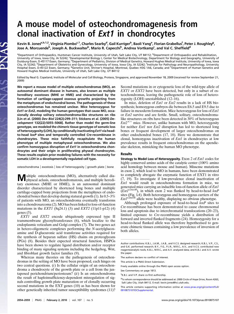
To determine if the previously described chromosomal lossand apoptosis from inverted loxP sites was applicable to ourallele, we crossed homozygous Ext1e2neofl/e2neofl mutants to micecarrying Cre-recombinase under the control of the collagen IIa1promoter (Col2-Cre) (25). No double-heterozygous Col2-cre;Ext1e2neofl/+ offspring (0 of 113) were produced. Similarly,Ext1e2neofl/e2neofl mutants crossed to mice expressing Cre-recombinase from the Hprt locus produced only very small litters.No double-heterozygous Hprt-Cre;Ext1e2neofl/+ mice were re-trieved even at E11.5 (zero of four matings) (Table 1).To test if chromosomal loss and apoptosis could be avoided
by limited exposures toCre-recombinase, we exposedE14.5mouseembryonic fibroblasts (MEFs) isolated from heterozygousExt1e2neofl/+ and homozygous Ext1e2neofl/e2neofl mutants to TAT-Cre (26). The cells remained healthy over at least five passages andhad normal cytogenetic profiles on standard chromosomalspreads. Fluorescent in situ hybridization (FISH) with a chromo-some 15 paint demonstrated the presence of the expected com-plement of two chromosomes 15 in homozygous Ext1e2neofl/e2neofl
cells after TATCre exposure (Fig. 1D). Genotyping of the
Ext1 allele detected all possible Cre-mediated recombinationproducts predictable from the design of the targeting construct(Fig. 1 B and C).Accordingly, introduction of a transgenic Cre-recombinase
under the control of a doxycycline-inducible Col2 promoter(Col2-rtTA-Cre) (27) generated viable Col2-rtTA-Cre;Ext1e2neofl/+
and Col2-rtTA-Cre;Ext1e2neofl/e2neofldoublemutants.Furthermore,inductionofCre activity during gestationafter twomatings betweenCol2-rtTA-Cre;Ext1e2neofl/+ mice resulted in one Col2-rtTA-Cre;Ext1e2neofl/+ and one Col2-rtTA-Cre;Ext1e2neofl/e2neofl offspring.PCR-based genotyping of tail and other cartilaginous tissues re-vealed the presence of all predictable recombinations. Sequencingof the individual PCR products confirmed the expected identity ofthe rearranged fragments.
Exon 2 Inversion Disrupts Ext1 Function. To simplify recombinationproducts of the allele, we crossed Ext1e2neofl/e2neofl males to Prx1-crefemales (28), which express Cre-recombinase in the female germline. By PCR we screened for mutants that had lost the neocassette and retained exon 2 in either wild-type (Ext1e2fl) or in-verted (Ext1e2flinv) orientation (Fig. 1B). Stable germ-line trans-mission of the generated alleles was confirmed by crossingfounders (F0) to C57B6 females. F1 males that produced off-spring (F2) consistently carrying the allele in the expected ori-entation were used for further breeding. Both heterozygousExt1e2fl/+ and Ext1e2flinv/+ mice were born at Mendelian ratios.To verify that exon 2 inversion generates a functionally null
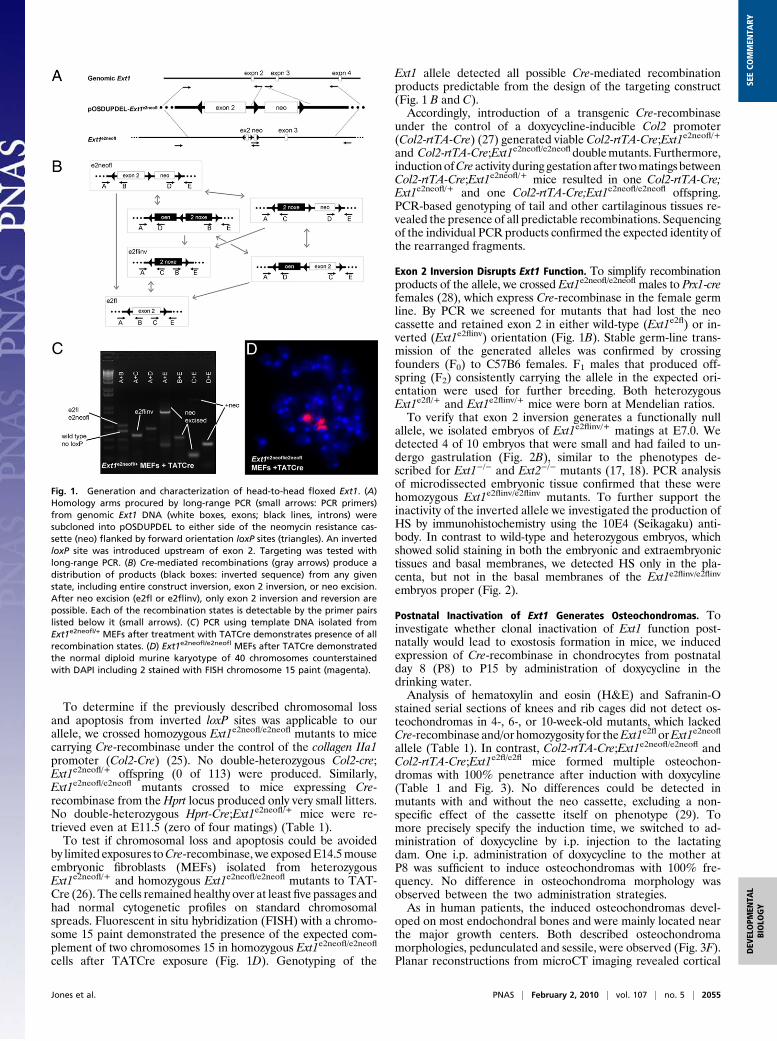
allele, we isolated embryos of Ext1e2flinv/+ matings at E7.0. Wedetected 4 of 10 embryos that were small and had failed to un-dergo gastrulation (Fig. 2B), similar to the phenotypes de-scribed for Ext1−/− and Ext2−/− mutants (17, 18). PCR analysisof microdissected embryonic tissue confirmed that these werehomozygous Ext1e2flinv/e2flinv mutants. To further support theinactivity of the inverted allele we investigated the production ofHS by immunohistochemistry using the 10E4 (Seikagaku) anti-body. In contrast to wild-type and heterozygous embryos, whichshowed solid staining in both the embryonic and extraembryonictissues and basal membranes, we detected HS only in the pla-centa, but not in the basal membranes of the Ext1e2flinv/e2flinvembryos proper (Fig. 2).
Postnatal Inactivation of Ext1 Generates Osteochondromas. Toinvestigate whether clonal inactivation of Ext1 function post-natally would lead to exostosis formation in mice, we inducedexpression of Cre-recombinase in chondrocytes from postnatalday 8 (P8) to P15 by administration of doxycycline in thedrinking water.Analysis of hematoxylin and eosin (H&E) and Safranin-O
stained serial sections of knees and rib cages did not detect os-teochondromas in 4-, 6-, or 10-week-old mutants, which lackedCre-recombinase and/or homozygosity for theExt1e2florExt1e2neofl
allele (Table 1). In contrast, Col2-rtTA-Cre;Ext1e2neofl/e2neofl andCol2-rtTA-Cre;Ext1e2fl/e2fl mice formed multiple osteochon-dromas with 100% penetrance after induction with doxycyline(Table 1 and Fig. 3). No differences could be detected inmutants with and without the neo cassette, excluding a non-specific effect of the cassette itself on phenotype (29). Tomore precisely specify the induction time, we switched to ad-ministration of doxycycline by i.p. injection to the lactatingdam. One i.p. administration of doxycycline to the mother atP8 was sufficient to induce osteochondromas with 100% fre-quency. No difference in osteochondroma morphology wasobserved between the two administration strategies.As in human patients, the induced osteochondromas devel-
oped on most endochondral bones and were mainly located nearthe major growth centers. Both described osteochondromamorphologies, pedunculated and sessile, were observed (Fig. 3F).Planar reconstructions from microCT imaging revealed cortical
Fig. 1. Generation and characterization of head-to-head floxed Ext1. (A)Homology arms procured by long-range PCR (small arrows: PCR primers)from genomic Ext1 DNA (white boxes, exons; black lines, introns) weresubcloned into pOSDUPDEL to either side of the neomycin resistance cas-sette (neo) flanked by forward orientation loxP sites (triangles). An invertedloxP site was introduced upstream of exon 2. Targeting was tested withlong-range PCR. (B) Cre-mediated recombinations (gray arrows) produce adistribution of products (black boxes: inverted sequence) from any givenstate, including entire construct inversion, exon 2 inversion, or neo excision.After neo excision (e2fl or e2flinv), only exon 2 inversion and reversion arepossible. Each of the recombination states is detectable by the primer pairslisted below it (small arrows). (C) PCR using template DNA isolated fromExt1e2neofl/+ MEFs after treatment with TATCre demonstrates presence of allrecombination states. (D) Ext1e2neofl/e2neofl MEFs after TATCre demonstratedthe normal diploid murine karyotype of 40 chromosomes counterstainedwith DAPI including 2 stained with FISH chromosome 15 paint (magenta).
Jones et al. PNAS | February 2, 2010 | vol. 107 | no. 5 | 2055
DEV
ELOPM
ENTA
LBIOLO
GY
SEECO
MMEN
TARY
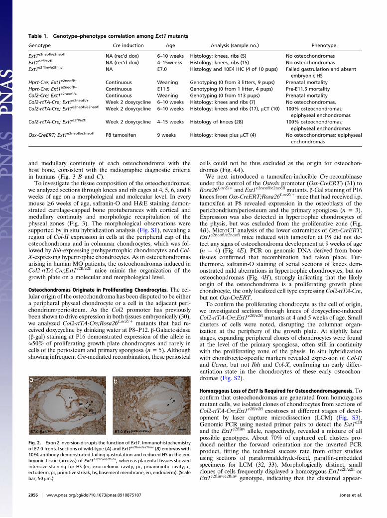
and medullary continuity of each osteochondroma with thehost bone, consistent with the radiographic diagnostic criteriain humans (Fig. 3 B and C).To investigate the tissue composition of the osteochondromas,
we analyzed sections through knees and rib cages at 4, 5, 6, and 8weeks of age on a morphological and molecular level. In everymouse ≥6 weeks of age, safranin-O and H&E staining demon-strated cartilage-capped bone protuberances with cortical andmedullary continuity and morphologic recapitulation of thephyseal zones (Fig. 3). The morphological observations weresupported by in situ hybridization analysis (Fig. S1), revealing aregion of Col-II expression in cells at the peripheral cap of theosteochondroma and in columnar chondrocytes, which was fol-lowed by Ihh-expressing prehypertrophic chondrocytes and Col-X-expressing hypertrophic chondrocytes. As in osteochondromasarising in human MO patients, the osteochondromas induced inCol2-rtTA-Cre;Ext1e2fl/e2fl mice mimic the organization of thegrowth plate on a molecular and morphological level.
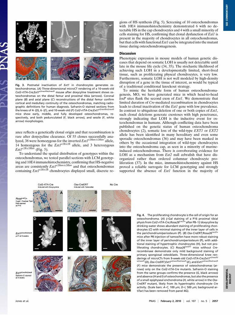
Osteochondromas Originate in Proliferating Chondrocytes. The cel-lular origin of the osteochondroma has been disputed to be eithera peripheral physeal chondrocyte or a cell in the adjacent peri-chondrium/periosteum. As the Col2 promoter has previouslybeen shown to drive expression in both tissues embryonically (30),we analyzed Col2-rtTA-Cre;Rosa26LacZ/+ mutants that had re-ceived doxycycline by drinking water at P8–P12. β-Galactosidase(β-gal) staining at P16 demonstrated expression of the allele in≈50% of proliferating growth plate chondrocytes and rarely incells of the periosteum and primary spongiosa (n = 5). Althoughshowing infrequentCre-mediated recombination, these periosteal
cells could not be thus excluded as the origin for osteochon-dromas (Fig. 4A).We next introduced a tamoxifen-inducible Cre-recombinase
under the control of the Osterix promoter (Osx-CreERT) (31) toRosa26LacZ/+ and Ext1e2neofl/e2neofl mutants. β-Gal staining of P16knees from Osx-CreERT;Rosa26LacZ/+ mice that had received i.p.tamoxifen at P8 revealed expression in the osteoblasts of theperichondrium/periosteum and the primary spongiosa (n = 3).Expression was also detected in hypertrophic chondrocytes ofthe physis, but was excluded from the proliferative zone (Fig.4B). MicroCT analysis of the lower extremities of Osx-CreERT;Ext1e2neofl/e2neofl mice induced with tamoxifen at P8 did not de-tect any signs of osteochondroma development at 9 weeks of age(n = 4) (Fig. 4E). PCR on genomic DNA derived from bonetissues confirmed that recombination had taken place. Fur-thermore, safranin-O staining of serial sections of knees dem-onstrated mild aberrations in hypertrophic chondrocytes, but noosteochondromas (Fig. 4H), strongly indicating that the likelyorigin of the osteochondroma is a proliferating growth platechondrocyte, the only localized cell type expressing Col2-rtTA-Cre,but not Osx-CreERT.To confirm the proliferating chondrocyte as the cell of origin,
we investigated sections through knees of doxycycline-inducedCol2-rtTA-Cre;Ext1e2fl/e2fl mutants at 4 and 5 weeks of age. Smallclusters of cells were noted, disrupting the columnar organ-ization at the periphery of the growth plate. At slightly laterstages, expanding peripheral clones of chondrocytes were foundat the level of the primary spongiosa, often still in continuitywith the proliferating zone of the physis. In situ hybridizationwith chondrocyte-specific markers revealed expression of Col-IIand Ucma, but not Ihh and Col-X, confirming an early differ-entiation state in the chondrocytes of these early osteochon-dromas (Fig. S2).
Homozygous Loss of Ext1 Is Required for Osteochondromagenesis. Toconfirm that osteochondromas are generated from homozygousmutant cells, we isolated clones of chondrocytes from sections ofCol2-rtTA-Cre;Ext1e2fl/e2fl exostoses at different stages of devel-opment by laser capture microdissection (LCM) (Fig. S3).Genomic PCR using nested primer pairs to detect the Ext1e2fl
and the Ext1e2flinv allele, respectively, revealed a mixture of allpossible genotypes. About 70% of captured cell clusters pro-duced neither the forward orientation nor the inverted PCRproduct, fitting the technical success rate from other studiesusing sections of paraformaldehyde-fixed, paraffin-embeddedspecimens for LCM (32, 33). Morphologically distinct, smallclones of cells frequently displayed a homozygous Ext1e2fl/e2fl orExt1e2flinv/e2flinv genotype, indicating that the clustered appear-
Table 1. Genotype–phenotype correlation among Ext1 mutants
Genotype Cre induction Age Analysis (sample no.) Phenotype
Ext1e2neofl/e2neofl NA (rec’d dox) 6–10 weeks Histology: knees, ribs (5) No osteochondromasExt1e2fl/e2fl NA (rec’d dox) 4–15weeks Histology: knees, ribs (15) No osteochondromasExt1e2flinv/e2flinv NA E7.0 Histology and 10E4 IHC (4 of 10 pups) Failed gastrulation and absent
embryonic HSHprt-Cre; Ext1e2neofl/+ Continuous Weaning Genotyping (0 from 3 litters, 9 pups) Prenatal mortalityHprt-Cre; Ext1e2neofl/+ Continuous E11.5 Genotyping (0 from 1 litter, 4 pups) Pre-E11.5 mortalityCol2-Cre; Ext1e2neofl/+ Continuous Weaning Genotyping (0 from 113 pups) Prenatal mortalityCol2-rtTA-Cre; Ext1e2neofl/+ Week 2 doxycycline 6–10 weeks Histology: knees and ribs (7) No osteochondromas.Col2-rtTA-Cre; Ext1e2neofl/e2neofl Week 2 doxycycline 6–10 weeks Histology: knees and ribs (17), μCT (10) 100% osteochondromas;
epiphyseal enchondromasCol2-rtTA-Cre; Ext1e2fl/e2fl Week 2 doxycycline 4–15 weeks Histology of knees (28) 100% osteochondromas;
epiphyseal enchondromasOsx-CreERT; Ext1e2neofl/e2neofl P8 tamoxifen 9 weeks Histology: knees plus μCT (4) No osteochondromas; epiphyseal
enchondromas
Fig. 2. Exon 2 inversion disrupts the function of Ext1. Immunohistochemistryof E7.0 frontal sections of wild-type (A) and Ext1e2flinv/e2flinv (B) embryos with10E4 antibody demonstrated failing gastrulation and reduced HS in the em-bryonic tissue (arrows) of Ext1e2flinv/e2flinv, whereas placental tissues showedintensive staining for HS (ec, exocoelomic cavity; pc, proamniotic cavity; e,ectoderm; ps, primitive streak; bs, basementmembrane; en, endoderm). (Scalebar, 50 μm.)
2056 | www.pnas.org/cgi/doi/10.1073/pnas.0910875107 Jones et al.
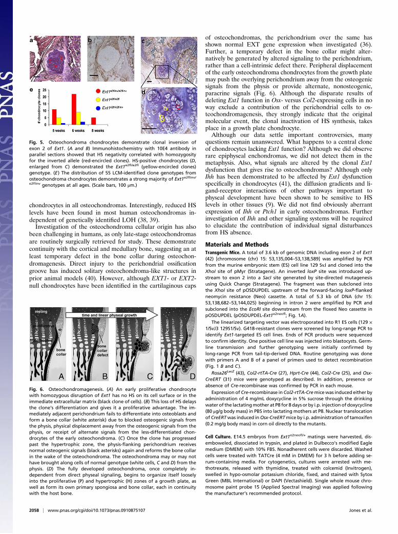
ance reflects a genetically clonal origin and that recombination israre after doxycycline clearance. Of 55 clones successfully ana-lyzed, 38 were homozygous for the inverted Ext1e2flinv/e2flinv allele,14 homozygous for the Ext1e2fl/e2fl allele, and 3 heterozygousExt1e2fl/e2flinv (Fig. 5).To understand the spatial distribution of genotypes within the
osteochondromas, we tested parallel sections with LCM genotyp-ing and 10E4 immunohistochemistry, confirming thatHS-negativeareas are consistently Ext1e2flinv/e2flinv and that osteochondromascontaining Ext1e2fl/e2fl chondrocytes displayed small, discrete re-
gions of HS synthesis (Fig. 5). Screening of 10 osteochondromaswith 10E4 immunohistochemistry demonstrated 6 with no de-tectable HS in the cap chondrocytes and 4 with a small minority ofcells staining for HS, confirming that clonal dysfunction of Ext1 ispresent in the majority of chondrocytes in all osteochondromas,but that cells with functionalExt1 can be integrated into themutanttissue during osteochondromagenesis.
DiscussionPhenotypic expression in mouse models of human genetic dis-eases that depend on somatic LOH is usually not detectable untillate in the mouse life cycle (34, 35). The stochastic likelihood ofachieving such LOH in a developmentally limited, short-livedtissue, such as proliferating physeal chondrocytes, is very low.Furthermore, somatic LOH is not well modeled by high-densitydisruption of a gene in the tissue of interest, as would be typicalof a traditional conditional knockout strategy.To mimic the heritable form of human osteochondroma-
genesis, MO, we have generated mice in which head-to-headloxP sites flank the second exon of Ext1. We demonstrate thatlimited duration of Cre-mediated recombination in chondrocytesleads to clonal inactivation of the Ext1 gene with low prevalence.In contrast to ubiquitous deletion of one or both copies of Ext1,such clonal deletions generate exostoses with high penetrance,strongly indicating that LOH is the inductive event for os-teochondromas in humans. Although conflicting data have beenpublished on the genetic status of human osteochondromachondrocytes (2), somatic loss of the wild-type EXT1 or EXT2allele has been identified in many hereditary and even somesporadic osteochondromas (36) and may have been masked inothers by the occasional integration of wild-type chondrocytesinto the osteochondroma cap, as seen in a minority of murine-induced osteochondromas. There is corroborating evidence forLOH as a mechanism from Ext2 null zebrafish that have dis-organized rather than ordered columnar chondrocyte pro-liferation (37). In the mice, immunohistochemistry against HSproved a reliable surrogate for LCM genotyping and stronglysupported the absence of Ext1 function in the majority of
Fig. 3. Postnatal inactivation of Ext1 in chondrocytes generates os-teochondromas. (A) Three-dimensional microCT rendering of a 10-week-oldCol2-rtTA-Cre;Ext1e2neofl/e2neofl mouse after doxycyline treatment shows os-teochondromas on the distal femur and proximal tibia (arrows). Coronalplane (B) and axial plane (C) reconstructions of the distal femur confirmcortical and medullary continuity of the osteochondromas, matching radio-graphic definitions for human diagnosis. Safranin-O stained sections fromthe knees of 4- (D), 6- (E), and 10-week-old (F) Col2-rtTA-Cre;Ext1e2neofl/e2neofl
mice show early, middle, and fully developed osteochondromas, re-spectively, and both pedunculated (F, black arrow), and sessile (F, whitearrow) morphologies.
Fig. 4. The proliferating chondrocyte is the cell of origin for anosteochondroma. (A) β-Gal staining of a P16 proximal tibialphysis fromCol2-rtTA-Cre;Rosa26LacZ/+after P8–12doxycyline bydrinking water shows abundant staining of proliferating chon-drocytes (C) with minimal staining of the inner layer of cells inthe perichondrium/periosteum (P). (B) Osx-CreERT;Rosa26LacZ/+
mice after P8 injection of tamoxifen have more robust stainingof the inner layer of perichondrium/periosteum (P), with addi-tional staining of hypertrophic chondrocytes (H), but not pro-liferating chondrocytes. (C) Rosa26LacZ/+ mice without Cre-recombinase demonstrate only mild background staining ofprimary spongiosal osteoblasts. Three-dimensional knee ren-derings of microCTs from 9-week-old Col2-rtTA-Cre;Ext1e2neofl/e2neofl (D),Osx-CreERT;Ext1e2neofl/e2neofl (E), and Ext1e2neofl/e2neofl
(F) mice demonstrate the presence of osteochondromas (ar-rows) only on the Col2-rtTA-Cre mutants. Safranin-O stainingfrom the same groups confirms the presence (G, black arrows)andabsence (Hand I) of osteochondromas, but also thepresenceof a small epiphyseal enchondroma (H, white arrow) in theOsx-CreERT mutant, likely from its hypertrophic chondrocyte Creactivity. (Scale bars: A–C, 100 μm; D–I, 500 μm; background ar-tifact has been removed from panel 4G).
Jones et al. PNAS | February 2, 2010 | vol. 107 | no. 5 | 2057
DEV
ELOPM
ENTA
LBIOLO
GY
SEECO
MMEN
TARY
chondrocytes in all osteochondromas. Interestingly, reduced HSlevels have been found in most human osteochondromas in-dependent of genetically identified LOH (38, 39).Investigation of the osteochondroma cellular origin has also
been challenging in humans, as only late-stage osteochondromasare routinely surgically retrieved for study. These demonstratecontinuity with the cortical and medullary bone, suggesting an atleast temporary defect in the bone collar during osteochon-dromagenesis. Direct injury to the perichondrial ossificationgroove has induced solitary osteochondroma-like structures inprior animal models (40). However, although EXT1- or EXT2-null chondrocytes have been identified in the cartilaginous caps
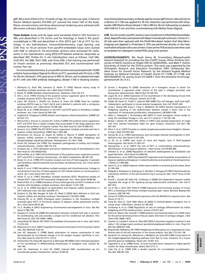
of osteochondromas, the perichondrium over the same hasshown normal EXT gene expression when investigated (36).Further, a temporary defect in the bone collar might alter-natively be generated by altered signaling to the perichondrium,rather than a cell-intrinsic defect there. Peripheral displacementof the early osteochondroma chondrocytes from the growth platemay push the overlying perichondrium away from the osteogenicsignals from the physis or provide alternate, nonosteogenic,paracrine signals (Fig. 6). Although the disparate results ofdeleting Ext1 function in Osx- versus Col2-expressing cells in noway exclude a contribution of the perichondrial cells to os-teochondromagenesis, they strongly indicate that the originalmolecular event, the clonal inactivation of HS synthesis, takesplace in a growth plate chondrocyte.Although our data settle important controversies, many
questions remain unanswered. What happens to a central cloneof chondrocytes lacking Ext1 function? Although we did observerare epiphyseal enchondromas, we did not detect them in themetaphysis. Also, what signals are altered by the clonal Ext1dysfunction that gives rise to osteochondromas? Although onlyIhh has been demonstrated to be affected by Ext1 dysfunctionspecifically in chondrocytes (41), the diffusion gradients and li-gand-receptor interactions of other pathways important tophyseal development have been shown to be sensitive to HSlevels in other tissues (9). We did not find obviously aberrantexpression of Ihh or Ptch1 in early osteochondromas. Furtherinvestigation of Ihh and other signaling systems will be requiredto elucidate the contribution of individual signal disturbancesfrom HS absence.
Materials and MethodsTransgenic Mice. A total of 3.6 kb of genomic DNA including exon 2 of Ext1(42) [chromosome (chr) 15: 53,135,004–53,138,589] was amplified by PCRfrom the murine embryonic stem (ES) cell line 129 SvJ and cloned into theXhoI site of pMyr (Stratagene). An inverted loxP site was introduced up-stream to exon 2 into a SacI site generated by site-directed mutagenesisusing Quick Change (Stratagene). The fragment was then subcloned intothe XhoI site of pOSDUPDEL upstream of the forward-facing loxP-flankedneomycin resistance (Neo) cassette. A total of 5.3 kb of DNA (chr 15:53,138,682–53,144,025) beginning in intron 2 were amplified by PCR andsubcloned into the EcoRI site downstream from the floxed Neo cassette inpOSDUPDEL (pOSDUPDEL-Ext1e2neofl; Fig. 1A).
The linearized targeting vector was electroporated into R1 ES cells (129 ×1/SvJ3 129S1/Sv). G418-resistant clones were screened by long-range PCR toidentify Ext1-targeted ES cell lines. Ends of PCR products were sequencedto confirm identity. One positive cell line was injected into blastocysts. Germ-line transmission and further genotyping were initially confirmed bylong-range PCR from tail-tip-derived DNA. Routine genotyping was donewith primers A and B of a panel of primers used to detect recombination(Fig. 1 B and C ).
Rosa26LacZ (43), Col2-rtTA-Cre (27), Hprt-Cre (44), Col2-Cre (25), and Osx-CreERT (31) mice were genotyped as described. In addition, presence orabsence of Cre-recombinase was confirmed by PCR in each mouse.
Expression of Cre-recombinase in Col2-rtTA-Cremicewas induced either byadministration of 4 mg/mL doxycycline in 5% sucrose through the drinkingwater of the lactatingmother at P8 for 8 days or by i.p. injection of doxycycline(80 μg/g body mass) in PBS into lactating mothers at P8. Nuclear translocationof CreERTwas induced inOsx-CreERTmice by i.p. administration of tamoxifen(0.2 mg/g body mass) in corn oil directly to the mutants.
Cell Culture. E14.5 embryos from Ext1e2neofl/+ matings were harvested, dis-emboweled, dissociated in trypsin, and plated in Dulbecco’s modified Eaglemedium (DMEM) with 10% FBS. Nonadherent cells were discarded. Washedcells were treated with TATCre (4 mM in DMEM) for 3 h before adding se-rum-containing media. For cytogenetics, cultures were arrested with me-thotrexate, released with thymidine, treated with colcemid (Invitrogen),swelled in hypo-osmolar potassium chloride, fixed, and stained with SytoxGreen (MBL International) or DAPI (Vectashield). Single whole mouse chro-mosome paint probe 15 (Applied Spectral Imaging) was applied followingthe manufacturer’s recommended protocol.
Fig. 5. Osteochondroma chondrocytes demonstrate clonal inversion ofexon 2 of Ext1. (A and B) Immunohistochemistry with 10E4 antibody inparallel sections showed that HS negativity correlated with homozygosityfor the inverted allele (red-encircled clones). HS-positive chondrocytes (D,enlarged from C) demonstrated the Ext1e2fl/e2fl (yellow-encircled clones)genotype. (E) The distribution of 55 LCM-identified clone genotypes fromosteochondroma chondrocytes demonstrates a strong majority of Ext1e2flinv/e2flinv genotypes at all ages. (Scale bars, 100 μm.)
Fig. 6. Osteochondromagenesis. (A) An early proliferative chondrocytewith homozygous disruption of Ext1 has no HS on its cell surface or in theimmediate extracellular matrix (black clone of cells). (B) This loss of HS delaysthe clone’s differentiation and gives it a proliferative advantage. The im-mediately adjacent perichondrium fails to differentiate into osteoblasts andform a bone collar (white asterisk) due to blocked osteogenic signals fromthe physis, physical displacement away from the osteogenic signals from thephysis, or receipt of alternate signals from the less-differentiated chon-drocytes of the early osteochondroma. (C) Once the clone has progressedpast the hypertrophic zone, the physis-flanking perichondrium receivesnormal osteogenic signals (black asterisks) again and reforms the bone collarin the wake of the osteochondroma. The osteochondroma may or may nothave brought along cells of normal genotype (white cells, C and D) from thephysis. (D) The fully developed osteochondroma, once completely in-dependent from direct physeal signaling, begins to organize itself looselyinto the proliferative (P) and hypertrophic (H) zones of a growth plate, aswell as form its own primary spongiosa and bone collar, each in continuitywith the host bone.
2058 | www.pnas.org/cgi/doi/10.1073/pnas.0910875107 Jones et al.
μCT.Micewere killed at 9 or 10weeks of age. No contrast was used. A GeneralElectric Medical Systems EVS-RS9 μCT scanned the lower half of the body.Planar reconstructions and three-dimensional rendering were achieved withMicroview software (General Electric).
Tissue Analysis. Knees and rib cages were harvested, fixed in 10% formalin inPBS, and decalcified in 5% formic acid for histology or fixed in 4% paraf-ormaldehyde in PBS and decalcified in 25% EDTA (pH 7.4) at 37°C for his-tology, in situ hybridization, immunohistochemistry, β-gal staining, andLCM. Five- to 10-μm sections from paraffin-embedded tissue were stainedwith H&E or safranin-O. Six-micrometer sections were processed for radio-active in situ hybridization using [P33]-UTP-labeled antisense riboprobes asdescribed (45). Probes for in situ hybridization were as follows: Col2 (46),ColX (47), Ihh (48), Ptch1 (49), and Ucma (50). β-Gal staining was performedon frozen sections as previously described (51) and counterstained withnuclear fast red.
For immunohistochemistry,6-μmparaffinsectionswerepretreatedwith10,000units/mLhyaluronidase I (Sigma) for30minat37°C,quenchedwith3%H2O2 inPBSfor 30min, blocked in 10%goat serum inPBS for 30min, and incubatedovernightat 4°C with anti-10E4 antibody (Seikagaku), diluted 1:100 in blocking solution.
Goat biotinylated secondary antibody againstmouse IgM (Vector Laboratories) ata dilution of 1:100 was applied for 30 min. Detection was performed with Strep-tavidin-HRP (Perkin-Elmer) diluted 1:100 in PBS for 30min followed by incubationwith DAB for 5 min and then counterstaining with Methyl Green (Sigma).
LCM. Six-micrometer paraffin sectionswere transferred to PALMMembraneSlides,dried,deparaffinized,andstainedwithMayer’shematoxylinandeosin.Clonesof1–20 cells were then captured with the PALM Microbeam System (Carl Zeiss Micro-Imaging) and digested in Proteinase K. PCR was performed directly on the heat-inactivatedcell lysatewithouterprimers.OuterprimerPCRproductswerethenusedas templates for subsequent nested PCRs using inner primers.
ACKNOWLEDGMENTS. We thank Henry M. Kronenberg (MassachusettsGeneral Hospital) for providing the Osx-CreERT mouse, Oliver Smithies (Uni-versity of North Carolina at Chapel Hill) for pOSDUPDEL, and Matt F. Hockin(University of Utah) for the TATCre protein. This work was supported by theOrthopedic Research and Education Foundation 2003 Centerpulse Res-ident Grant for Growth Factor Research; by the Howard Hughes MedicalInstitute; by National Institutes of Health Grants EY-11298, EY-17168, and5R37HD030701-16; and by Grant VO 620/9-1 from the Deutsche Forschungs-gemeinschaft (to A.V.).
1. Wicklund CL, Pauli RM, Johnston D, Hecht JT (1995) Natural history study ofhereditary multiple exostoses. Am J Med Genet 55:43–46.
2. Bovée JV (2008) Multiple osteochondromas. Orphanet J Rare Dis 3:3.3. Ahn J, et al. (1995) Cloning of the putative tumour suppressor gene for hereditary
multiple exostoses (EXT1). Nat Genet 11:137–143.4. Ligon AH, Potocki L, Shaffer LG, Stickens D, Evans GA (1998) Gene for multiple
exostoses (EXT2) maps to 11(p11.2p12) and is deleted in patients with a contiguousgene syndrome. Am J Med Genet 75:538–540.
5. Wuyts W, et al. (1998) Mutations in the EXT1 and EXT2 genes in hereditary multipleexostoses. Am J Hum Genet 62:346–354.
6. Sugahara K, Kitagawa H (2002) Heparin and heparan sulfate biosynthesis. IUBMB Life54:163–175.
7. McCormick C, Duncan G, Goutsos KT, Tufaro F (2000) The putative tumor suppressorsEXT1 and EXT2 form a stable complex that accumulates in the Golgi apparatus andcatalyzes the synthesis of heparan sulfate. Proc Natl Acad Sci USA 97:668–673.
8. Senay C, et al. (2000) The EXT1/EXT2 tumor suppressors: Catalytic activities and role inheparan sulfate biosynthesis. EMBO Rep 1:282–286.
9. Bornemann DJ, Duncan JE, Staatz W, Selleck S, Warrior R (2004) Abrogation ofheparan sulfate synthesis in Drosophila disrupts the Wingless, Hedgehog andDecapentaplegic signaling pathways. Development 131:1927–1938.
10. Porter DE, Simpson AH (1999) The neoplastic pathogenesis of solitary and multipleosteochondromas. J Pathol 188:119–125.
11. Knudson AG, Jr. (1971) Mutation and cancer: Statistical study of retinoblastoma. ProcNatl Acad Sci USA 68:820–823.
12. Bernard MA, et al. (2001) Diminished levels of the putative tumor suppressor proteinsEXT1 and EXT2 in exostosis chondrocytes. Cell Motil Cytoskeleton 48:149–162.
13. Bovée JV, et al. (1999) EXT-mutation analysis and loss of heterozygosity in sporadicand hereditary osteochondromas and secondary chondrosarcomas. Am J Hum Genet65:689–698.
14. Hecht JT, et al. (1995) Hereditary multiple exostosis and chondrosarcoma: Linkage tochromosome II and loss of heterozygosity for EXT-linked markers on chromosomes IIand 8. Am J Hum Genet 56:1125–1131.
15. Hecht JT, et al. (1997) Hereditary multiple exostoses (EXT): Mutational studies offamilial EXT1 cases and EXT-associated malignancies. Am J Hum Genet 60:80–86.
16. Raskind WH, et al. (1998) Evaluation of locus heterogeneity and EXT1 mutations in 34families with hereditary multiple exostoses. Hum Mutat 11:231–239.
17. Lin X, et al. (2000) Disruption of gastrulation and heparan sulfate biosynthesis inEXT1-deficient mice. Dev Biol 224:299–311.
18. Stickens D, Zak BM, Rougier N, Esko JD, Werb Z (2005) Mice deficient in Ext2 lackheparan sulfate and develop exostoses. Development 132:5055–5068.
19. Cheung PK, et al. (2001) Etiological point mutations in the hereditary multipleexostoses gene EXT1: A functional analysis of heparan sulfate polymerase activity.Am J Hum Genet 69:55–66.
20. Yu Y, Bradley A (2001) Engineering chromosomal rearrangements in mice. Nat RevGenet 2:780–790.
21. Grégoire D, Kmita M (2008) Recombination between inverted loxP sites is cytotoxicfor proliferating cells and provides a simple tool for conditional cell ablation. ProcNatl Acad Sci USA 105:14492–14496.
22. Lewandoski M, Martin GR (1997) Cre-mediated chromosome loss in mice. Nat Genet17:223–225.
23. Matsumura H, et al. (2007) Targeted chromosome elimination from ES-somatic hybridcells. Nat Methods 4:23–25.
24. Lam KP, Rajewsky K (1998) Rapid elimination of mature autoreactive B cellsdemonstrated by Cre-induced change in B cell antigen receptor specificity in vivo.Proc Natl Acad Sci USA 95:13171–13175.
25. Ovchinnikov DA, Deng JM, Ogunrinu G, Behringer RR (2000) Col2a1-directed expressionof Cre recombinase in differentiating chondrocytes in transgenic mice. Genesis 26:145–146.
26. Joshi SK, Hashimoto K, Koni PA (2002) Induced DNA recombination by Crerecombinase protein transduction. Genesis 33:48–54.
27. Grover J, Roughley PJ (2006) Generation of a transgenic mouse in which Crerecombinase is expressed under control of the type II collagen promoter anddoxycycline administration. Matrix Biol 25:158–165.
28. Logan M, et al. (2002) Expression of Cre recombinase in the developing mouse limbbud driven by a Prxl enhancer. Genesis 33:77–80.
29. Haldar M, Karan G, Tvrdik P, Capecchi MR (2008) Two cell lineages, myf5 and myf5-independent, participate in mouse skeletal myogenesis. Dev Cell 14:437–445.
30. Long F, Zhang XM, Karp S, Yang Y, McMahon AP (2001) Genetic manipulation ofhedgehog signaling in the endochondral skeleton reveals a direct role in theregulation of chondrocyte proliferation. Development 128:5099–5108.
31. Maes C, Kobayashi T, Kronenberg HM (2007) A novel transgenic mouse model tostudy the osteoblast lineage in vivo. Ann N Y Acad Sci 1116:149–164.
32. Becker I, Becker KF, Röhrl MH, Höfler H (1997) Laser-assisted preparation of singlecells from stained histological slides for gene analysis. Histochem Cell Biol 108:447–451.
33. Ohno T, et al. (1997) Clonality in nodular lymphocyte-predominant Hodgkin’s disease.N Engl J Med 337:459–465.
34. Harvey M, et al. (1993) Spontaneous and carcinogen-induced tumorigenesis in p53-deficient mice. Nat Genet 5:225–229.
35. Lu W, et al. (1999) Late onset of renal and hepatic cysts in Pkd1-targetedheterozygotes. Nat Genet 21:160–161.
36. Hameetman L, et al. (2007) The role of EXT1 in nonhereditary osteochondroma:Identification of homozygous deletions. J Natl Cancer Inst 99:396–406.
37. Clément A, et al. (2008) Regulation of zebrafish skeletogenesis by ext2/dackel andpapst1/pinscher. PLoS Genet 4:e1000136.
38. Hameetman L, et al. (2007) Decreased EXT expression and intracellular accumulation ofheparan sulphate proteoglycan in osteochondromas and peripheral chondrosarcomas.J Pathol 211:399–409.
39. Hecht JT, et al. (2002) Heparan sulfate abnormalities in exostosis growth plates. Bone31:199–204.
40. Delgado E, Rodríguez JI, Rodríguez JL, Miralles C, Paniagua R (1987) Osteochondromainduced by reflection of the perichondrial ring in young rat radii. Calcif Tissue Int 40:85–90.
41. Koziel L, Kunath M, Kelly OG, Vortkamp A (2004) Ext1-dependent heparan sulfateregulates the range of Ihh signaling during endochondral ossification. Dev Cell 6:801–813.
42. Lin X, Gan L, Klein WH, Wells D (1998) Expression and functional analysis of mouseEXT1, a homolog of the human multiple exostoses type 1 gene. Biochem Biophys ResCommun 248:738–743.
43. Soriano P (1999) Generalized lacZ expression with the ROSA26 Cre reporter strain. NatGenet 21:70–71.
44. Tang SH, Silva FJ, Tsark WM, Mann JR (2002) A Cre/loxP-deleter transgenic line inmouse strain 129S1/SvImJ. Genesis 32:199–202.
45. Vortkamp A, et al. (1996) Regulation of rate of cartilage differentiation by Indianhedgehog and PTH-related protein. Science 273:613–622.
46. Kohno K, Martin GR, Yamada Y (1984) Isolation and characterization of a cDNA clonefor the amino-terminal portion of the pro-alpha 1(II) chain of cartilage collagen. J BiolChem 259:13668–13673.
47. Jacenko O, LuValle P, Solum K, Olsen BR (1993) A dominant negative mutation in thealpha 1 (X) collagen gene produces spondylometaphyseal defects in mice. Prog ClinBiol Res 383B:427–436.
48. Bitgood MJ, McMahon AP (1995) Hedgehog and Bmp genes are coexpressed at manydiverse sites of cell-cell interaction in the mouse embryo. Dev Biol 172:126–138.
49. Goodrich LV, Johnson RL, Milenkovic L, McMahon JA, Scott MP (1996) Conservation ofthe hedgehog/patched signaling pathway from flies to mice: Induction of a mousepatched gene by Hedgehog. Genes Dev 10:301–312.
50. Tagariello A, et al. (2008) Ucma—A novel secreted factor represents a highly specificmarker for distal chondrocytes. Matrix Biol 27:3–11.
51. Lobe CG, et al. (1999) Z/AP, a double reporter for cre-mediated recombination.Dev Biol 208:281–292.
Jones et al. PNAS | February 2, 2010 | vol. 107 | no. 5 | 2059
DEV
ELOPM
ENTA
LBIOLO
GY
SEECO
MMEN
TARY