a molecular and microscopically studies of calicophoron ...egyptianjournal.xyz/66_3.pdf · a...
TRANSCRIPT

The Egyptian Journal of Hospital Medicine (Jan. 2017) Vol. 66, Page 28- 39
28
Received:121/0/2016 DOI: 10.12816/0034630
Accepted:22/10/2016
A Molecular and Microscopically Studies of Calicophoron Microbothrium
(Paramphistomatidae) Alaa Abdel-Aziz Mohamed Samn
Zoology Department, Faculty of Science (Boys),Al-Azhar University, Cairo, Egypt
[email protected], [email protected]
ABSTRACT
Background: Paramphistomiasis is a parasitic disease of livestock animals and humans, which causes
heavy economic black lashes especially in countries with advanced animal industry
Aim of Study: the current study aimed to add more information about Calicophoron microbothrium
(C. microbothrium) and clarify its biological role and how its miracidia infecte the molluscan
intermediate host. In addition, a brief description to Bullins truncates; the morphological, structural and
chronological characteristics of the various intermolluscan stages of the parasite are studied in detail.
Moreover, the present work showed the effective role of physical parameters (light, temperature,
salinity and gas-phase (aerobic versus anaerobic)) on egg development and hatching and the biological
activities of cercaria and metacercaria. Beside these routine techniques, PCR also was used as more
advanced and accurate diagnostic technique based on the detection of nucleic acid. Where, 34 larvae
and adult worms of Calicophoron microbothrium were isolated from naturally infected buffaloes. The
results of the present study will facilitate the identification of this despise secular group of digeneans
although its bad effect not only affect animal industry but also human health. Furthermore, the current
research clears the weak points in its life cycle to aperient settling this parasite.
Keywords: Calicophoron microbothrium, Bullins truncates, paramphistome, biology, histology, Mas–
PCR technique.
INTRODUCTION
Paramphistomes are spreading worldwide,
especially in the warmer regions such as:
Australia, Africa and India, and mainly infect
cattle, goats and sheep. In addition, there are
certain species of paramphistomes that infect
human it has medical importance beside their
economic one. Calicophoron microbothrium
(Paramphistomum microbothrium) is one of
two of these parasites in Egypt as reported by
Ashour (1)
; the other species was Calicophoron
gregarius. They were the most important flukes
identified in domestic Egyptian ruminants.
This C. microbothrium has a life cycle
similar to that of the gastrointestinal trematodes.
Where, the adult flukes reside in the host's
rumen and reticulum; their eggs are passed
through host's faeces. When eggs reach water
the miracidia hatch within 12-16 days and
penetrate suitable intermediate host. The
parasite has a number of generations of rediae
before the production of free swimming
cercariae that are changed to metacercariae after
encysting on water plants; these plants become
the source of the ruminant food causing the
infection and beginning a new cycle (2)
.
Seriously, these parasites may survive up to
years, so that they become a virtually constant
infestation source for numerous snail
generations. As a result to that, the intermediate
host with its ability of widespread, surviving for
several months and shedding numerous
cercariae, and then so dangerous threaten to the
domestic animals (3)
.
The paramphistomes are conical or
cylindrical digenean with thick bodies. They are
distinguished from other flukes by the
possession of a posteriorly located acetabulum.
The most familiar species are parasites of
domesticated livestock. It has been considered,
for a long time, that paramphistomes are
completely non-injurious to their vertebrate
hosts, but this view has been challenged by
many authors (4-7)
. Where, an acute infection of
calves and sheep had been caused by the
immature conical flukes in the small intestine,
particularly in advanced cattle raising regions
causing elevation of mortality rate in sheep
about 30% as reported by Chauhan et al. (8)
and
21%-37.4% in cattle by Pande (9)
. In addition,
Katlyar and Varsheney (10)
recorded an
average percentage morbidity and mortality
41.24% and 57.62% in sheep and 68.59% and
75.53% in goats respectively. Moreover, Horak (6)
recorded a loss of 11.4 kg of sheep's weight
within 52 days and 27.3 kg in bovines after 140
days, while an uninfected bovine gained 50.9 kg
during the same period.
The clinical signs of acute paramphistomiasis in
sheep, goats and cattle have been represented by

A Molecular and Microscopically Studies of Calicophoron Microbothrium
29
restless and progressive decrease in appetite
developed to complete anorexia, small
quantities of water are taken and the animals
might stand with their muzzles in water for a
long time; a condition known as polydipsia.
Diarrhoea developed 2-4 weeks after infection,
with fetid faeces and rectal hemorrhage.
Submandibular oedema (bottle jaw) has been
noted in a number of outbreaks. Oedema of
lungs, hydrothorax, hydropsyeasdium and
ascites were reported by [Baldrey (11)
, Walker (12)
, Chauhan et al. (8)
, Pande (9)
, Haji (13)
,
Bawa (14)
, Boray (15)
, Varma (16)
, Horak and
Clank (17)
, Katiyar and Vershney (18)
and
Horak (6)
].
About how the parasite causing its disorder
effects in animals, Horak (7)
stated that
immature paramphistomes might penetrate the
intestinal wall to just below the serosa and can
be seen from the peritoneal side of the intestine.
On rare occasions, the parasites perforated the
intestine and were found in the abdominal
cavity. While, Dinnik (4)
and Horak (6)
reported
that many cattle and sheep were infected with
adult paramphistomes and the infection was
acquired by ingestion of few numbers of
metacercariae on one or several occasions,
didn’t cause an obvious harm to the host. The
ingested metacercarine mature rapidly and serve
as a source of infection for successive
generations of snails. These paramphistomes
can survive for some years, where the source of
infection is virtually consistent.
The average daily egg produced by a single C.
microbothrium in infected sheep was estimated
about 75 eggs (6)
. Although this was not a large
output but the large number of parasites was
usually found in host with large number
according to Boray (19)
who found 60.000
worms or excess of C. ichikawai/ 3-8 naturally
infected sheep and Dinnik (4)
returned that to
long life span of C. microbothrium in cattle
which maintains its egg production for many
years, that appearing the severity of this
parasite.
Surprising, it was possible that some species of
paramphistomes which are parasites of wild
ruminants were occasionally encountered in
domestic ruminants if wild ruminants
occasionally infect domestic ruminants' places
and stocks live closely together (20&21)
.
Notably, there was asymptomatic in
paramphistomes' natural hosts such as pigs and
monkeys, while in human causes serious health
problems like diarrhea, fever, abdominal pain,
colic, and an increased mucous production. In
addition, in extreme situations this disease leads
to a number of mortality among children like
happened in Assam, India (22)
.
Although the dangerous effects of this parasite
on humans and economic animals, there were
limited number of papers has been studied the
paramphistomes in Egypt. Ezzat (23)
described
specimens from Gazella dorcas which were
identified as Paramphistomum cervi. Tadros (24)
reported the presence of paramphistomes in
cows, buffaloes, sheep and camels in Shebin El-
Kanater district in the Nile Delta, but he did not
designate these to known genera or species.
Abdel-Ghani (25&26)
described specimens,
which were identified as P. cervi but his
description was not based on anatomical and
histological features which were considered
necessary for the specific identification of
paramphistomes. Also, he described briefly the
eggs and miracidia of these flukes and
conducted some experiments to determine the
susceptibility of small laboratory animals to
infection. Ashour (1)
gave the first detailed
morphological, anatomical and histological
account of paramphistomes in Egypt. He
recorded two species under the genus
Paramphistomum, namely, C. (= Calicophoron)
microbothrium and C. khalili as well as one
species in the genus Carmyerus, C. gregarius.
Moreover, he conducted comparative
morphological and experimental studies on the
eggs and miracadia of both P. microbothrium
and P. gregarious. Elkabbany (27)
gave more
information about scanning and transmission
electron microscope of two species of
paramphistomes, C. microbothrium and C.
gregarious.
In addition to the previous causes, the difficulty
of paramphistomes' identification
morphologically and histologically leads to find
new method easier. Therefore, the current study
used the PCR based techniques beside the
routine techniques which should be a suitable
tool because it was providing rDNA ITS2
sequences that have proven as a suitable marker
for identification of paramphistomes species (28)
.
MATERIAL AND METHODS 1 Field studies:
1.1 Collection of paramphistomes:
Mature specimens of paramphistomes
were collected from cows, buffaloes, sheep, and
goats, which slaughtered at Sharkiya and Mit
Ghamre Abattoirs. The flukes were found
attached by their strong acetabula to the inner
surfaces of the rumen and the reticulum of their
hosts. They appear in groups as pinkish to red

Alaa Samn
30
flesh-likes patches in case of Calicophoron;
however, Few flukes were seen free in the
lumen of the stomach.
After the removal of the flukes from the
stomach, the flukes being attached to each
other, therefore, they were put in normal saline
solution (0.7% NaCl). Where, saline make the
fluke’s exhibit slow movement, in which the
body becomes narrower, elongated and the
anterior end of the body moves in different
directions, while the posterior end is usually
fixed to another fluke. Then all these samples
were stored in 95% ethanol.
1.2 Morphological identification:
Some fresh flukes were pressed between two
glass slides and fixed in 70% alcohol for two
days. The relaxed flukes were stained for
identification according to Eduardo technique
(29). All specimens were examined
morphologically and confirmed as
paramphistomes before they included in
molecular study.
1.3 DNA extraction:
Genomic DNA collected from a small part of
adult flukes was extracted by the alkaline-lysis
(Hot-SHOT) method (30)
. Mass-PCR technique
for detection of C. microbothrium was carried
out at molecular unit of Microbiology
Department, Faculty of Medicine Zagazig
University.
1.4 Molecular analysis
The ITS2 (internal transcribed spacer region 2)
region was amplified using the primers GA1
[5_-AGA ACA TCG ACA TCT TGA AC-3_] (31)
and BD2 [5_-TAT GCT TAA ATT CAG
CGG GT- 3_] (32)
. Polymerase chain reaction
(PCR) cycles were performed on Eppendorf
Mastercycler epigradient machines. After the
thermocycler process was finished, PCR
products were purified using PCR Microcon
columns and both strands were sequenced using
an Applied Biosystems 3100 automated
sequencer.
1.5 Agarose gel electrophoresis:
Amplification products of C. microbothrium
adult flukes were visualized by electrophoresis
on 1.5% agarose agar and staining by 0.5 µg/ml
ethidium bromide in the running buffer
procedure of Viljoen et al. (33)
.
2 Laboratory studies:
The other part of the current study was carried
out in the Labe. Amounts of Calicophoron's
mature parasites and eggs were collected from
infected sheep. In addition, snails those were
controlled under the stereomicroscope for
determination of natural infection.
2.1 Preparation of tissue sections for
histological studies:
Snail's tissues for histological studies, using
haematoxylin, eosin and toluidine blue as the
method of Krichesky (34)
.
2.2 Follow up the life cycle of C.
microbothrium:
Using both morphological and histological
examination to all stages of the parasite life
cycle in the intermediate host and adult flocks.
RESULTS
Examination of C. microbothrium's eggs
revealed they are oval operculated, smooth-
surfaced and white-grayish in color; their length
130-170 m and width 70-100 m (Fig 1). In
the central of the egg, embryo was seen in an
early stage and surrounded by yolk cells (Figs 2
& 3). While, Figure 4 was showed the escaping
process of miracidium and showing operculum
and egg opening. Notably, the hatching of C.
microbothrium's eggs occurred more on the
same periods in variable water mediums at the
same temperature.
C. microbothrium's miracidium
shape like a torpedo; it was covered with cilia
and carried on eye spots (Fig 5). The average
size of miracidium was measured about 199-
270 x 50-60 m. Figs 6 & 7 showed miracidium
penetrated the Bulinus truncatus snail through
the mantle cavity.
By following up the infection stages of
snail by miracidium, using dissection of snail at
different periods to detect the stages of
Sporocyst, it was found that Sporocyst was
sacculer, elongated and curved in shape and
measured 200 m x 100 m (Fig. 8, 9, 10 &
11). Interestingly, Sporocysts were detected
mainly around the intestines and in mantle
tissue of snails.
While, rediae were opaque,
slightly curved in shape and had a limited cavity
as observed in Fig 12. In the young rediae the
pharynx and the intestine were visible and they
contain embryo balls from which cercariae or
daughter rediae would develop (Figs 12 & 13).
Rediae were detected around the intestines,
hepato-pancreas and in the mantle tissue of
Bulinus truncatus. The developing rediae
showed great variations in size and the mature
rediae measured 700-1170 m in length and
130-260 m in width (Fig. 14).
Otherwise, immature cercariae were
liberated from rediae before their complete
development. In the young cercariae the body
was small, eye spots were prominent and

A Molecular and Microscopically Studies of Calicophoron Microbothrium
31
pigment and the tail was short and wide rather
than long (Figs. 15, 16, 17 & 18). But the
mature cercariae were large, active, and dark
pigmented and their body was 500-550 m long
and 200-400 wide; while their tail was 550-560
long and 80-90 m wide (Figs 18, 19, 20, 21 &
22).
Notably, it was seen that light was a great
factors in shedding cercariae, infected snail kept
in the dark started shedding cercariae again after
being exposed to light. Cercariae were collected
from the snails only one day in a week in order
to regulate metacercarial age conformity and to
obtain then in high numbers.
Under the light source and in the
presence of vegetation in the water most
cercariae encyst on the vegetation. During the
encystment cercariae attaches with its ventral
surface, and the material for cystation being to
secrete from the pores all over the body (Figs 23
& 24). Metacercaria was in the form of a half
sphere and was surrounded with a thick layer of
a cyst wall. These metacercariae were measured
240-260 m in diameter (Figs 25, 26 & 27).
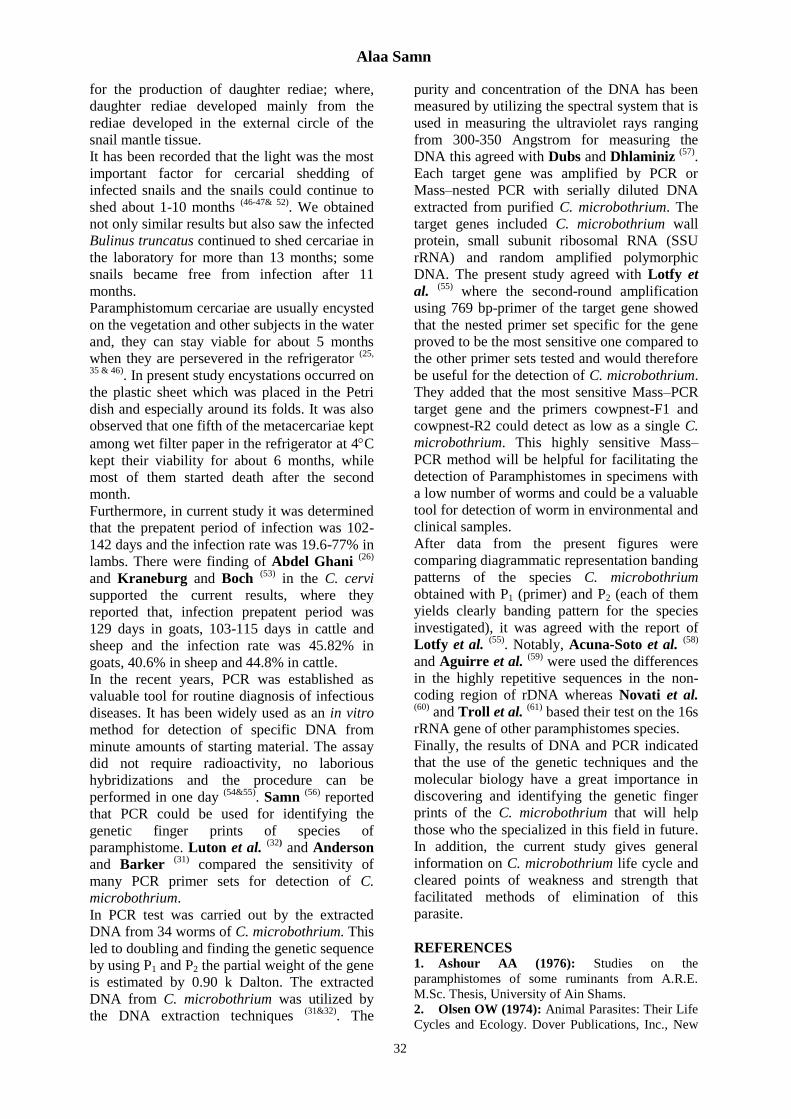
The sensitivity of polymerase chain
reaction (PCR) was measured using various
number of lysate cells of C. microbothrium.
After 22 cycles of PCR as little as one copy of
the target gene was detracted by specific primer
on agarose gel electrophoresis (Fig. 28). Each
primer (P1 and P2) amplified several DNA
fragments that were polymorphic the species.
The results which obtained with primer 1 and 2
produced characteristic intense band patterns of
C. microbothrium (769 bp) (Fig. 28).
DISCUSSION
It is well known that the intermediate host of C.
microbothrium varies according to the
geographical regions; however, many types of
snails play the main role as intermediate host (25
& 35-41), Bulinus truncates were recorded as an
intermediate host for heavy infestation of the
snails with various paramphistomes is reported
mainly in the late summer and autumn months
and the incidence varies between 3 to 75% in
the infected areas (36-38 & 42-44)
. In the present
study naturally infected Bulinus truncatus were
found at each month between April and
November too.
Otherwise, it was noted that many factors affect
the development of Paramphistome eggs such
as temperature and light; but the effect of
temperature rather than that of light (45-47)
.
Similar results were obtained from the current
study, where the development of C.
microbothrium eggs in various mediums and
temperatures did not show any variation; while,
light was found to be the most effective
influence factor for hatching.
On the other hand, it was noted that tap water is
a good medium than distilled water and saline
solution (0.9%) for several of Paramphistomum
miracidia and some miracidia of
Paramphistomum species kept their viability
longer in lakes and pools compared with other
media (46-48)
. The previous results supported our
finding that newly emerged miracidia swam
more active in distilled water and spring water.
Otherwise, it was recorded that too many
miracidia had a negative effect on the
experimental infection of snails and only the
young or medium sized snails were suitable for
infection (47&49)
. During the current experiment,
it was observed that the infection rate increased
with the number of miracidia used; but contrast
to this, it was seen that the large proportions of
miracidia had a negative effect. Concerning
snail size, it was seen that the infection rate
among the small and middle sized Bulinus
truncatus is elevated their mortality rate is
increased too.
Generally, it was accepted that the miracidia
lost their cilia during the penetration and
entered the snail host tissues as a young
sporocyst. On the observation of miracidial
penetration into the snails, both mechanical and
enzymatic effects together play a role (46-47)
. The
transformation of the miracidium into a
sporocyst is a gradual process and no clear line
can be drawn between the invading miracidium
and young sporocyst (50)
. In the present study
persistence of cilia of the anterior part of the
miracidium was also observed, 1-2 hours after
the infection of snails.
Available information on the development of C.
microbothrium in the snail is very limited (46)
.
Kıselev (51)
reported that, in some
Paramphistomum species, the occurrence of
daughter rediae were recorded only in autumn
months. Moreover, the production of redia was
less in number compared to the regular
production of cercariae and they were observed
after the 40th days of infection yet. The current
experiment showed the daughter rediae in
Bulinus truncatus not only in autumn months
but also in different months of the year.
Interestingly, in the current research, it is
possible to detect the rediae which caring
daughter rediae from the external lateral parts of
the mantle tissue of Bulinus truncatus before its
dissection on the examination under the
stereomicroscope. Notably, the location of
rediae in the snail tissue was an important factor
Alaa Samn
32
for the production of daughter rediae; where,
daughter rediae developed mainly from the
rediae developed in the external circle of the
snail mantle tissue.
It has been recorded that the light was the most
important factor for cercarial shedding of
infected snails and the snails could continue to
shed about 1-10 months (46-47& 52)
. We obtained
not only similar results but also saw the infected
Bulinus truncatus continued to shed cercariae in
the laboratory for more than 13 months; some
snails became free from infection after 11
months.
Paramphistomum cercariae are usually encysted
on the vegetation and other subjects in the water
and, they can stay viable for about 5 months
when they are persevered in the refrigerator (25,
35 & 46). In present study encystations occurred on
the plastic sheet which was placed in the Petri
dish and especially around its folds. It was also
observed that one fifth of the metacercariae kept
among wet filter paper in the refrigerator at 4C
kept their viability for about 6 months, while
most of them started death after the second
month.
Furthermore, in current study it was determined
that the prepatent period of infection was 102-
142 days and the infection rate was 19.6-77% in
lambs. There were finding of Abdel Ghani (26)
and Kraneburg and Boch (53)
in the C. cervi
supported the current results, where they
reported that, infection prepatent period was
129 days in goats, 103-115 days in cattle and
sheep and the infection rate was 45.82% in
goats, 40.6% in sheep and 44.8% in cattle.
In the recent years, PCR was established as
valuable tool for routine diagnosis of infectious
diseases. It has been widely used as an in vitro
method for detection of specific DNA from
minute amounts of starting material. The assay
did not require radioactivity, no laborious
hybridizations and the procedure can be
performed in one day (54&55)
. Samn (56)
reported
that PCR could be used for identifying the
genetic finger prints of species of
paramphistome. Luton et al. (32)
and Anderson
and Barker (31)
compared the sensitivity of
many PCR primer sets for detection of C.
microbothrium.
In PCR test was carried out by the extracted
DNA from 34 worms of C. microbothrium. This
led to doubling and finding the genetic sequence
by using P1 and P2 the partial weight of the gene
is estimated by 0.90 k Dalton. The extracted
DNA from C. microbothrium was utilized by
the DNA extraction techniques (31&32)
. The
purity and concentration of the DNA has been
measured by utilizing the spectral system that is
used in measuring the ultraviolet rays ranging
from 300-350 Angstrom for measuring the
DNA this agreed with Dubs and Dhlaminiz (57)
.
Each target gene was amplified by PCR or
Mass–nested PCR with serially diluted DNA
extracted from purified C. microbothrium. The
target genes included C. microbothrium wall
protein, small subunit ribosomal RNA (SSU
rRNA) and random amplified polymorphic
DNA. The present study agreed with Lotfy et
al. (55)
where the second-round amplification
using 769 bp-primer of the target gene showed
that the nested primer set specific for the gene
proved to be the most sensitive one compared to
the other primer sets tested and would therefore
be useful for the detection of C. microbothrium.
They added that the most sensitive Mass–PCR
target gene and the primers cowpnest-F1 and
cowpnest-R2 could detect as low as a single C.
microbothrium. This highly sensitive Mass–
PCR method will be helpful for facilitating the
detection of Paramphistomes in specimens with
a low number of worms and could be a valuable
tool for detection of worm in environmental and
clinical samples.
After data from the present figures were
comparing diagrammatic representation banding
patterns of the species C. microbothrium
obtained with P1 (primer) and P2 (each of them
yields clearly banding pattern for the species
investigated), it was agreed with the report of
Lotfy et al. (55)
. Notably, Acuna-Soto et al. (58)
and Aguirre et al. (59)
were used the differences
in the highly repetitive sequences in the non-
coding region of rDNA whereas Novati et al. (60)
and Troll et al. (61)
based their test on the 16s
rRNA gene of other paramphistomes species.
Finally, the results of DNA and PCR indicated
that the use of the genetic techniques and the
molecular biology have a great importance in
discovering and identifying the genetic finger
prints of the C. microbothrium that will help
those who the specialized in this field in future.
In addition, the current study gives general
information on C. microbothrium life cycle and
cleared points of weakness and strength that
facilitated methods of elimination of this
parasite.
REFERENCES 1. Ashour AA (1976): Studies on the
paramphistomes of some ruminants from A.R.E.
M.Sc. Thesis, University of Ain Shams.
2. Olsen OW (1974): Animal Parasites: Their Life
Cycles and Ecology. Dover Publications, Inc., New

A Molecular and Microscopically Studies of Calicophoron Microbothrium
33
York/University Park Press, Baltimore, US. (3 ed.),
Pp.:273–274.
3. Jones A (2005): Superfamily
Paramphistomoidea Fischoeder, 1901. In: Jones A,
Bray RA, Gibson DI (Eds.), Keys to the Trematoda.
CABI Publishing and the Natural History Museum,
New York, Pp.: 221–327.
4. Dinnik JA (1964): Paramphistomum sukumum
Nov. and other stomach flukes from cattle in the
Sukumaland area of the Lake Region. Tanganyika.
Parasitology, 54:201-209
5. Horak IG (1962): Studies on
paramphistomiasis. Part IV modified critical and
controlled anthelmintic test on the conical fluke
Paramphistomum microbothrium. Jour. South Afr.
Vet. Med. Ass., 33, 203-208.
6. Horak IG (1967): Host parasite relationships of
Paramphistomum microbothrium in experimentally
infested ruminants, with particular reference to sheep
Onderstepoort. J. Vet. Res. 34:451-540.
7. Horak IG (1971): Paramphistomiasis of
domestic ruminants. In: Advances in parasitology
Dawes. B (Editor), 9: 33-72.
8. Chauhan PPS, Pande BP and Singh M
(1972): A new species of Ashworthius Le Roux,
1930 (Haemonchinae: Trichostrongylidae) from two
wild ruminants with a note on associated lesions.
Journal of Helminthology, 46:149–155.
9. Pande PG (1935): Acute amphistomiasis
among cattle in Assam. A preliminary report. Indian.
J. Vet. Sci., 5: 364-375.
10. Katiyar RD and Varshney RT (1963): Amphistomiasis in sheep and goats in Uttar Pradesh.
Indian J. Vest. Sci., 33: 94-98.
11. Baldre FSH (1906): Some problems in sheep
diseases. J. Trop Vet. Sci., 1: 387.
12. Walker GK (1906): A preliminary note on
gillar, a disease affecting sheep and goats. J. Trop.
Vet. Sci., 1: 410.
13. Haji CSG (1935): Preliminary note on a disease
of sheep and goats locally known as Phaet or Pillo-
Indian Vet. J., (12):18.
14. Bawa MS (1939): Intestinal Paramphistomiasis
of sheep in Sind. A preliminary report. Indian J. Vet.
Sci., 9: 425.
15. Boray JC (1959): Studies on intestinal
amphistomiasis in cattle Aust. Vet. J., 35:282-287.
16. Varma AK (1961): Observation on the biology
and pathogenicity of Calicophoron microbothrium
(Flaschoeder, 1910) J. of Helminth., 33:161-168.
17. Horak IG and Clark R (1963): Studies on
paramphistomiasis. V. The pathological physiology
of acute disease in sheep. Onderste-poort J. Vet.
Res., 30:145-160.
18. Katiyar RD and Varshney RT (1963): Amphistomiasis in sheep and goats in Uttar Pradesh.
Indian J. Vest. Sci., 33, 94-98.
19. Boray JC (1969): Experimental Fascioliasis in
Australia. In "Advances in Parasitology" 7 ed. 95-
210 Academic presses London and New York.
20. Gretillat S (1960): Amphistomes (Trematodes)
Des Ruminants domestiques de la Republique du
Tchad. Description d`un Gastrothylacidae Nouveau,
Carmyerius graberi. Ann. Parasit. Hum. Comp.,
35:509-527.
21. Gretillat S (1962): Carmyerius Papillatus Et
Carmyerius Parvipapillatus (Trematoda:
Gastrothylacidae) parasites des reservoirs gastriques
de I, antilope Kobus defassa Rupp. Ann. Parasit.
Hum. Comp., 37, 121-139.
22. Mas-Coma S, Bargues MD, Valero MA
(2006): Gastrodiscoidiasis, a plant-borne zoonotic
disease caused by the intestinal amphistome fluke
Gastrodiscoides hominis
(Trematoda:Gastrodiscidae). Revista Ibérica de
Parasitología, 66 (1-4): 75–81.
23. Ezzat AM (1943): Helminth parasites of some
ungulates from Giza Zoological Gardnes, Egypt
M.V.Sc. Thesis. Faculty of Veterinary Medicine,
Cairo University.
24. Tadros G (1958): Helminthological
investigation on livestock in Shebin El Kanatir
district. Agr. Res. Rev. Cairo, 36:619-623.
25. Abdel Ghani A F (1960a): Experimental
infection of animals with with Fasciola and
Paramphistomum cyst. Agric. Res. Rev., Cairo (38):
275-287.
26. Abdel Ghani AF (1960b): The cercaria and the
metacercaria of Paramphistomum cervi. Agric. Res.
Rev., Cairo (38): 237-243.
27. El–Kabbany M (2016): Studies on
paramphistome of some Egyptian Ruminants. Thesis
submitted for the Ph.D. Degree, Faculty of Science,
Zagazig Univ.
28. Rinaldi L, Perugini G, Capuano F and
Cringoli G (2005): Charac-terization of the second
internal transcribed spacer of ribosomal DNA of
Calicophoron daubneyi from various hosts and
locations in southern Italy. In Vet. Parasito, 131(3-
4):247-253.
29. Eduardo SL (1983): The taxonomy of the
family paramphisto-midae Fischoeder, 1901 with
special reference to the morphology of species
occurring in ruminants. III. Revision of the genus
Calicophoron Nasmark, 1937. Systematic Parasito.,
5: 25-79.
30. Truett G E, Heeger P, Mynatt R L, Truett A
A, Walker J A, and Warman M L (2000): Preparation of PCR-quality mouse genomic DNA
with hot sodium hydroxide and Tris (HotSHOT).
Bio. Techniques, 29:52-54.
31. Anderson GR and Barker SC (1998): Inference of phylogeny and taxonomy within the
Didymozoidae (Digenea) from the second internal
transcribed spacer (ITS2) of ribosomal DNA. Syst.
Parasitol., 41: 87–94.
32. Luton K, Walker D and Blair D (1992): Comparisons of ribosomal internal transcribed
spacers from two congeneric species of flukes
(Platyhelminths: Trematoda Digenea). Mol.
Biochem. Parasitol., 56:323–328.
33. Viloen GJ, Nel LH and Crowther JR (2005): Molecular Diagnostic PCR Handbook, Dondrecht.
The Netherlands, Springer.
34. Krchesky B (1931): A modification of
Mallory`s is triple stain. Stain Techno, 6:97-100.

Alaa Samn
34
35. Altaif KI, Al-Abbassy N, Al-Saqur IM and
Jawad AK (1978): Experimental studies on the
suitability of aquatic snails as intermediate hosts for
Paramphistomum cervi in Iraq. Ann. Trop. Med.
Parasit., 72:151-155.
36. Arru E and Deiana S (1962): Sull' andamento
stagianale della paramfistomosi intestinale
(Paramphistomum cervi) dei caprini e degli ovini in
Sardegna. Atti. Soc. Ital. Sci. Vet., 16: 231-232.
37. Arru E and Deiana S (1969): Gli ospiti
intermediti Paramphistomum (P. cervi) in Sardegna.
Atti. Soc. Ital. Sci. Vet., 23: 909-912.
38. Deiana S (1953): L`infestione del Bulinus
contortus da cercarie di Schistosoma bouis (Sonsino,
1876) e del Paramphistomum cervi (Schrank, 1790)
in diverse stagioni dell`anno. Biol. Sper., 5: 1939-
1940.
39. Kamburov P, Vassilev I and Samnaliev P
(1976): The species composition of
Paramphistomum Fischoeder, 1901, in Bulgaria.
Khelmintologiya, Sof., 1, 19-22 (Ref. : Helminth
Abst. (1978), 47: 3497).
40. Sey O (1977): Examination of amphistomes
(Tremotoda: Paramphistomata) parasitizing in
Egyptian ruminant, 10: 47-50 (Ref.: Helminth, Abst.
(1979), 48: 1635).
41. Sey O and Arru E (1977): A review of species
of Paramphistomum Fischoeder, 1901 occurring in
Sardinian domestic ruminants. Riv. Parasit., 38: 295-
301.
42. Arfaa F (1962): A study on Paramphistomum
microbothrium in Khazistan S.W. Iran. Annals
Parasit. Hump. Comp., 37: 519-555.
43. Katiyar RD and Varshney RT (1963): Amphistomiasis in sheep and goats in Uttar Pradesh.
Indian J. Vest. Sci., 33: 94-98.
44. Zharikov IS (1964): Ecological and
parasitological study of mollusks of the genus
Planorbis in the Minsk region, Sb. Mauch. Trud.
Belorussk. Nauchno-issled. Vet. Inst., 99-10. (Ref.
Helminth. Abst. (1965), 34: 1734).
45. Dinnik JA (1958): Identification of liver fluke
and stomach fluke eggs recovered from faeces of
infested animals. Bull. Epizoot. Dis. Afr., 6: 135-
138.
46. Kraneburg W (1977): Beitrige zur Biologie
and Pathogenitat des einheimischen Pansenegels
Paramphistomum cervi. I. Entwicklungsstadien in
der Aussenwelt und im Zwischenwirt, Berl-Munch,
Tierarztl. Wschr., 90: 316-320.
47. Lengy J (1960): Study of Paramphistomum
microbothrium Fischoeder 1901, A. rumen parasite
of cattle in Israel. Bull. Res. Coun. Israel, B (9): 71-
130.
48. Colvin Jr (1962): The longevity of mirecidia of
Paramphistomum microbothrium in various media.
Proc. La Acad. Sci., 25: 109-116 (Ref: Helminth
Abst. (1966), 35: 1349).
49. Dinnik JA and Dinnik NN (1962): The growth
of Paramphistomum microbothrium Fischoeder to
maturity and its longevity in cattle. Bull. Epizoot.
Dis. Afr., 10: 27-31.
50. Dawes B (1960): The penetration of Fasciola
hepatica into Limnea truncatula and of F. gigantica
into I. auricularia. Trans. Soc. Trop. Med. Hyg., 54:
9-10.
51. Kiselev NP (1967): The biology of
Paramphistomum ichikawai Fukui, Veterinariya,
Moscow 44: 51-53 (Ref: Helminth. Abst. (1969), 38:
1035).
52. Varma, A.K. (1961): Observation on the biology
and pathogenicity of Calicophoron microbothrium
(Flaschoeder, 1910) Journal of Helminthology,
33:161-168.
53. Kraneburg W and Boch J (1978): Beinlge zer
Biologic und Pathogeitae des einheimischen
Pansenegels Paramphistomum cervi. 3, Eutwicklung
in Rind, Schaf und Reh. Berl. Munchtrachticarztl.
Wschr., 91: 71-75.
54. Haque R, Ali IK, Akther S and Petri AW
(1998): Comparison of PCR. Isoenzyme analysis and
antigen detection for diagnosis of Calicophoron
microbothrium infection. J. Clini. Micro. 63(2):449-
452.
55. Lotfy W M, Brant S V, Ashmawy K I,
Devkota R, Mkoji G M and Loker E S (2010): A
molecular approach for identification of
paramphistomes from Africa and Asia. Vet.
Parasito., 174 :234–240.
56. Samn A (2007): Molecular, Electrophoresis
patterns and microscopical studies for identification
and diagnosis of some parasitic. Trematodes in
Egypt. Thesis submitted degree of Faculty, Science,
PhD Al–Azhar Univ.
57. Dubs I, Sibula M and Dhlaminiz S (2015):
Molecular analysis of selected paramphistome
isolates from cattle in South Africa. J. Helminthol.,
(2): 65-75.
58. Acuna-Soto R, Samuelson J, GirulamI P and
Wirth d (1993): Application nof polymerase chain
reaction to the epidemiology of Calicophoron
microbothrium. Am. J. Trop. Med. Hyg., 49(1):58-
70.
59. Aguirre A, Warthurst DC, Guhi F and
Frame JA (1995): Polymerase chain reaction –
solution hybridization enzyme – linked
immunoassay (PCR-SHELA) for the differential
diagnosis of Calicophoron spp. Trans. Royal. Soc.
Trop. Med. and Hyg., 89:187-188.
60. Novati S, Sironi S, Granata A, Bruno S,
Gatti, Seaglia M and Brandi C (1996): Direct
sequencing of the PCR amplified SSU rRNA gene of
Calicophoron microbothrium and the design of
primers for rapid differentiation from Calicophoron
microbothrium. Parasito., 112:363-369.
61. Troll H, Marti H and Weiss N (1997): Simple
differential detection of Calicophoron
microbothrium in fresh stool specimens by sodium
acetate – acetic acid-formaline concentration and
PCR J. of Clinic. Microbio., 35(7):1701-1705.

A Molecular and Microscopically Studies of Calicophoron Microbothrium
35
Fig (1): Photomicrograph of
permanent preparation of egg
stained with aceto-carmine,
showing the operculum (O) and
vitelline cells (V.C.). X:600
Fig (2): Photomicrograph of
Egg with fully formed
miracidium (Mi) during
rupture of the vitelline
membrane (V.M.). X:500
Fig (3): Photomicrograph
of The mira-cidium (Mi)
leaving the shell. X:400
Fig (4): Photomicrograph
of an empty egg after the
miracidium has escaped
showing operculum (O)
and egg opening (E.Op).
X:500
Fig (5): Photomicrograph of
perma-nent preparation of the
miracidium stained by aceto-
carmine, showing the
terebratorium (Tr.), the shoulder
line (Sh.L), cilia (Ci), and nuclei
of the epidermal cells (Nu.E) and
germinal cells (G.C). X:400
Fig (6): Photomicrograph of
histolo-gical section through
the mantle cavity of Bulinus
truncatus showing (Mi) 30-
60 minutes after
penetration. X:270

Alaa Samn
36
Fig (7): Photomicrograph of
histo-logical section through
Bulinus truncatus six hours
after penetra-tion, showing
the (Mi) closely lodged within
the tissue, (Ci), apical gland
(Ap. G.) and germ cells (G.C.).
X:380
Fig (8): Photomicrograph of
section through the mantle
showing the young
sporocyst (Sp.) 24 hours
post-infection. X:400
Fig (10): Photomicrograph of
section through the
hemolymph space showing
the young (Sp.) 4 days post-
infection. X:400
Fig (11): Photomicrograph of
sec-tion through the
hemolymph space showing part
of the mature (Sp.) 9-10 days
post-infection. X:250
Fig (9): Photomicrograph of
section through the hemolymph
space showing the young (Sp.)
72 hours post-infect ion,
germinal cell in mitotic status.
X:400
Fig (12): Photomicrograph
of mature radia showing
the birth pore (B.P.) and
the distribution of the
germinal balls. X:325

A Molecular and Microscopically Studies of Calicophoron Microbothrium
37
Fig (13): Photomicrograph
of section of the mature
mother radia showing the
mouth (M), pharynx (P),
gut (Gu) and (G.B.). X:450
Fig (14): Photomicrograph of
inter-radial stage of immature
cercaria (I.C.) representing
stage II of the cercarial
development showing the
primordia of the eye spots
(P.E.S.) and primordial of the
tail (P.T.). X:170
Fig (16): Photomicrograph of
sec-tion through the infected
snail showing the internal
structure of immature
cercaria (I.C.) cystogenous
cells (C.C.) and T.Bu. X:150
Fig (15): Photomicrograph of
extra-radial stage of immature
cercaria representing stage III
of cercarial development,
showing the primordial of oral
sucker (O.Su.), eye spots (E.S.)
and tail bud (T.Bu.). X:170
Fig (18): Photomicrograph of
extra-radial stage of immature
cercaria representing stage IX
of cercarial development,
showing the branches of eye
spots-pigment (Br.E), the bud
of the tail fin (B.F.). X:180
Fig (17): Photomicrograph of
extra-radial stage of immature
cercaria representing stage
VII of cercarial development,
showing the oral sucker
(ACE), acetabulum (A) and
Tail (T). X:170

Alaa Samn
38
Fig (19): Photomicrograph of
section through infected snail
showing the mature cercariae
with oral sucker (O.Su.) and
ventral sucker (V.Su.), eye spots
(E.S.). X:350
Fig (20): Photomicrograph of
section through the body of
mature cercariae showing the
superficial pigment (S.P.) of
tegument rods (RO.) and eye
spots (E.S.). X:580
Fig (22): Photomicrograph
of mature cercariae showing
extrusion of cysto-genous
rods (R.O) through the
whole surface. X:120
Fig (23): Photomicrograph of
un-stained whole-mounted
metacercariae on the lettuce
leaves. X:45
Fig (24): Photomicrograph
of top view of metacercariae
stained with aceta-carmine
showing the body, and the
eye spots. X:300
Fig (21): Photomicrograph of
mature cercariae showing the
tail (T) and mucoid tail fins
(F1, F2, and F3). X:120

A Molecular and Microscopically Studies of Calicophoron Microbothrium
39
Fig (25): Photomicrograph
of lateral view of
metacercaria, stained with
acetocarmine showing the
dome shape (Do.), the base
(Ba.) and the wall of the
cyst (W). X:350
Fig (26): Photomicrograph
of frozen section of
metacercaria stained with
acetocarmine showing the
cyst wall (W), (E.S.),
(O.Su.), (V.Su) and
pigmented tegument (Pi).
X:320
Fig. (27): Photomicrograph of
Frozen section of the cyst
stained with toluidine blue
showing the various layers of
the cyst including outer layer
(O.L.), inner layer (I.L.) as
well as pigmented wall (P.W.),
the body, (V.Su.) and ventral
blug (V.P.). X:300
Fig. (28): Photomicrograph of detection of Calicophoron
microbothrium (paramphistomatidae) DNA using polymerase chain
reaction Mass–PCR. Lane m = molecular size ladder; lane 1 : Control,
Lane 6 and 14 [ positive samples by Mass–PCR at 796 bp.