a membrane protein complex mediates retro-translocation from the er lumen into the cytosol
TRANSCRIPT

A membrane protein complex mediatesretro-translocation from the ER lumeninto the cytosolYihong Ye1, Yoko Shibata1, Chi Yun2, David Ron2 & Tom A. Rapoport1
1Howard Hughes Medical Institute and Department of Cell Biology, Harvard Medical School, 240 Longwood Avenue, Boston, Massachusetts 02115, USA2Skirball Institute, New York University School of Medicine, New York, New York 10016, USA
...........................................................................................................................................................................................................................
Elimination of misfolded proteins from the endoplasmic reticulum (ER) by retro-translocation is an important physiologicaladaptation to ER stress. This process requires recognition of a substrate in the ER lumen and its subsequent movement through themembrane by the cytosolic p97 ATPase. Here we identify a p97-interacting membrane protein complex in the mammalian ER thatlinks these two events. The central component of the complex, Derlin-1, is a homologue of Der1, a yeast protein whose inactivationprevents the elimination of misfolded luminal ER proteins. Derlin-1 associates with different substrates as they move through themembrane, and inactivation of Derlin-1 in C. elegans causes ER stress. Derlin-1 interacts with US11, a virally encoded ER proteinthat specifically targets MHC class I heavy chains for export from the ER, as well as with VIMP, a novel membrane protein thatrecruits the p97 ATPase and its cofactor.
Many proteins of eukaryotic cells undergo folding andmodificationin the lumen of the endoplasmic reticulum (ER). Properly foldedpolypeptides leave the ER along the secretory pathway, whereasmisfolded proteins or unassembled protein complexes are retained1.These proteins are eventually degraded by the proteasome andmusttherefore be transported back into the cytosol by a multi-stepprocess called retro-translocation, dislocation, or ERAD (for ER-associated protein degradation) (for review, see ref. 2). Blockingretro-translocation induces the unfolded protein response (UPR), acollection of signalling pathways that adapt cells to ER stress. Theretro-translocation pathway has been co-opted by certain viruses toselectively destroy cellular proteins required for the immune defenceof the host. For example, the US11 protein of human cytomegalo-virus (HCMV) targets newly synthesized major histocompatibilitycomplex (MHC) class I heavy chains for retro-translocation3. Theseobservations, and the role played by ER stress in the pathophysiol-ogy of important human diseases4, emphasize the need for anunderstanding of the mechanism of retro-translocation.
One of the best established components of the retro-translocationmachinery is the cytosolic ATPase p97 (also called VCP or, in yeast,Cdc48), which interacts with a cofactor complex consisting of Ufd1and Npl4 (ref. 5). A retro-translocation substrate emerging on thecytosolic side of the ER membrane is poly-ubiquitinated andrecognized by p97 (ref. 6). The ATPase probably then ‘pulls’ thesubstrate out of the ER membrane by a mechanism similar to thatproposed for AAA proteases in mitochondria and bacteria (forreview, see ref. 7). In yeast, mutations in constituents of the ATPasecomplex block the degradation of all misfolded ER substratestested8–12. In mammals, the complex is required for the US11-mediated retro-translocation of MHC class I heavy chains6,9.
Other steps in retro-translocation are less well understood.Substrate recognition must occur in the ER lumen. It appears thatUS11 binds specifically to MHC class I heavy chains13 and thatcertain ER chaperones recognize misfolded proteins12,14–18, but howthese substrates are subsequently targeted to the retro-translocationmachinery and cross the ER membrane is unclear. Some evidencesuggests that they may be transported through the Sec61 channel,which is responsible for transporting proteins from the cytosol intothe ER, but the data are inconclusive (for discussion, see ref. 2).Importantly, there is as yet no link between substrate recognition in
the ER lumen and the function of p97 in the cytosol. Here we reportthe identification of a membrane protein complex that provides thislink.
A membrane receptor for p97Previous experiments had shown membrane association of p97 andits cofactor Ufd1/Npl4 and suggested the existence of a membranereceptor6. To identify the receptor, we first demonstrated thatpurified, recombinant p97 associates and co-sediments with iso-lated ER membranes from dog pancreas (Fig. 1a, lane 2). A p97mutant lacking the amino-terminal domain (p97DN)6 interactedonly weakly (lane 4). The membrane interaction of p97 is notmediated by its cofactor Ufd1/Npl4. After washing the membraneswith high salt, Ufd1 was removed, while ,30% of the endogenousp97 remained membrane-bound (Fig. 1a, lane 10 versus 9). Thecofactor-depleted membranes still bound recombinant p97 (lane 6)and again the p97DN had a much reduced affinity (lane 8).Together, these results indicate that p97 binds directly to ERmembranes, and that high affinity binding requires its N-domain.Protease pretreatment of the salt-washed membranes abolished theinteraction (data not shown), suggesting that p97 binds to anintegral membrane protein receptor.We next labelled p97 with an amino-reactive biotin derivative,
containing a disulphide bridge between the reactive group andbiotin. The modified p97 protein was added to salt-washed ERmembranes, unbound material was removed by sedimentation, andthe membranes were solubilized in digitonin. The detergent extractwas incubated with streptavidin beads and bound proteins wereeluted by reducing the disulphide linker. The Coomassie blue-stained gel showed two bands that were absent from the inputp97 preparation (Fig. 1b, lane 2 versus 3). Because staining intensityis approximately proportional to molecular mass, it seems that thetwo proteins are present in near-stoichiometric amounts comparedwith membrane-bound p97. These proteins were not seen whenp97DN was used, even though a small amount of p97DN bound tothe membrane (lane 1). Sequencing by mass spectrometry gaveseveral peptides that allowed unambiguous identification of theproteins (Supplementary Table 1).The upper band corresponds to a homologue of S. cerevisiaeDer1
(for ‘degradation in the ER’), a protein identified in a genetic screen
articles
NATURE |VOL 429 | 24 JUNE 2004 | www.nature.com/nature 841© 2004 Nature Publishing Group

for components required for the degradation of amisfolded luminalER protein19. Essentially nothing is known about Der1’s function,except that it is required for the degradation of other substrates20,and that its expression is upregulated under ER stress21. We call theDer1 homologue Derlin-1 to indicate that it is Der1-like. Homo-logues of Derlin-1 are found in every eukaryotic organism (Fig. 1c;Supplementary Fig. S1). All species contain at least one other relatedprotein, called Derlin-2, that belongs to a distinct group (Fig. S1). Itis unclear which of the two classes of Der1 homologues is moreclosely related to yeast Der1 (E. Hartmann, personal communi-cation). Mammalian Derlin-1 is predicted to have four trans-membrane segments with both the amino and carboxy termini inthe cytosol (Fig. 1c), consistent with experimental results on yeastDer1 (ref. 22).The smaller p97-interacting protein does not have identifiable
homologues outside vertebrates. It had previously turned up in ascreen for selenocysteine-containing proteins23, and is predicted tospan themembrane once, with a short luminal segment and a longercytosolic domain of,132 amino acids (Supplementary Fig. S2). Onthe basis of additional data (see below), we have named this proteinVIMP: for VCP (another name for p97)-interacting membraneprotein (Gene bank ID: AY618665).To confirm the interactions between Derlin-1, VIMP, and p97, we
used peptide-specific antibodies to human Derlin-1 (hDerlin-1)and VIMP (see Supplementary Fig. S3). A detergent extract of HeLacell membranes was subjected to immunoprecipitation with VIMPantibodies. Immunoblots showed that endogenous p97 and Derlin-1
were co-precipitated (Fig. 1d, lanes 2, 3). No precipitation wasobserved with a pre-immune serum (lane 1) or with an antiserumdepleted of VIMP antibodies (lane 4). WhenMyc-tagged VIMP andp97 were co-expressed in HEK293 cells, the two proteins could beco-immunoprecipitated with Myc antibodies (SupplementaryFig. S4). A similar result was obtained with p97AA, a mutantdefective in substrate binding6, indicating that VIMP does notserve as substrate for p97 (Supplementary Fig. S4). p97DN didnot co-precipitate withMyc-VIMP. Together, these data suggest thatDerlin-1 and VIMP form a membrane protein complex that servesas a receptor for p97.
VIMP links Derlin-1 with the p97 ATPase complexGiven that VIMP has a sizeable cytosolic domain, we suspected thatit may be responsible for the interactionwith p97. Indeed, a purifiedfusion protein containing glutathione S-transferase (GST) and thecytosolic domain of VIMP (GST–VIMPc) bound recombinant p97(data not shown). Interestingly, when GST-VIMPc was added to asolution of p97, a complex containing both proteins precipitated(Fig. 2a, lanes 7–12). His-tagged VIMP also precipitated p97 (datanot shown). Because both p97 and VIMPare able to form oligomers(ref. 24, and Supplementary Fig. S5), precipitation is probablycaused by extensive multivalent interactions, analogous to thosebetween a polyclonal antibody and its antigen.
To test whether VIMP can bind p97 together with its cofactorUfd1/Npl4, we added GST–VIMPc to rat liver cytosol and analysedthe resulting precipitate by immunoblotting. Indeed, the pellet
Figure 1 Identification of the Derlin-1/VIMP complex. a, Microsomes, washed with low
(control) or high salt, were incubated with purified His-tagged wild-type p97 (p97wt) or
p97 lacking the N-domain (p97DN). The membranes were sedimented, and the pellet (P)
and supernatant (S; 20%) fractions were analysed by immunoblotting (IB) with His and
Sec61b antibodies. p97 in the supernatant runs a little more slowly because of the
presence of sucrose. Endogenous p97, Ufd1 and Sec61b were detected by
immunoblotting (right panel). b, Biotinylated p97wt or p97DN were incubated with salt-
washed microsomes. A membrane extract was incubated with streptavidin beads, and
bound proteins were analysed by SDS–PAGE and Coomassie staining. An aliquot of p97wt
was loaded in parallel (Input). c, Sequence alignment of some Der1/Derlin homologues (H.
s., Homo sapiens; ID:NP_077271; M. m., Mus musculus; ID: NP_077169; C. e.,
Caenorhabditis elegans; ID: NP_492721; D. m., Drosophila melanogaster; ID:
NP_608632; S. c., Saccharomyces cerevisiae). TM1 to TM4, predicted trans-membrane
segments. d, A HeLa cell membrane extract was subjected to immunoprecipitation (IP)
with preimmune (Pre-im) or VIMP antisera (depl, serum depleted of VIMP antibodies by
GST–VIMPc; mock depl, mock-depleted), followed by immunoblotting with the indicated
antibodies.
articles
NATURE | VOL 429 | 24 JUNE 2004 | www.nature.com/nature842 © 2004 Nature Publishing Group

contained both p97 and the cofactor Ufd1 (Fig. 2b, lane 4), but notother abundant cytosolic proteins, such as proteasome subunits,Mss1 and a7, or Hsp70 (lanes 6–8). To test whether precipitation ofthe cofactor depends on p97, we depleted endogenous p97 from ratliver cytosol, using beads containing the p97-binding domain ofUfd1; this procedure removes p97, but not its cofactors, as shown byimmunoblotting (Fig. 2c, lane 1). GST-VIMPc did not precipitateUfd1 from the depleted extract (lane 4), but re-addition of p97restored the interaction (lane 5). As expected, re-addition of thep97DNmutant did not result in precipitation of the cofactor but, asbefore (Fig. 1b), a weak interaction with VIMP was observed (lane6). These results show that the cytosolic domain of VIMP can bindthe entire p97 ATPase complex.
Next we tested by immunofluorescence microscopy the inter-actions of VIMP, p97, and Derlin-1 in COS cells transiently expres-sing tagged versions of the proteins. When expressed individually,
HA-tagged hDerlin-1 (hDerlin-1-HA) showed a reticular patterntypical of ER localization, and a variable number of dots, presum-ably caused by its aggregation (Fig. 3a, panel 1, SupplementaryFig. S6). Myc-tagged VIMP (Myc-VIMP) was also often reticular(Fig. 3a, panel 2), but in a significant number of cells, its over-expression altered ER morphology, resulting in long filaments(Fig. 3b, panel 8, Supplementary Fig. S7). His-tagged p97 (His-p97) was localized throughout the cell (Fig. 3a, panel 3).When Myc-VIMP and His-p97 were expressed together, the
staining pattern of both proteins was changed; they co-localizedin large punctae around the nucleus (Fig. 3b, panels 1–3). Althoughthe re-localization of VIMP and p97 is probably the result of theiroverexpression, it demonstrates that they interact with one another.Consistent with the co-immunoprecipitation results (Supplemen-tary Fig. S4), expression of Myc-VIMP with His-p97AA (Fig. 3b,panels 4–6), but not p97DN (panels 7–9), gave rise to punctae.When hDerlin-1-HA was expressed together with Myc-VIMP andHis-p97, it also relocalized to these structures (Fig. 3c, panels 1–3).In contrast, other ER membrane proteins expressed at about thesame level, such as ATF6, and Sec61b, were not recruited to them(Fig. 3c, panels 4–6, and data not shown). When Myc-VIMP wasomitted, hDerlin-1 and p97 localized as when expressed alone (Fig.3c, panels 7–9). Together, these results suggest that VIMP recruitsp97 to Derlin-1, although our data do not exclude an additionalinteraction between p97 and Derlin-1.
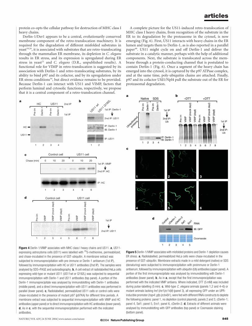
Derlin-1/VIMP is involved in US11-induced retro-translocationNext we asked whether the Derlin-1/VIMP complex is involved inretro-translocation. We first tested whether it associates with retro-translocating MHC class I heavy chains in US11-expressing astro-cytoma cells. The cells were pulse-labelled with 35S-methionine,permeabilized with digitonin, and chase-incubated in the presenceof a GST–ubiquitin fusion protein (GST–Ub). The modification ofretro-translocating substrates with poly(GST–Ub), instead of poly-ubiquitin, prevents their release from the ER membrane, resultingin a translocation intermediate25. The membrane fraction wassolubilized and subjected to sequential immunoprecipitation withDerlin-1 and heavy chain (HC) antibodies. A significant amount ofheavy chains (5–10%) was found associated with Derlin-1 (Fig. 4a,lane 2), while little was precipitated with a pre-immune serum(lane 1). The immunoprecipitatedmaterial co-migrated in SDS-gelswith glycosylated heavy chains (data not shown), indicating that theDerlin-1-interacting substrate had not yet reached the cytosolicN-glycanase26.Retro-translocating heavy chains could also be immunoprecipi-
tated with VIMP antibodies. In this case, the chase incubation wascarried out in the presence of a p97 mutant (p97KA) defective inATP hydrolysis to accumulate retro-translocation intermediates onthe membrane6. Sequential immunoprecipitation showed thatincreasing amounts of heavy chains became associated with VIMPduring the chase period in US11-expressing cells (Fig. 4c, lanes 4–6),while little was seen in control cells (lanes 1–3).Whenwild-type p97instead of p97KA was added during the chase, less substrateprecipitated with VIMP antibodies (data not shown), consistentwith the assumption that retro-translocating polypeptides onlytransiently interact with Derlin-1/VIMP.Because the US11 protein initiates the retro-translocation of
MHC class I heavy chains, we tested whether it can interact withDerlin-1. Sequential immunoprecipitation experiments with Der-lin-1 and US11 antibodies did indeed show an association (Fig. 4a,lane 4 versus 3). Similar results were obtained when US11 wastransiently expressed in HeLa cells; again US11 co-precipitated withDerlin-1 (Fig. 4b, lane 1). AUS11 mutant (Q192L) that binds MHCclass I heavy chains but cannot target them for retro-translocation27
showed a reduced affinity for Derlin-1 (lane 2), indicating that theUS11-Derlin-1 interaction is functionally important. An inter-action between these proteins could also be seen by immunofluor-
Figure 2 In vitro association of VIMP with p97 complexes. a, Purified p97 (80 nM) was
incubated with the indicated amounts of a GST fusion to the cytosolic domain of VIMP
(GST–VIMPc). Following centrifugation, supernatant and pellet fractions were analysed by
SDS–PAGE and Coomassie staining. b, As in a, except that rat liver cytosol (250mg) was
used and the analysis was performed by immunoblotting with the indicated antibodies.
c, As in b, except that cytosol depleted of p97 was used. Where indicated, p97 (p97wt) or
p97 mutant (p97DN) were added back to depleted cytosol (at 80 nM), before addition of
GST–VIMPc.
articles
NATURE |VOL 429 | 24 JUNE 2004 | www.nature.com/nature 843© 2004 Nature Publishing Group

escence microscopy in cells overexpressing Derlin-1 and US11; theyco-localized in dotted structures (Supplementary Fig. S6).US11 stably expressed in astrocytoma cells could also be immu-
noprecipitated with VIMP antibodies (Fig. 4d, lanes 6 and 7),whereas a pre-immune serum or an antiserum depleted of VIMPantibodies precipitated only small amounts (lanes 5 and 8). Nolabelled protein was precipitated from control cells that did notexpress US11 (lanes 1–4). The US11–VIMP interaction is probablymediated by Derlin-1, because US11 was not recruited to thepunctate structures formed by co-expression of VIMP and p97(Supplementary Fig. S6). Taken together, these results indicate thatDerlin-1/VIMP associates with US11 and retro-translocating MHCclass I heavy chains.
A general role for Derlin-1/VIMP in ER protein degradationTo investigate whether Derlin-1/VIMP has a general function in theretro-translocation of misfolded ER proteins, we tested its associ-ation with substrates. Pulse-labelled HeLa cells were permeabilizedwith digitonin, and chase-incubated in the presence of GST–Ubfusion protein. The membrane fraction was solubilized andsubjected to sequential immunoprecipitation with Derlin-1 andubiquitin antibodies. Poly(GST–Ub) modified proteins were co-precipitated with Derlin-1 under native conditions, but not afterdenaturation in SDS (Fig. 5a, lane 2 versus 4), although theantibodies work even better under denaturing conditions (lowerpanel). No precipitation was seen with the pre-immune serum(lanes 1 and 3). The association between poly(GST–Ub)-modifiedproteins and the Derlin-1/VIMP complex could also be seen byimmunoprecipitation with VIMP antibodies under native con-ditions (Fig. 5b, lanes 2 and 3). Much less material was precipitatedwith pre-immune serum (lane 1) or an antiserum depleted of VIMPantibodies (lane 4). When HeLa cells were treated during the pulse-labelling period with dithiothreitol (DTT) to increase the amountof misfolded ER proteins28, more poly(GST–Ub) modified materialwas found associated with VIMP (Fig. 5b, lanes 6, 7 versus lanes 2,3). Together, these results suggest that Derlin-1/VIMP associates
with a large set of substrates undergoing retro-translocation.If Derlin-1 plays a general role in retro-translocation, one would
expect that its depletion by RNA interference (RNAi) would lead tothe accumulation of misfolded proteins in the ER and elicit theUPR. To test this assumption, we used a strain of C. elegans, whichexpresses the green fluorescent protein (GFP) under the control ofthe UPR-inducible hsp4 promoter (hsp4::gfp)29. Inactivation ofC. elegansDerlin-1 (cDerlin-1) by RNAi activated the UPR reporterin many different cells of the animal, as demonstrated by fluor-escence microscopy (Fig. 5c, panel 2 versus 1) and immunoblottingfor GFP (Fig. 5d, lane 2 versus 1). The expression of hsp4::gfp wasblocked in mutant ire1 animals (Fig. 5c, panel 3), as predicted fromthe essential role of the Ire1 kinase in UPR29. The level of hsp4::gfpexpression by cDerlin-1-depletion was comparable to that observedafter inactivation of Sel1 (Fig. 5c, panel 2 versus 4; Fig. 5d, lane 2versus 4), which encodes a homologue of yeast Hrd3 involved in thepoly-ubiquitination of a subset of retro-translocation substrates30,31.RNAi-mediated depletion of Ero1, a protein required for disulphidebond formation in the ER32,33, led to an even stronger activation ofUPR (Fig. 5c, panel 5; Fig. 5d, lane 5). The lower level of hsp-4::gfpactivation observed following cDerlin-1 or Sel1 depletion is con-sistent with their function in removingmisfolded ER proteins underphysiological conditions, rather than in serving as general foldingcatalysts. As in yeast19,30,31, neither cDerlin-1 nor Sel1 is essential forviability. Depletion of Derlin-2 (cDerlin-2; Supplementary Fig. S1)in wild-type animals did not elicit the UPR (Fig. 5c, panel 6; Fig. 5d,lane 3), suggesting that it may not be involved in retro-translocationor may be required for the degradation of a restricted subset ofsubstrates.
DiscussionThe discovery of the Derlin-1/VIMP membrane protein complex inmammalian cells provides a link between substrate recognition inthe ER and the function of the p97 ATPase in the cytosol. It explainshow the p97 complex is recruited to the ER membrane for retro-translocation, and suggests a mechanism by which the viral US11
Figure 3 VIMP mediates p97 binding to hDerlin-1. a, HA-tagged Derlin-1 (hDerlin-1-HA,
panel 1), Myc-tagged VIMP (Myc-VIMP, panel 2), or His-tagged p97 (His-p97, panel 3)
were expressed individually in COS cells, stained with the corresponding antibodies, and
visualized by fluorescence microscopy. b, Myc-VIMP was expressed together with His-
tagged p97 (His-p97wt), p97 lacking the N-domain (His-p97DN), or p97 defective in ATP
and substrate binding (His-p97AA). The cells were stained with His or Myc antibodies, as
indicated. Panels 3, 6 and 9 were also stained with 4,6-diamidino-2-phenylindole (DAPI)
and show merged images. c, His-p97wt, Myc-VIMP, and hDerlin-1-HA were coexpressed
(panels 1–3). Controls were performed by replacing hDerlin-1-HA with FLAG-tagged
ATF6 (FLAG-ATF6, panels 4–6), or by omitting Myc-VIMP (panels 7–9). The cells were
stained with HA, FLAG or Myc antibodies, as indicated. Panels 3, 6 and 9 show merged
images.
articles
NATURE | VOL 429 | 24 JUNE 2004 | www.nature.com/nature844 © 2004 Nature Publishing Group
protein co-opts the cellular pathway for destruction of MHC class Iheavy chains.
Derlin-1/Der1 appears to be a central, evolutionarily conservedmembrane component of the retro-translocation machinery. It isrequired for the degradation of different misfolded substrates inyeast19,20, it is associated with substrates that are retro-translocatingthrough the mammalian ER membrane, its depletion in C. elegansresults in ER stress, and its expression is upregulated during ERstress in yeast21 and C. elegans (D.R., unpublished results). Afunctional role for VIMP in retro-translocation is suggested by itsassociation with Derlin-1 and retro-translocating substrates, by itsability to bind p97 and its cofactor, and by its upregulation underER stress conditions34, but direct evidence remains to be provided.Because Derlin-1 can interact with US11 and VIMP, factors thatperform luminal and cytosolic functions, respectively, we proposethat it is a central component of a retro-translocation channel.
A complete picture for the US11-induced retro-translocation ofMHC class I heavy chains, from recognition of the substrate in theER to its degradation by the proteasome in the cytosol, is nowemerging (Fig. 6). First, US11 interacts with heavy chains in the ERlumen and targets them to Derlin-1, as is also reported in a parallelpaper40. US11 might cycle on and off Derlin-1 and deliver thesubstrate in a catalytic manner, perhaps with the help of additionalcomponents. Next, the substrate is translocated across the mem-brane through a protein-conducting channel that is postulated tocontain Derlin-1 (Fig. 6). Once a segment of the heavy chain hasemerged into the cytosol, it is captured by the p97 ATPase complex,and at the same time, poly-ubiquitin chains are attached. Finally,p97 and its cofactor Ufd1/Npl4 pull the substrate out of the ER forproteasomal degradation.
Figure 4 Derlin-1/VIMP associates with MHC class I heavy chains and US11. a, US11-
expressing astrocytoma cells (US11) were labelled with 35S-methionine, permeabilized,
and chase-incubated in the presence of GST-ubiquitin. A membrane extract was
subjected to immunoprecipitation with pre-immune or Derlin-1 antiserum (1st IP),
followed by immunoprecipitation with HC or US11 antibodies (2nd IP). The samples were
analysed by SDS–PAGE and autoradiography. b, A cell extract of radiolabelled HeLa cells
expressing wild-type or mutant US11 (US11wt or Q192L) was subjected to sequential
immunoprecipitation with Derlin-1 and US11 antibodies (top panel). A portion of the
Derlin-1 immunoprecipitate was analysed by immunoblotting with Derlin-1 antibodies
(middle panel), and a direct immunoprecipitation with US11 antibodies was performed in
parallel (lower panel). c, Radiolabelled, permeabilized US11 cells or control cells were
chase-incubated in the presence of mutant p97 (p97KA) for different time periods. A
membrane extract was subjected to sequential immunoprecipitation with VIMP and HC
antibodies (upper panel) or to direct immunoprecipitation with HC antibodies (lower panel).
d, As in c, with the sequential immunoprecipitation performed with the indicated
antibodies.
Figure 5 Derlin-1/VIMP associates with misfolded proteins and Derlin-1 depletion causes
ER stress. a, Radiolabelled, permeabilized HeLa cells were chase-incubated in the
presence of GST-ubiquitin. Membrane extracts made in a mild detergent (native) or SDS
(denaturing) were subjected to immunoprecipitation with preimmune or Derlin-1
antiserum, followed by immunoprecipitation with ubiquitin (Ub) antibodies (upper panel). A
portion of the first immunoprecipitate was analysed by immunoblotting with Derlin-1
antibodies (lower panel). b, As in a, except that the first immunoprecipitation was
performed with the indicated VIMP antisera. Where indicated, DTT (5mM) was included
during pulse-labelling (5 min). c, Wild-type C. elegans animals (panels 1,2 and 4–6) or
mutant animals lacking Ire1(ire1(zc14)II) (panel 3), all expressing GFP under an UPR-
inducible promoter (hsp4::gfp (zcls4)V ), were fed with different RNAi constructs to deplete
the following proteins: panel 1, no depletion (control plasmid); panels 2 and 3, cDerlin-1;
panel 4, Sel1; panel 5, Ero1; panel 6, cDerlin-2. d, Extracts of different animals were
analysed by immunoblotting with GFP antibodies (top panel) or Coomassie staining
(bottom panel).
articles
NATURE |VOL 429 | 24 JUNE 2004 | www.nature.com/nature 845© 2004 Nature Publishing Group

The retro-translocation of misfolded ER proteins probablyoccurs by the same pathway, the major difference being in substraterecognition. Initial recognition may be mediated by certain ERchaperones, such as protein disulphide isomerase or calnexin/calreticulin12,14,16–18, but targeting to Derlin-1 may requireadditional shuttle proteins that can interact with both the chaper-one-associated substrate and Derlin-1/VIMP. The viral US11 pro-tein appears to combine both functions. The retro-translocation ofmisfolded proteins in lower organisms also requires a different‘cytosolic adaptor’ to recruit the p97 ATPase, because homologuesof VIMP appear to exist only in vertebrates.Experiments in yeast show that Der1 is only required for the
degradation of ER proteins with misfolded luminal domains20.Membrane proteins with misfolded cytosolic or intra-membranedomains do not require Der1 and might move through a differenttranslocation channel, perhaps the Sec61 channel. Alternatively,they may not need a channel at all, as suggested by the bacterial AAAprotease FtsH, which can degrade a misfoldedmembrane protein ina purified reconstituted system lacking any obvious channel35. Allpathways of retro-translocation, whether Der1/Derlin-1-dependentor not, appear to require the function of the p97 ATPase complex,which may provide the general driving force for the movement ofproteins into the cytosol. A
MethodsConstructsConstructs that express His-tagged versions of wild type p97 (His-p97wt), a mutantlacking the N domain (His-p97DN), and a mutant defective in ATP and substrate binding(His-p97AA) in E. coli were described previously6. To generate mammalian expressionconstructs containing these p97 variants, the coding regions in the bacterial expressionplasmids were amplified by polymerase chain reaction (PCR) and inserted into thepcDNA3.1 vector using the TOPO cloning technology (Invitrogen). Human Derlin-1(hDerlin-1) (Gene bank ID: NP_077271) and VIMP (Gene bank ID: AY618665)complementary DNAs were isolated from a cDNA library (Panomics) by PCR and clonedinto the pcDNA3.1 vector. Sequencing of several independent PCR clones of hDerlin-1revealed a single base variation from the reported sequence, resulting in a G to Rsubstitution at residue 245. To generate a construct expressing a GST fusion to thecytosolic domain of VIMP (GST–VIMPc), a DNA segment encoding amino acids 49 to187 of VIMP was inserted into the pET42 vector (Novagen). The RNAi constructs for C.elegans Derlin-1 homologues were generated by cloning a cDNA segment encodingresidues 68 to 242 of cDerlin-1 (F25D7.1; ID: NP_492721) and a segment encodingresidue 24 to 221 of cDerlin-2 (R151.6; ID: T16766) into the pPD129.36 vector36. The Sel1and Ero1 RNAi constructs were described37,38. The plasmid pCMV-Flag-ATF6 wasprovided by R. Prywes. The plasmid pCMV-US11 was described previously3. The US11Q192L mutant was made by site-directed mutagenesis.
Antibodies and proteinsAntibodies to MHC class I heavy chain, Sec61b, p97, His-tag, and ubiquitin weredescribed previously6. Antibodies to mammalian Ufd1 were described5. Myc antibodies
(9E10), HA antibodies (12CA5), and FLAG M2 antibodies were purchased from SantaCruz, Roche and Sigma, respectively. Antibodies to proteasome subunits (a7 and Mms1),to the cytosolic chaperone Hsp70, and to GFP were from Affiniti Bioreagents, StressGenBiotechnology and Molecular Probes, respectively. Peptide-specific antibodies againsthDerlin-1 and VIMPwere raised in rabbits. Two peptides corresponding to residues 204 to216 and 238 to 251 of hDerlin-1, and a peptide corresponding to residues 174 to 187 ofhuman VIMP were chosen by Zymed for immunization.
The purification of His-tagged p97 variants was described in ref. 6. GST-VIMPc wasexpressed in E. coli and purified with glutathione beads. The eluted protein was furtherpurified on a Superdex 200 HR (10/30) column in 50mM Tris/HCl pH 8.0, 150mMpotassium chloride, 5% glycerol and 2mM magnesium chloride. His-VIMPc wasexpressed in E. coli and purified by Ni-NTA chromatography. GST-ubiquitin waspurchased from BostonBiochem.
Membrane binding assayPurified p97 variants (250 nM) were incubated with 60 equivalents of dog pancreaticmicrosomes for 20min at 4 8C. The membranes were layered on top of a sucrose cushion(50mM HEPES pH 7.4, 150mM potassium acetate, 10mM magnesium acetate, 1.2Msucrose) and sedimented by centrifugation in a TLA100 rotor for 15min at 75,000 r.p.m.Supernatant fractions were analysed directly, while the pellets were washed withmembrane buffer (50mMHEPES pH 7.4, 150mM potassium acetate, 10mMmagnesiumacetate and 250mM sucrose) before analysis by SDS–polyacrylamide gel electrophoresis(PAGE) and immunoblotting. Where indicated, peripheral membrane proteins wereremoved from microsomes by washing the membranes twice with membrane buffercontaining 800mM sodium chloride. Salt-washed membranes were resuspended inmembrane buffer and stored at 280 8C.
Purification of Derlin-1/VIMP complexBiotinylation of purified p97 variants was performed using EZ-link sulfo-NHS-SS-Biotin(Pierce) according to the instructions of the manufacturer. Modified p97 or p97DN(100 mg) was incubated with 6,000 equivalents salt-washed dog pancreatic microsomes for30min at 4 8C. Unbound proteins were removed by sedimentation of the membranesthrough a sucrose cushion. Membranes were solubilized in 50mM HEPES pH 7.4,200mM potassium acetate and 10mM magnesium, 2.5% digitonin plus proteaseinhibitor cocktail. The extract was incubated with streptavidin beads and bound p97 waseluted in solubilization buffer containing 50mM DTT. The proteins were subjected toSDS–PAGE and Coomassie staining. Two visible bands were cut out and analysed by massspectrometry. Sequences of five peptides from the 28-kD bands and three peptides fromthe 25-kD bands were obtained (Supplementary Table 1). These peptides match thesequences of two proteins in the human genome sequence database (ID: NP_077271 andNP_060915, respectively).
In vitro co-precipitation experimentsTo determine the association of VIMP with p97 and its cofactor, the indicated amount ofGST-VIMPc was incubated with either 80 nM purified p97 or with 100 ml rat liver cytosol.The samples were centrifuged for 20min at 20,000 g, and the resulting supernatant andpellet fractions were analysed by SDS–PAGE, followed by Coomassie staining orimmunoblotting.
Mammalian cell culture and transient gene expressionHeLa cells, HEK293 cells, and COS cells were maintained according to standardprocedures. Transient transfections were performed either following the standard calciumphosphate precipitation protocol for HEK293 cells, or using the FuGENE 6 reagent(Roche) for HeLa and COS cells. For immunofluorescence microscopy, cells were fixedwith 3% paraformaldehyde and stained with antibodies to Myc (1:1000), His (1:100), HA(1:1000), FLAG (1:1000) or US11 (1:2000).
Association of Derlin-1/VIMP with retro-translocating substrates and US11The use of permeabilized US11-expressing astrocytoma cells to study the retro-translocation of MHC class I heavy chains was described39. To analyse the association ofDerlin-1/VIMP with retro-translocating HC, 35S-methinione labelled cells werepermeabilized and chase-incubated for 40min in the presence of either 20 mM GST-ubiquitin or 300 nMmutant p97 protein (p97KA)6,25. The membranes were solubilized inbuffer N (1%DeoxyBig CHAP, 30mMTris/HCl pH 7.4, 150mMpotassium acetate, 4mMmagnesium acetate, 1mM ATP and protease inhibitor cocktail), and extracts weresubjected to immunoprecipitation with antibodies to Derlin-1 or VIMP, followed byimmunoprecipitation with HC antibodies. To determine the interaction of Derlin-1/VIMP with poly-ubiquitinated substrates, HeLa cells were labelled with 35S-methionineand permeabilized as was done with US11 cells. GST–Ub (20 mM)was included during thechase incubation. Membranes were solubilized in buffer N and subjected toimmunoprecipitation with antibodies to Derlin-1 or VIMP, followed byimmunoprecipitation with ubiquitin antibodies. The association of Derlin-1/VIMP withUS11 was determined in a similar way, except that the second immunoprecipitation wasperformed with US11 antibodies.
RNAi experiments in C. elegansC. elegans were maintained on nematode growing media (NGM) agar plates according tostandard protocols. For RNAi experiments, 3–6 animals at L4 stage were placed on anisopropyl-b-D-thiogalactopyranoside-containing NGM plate pre-seeded with bacteriacarrying an RNAi feeding construct. Phenotypes were examined three days later with afluorescent microscope. To verify the UPR induction by immunoblotting, animals fromthree plates were washed off withM3medium and homogenized in 20mMHEPES pH 7.4,
Figure 6Model for US11-mediated retro-translocation of MHC class I heavy chains. US11
recognizes HC in the ER lumen and targets it to Derlin-1, a proposed component of the
retro-translocation channel. The p97 ATPase complex is recruited to Derlin-1 by VIMP. HC
emerging into the cytosol is bound by p97. Poly-ubiquitin chains (Poly-Ub, red) are
attached and recognized by both the N-domain (N) of p97 and the cofactor Ufd1/Npl4 (U/
N). ATP hydrolysis by p97 moves HC into the cytosol. The retro-translocation of misfolded
ER proteins may occur similarly, with US11 being replaced by other targeting
components.
articles
NATURE | VOL 429 | 24 JUNE 2004 | www.nature.com/nature846 © 2004 Nature Publishing Group

50mM sodium chloride, 10% glycerol, 1% Triton X-100 and 1mM EDTA plus proteaseinhibitors. Extracts corresponding to 20 mg proteins were fractionated on a SDS–PAGE geland analysed by either immunoblotting with GFP antibodies or Coomassie staining.
Received 5 March; accepted 17 May 2004; doi:10.1038/nature02656.
1. Ellgaard, L. & Helenius, A. ER quality control: towards an understanding at the molecular level. Curr.
Opin. Cell Biol. 13, 431–437 (2001).
2. Tsai, B., Ye, Y. & Rapoport, T. A. Retro-translocation of proteins from the endoplasmic reticulum into
the cytosol. Nature Rev. Mol. Cell Biol. 3, 246–255 (2002).
3. Wiertz, E. J. H. J. et al. The human cytomegalovirus US11 gene product dislocates MHC class I heavy
chains from the endoplasmic reticulum to the cytosol. Cell 84, 769–779 (1996).
4. Aridor, M. & Balch, W. E. Integration of endoplasmic reticulum signaling in health and disease.
Nature Med. 5, 745–751 (1999).
5. Meyer, H. H., Shorter, J. G., Seemann, J., Pappin, D. &Warren, G. A complex of mammalian ufd1 and
npl4 links the AAA-ATPase, p97, to ubiquitin and nuclear transport pathways. EMBO J. 19,
2181–2192 (2000).
6. Ye, Y., Meyer, H. H. & Rapoport, T. A. Function of the p97-Ufd1-Npl4 complex in retrotranslocation
from the ER to the cytosol: dual recognition of nonubiquitinated polypeptide segments and
polyubiquitin chains. J. Cell Biol. 162, 71–84 (2003).
7. Langer, T. AAA proteases: cellular machines for degrading membrane proteins. Trends Biochem. Sci.
25, 247–251 (2000).
8. Bays, N. W., Wilhovsky, S. K., Goradia, A., Hodgkiss-Harlow, K. & Hampton, R. Y. HRD4/NPL4 is
required for the proteasomal processing of ubiquitinated ER proteins.Mol. Biol. Cell 12, 4114–4128
(2001).
9. Ye, Y., Meyer, H. H. & Rapoport, T. A. The AAAATPase Cdc48/p97 and its partners transport proteins
from the ER into the cytosol. Nature 414, 652–656 (2001).
10. Jarosch, E. et al. Protein dislocation from the ER requires polyubiquitination and the AAA-ATPase
Cdc48. Nature Cell Biol. 4, 134–139 (2002).
11. Rabinovich, E., Kerem, A., Frohlich, K. U., Diamant, N. & Bar-Nun, S. AAA-ATPase p97/Cdc48p, a
cytosolic chaperone required for endoplasmic reticulum-associated protein degradation. Mol. Cell.
Biol. 22, 626–634 (2002).
12. Wang, Q. & Chang, A. Substrate recognition in ER-associated degradation mediated by Eps1, a
member of the protein disulfide isomerase family. EMBO J. 22, 3792–3802 (2003).
13. Story, C. M., Furman, M. H. & Ploegh, H. L. The cytosolic tail of class I MHC heavy chain is required
for its dislocation by the human cytomegalovirus US2 and US11 gene products. Proc. Natl Acad. Sci.
USA 96, 8516–8521 (1999).
14. Gillece, P., Luz, J. M., Lennarz,W. J., de La Cruz, F. J. & Romisch, K. Export of a cysteine-freemisfolded
secretory protein from the endoplasmic reticulum for degradation requires interaction with protein
disulfide isomerase. J. Cell Biol. 147, 1443–1456 (1999).
15. Nishikawa, S. I., Fewell, S. W., Kato, Y., Brodsky, J. L. & Endo, T. Molecular chaperones in the yeast
endoplasmic reticulum maintain the solubility of proteins for retrotranslocation and degradation.
J. Cell Biol. 153, 1061–1070 (2001).
16. Tsai, B., Rodighiero, C., Lencer, W. I. & Rapoport, T. A. Protein disulfide isomerase acts as a redox-
dependent chaperone to unfold cholera toxin. Cell 104, 937–948 (2001).
17. Molinari, M., Calanca, V., Galli, C., Lucca, P. & Paganetti, P. Role of EDEM in the release of misfolded
glycoproteins from the calnexin cycle. Science 299, 1397–1400 (2003).
18. Oda, Y., Hosokawa, N., Wada, I. & Nagata, K. EDEM as an acceptor of terminally misfolded
glycoproteins released from calnexin. Science 299, 1394–1397 (2003).
19. Knop, M., Finger, A., Braun, T., Hellmuth, K. &Wolf, D. H. Der1, a novel protein specifically required
for endoplasmic reticulum degradation in yeast. EMBO J. 15, 753–763 (1996).
20. Vashist, S. & Ng, D. T. Misfolded proteins are sorted by a sequential checkpoint mechanism of ER
quality control. J. Cell Biol. 165, 41–52 (2004).
21. Travers, K. J. et al. Functional and genomic analyses reveal an essential coordination between the
unfolded protein response and ER-associated degradation. Cell 101, 249–258 (2000).
22. Hitt, R. & Der Wolf, D. H. 1p, a protein required for degradation of malfolded soluble proteins of the
endoplasmic reticulum: topology and Der1-like proteins. FEMS Yeast Res. 4, 721–729 (2004).
23. Kryukov, G. V. et al. Characterization of mammalian selenoproteomes. Science 300, 1439–1443
(2003).
24. DeLaBarre, B. & Brunger, A. T. Complete structure of p97/valosin-containing protein reveals
communication between nucleotide domains. Nature Struct. Biol. 10, 856–863 (2003).
25. Flierman, D., Ye, Y., Dai, M., Chau, V. & Rapoport, T. A. Polyubiquitin serves as a recognition signal,
rather than a ratcheting molecule, during retrotranslocation of proteins across the endoplasmic
reticulum membrane. J. Biol. Chem. 278, 34774–34782 (2003).
26. Wiertz, E. J. H. J. et al. Sec61-mediated transfer of a membrane protein from the endoplasmic
reticulum to the proteasome for destruction. Nature 384, 432–438 (1996).
27. Lilley, B. N., Tortorella, D. & Ploegh, H. L. Dislocation of a type I membrane protein requires
interactions between membrane-spanning segments within the lipid bilayer. Mol. Biol. Cell 14,
3690–3698 (2003).
28. Braakman, I., Helenius, J. & Helenius, A. Manipulating disulfide bond formation and protein folding
in the endoplasmic reticulum. EMBO J. 11, 1717–1722 (1992).
29. Calfon, M. et al. IRE1 couples endoplasmic reticulum load to secretory capacity by processing the
XBP-1 mRNA. Nature 415, 92–96 (2002).
30. Plemper, R. K. et al. Genetic interactions of Hrd3p and Der3p/Hrd1p with Sec61p suggest a retro-
translocation complex mediating protein transport for ER degradation. J. Cell Sci. 112, 4123–4134
(1999).
31. Gardner, R. G. et al. Endoplasmic reticulum degradation requires lumen to cytosol signaling.
Transmembrane control of Hrd1p by Hrd3p. J. Cell Biol. 151, 69–82 (2000).
32. Frand, A. R. & Kaiser, C. A. The ERO1 gene of yeast is required for oxidation of protein dithiols in the
endoplasmic reticulum. Mol. Cell 1, 161–170 (1998).
33. Pollard, M. G., Travers, K. J. &Weissman, J. S. Ero1p: a novel and ubiquitous protein with an essential
role in oxidative protein folding in the endoplasmic reticulum. Mol. Cell 1, 171–182 (1998).
34. Gao, Y. et al. Regulation of the selenoprotein SelS by glucose deprivation and endoplasmic reticulum
stress—SelS is a novel glucose-regulated protein. FEBS Lett. 563, 185–190 (2004).
35. Akiyama, Y. & Ito, K. Reconstitution of membrane proteolysis by FtsH. J. Biol. Chem. 278,
18146–18153 (2003).
36. Timmons, L., Court, D. L. & Fire, A. Ingestion of bacterially expressed dsRNAs can produce specific
and potent genetic interference in Caenorhabditis elegans. Gene 263, 103–112 (2001).
37. Urano, F. et al. A survival pathway for Caenorhabditis elegans with a blocked unfolded protein
response. J. Cell Biol. 158, 639–646 (2002).
38. Harding, H. P. et al. An integrated stress response regulates amino acid metabolism and resistance to
oxidative stress. Mol. Cell 11, 619–633 (2003).
39. Shamu, C. E., Story, C. M., Rapoport, T. A. & Ploegh, H. L. The pathway of US11-dependent
degradation of MHC class I heavy chains involves a ubiquitin-conjugated intermediate. J. Cell Biol.
147, 45–58 (1999).
40. Lilley, B. N. & Ploegh, H. L. A membrane protein required for dislocation of misfolded proteins from
the ER. Nature (this issue).
Supplementary Information accompanies the paper on www.nature.com/nature.
Acknowledgements We thank R. Prywes for ATF6 plasmid, the TaplinMass Spectrometry facility
(Harvard Medical School) for protein identification, and the Nikon imaging facility (Harvard
Medical School) for assistance in microscopy. We thank E. Hartmann for help with sequence
analysis, D. Flierman for help with the Derlin-1 immunoprecipitation, and C. Shamu, D. Finley
and K. Cannon for critical reading of the manuscript. Y.Y. is supported by a Helen Hay Whitney
fellowship and T.A.R. by an NIH grant. C.Y is supported by an NIH fellowship and D.R. by NIH
grants and the Ellison Medical Foundation. T.A.R. is a Howard Hughes Medical Institute
investigator.
Competing interests statement The authors declare that they have no competing financial
interests.
Correspondence and requests for materials should be addressed to T.A.R.
articles
NATURE |VOL 429 | 24 JUNE 2004 | www.nature.com/nature 847© 2004 Nature Publishing Group